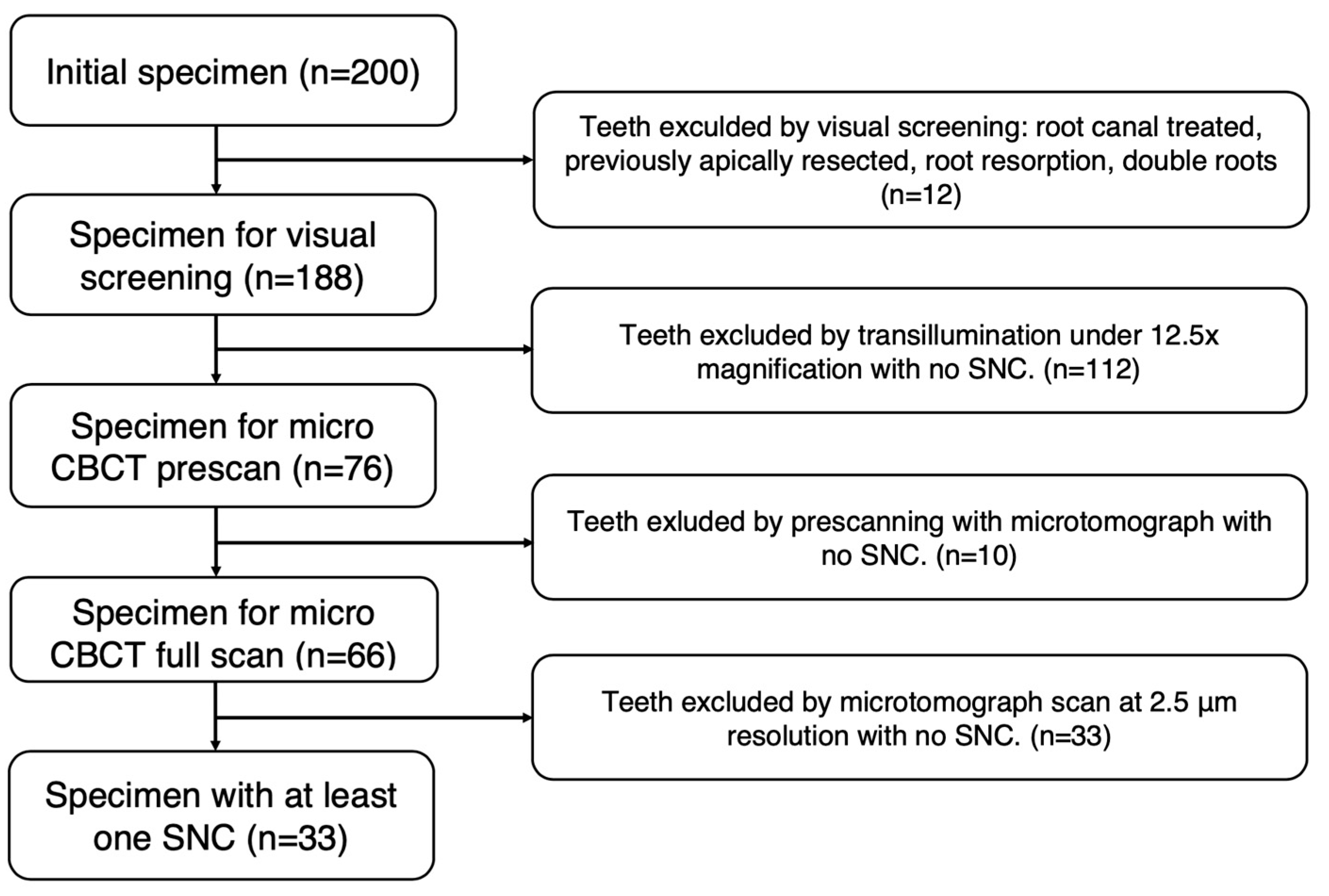
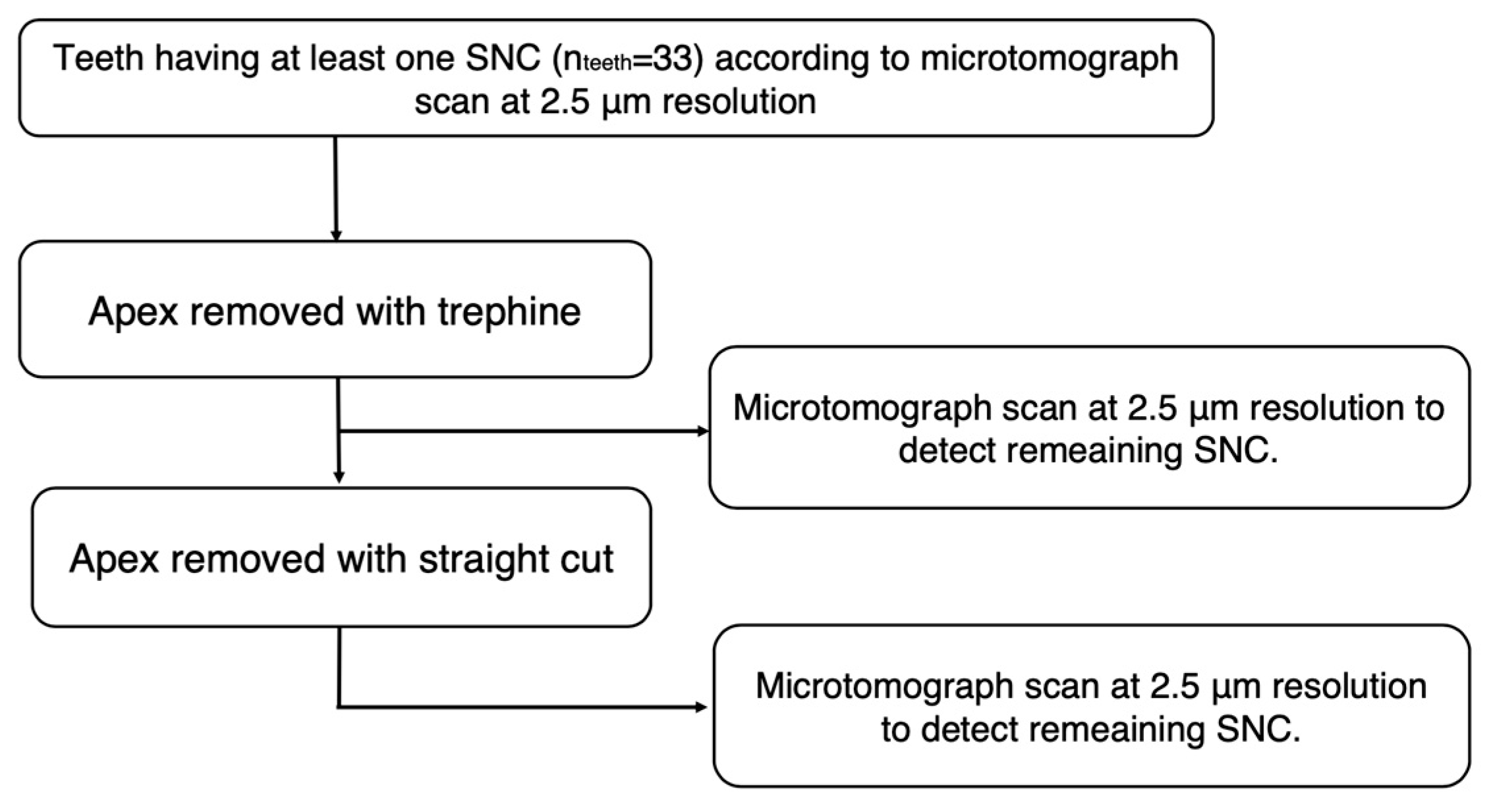
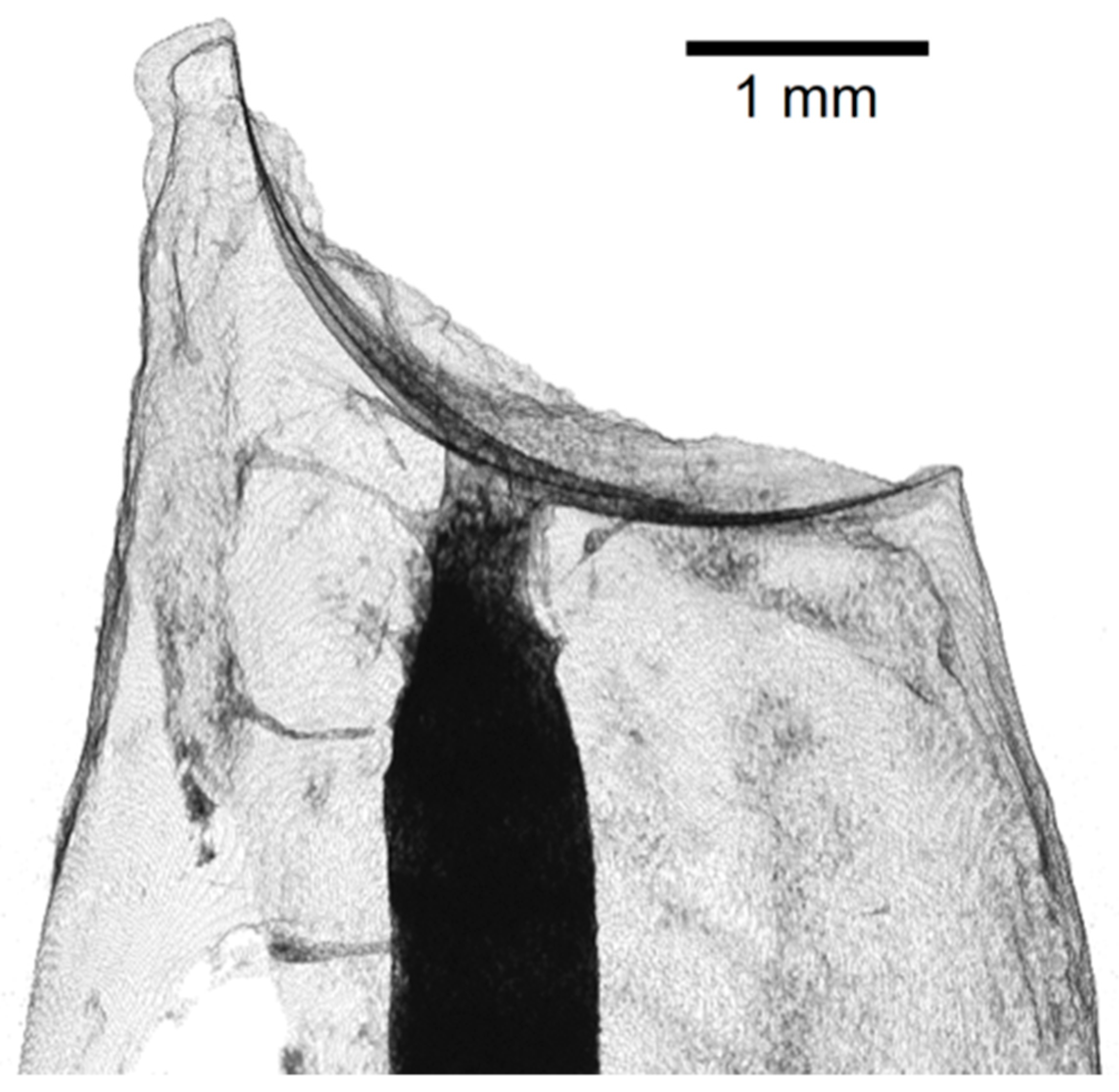
An Exploratory In Vitro Microcomputed Tomographic Investigation of the Efficacy of Semicircular Apicoectomy Performed with Trephine Bur
Abstract
1. Introduction
2. Materials and Methods
3. Results
4. Discussion
5. Conclusions
Author Contributions
Funding
Institutional Review Board Statement
Informed Consent Statement
Data Availability Statement
Conflicts of Interest
References
- Tabassum, S.; Khan, F.R. Failure of endodontic treatment: The usual suspects. Eur. J. Dent. 2016, 10, 144–147. [Google Scholar] [CrossRef] [PubMed]
- Meder-Cowherd, L.; Williamson, A.E.; Johnson, W.T.; Vasilescu, D.; Walton, R.; Qian, F. Apical morphology of the palatal roots of maxillary molars by using micro-computed tomography. J. Endod. 2011, 37, 1162–1165. [Google Scholar] [CrossRef] [PubMed]
- Gao, X.; Tay, F.R.; Gutmann, J.L.; Fan, W.; Xu, T.; Fan, B. Micro-CT evaluation of apical delta morphologies in human teeth. Sci. Rep. 2016, 6, 36501. [Google Scholar] [CrossRef] [PubMed]
- Ahmed, H.M.A.; Versiani, M.A.; De-Deus, G.; Dummer, P.M.H. A new system for classifying root and root canal morphology. Int. Endod. J. 2017, 50, 761–770. [Google Scholar] [CrossRef]
- Karobari, M.I.; Parveen, A.; Mirza, M.B.; Makandar, S.D.; Nik Abdul Ghani, N.R.; Noorani, T.Y.; Marya, A. Root and Root Canal Morphology Classification Systems. Int. J. Dent. 2021, 2021, 6682189. [Google Scholar] [CrossRef] [PubMed]
- Vertucci, F.J. Root canal anatomy of the human permanent teeth. Oral Surg. Oral Med. Oral Pathol. 1984, 58, 589–599. [Google Scholar] [CrossRef]
- Nair, P.N. On the causes of persistent apical periodontitis: A review. Int. Endod. J. 2006, 39, 249–281. [Google Scholar] [CrossRef]
- Kim, S.; Kratchman, S. Modern endodontic surgery concepts and practice: A review. J. Endod. 2006, 32, 601–623. [Google Scholar] [CrossRef]
- Morfis, A.; Sylaras, S.N.; Georgopoulou, M.; Kernani, M.; Prountzos, F. Study of the apices of human permanent teeth with the use of a scanning electron microscope. Oral Surg. Oral Med. Oral Pathol. 1994, 77, 172–176. [Google Scholar] [CrossRef]
- Mjor, I.A.; Nordahl, I. The density and branching of dentinal tubules in human teeth. Arch. Oral Biol. 1996, 41, 401–412. [Google Scholar] [CrossRef]
- Kim, S.; Pecora, G.; Rubinstein, R.; Dorschr-Kim, J. Color Atlas of Microsurgery in Endodontics; BW Saunders: Philadelphia, PA, USA, 2001. [Google Scholar]
- Tortorici, S.; Difalco, P.; Caradonna, L.; Tete, S. Traditional endodontic surgery versus modern technique: A 5-year controlled clinical trial. J. Craniofacial Surg. 2014, 25, 804–807. [Google Scholar] [CrossRef] [PubMed]
- Frater, M.; Antal, M.; Braunitzer, G.; Joob-Fancsaly, A.; Nagy, K. An update on endodontic microsurgery—A literature review. Fogorv. Sz. 2017, 110, 43–48. (In Hungarian) [Google Scholar]
- Von Arx, T.; Hanni, S.; Jensen, S.S. Correlation of bone defect dimensions with healing outcome one year after apical surgery. J. Endod. 2007, 33, 1044–1048. [Google Scholar] [CrossRef] [PubMed]
- Pinsky, H.M.; Champleboux, G.; Sarment, D.P. Periapical surgery using CAD/CAM guidance: Preclinical results. J. Endod. 2007, 33, 148–151. [Google Scholar] [CrossRef] [PubMed]
- Yunfeng, L.; Guangsheng, J.; Quanming, Y.; Wei, P. Additive manufacturing and digital design assisted precise apicoectomy: A case study. Rapid Prototyp. J. 2014, 20, 33–40. [Google Scholar]
- Patel, S.; Aldowaisan, A.; Dawood, A. A novel method for soft tissue retraction during periapical surgery using 3D technology: A case report. Int. Endod. J. 2017, 50, 813–822. [Google Scholar] [CrossRef] [PubMed]
- Ye, S.; Zhao, S.; Wang, W.; Jiang, Q.; Yang, X. A novel method for periapical microsurgery with the aid of 3D technology: A case report. BMC Oral Health 2018, 18, 85. [Google Scholar] [CrossRef] [PubMed]
- Kim, J.E.; Shim, J.S.; Shin, Y. A new minimally invasive guided endodontic microsurgery by cone beam computed tomography and 3-dimensional printing technology. Restor. Dent. Endod. 2019, 44, e29. [Google Scholar] [CrossRef]
- Lai, P.T.; Yang, S.F.; Lin, Y.M.; Ho, Y.C. Computer-aided design-guided endodontic microsurgery for a mandibular molar with hypercementosis. J. Formos. Med. Assoc. 2019, 118, 1471–1472. [Google Scholar] [CrossRef]
- Giacomino, C.M.; Ray, J.J.; Wealleans, J.A. Targeted Endodontic Microsurgery: A Novel Approach to Anatomically Challenging Scenarios Using 3-dimensional-printed Guides and Trephine Burs-A Report of 3 Cases. J. Endod. 2018, 44, 671–677. [Google Scholar] [CrossRef]
- Ahn, S.Y.; Kim, N.H.; Kim, S.; Karabucak, B.; Kim, E. Computer-aided Design/Computer-aided Manufacturing-guided Endodontic Surgery: Guided Osteotomy and Apex Localization in a Mandibular Molar with a Thick Buccal Bone Plate. J. Endod. 2018, 44, 665–670. [Google Scholar] [CrossRef] [PubMed]
- Antal, M.; Nagy, E.; Sanyo, L.; Braunitzer, G. Digitally planned root end surgery with static guide and custom trephine burs: A case report. Int. J. Med. Robot. Comput. Assist. Surg. 2020, 16, e2115. [Google Scholar] [CrossRef] [PubMed]
- Popowicz, W.; Palatynska-Ulatowska, A.; Kohli, M.R. Targeted Endodontic Microsurgery: Computed Tomography-based Guided Stent Approach with Platelet-rich Fibrin Graft: A Report of 2 Cases. J. Endod. 2019, 45, 1535–1542. [Google Scholar] [CrossRef] [PubMed]
- Buniag, A.G.; Pratt, A.M.; Ray, J.J. Targeted Endodontic Microsurgery: A Retrospective Outcomes Assessment of 24 Cases. J. Endod. 2021, 47, 762–769. [Google Scholar] [CrossRef] [PubMed]
- Hawkins, T.K.; Wealleans, J.A.; Pratt, A.M.; Ray, J.J. Targeted endodontic microsurgery and endodontic microsurgery: A surgical simulation comparison. Int. Endod. J. 2020, 53, 715–722. [Google Scholar] [CrossRef] [PubMed]
- Ray, J.J.; Giacomino, C.M.; Wealleans, J.A.; Sheridan, R.R. Targeted Endodontic Microsurgery: Digital Workflow Options. J. Endod. 2020, 46, 863–871. [Google Scholar] [CrossRef] [PubMed]
- Zubizarreta-Macho, A.; Castillo-Amature, C.; Montiel-Company, J.M.; Mena-Alvarez, J. Efficacy of Computer-Aided Static Navigation Technique on the Accuracy of Endodontic Microsurgery. A Systematic Review and Meta-Analysis. J. Clin. Med. 2021, 10, 313. [Google Scholar] [CrossRef] [PubMed]
- Benjamin, G.; Ather, A.; Bueno, M.R.; Estrela, C.; Diogenes, A. Preserving the Neurovascular Bundle in Targeted Endodontic Microsurgery: A Case Series. J. Endod. 2021, 47, 509–519. [Google Scholar] [CrossRef]
- Smith, B.G.; Pratt, A.M.; Anderson, J.A.; Ray, J.J. Targeted Endodontic Microsurgery: Implications of the Greater Palatine Artery. J. Endod. 2021, 47, 19–27. [Google Scholar] [CrossRef]
- Nagy, E.; Fráter, M.; Antal, M. Guided modern endodontic microsurgery by use of a trephine bur. Orvosi Hetil. 2020, 161, 1260–1265. [Google Scholar] [CrossRef]
- Antal, M.; Nagy, E.; Braunitzer, G.; Fráter, M.; Piffkó, J. Accuracy and clinical safety of guided root end resection with a trephine: A case series. Head Face Med. 2019, 15, 30. [Google Scholar] [CrossRef] [PubMed]
- Tavares, W.L.F.; Fonseca, F.O.; Maia, L.M.; de Carvalho Machado, V.; Franca Alves Silva, N.R.; Junior, G.M.; Ribeiro Sobrinho, A.P. 3D Apicoectomy Guidance: Optimizing Access for Apicoectomies. J. Oral Maxillofac. Surg. 2020, 78, 357.e1–357.e8. [Google Scholar] [CrossRef] [PubMed]
- Tidmarsh, B.G.; Arrowsmith, M.G. Dentinal tubules at the root ends of apicected teeth: A scanning electron microscopic study. Int. Endod. J. 1989, 22, 184–189. [Google Scholar] [CrossRef] [PubMed]
- Gilheany, P.A.; Figdor, D.; Tyas, M.J. Apical dentin permeability and microleakage associated with root end resection and retrograde filling. J. Endod. 1994, 20, 22–26. [Google Scholar] [CrossRef] [PubMed]
- Gagliani, M.; Taschieri, S.; Molinari, R. Ultrasonic root-end preparation: Influence of cutting angle on the apical seal. J. Endod. 1998, 24, 726–730. [Google Scholar] [CrossRef] [PubMed]
- Tahmaseb, A.; Wu, V.; Wismeijer, D.; Coucke, W.; Evans, C. The accuracy of static computer-aided implant surgery: A systematic review and meta-analysis. Clin. Oral Implant. Res. 2018, 29 (Suppl. 16), 416–435. [Google Scholar] [CrossRef] [PubMed]
- Adorno, C.G.; Yoshioka, T.; Suda, H. Incidence of accessory canals in Japanese anterior maxillary teeth following root canal filling ex vivo. Int. Endod. J. 2010, 43, 370–376. [Google Scholar] [CrossRef]
- Kasahara, E.; Yasuda, E.; Yamamoto, A.; Anzai, M. Root canal system of the maxillary central incisor. J. Endod. 1990, 16, 158–161. [Google Scholar] [CrossRef]
- Pan, J.Y.Y.; Parolia, A.; Chuah, S.R.; Bhatia, S.; Mutalik, S.; Pau, A. Root canal morphology of permanent teeth in a Malaysian subpopulation using cone-beam computed tomography. BMC Oral Health 2019, 19, 14. [Google Scholar] [CrossRef]
- Sert, S.; Bayirli, G.S. Evaluation of the root canal configurations of the mandibular and maxillary permanent teeth by gender in the Turkish population. J. Endod. 2004, 30, 391–398. [Google Scholar] [CrossRef]
- Weng, X.L.; Yu, S.B.; Zhao, S.L.; Wang, H.G.; Mu, T.; Tang, R.Y.; Zhou, X.D. Root canal morphology of permanent maxillary teeth in the Han nationality in Chinese Guanzhong area: A new modified root canal staining technique. J. Endod. 2009, 35, 651–656. [Google Scholar] [CrossRef]
- Somalinga Amardeep, N.; Raghu, S.; Natanasabapathy, V. Root canal morphology of permanent maxillary and mandibular canines in Indian population using cone beam computed tomography. Anat. Res. Int. 2014, 2014, 731859. [Google Scholar] [CrossRef]
- Plascencia, H.; Cruz, A.; Palafox-Sanchez, C.A.; Diaz, M.; Lopez, C.; Bramante, C.M.; Moldauer, B.I.; Ordinola-Zapata, R. Micro-CT study of the root canal anatomy of maxillary canines. J. Clin. Exp. Dent. 2017, 9, e1230–e1236. [Google Scholar] [CrossRef][Green Version]


| INIT | VIS | TI | PS | FS | |
|---|---|---|---|---|---|
| Teeth (N) | 200 | 188 | 76 | 66 | 33 |
| Percentage (%) | NA | 100% | 40.4% | 35.1% | 17.6% |
| SNC Count | Central Incisor (N = 14) | Lateral Incisor (N = 7) | Canine (N = 12) | Total (N = 33) | ||||||||||||
|---|---|---|---|---|---|---|---|---|---|---|---|---|---|---|---|---|
| NTEETH | % | NSNC | % | NTEETH | % | NSNC | % | NTEETH | % | NSNC | % | NTEETH | % | NSNC | % | |
| 1 | 5 | 35.7 | 5 | 19.2 | 3 | 42.9 | 3 | 23.1 | 6 | 50.0 | 6 | 18.2 | 14 | 42.4 | 14 | 19.4 |
| 2 | 6 | 42.9 | 12 | 46.2 | 3 | 42.9 | 6 | 46.2 | 0 | 0.0 | 0 | 0.0 | 9 | 27.3 | 18 | 25.0 |
| 3 | 3 | 21.4 | 9 | 34.6 | 0 | 0.0 | 0 | 0.0 | 3 | 25.0 | 9 | 27.3 | 6 | 18.2 | 18 | 25.0 |
| 4 | 0 | 0.0 | 0 | 0.0 | 1 | 14.3 | 4 | 30.8 | 1 | 8.3 | 4 | 12.1 | 2 | 6.1 | 8 | 11.1 |
| 5 | 0 | 0.0 | 0 | 0.0 | 0 | 0.0 | 0 | 0.0 | 0 | 0.0 | 0 | 0.0 | 0 | 0.0 | 0 | 0.0 |
| 6 | 0 | 0.0 | 0 | 0.0 | 0 | 0.0 | 0 | 0.0 | 1 | 8.3 | 6 | 18.2 | 1 | 3.0 | 6 | 8.3 |
| 7 | 0 | 0.0 | 0 | 0.0 | 0 | 0.0 | 0 | 0.0 | 0 | 0.0 | 0 | 0.0 | 0 | 0.0 | 0 | 0.0 |
| 8 | 0 | 0.0 | 0 | 0.0 | 0 | 0.0 | 0 | 0.0 | 1 | 8.3 | 8 | 24.2 | 1 | 3.0 | 8 | 11.1 |
| Total | 14 | 100 | 26 | 100 | 7 | 100 | 13 | 100 | 12 | 100 | 33 | 100 | 33 | 100 | 72 | 100 |
| Number of Teeth with SNC | Number of SNC | Number of Teeth without SNC | Eliminated SNC | |
|---|---|---|---|---|
| Teeth included in the last phase | 33 | 72 | NA | NA |
| Microtomograph results after semi-curcular cut | 2 | 2 | 31 | 70 |
| Microtomograph results after straight cut | 1 | 1 | 32 | 71 |
Disclaimer/Publisher’s Note: The statements, opinions and data contained in all publications are solely those of the individual author(s) and contributor(s) and not of MDPI and/or the editor(s). MDPI and/or the editor(s) disclaim responsibility for any injury to people or property resulting from any ideas, methods, instructions or products referred to in the content. |
© 2023 by the authors. Licensee MDPI, Basel, Switzerland. This article is an open access article distributed under the terms and conditions of the Creative Commons Attribution (CC BY) license (https://creativecommons.org/licenses/by/4.0/).
Share and Cite
Nagy, E.; Vőneki, B.; Vásárhelyi, L.; Szenti, I.; Fráter, M.; Kukovecz, Á.; Antal, M.Á. An Exploratory In Vitro Microcomputed Tomographic Investigation of the Efficacy of Semicircular Apicoectomy Performed with Trephine Bur. Appl. Sci. 2023, 13, 9431. https://doi.org/10.3390/app13169431
Nagy E, Vőneki B, Vásárhelyi L, Szenti I, Fráter M, Kukovecz Á, Antal MÁ. An Exploratory In Vitro Microcomputed Tomographic Investigation of the Efficacy of Semicircular Apicoectomy Performed with Trephine Bur. Applied Sciences. 2023; 13(16):9431. https://doi.org/10.3390/app13169431
Chicago/Turabian StyleNagy, Eszter, Brigitta Vőneki, Lívia Vásárhelyi, Imre Szenti, Márk Fráter, Ákos Kukovecz, and Márk Ádám Antal. 2023. "An Exploratory In Vitro Microcomputed Tomographic Investigation of the Efficacy of Semicircular Apicoectomy Performed with Trephine Bur" Applied Sciences 13, no. 16: 9431. https://doi.org/10.3390/app13169431
APA StyleNagy, E., Vőneki, B., Vásárhelyi, L., Szenti, I., Fráter, M., Kukovecz, Á., & Antal, M. Á. (2023). An Exploratory In Vitro Microcomputed Tomographic Investigation of the Efficacy of Semicircular Apicoectomy Performed with Trephine Bur. Applied Sciences, 13(16), 9431. https://doi.org/10.3390/app13169431


