Abstract
Cobalt oxide, a multifunctional, anti-ferromagnetic p-type semiconductor with an optical bandgap of ~2.00 eV, exhibits remarkable catalytic, chemical, optical, magnetic, and electrical properties. In our study, cobalt oxide nanoparticles (Co3O4 NPs) were prepared by the green synthesis method using dried fruit extracts of Hyphaene thebaica (doum palm) as a cost-effective reducing and stabilizing agent. Scanning electron microscopy (SEM) depicts stable hollow spherical entities which, consist of interconnected Co3O4 NPs, while energy-dispersive X-ray spectroscopy (EDS) indicates the presence of Co and O. The obtained product was identified by X-ray diffraction (XRD) that showed a sharp peak at (220), (311), (222), (400), (511) indicating the high crystallinity of the product. The Raman peaks indicate the Co3O4 spinel structure with an average shift of Δν~9 cm−1 (191~470~510~608~675 cm−1). In the Fourier transform infrared spectroscopy (FT-IR) spectrum, the major bands at 3128 cm−1, 1624 cm−1, 1399 cm−1, 667 cm−1, and 577 cm−1 can be attributed to the carbonyl functional groups, amides, and Co3O4 NPs, respectively. The photocatalytic activity of the synthesized NPs was evaluated by degrading methylene blue dye under visible light. Approximately 93% degradation was accomplished in the reaction time of 175 min at a catalyst loading of 1 g/L under neutral pH. This study has shown that Co3O4 is a promising material for photocatalytic degradation.
1. Introduction
As the world’s population continues to grow, the demand for various raw materials and finished products is increasing rapidly. This trend has been driven by global economic growth and industrial revolutions, which have led to a phase of rapid industrialization. Due to this rapid pace of industrialization and urbanization, water pollution is a top priority concern globally [1]. Approximately 2.3 million individuals lose their lives every year due to water pollution-related diseases, including typhoid, diarrhea, cholera, hepatitis, and cancer, as reported by the World Health Organization (WHO) [2]. Per annum, around 100,000 various dyes are used, and approximately 10–15% of these dyes are discharged directly or indirectly into the environment, resulting in detrimental consequences for ecosystems and living organisms [3]. The presence of nitrogenous chemicals in the environment can have detrimental effects on human health, including an increased risk of cancer and the potential development of conditions such as blue baby syndrome [4]. Additionally, exposure to water contaminants during pregnancy can lead to elevated rates of low birth weight, negatively impacting fetal health [5]. Water pollution poses significant threats to marine life, including seaweed, mollusks, birds, fish, and crustaceans, all of which serve as crucial food sources for humans. Moreover, the accumulation of insecticides such as dichloro-diphenyl-trichloroethane (DDT) in the food chain poses hazards to human health [6]. To keep up with the latest technological advances, synthetic dyes are widely used in various industries due to their significant structural diversity [7,8]. The textile industry annually uses a substantial quantity of carcinogenic organic dyes such as azo dyes, volatile organic chemicals (formaldehyde), lead, and chromium, that ultimately find their way into aquatic ecosystems [9]. Therefore, it is crucial to eliminate these particles before discharging them into rivers and lakes to prevent health risks. Most of these dyes are toxic to the plants, microbes, and protozoa in the water and are resistant to discoloration and degradation, making it necessary to take action [10,11].
The presence of certain dyes in water, such as methylene blue, malachite green, direct black 38, rhodamine blue, and orange II, can impede the passage of light, which can negatively impact the development and viability of aquatic plants and algae [12,13]. This, in turn, can disrupt the oxygen levels in the water and cause harm to aquatic animals [14]. Furthermore, dyes have the potential to influence the pH and temperature of water, which may elevate concentrations of suspended solids, chlorides, nitrates, and various metals, including manganese, sodium, lead, copper, chromium, and iron. Moreover, they contribute to higher levels of biological oxygen demand (BOD) and chemical oxygen demand (COD), ultimately affecting water quality and exerting effects on the behavior, reproductive success, and survival of aquatic organisms [15]. In some cases, dyes can accumulate in the tissues of these organisms, resulting in the buildup of harmful levels of the dye in the food chain and ultimately causing the death of affected organisms [16]. Humans who consume contaminated marine life can also be adversely affected by these toxic substances [17,18,19]. A sustainability plan based on green chemistry is being scrutinized for its potential to protect and preserve the environment and its inhabitants.
One of the dyes that is known to be toxic to aquatic organisms is methylene blue (MB). It is also known as [7-(dimethylamino)phenothiazin-3-ylidene]dimethylazanium chloride, an organic dye with a positive charge that finds widespread use in dyeing various items such as paper, clothes, office supplies, and leather [20]. Most of the MB produced, around 10–15%, is typically discharged into the environment through industrial wastewater [21]. The presence of dye in industrial wastewater, even at low concentrations, can visibly hinder sunlight from penetrating the water, causing a reduction in photosynthesis and imposing strict regulations on the organic content of polluted water. Additionally, long-term exposure to MB can be detrimental to human health, leading to gastrointestinal issues, central nervous system dysfunction, and kidney damage [22]. It is worth noting that MB exhibits high resistance to degradation from environmental factors such as light, temperature, and biological activity [23]. Hence, it is of utmost importance to identify the most efficient method for eliminating MB from water to safeguard both the environment and human well-being.
Wastewater treatment techniques for dye removal are typically grouped into three categories: physical, chemical, and biological. Physical methods such as adsorption, ion exchange, oxidation, and irradiation are commonly used but can be labor-intensive, consume a lot of power, and require multistage processing [24]. However, the photocatalytic degradation process is becoming increasingly popular as an effective dye removal method [25]. It is an effective and environmentally friendly method for the degradation of dyes in wastewater. Its selectivity, efficiency, versatility, and cost-effectiveness make it a promising option for the treatment of colored effluents from different industries. This process oxidizes the large molecules of dyes into smaller by-products such as water, carbon dioxide, and minerals. It is a contemporary technique extensively utilized for the breakdown or whitening of dyes [26]. This method entails the transfer of electrons from the valence band to the conduction band on a semiconductor surface when illuminated with light of a suitable wavelength. The excitons produced then interact with water or oxygen, generating hydroxide radicals and superoxide anions, which have potent oxidizing properties for breaking down various molecules, including dyes [27]. These decontamination procedures utilizing reactive oxygen species and other reactive species are collectively known as Advanced Oxidation Processes (AOPs) [28].
Conventional methods have been effective in removing dyes from water, but they often have limitations in terms of efficiency, selectivity, and cost [29]. Among the existing chemical, physical, and biological methods for the degradation of harmful dyes, nanotechnology reductive degradation has demonstrated significant qualities [30]. Nanotechnology-based approaches for the removal of dyes from water include the use of nanomaterials such as nanoparticles, nanotubes, and nanofibers. These materials offer a high surface area-to-volume ratio, which enhances their reactivity and makes them more effective in removing dyes from water. They can also be designed to be selective towards specific dyes, which increases their efficiency and specificity [31]. Recent studies have found that transition metal oxides have drawn research interest because of their compact size and large reactive surface area, which paved their way in various fields, including catalytic, chemical, optical, magnetic, and electrical [32,33,34]. In metal oxide NPs, the cobalt oxide (Co3O4) NPs and their composites are well known to be suitable catalysts for water oxidation and as light absorbers with wide band gaps because of their dual activity [35,36]. The non-stoichiometric polymorph Co3O4 has drawn significant interest among the other numerous cobalt oxidation states that are possible, including cobaltous oxide (CoO), cobaltic oxide (Co2O3), and cobaltosic oxide (Co3O4), because of its structural, redox and highest stability characteristics [37]. Cobalt oxide (Co3O4) is a significant p-type semiconductor with a band gap of 3.95–2.13 eV that comprises a spinel structure with tetrahedral sites (8a) occupied by Co2+ ions and octahedral sites (16d) by Co3+ ions [38], which makes it highly stable and anti-ferromagnetic [39]. Therefore, Co3O4-NPs have vast applications, including lithium-ion batteries, electrochromic devices, magneto-resistive devices, sensors for different organic and inorganic substrates, pigments and dyes, supercapacitors, and selective solar absorbers [40,41]. Recently, Sonkusare et al. reported a facile approach to prepare mesoporous octahedral Co3O4 NPs with a very narrow size distribution as an eco-friendly catalyst for the photo-degradation of toxic dyes under visible light [42]. Immobilization of the metal oxide particles on the surface of a natural polymer, such as chitosan (CS), is one of the ways to prevent aggregation, increase their catalytic efficiency, and improve their biocompatibility properties. The hydroxyl and amino groups on the surface of CS can adsorb the dye molecules, which facilitates the photocatalytic degradation reaction [43,44].
Utilizing plants and microorganisms to synthesize NPs is a promising alternative method for producing functional materials, thanks to their diverse structures and energy storage capacity. Specifically, the use of fruit extracts that are abundant in reducing agents has recently garnered attention due to their functional versatility and accessibility [28]. Phytochemicals, including primary and secondary metabolites such as antioxidants, flavonoids, catechins, flavones, isoflavones, anthocyanidins, isothiocyanates, carotenoids, and polyphenols, play a crucial role in reduction [45]. These compounds possess strong reducing properties that enable them to facilitate the conversion of metal ions into nanoparticles [46,47,48]. Additionally, these metabolites serve as capping agents to thwart potential NPs aggregation throughout the reaction process [49]. Various chemical and physical methods have been described for the synthesis of metal oxide NPs [50]. Therefore, the process of making Co3O4 NPs involves several common chemical and physical techniques, including the sol–gel method, sonochemical techniques, co-precipitation, wet polymerization, thermal decomposition, micro-emulsion, hydrothermal method, and spray pyrolysis [51]. However, these methods necessitate toxic and expensive chemicals, high temperatures, and pricey and complicated equipment; they also produce hazardous by-products that adversely affect the environment and are energy-intensive [52,53]. Therefore, they demand the development of clean, sustainable, and environmentally friendly synthesis methods [54,55]. The synthesis of NPs using plants and microorganisms is a promising alternative for making Co3O4 NPs because of their structural diversity and capacity for energy storage [56]. Due to diverse chemical quantities and mixtures in different plant extracts, the features of the produced NPs differed depending on the source of the plant [57,58]. Successful green synthesis of Co3O4 NPs has been reported using a variety of plant components, including leaves, stems, roots, seeds, latex, fruits, interior plant parts, peels, and shells. Rajeswari et al. synthesized eco-friendly Co3O4 NPs using the aqueous extract of P. dactylifera L seeds to evaluate the antimicrobial and photocatalytic activity [59]. According to Dubey et al., Calotropis Procera latex was used to manufacture Co3O4 NPs [60]. Punica granatum peel extract was utilized by Bibi et al. to create Co3O4 NPs from cobalt nitrate hexahydrate [61].
In an effort to produce Co3O4 NPs without the use of acid or base, Hyphaene thebaica L. fruit extracts were used as a bio reductant, taking into account the different physicochemical nature of Co3O4 NPs biosynthesized utilizing plant extracts [62]. Doum is the popular name for a type of palm tree, Hyphaene thebaica, a mint family member with edible oval fruit (Arecaceae) [45,63]. Common names include gingerbread palm, doum palm, zembaba, doom palm, mkoma, kam bash, and arkobkobai [64]. The northern portion of Africa is the doom palm’s natural habitat. Hyphaene thebaica is a plant recognized for its antioxidant, anticancer, anti-inflammatory, and antimicrobial properties due to its phenolic and flavonoid content [63]. Numerous preparations of H. thebaica have been used to treat hypertension, bleeding, dyslipidemia, haematuria, and decreasing blood pressure [65,66]. The fruit of H. thebaica was chosen for green synthesis because of its therapeutic applications and ethnopharmacological significance [67]. In the current study, Co3O4 NPs were produced using the plant extract of H. thebaica as a natural source for the synthesis, which addresses the environmentally friendly procedure. The physical properties of the synthesized nanoparticles were studied using diverse characterization techniques such as XRD, FTIR, UV–vis, EDS, and TEM/SEM. Furthermore, their photocatalytic application was measured under visible light irradiation.
2. Materials and Methods
2.1. Materials
Cobalt (II) nitrate hexahydrate (Co(NO3)2·6H2O) was obtained from Sigma-Aldrich (Cape Town, South Africa). Distilled water was used to prepare aqueous solutions.
2.2. Preparation of Hyphaene thebaica Fruit Extract
The fruits of H. thebaica were obtained from (Aswan) Egypt, from its native habitat. The collected fruit was first washed with tap water to remove any pollutants on the fruit. After that, they were repeatedly rinsed to remove the remaining moisture or unwanted impurities such as dust and other particulates. The dry material was finely milled into a powder. The plant extract was prepared as previously reported by Mohamed et al. [62]. Five grams of fruit powder was added to a 100 mL beaker of distilled water after being washed and dried. After that, the solution was heated to 80 °C on a hot plate while constantly stirring for two hours. A pale-yellow fruit extract was filtered and cooled at room temperature. The cooled fruit extract was filtered three times with Whatman filter paper to separate the macromolecules or residual material and stored in amber glass bottles to bio-synthesize Co3O4 NPs.
2.3. Synthesis of Cobalt Oxide Nanoparticles (Co3O4 NPs)
Co3O4 NPs were synthesized using the green synthesis method, which used Hyphaene thebaica aqueous extract as a reducing and capping agent. Accordingly, 0.2 M solution of Co(NO3)2·6H2O was prepared with 100 mL fruit extract of Hyphaene thebaica in a flask. Upon mixing, the solution turned from yellow to a red-brown color. After overnight aging, the solution was transferred into an oven at 130 °C for 5 h to obtain the dried precipitate. The residue was further crushed and annealed at 500 °C for 2 h. Finally, a dark brown colored powder was obtained, and the particles were stored in an air-tight container for further characterization and photocatalytic analysis. The step-by-step process is shown in Figure 1.
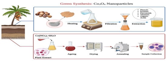
Figure 1.
Schematic diagram of green synthesis of Co3O4 NPs.
2.4. Characterization of H. thebaica Co3O4 NPs
After the synthesis of Co3O4 NPs, the nanoparticles were characterized using various techniques. The crystalline structure of the synthesized nanoparticles was analyzed using X-ray diffraction (XRD). XRD patterns were recorded in the 2θ range of 20–70°. The Debye–Scherrer equation was used to estimate crystallite sizes based on the width at half maxima. In order to find the functional groups, present in the samples, an FTIR analysis was carried out within the region of 4000–400 cm−1. The surface morphology and shape of the Co3O4 NPs were confirmed using scanning electron microscopy coupled with energy-dispersive X-ray spectroscopy (SEM-EDX). The internal morphology, size distribution, and particle size analysis of the nanoparticles were carried out using transmission electron microscopy (TEM). Image J software (1.53v) was used to perform TEM diameter measurements. Finally, the optical properties were characterized using the Raman spectroscopy technique by a Thermo Fisher DXR with a laser wavelength of 780 nm and a spot size of 0.5 nm.
2.5. Photocatalytic Tests
The photocatalytic activity of the synthesized Co3O4 NPs was evaluated by degrading methylene blue dye. One hundred milliliters of 10 ppm methylene blue (M.B.) aqueous solution was used for the photocatalytic experiment. A total of 1 g/L of the Co3O4 NPs was added to M.B. aqueous solution and stirred with a magnetic stirrer in the dark for 30 min to establish adsorption–desorption equilibrium between the solution and catalyst before irradiation from the 400 W high-pressure mercury lamp (λ ≥ 420 nm). The lamp was turned on, and after particular time intervals (0, 25, 50, 75, 100, 125, 150, and 175 min), the samples were filtered using centrifugation to settle down the suspended catalyst and then examined by a UV–visible spectrophotometer in the range of 400–800 nm. The absorbance measurements estimated the degradation degree of M.B. in the aqueous solution at about 665 nm. All the aqueous samples were at neutral pH, and all experiments were carried out at room temperature.
3. Results and Discussions
3.1. XRD Analysis
X-ray diffraction patterns (XRD) for samples of H. thebaica Co3O4 NPs were carried out using a Bruker Advanced D8 diffractometer with monochromatic Cu Kα radiation of wavelength 1.5406 Å operating at a current of 40 mA and a voltage of 40 kV in the Bragg–Brentano geometry. The observed intensity peaks at 31.50°, 37.05°, 38.78°, 44.98°, and 59.53° represent the corresponding planes at (220), (311), (222), (400), (511), respectively. These sharp peaks confirm that the synthesized Co3O4 NPs are crystalline in structure as shown in Figure 2. The fabricated Co3O4 NPs diffraction 2θ values correspond to the face-centered cubic crystalline phase of cobalt oxide (JCPDS card 073–1701) [68]. The average crystallite size of Co3O4 NPs was calculated by using Debye–Scherrer’s formula.
where λ is the wavelength of Cu-kα radiation, β is the full-width half-maximum (FWHM) in radians, and θ is the angle of diffraction (in radians). The average particle size and the lattice parameter (a = b = c) were found to be 11.87 nm and 8.082 Ǻ, respectively. No peaks of contaminants were observed, indicating the pure crystal structure of Co3O4. Thus, XRD confirms the formation of Co3O4 NPs with desired crystalline structure.
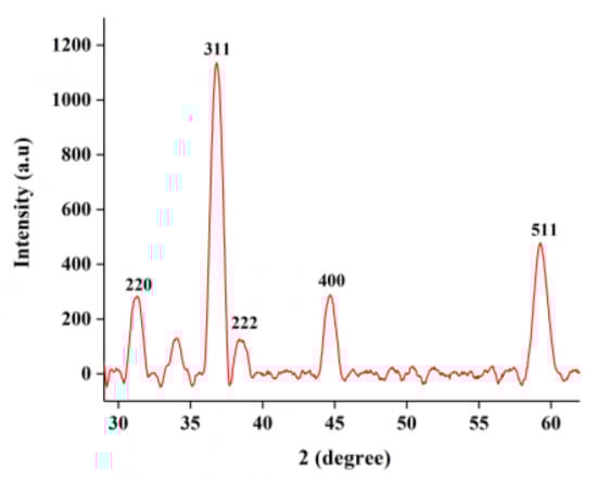
Figure 2.
XRD analysis of H. thebaica Co3O4 NPs.
3.2. Microscopic Analysis
Scanning electron microscopy (SEM)—electron dispersive X-ray spectroscopy (EDS) was carried out on a Tescan MIRA3 GMU Univac SEM with a secondary electron detector and Drawbeam advanced/offline software to examine the Co3O4 NPs’ morphology and structural configuration. Figure 3a,b depicts stable hollow spherical entities which consist of interconnected nanoparticles. It was observed that the Co3O4 NPs spontaneously arranged themselves in a sieve-like pattern and adhered to the surface, creating interior cavities and loose structures. Agglomeration could also be observed because of the reduced size, greater surface area, and the presence of biomolecules [50]. Image J software (1.53v) was also used to identify the particle size by the histogram, as shown in Figure 3c, with an average diameter of about 42.7 nm.
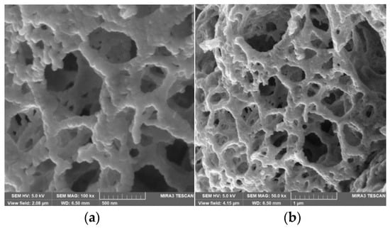
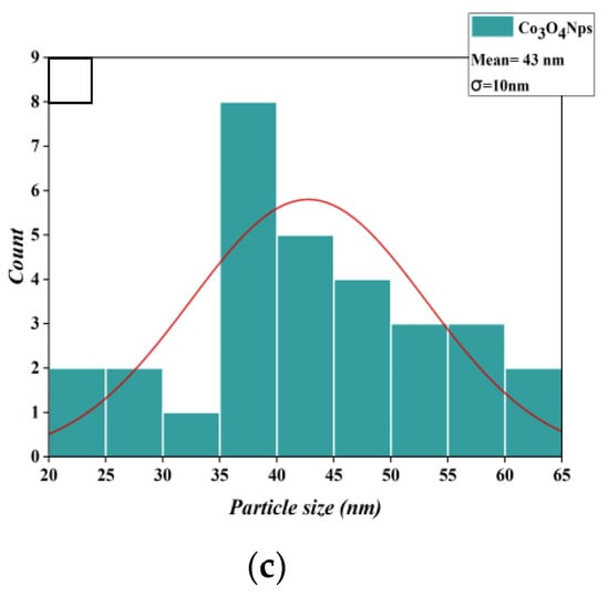
Figure 3.
SEM images of Co3O4 NPs at a magnification of (a) 100 kx and (b) 50 kx. (c) Histogram of Particle Size Distribution Curve.
Moreover, in order to further confirm the pores’ structure, TEM observations were carried out using an FEI Tecnai 20 transmission electron microscope, operating at 200 kV (Lab6 emitter). The high-magnification TEM images (Figure 4a–g) indicate that the pores structure consists of interconnected nanoparticles with an average diameter of about 42.7 nm. In the large area TEM image, it can be seen that a few nanoparticles are aggregated together in the form of chains.
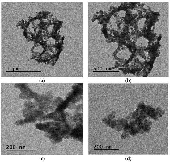
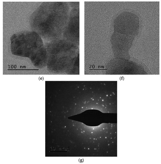
Figure 4.
TEM images of the Co3O4 NPs at a resolution of (a) 1 μm, (b) 500 nm, (c,d) 200 nm, (e) 100 nm, and (f) 20 nm; (g) diffraction pattern.
3.3. Chemical Composition of Co3O4 NPs
Furthermore, EDS was performed parallel to SEM for the elemental analysis of Co3O4 NPs. The EDS range was between 0 and 8 keV. Observed peaks, as depicted in Figure 5, indicate the presence of Co and O. The spectra also reveal the presence of residual chloride ion impurities. Moreover, the spectra depicted the potassium signal due to the presence of bioactive compounds in the extracts. The elemental composition of the nanoparticles shows 72.07 weight percent cobalt and 25.85 weight percent oxygen corresponding to Co3O4. The sharp peaks between 0 and 2 KeV and between 6 and 8 KeV correlate to crystalline Co3O4 NPs. All these characterizations confirm the biosynthesized nanopowder to be Co3O4 NPs, with a porous surface, which enhances the surface area of the biogenic Co3O4 NPs.
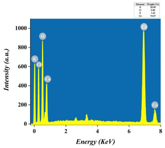
Figure 5.
EDS spectra of Co3O4 NPs.
3.4. Raman Spectroscopy
Raman scattering spectrometer was used to obtain the Raman spectra, which were recorded between 130 and 700 cm−1 (Figure 6). Six Raman bands are identified at 189.3, 345.2, 478.3, 522.6, 615.6, and 655.8 cm−1. According to the research on Co3O4 NPs characterization [69,70], these peaks indicate the Co3O4 spinel structure with an average shift of Δν∼9 cm−1 (191~470~510~608~675 cm−1). This shift may be attributed to surface strain or size effects. The notable Raman peaks correspond to the F2g (189.3 cm−1) Eg (345.2 and 478.3 cm−1), F2g (522.6 and 615 cm−1), A1g (655.8 cm−1) modes of the Co3O4 NPs crystalline phase. The Raman mode at 655.8 cm−1 is attributed to characteristics of the octahedral sites (CoO6) associated with the A1g symmetry, whereas the Eg and F2g modes are likely related to the combined vibrations of the tetrahedral site (CoO4) and octahedral oxygen motions.
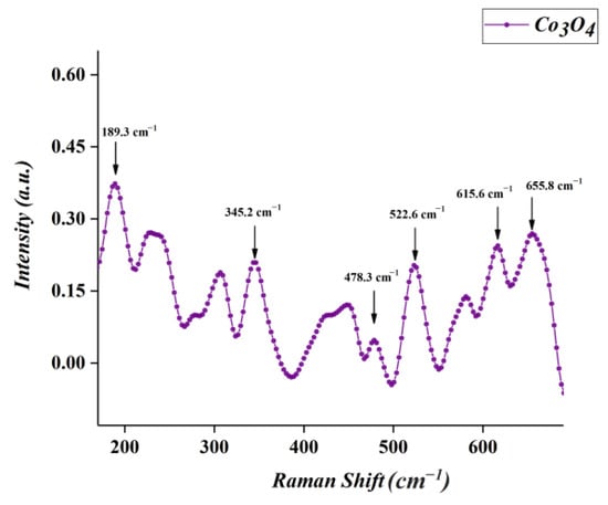
Figure 6.
Raman spectrum of Co3O4 NPs.
3.5. FTIR Analysis
Figure 7 reports the FTIR spectrum of the Co3O4 NPs in the spectral range of 400–4000 cm−1. The major bands at 3128 cm−1, 1624 cm−1, and 1399 cm−1 can be attributed to the hydroxyl, carbonyl, and functional groups of C–N, respectively [71]. The I.R. spectrum displays two strong bands due to vibration υ(Co–O) modes at ~667 cm−1 and ~577 cm−1, which originate from the fingerprint stretching vibrations of the cobalt–oxygen bond of the Co3O4 spinel oxide [59]. More precisely, the first band at 577 cm−1 is a characteristic of OB3 vibration in the spinel lattice, with B signifying the Co3+ in the octahedral site (O–Co), while the 667 cm−1 band is attributed to the ABO3 vibration where A denotes the Co2+ in a tetrahedral hole (Co2+ Co3+O3). In addition, some representative peaks of aromatic rings centered at about 1624 cm−1 and 1399 cm−1 could be attributed to the presence of hydroxyl group (O–H) or C–O stretching of alcohols and carboxylic acids due to the absorption of moisture by the nanoparticles [72]. The peak around 3128 cm−1 is the distinctive peak of the hydroxyl group of polyphenolic compounds. These results were consistent with other reports in which the FTIR spectra revealed the same characteristic peaks of Co3O4 NPs [61].
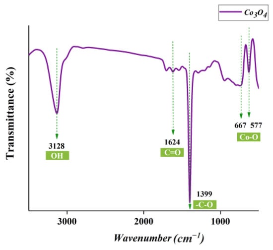
Figure 7.
FTIR spectrum of Co3O4 NPs.
3.6. Photocatalytic Activity
Under visible light irradiation at ambient temperature, the photocatalytic activity of the as-synthesized Co3O4 NPs has been tested for the oxidation of organic dyes such as methylene blue (M.B.). Figure 8 displays the UV–vis spectra of M.B. aqueous solution under visible light irradiation (>420 nm) in the presence of Co3O4 NPs as a photocatalyst over various time intervals. The UV–visible spectrum of M.B. dye showed a maximum absorption peak at 662 nm. However, in the presence of Co3O4 NPs, the absorption peak of the M.B. dye solution decreased with the increase in time due to the continuous photocatalytic degradation of M.B. molecules in the system. The dye color started to fade considerably after 25 min of visible light irradiation, and after 175 min of exposure, the color reduction reached 93.45%. Therefore, the photocatalytic experiments show that the as-prepared Co3O4 NPs have high visible-light photocatalytic activity and can treat dyes in textile wastewater.
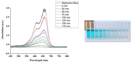
Figure 8.
Evolution of UV–vis absorption spectrum of methylene blue (M.B.) under visible light irradiation using Co3O4 NPs.
The photocatalytic degradation process of Co3O4 NPs involves the use of light energy to initiate a chemical reaction that breaks down organic compounds into smaller, harmless molecules. This is associated with the breakdown of the chromophoric group and the transformation of dye into low-molecular-weight by-products [73]. The dye degradation is mainly due to generating electrons and holes (e− & h+) on the catalyst surface under irradiation [74]. Under visible light, Co3O4 NPs undergo charge separation, and electrons in the valence band of Co3O4 can be excited to its conduction band (e− C.B.), causing the generation of holes in the valance band (h+ V.B.) simultaneously (Equation (2)). Thus, the OH· generated from the reaction (Equation (3)) is the main factor for the photodegradation of the dye because it is a strong oxidizing species, known to be an oxidation process. These ROSs can then react with organic molecules, such as pollutants or contaminants, breaking them down into smaller, less harmful compounds such as carbon dioxide and water.
Co3O4 + hv → e− CB + h+ VB
H2O + h+ VB → H+ + OH
MB + OH·→ degradation products (CO2, H2O, NO3, etc.)
The average C.R. degradation percentage was calculated using the following equation.
where Co was the initial concentration of the M.B. dye and Ct was the concentration of the dye after photocatalytic degradation. To optimize the photocatalytic degradation process of Co3O4 NPs, several factors need to be considered, including the type of organic compound being degraded, the intensity and wavelength of the light source, the size and shape of the nanoparticles, and the presence of other compounds in the environment.
Degradation efficiency (%) = (Co − Ct)/Co × 100
4. Conclusions
In conclusion, Co3O4 NPs with an average particle size of 42.7 nm have been successfully prepared by a cost-effective and eco-friendly green synthesis method using Hyphaene thebaica L. fruit extracts. The prepared nanoparticles were analyzed by various techniques such as FTIR, XRD, Raman, TEM, and SEM. These techniques revealed the successful synthesis of Co3O4 NPs. The photocatalytic activity of synthesized Co3O4 NPs was analyzed. The as-prepared Co3O4 NPs exhibit excellent photocatalytic activity for the degradation of M.B. dye under visible light irradiation. Hence, it can be successfully employed in the treatment of textile and pharmaceutical industrial wastewater to remove the dye.
Author Contributions
Data curation, A.S. and K.H.; Writing—original draft, A.S.; Writing—review & editing, A.M.; Supervision, H.E.A.M. and M.M.; Project administration, M.M. All authors have read and agreed to the published version of the manuscript.
Funding
This research received no external funding.
Institutional Review Board Statement
Not applicable.
Informed Consent Statement
Not applicable.
Data Availability Statement
Data will be made available on request.
Acknowledgments
This research was supported by the UNESCO-UNISA Africa Chair in Nanoscience and Nanotechnology (U2ACN2), the National Research Foundation of South Africa (NRF), the Abdus Salam International Centre for Theoretical Physics (ICTP), iThemba LABS-NRF, and the Abdus Salam International Centre for Theoretical Physics (ICTP) via the Nanosciences African Network (NANOAFENT) to whom we are all grateful.
Conflicts of Interest
The authors declare no conflict of interest.
References
- Bouras, D.; Fellah, M.; Mecif, A.; Barillé, R.; Obrosov, A.; Rasheed, M. High Photocatalytic Capacity of Porous Ceramic-Based Powder Doped with MgO. J. Korean Ceram. Soc. 2023, 60, 155–168. [Google Scholar] [CrossRef]
- Riaz, S.; Ikram, M.; Naz, S.; Shahzadi, A.; Nabgan, W.; Ul-Hamid, A.; Haider, A.; Haider, J.; Al-Shanini, A. Bactericidal Action and Industrial Dye Degradation of Graphene Oxide and Polyacrylic Acid-Doped SnO2 Quantum Dots: In Silico Molecular Docking Study. ACS Omega 2023, 8, 5808–5819. [Google Scholar] [CrossRef] [PubMed]
- Rafiq, A.; Imran, M.; Aqeel, M.; Naz, M.; Ikram, M.; Ali, S. Study of Transition Metal Ion Doped CdS Nanoparticles for Removal of Dye from Textile Wastewater. J. Inorg. Organomet. Polym. Mater. 2020, 30, 1915–1923. [Google Scholar] [CrossRef]
- Qadri, R.; Faiq, M.A. Freshwater Pollution: Effects on Aquatic Life and Human Health. Fresh Water Pollut. Dyn. Remediat. 2020, 15–26. [Google Scholar] [CrossRef]
- Currie, J.; Zivin, J.; Meckel, K.; Neidell, M.; Schlenker, W. Something in the Water: Contaminated Drinking Water and Infant Health. Can. J. Econ. Can. Déconomique 2013, 46, 791–810. [Google Scholar] [CrossRef]
- Haseena, M.; Malik, M.; Javed, A.; Arshad, S.; Asif, N.; Zulfiqar, S.; Hanif, J. Water Pollution and Human Health. Environ. Risk Assess. Remediat. 2017, 1, 16–19. [Google Scholar] [CrossRef]
- Bharagava, R.N.; Saxena, G.; Mulla, S.I. Introduction to Industrial Wastes Containing Organic and Inorganic Pollutants and Bioremediation Approaches for Environmental Management. In Bioremediation of Industrial Waste for Environmental Safety; Springer: Berlin/Heidelberg, Germany, 2020; pp. 1–18. [Google Scholar]
- Khan, S.; Malik, A. Toxicity Evaluation of Textile Effluents and Role of Native Soil Bacterium in Biodegradation of a Textile Dye. Environ. Sci. Pollut. Res. 2018, 25, 4446–4458. [Google Scholar] [CrossRef]
- Ansari, A.; Siddiqui, V.U.; Rehman, W.U.; Akram, M.K.; Siddiqi, W.A.; Alosaimi, A.M.; Hussein, M.A.; Rafatullah, M. Green Synthesis of TiO2 Nanoparticles Using Acorus Calamus Leaf Extract and Evaluating Its Photocatalytic and in Vitro Antimicrobial Activity. Catalysts 2022, 12, 181. [Google Scholar] [CrossRef]
- Madhav, S.; Ahamad, A.; Singh, P.; Mishra, P.K. A Review of Textile Industry: Wet Processing, Environmental Impacts, and Effluent Treatment Methods. Environ. Qual. Manag. 2018, 27, 31–41. [Google Scholar] [CrossRef]
- Thakur, S.; Chauhan, M.S. Treatment of Dye Wastewater from Textile Industry by Electrocoagulation and Fenton Oxidation: A Review. In Water Quality Management: Select Proceedings of ICWEES-2016; Springer: Berlin/Heidelberg, Germany, 2018; pp. 117–129. [Google Scholar]
- Bouras, D.; Rasheed, M.; Barille, R.; Aldaraji, M.N. Efficiency of Adding DD3+ (Li/Mg) Composite to Plants and Their Fibers during the Process of Filtering Solutions of Toxic Organic Dyes. Opt. Mater. 2022, 131, 112725. [Google Scholar] [CrossRef]
- Siddiqui, V.U.; Ansari, A.; Ansari, M.T.; Akram, M.K.; Siddiqi, W.A.; Alosaimi, A.M.; Hussein, M.A.; Rafatullah, M. Optimization of Facile Synthesized ZnO/CuO Nanophotocatalyst for Organic Dye Degradation by Visible Light Irradiation Using Response Surface Methodology. Catalysts 2021, 11, 1509. [Google Scholar] [CrossRef]
- Lu, F.; Astruc, D. Nanocatalysts and Other Nanomaterials for Water Remediation from Organic Pollutants. Coord. Chem. Rev. 2020, 408, 213180. [Google Scholar] [CrossRef]
- Uddin, F. Environmental Hazard in Textile Dyeing Wastewater from Local Textile Industry. Cellulose 2021, 28, 10715–10739. [Google Scholar] [CrossRef]
- Siddiqui, V.U.; Ansari, A.; Ansari, M.T.; Akram, M.K.; Siddiqi, W.A. Fabrication of a Zinc Oxide/Alginate (ZnO/Alg) Bionanocomposite for Enhanced Dye Degradation and Its Optimization Study. RSC Adv. 2022, 12, 7210–7228. [Google Scholar] [CrossRef] [PubMed]
- Anandan, S.; Kumar Ponnusamy, V.; Ashokkumar, M. A Review on Hybrid Techniques for the Degradation of Organic Pollutants in Aqueous Environment. Ultrason. Sonochem. 2020, 67, 105130. [Google Scholar] [CrossRef]
- Abdi, J.; Vossoughi, M.; Mahmoodi, N.M.; Alemzadeh, I. Synthesis of Metal-Organic Framework Hybrid Nanocomposites Based on GO and CNT with High Adsorption Capacity for Dye Removal. Chem. Eng. J. 2017, 326, 1145–1158. [Google Scholar] [CrossRef]
- Bhatia, D.; Sharma, N.R.; Singh, J.; Kanwar, R.S. Biological Methods for Textile Dye Removal from Wastewater: A Review. Crit. Rev. Environ. Sci. Technol. 2017, 47, 1836–1876. [Google Scholar] [CrossRef]
- Nuengmatcha, P.; Chanthai, S.; Mahachai, R.; Oh, W.-C. Sonocatalytic Performance of ZnO/Graphene/TiO2 Nanocomposite for Degradation of Dye Pollutants (Methylene Blue, Texbrite BAC-L, Texbrite BBU-L and Texbrite NFW-L) under Ultrasonic Irradiation. Dyes Pigments 2016, 134, 487–497. [Google Scholar] [CrossRef]
- Salami, R.; Amini, M.; Bagherzadeh, M.; Hosseini, H. Vanadium Supported on Spinel Cobalt Ferrite Nanoparticles as an Efficient and Magnetically Recoverable Catalyst for Oxidative Degradation of Methylene Blue. Appl. Organomet. Chem. 2019, 33, e5127. [Google Scholar] [CrossRef]
- Molina Higgins, M.; Toro González, M.; Rojas, J. Enhanced X-RAYS Degradation of Methylene Blue in the Presence of Gold Microspheres. Radiat. Phys. Chem. 2019, 156, 73–80. [Google Scholar] [CrossRef]
- Edison, T.N.J.I.; Atchudan, R.; Kamal, C.; Lee, Y.R. Caulerpa Racemosa: A Marine Green Alga for Eco-Friendly Synthesis of Silver Nanoparticles and Its Catalytic Degradation of Methylene Blue. Bioprocess Biosyst. Eng. 2016, 39, 1401–1408. [Google Scholar] [CrossRef]
- Brillas, E.; Martínez-Huitle, C.A. Decontamination of Wastewaters Containing Synthetic Organic Dyes by Electrochemical Methods. An Updated Review. Appl. Catal. B Environ. 2015, 166, 603–643. [Google Scholar] [CrossRef]
- Vasiljevic, Z.Z.; Dojcinovic, M.P.; Vujancevic, J.D.; Jankovic-Castvan, I.; Ognjanovic, M.; Tadic, N.B.; Stojadinovic, S.; Brankovic, G.O.; Nikolic, M.V. Photocatalytic Degradation of Methylene Blue under Natural Sunlight Using Iron Titanate Nanoparticles Prepared by a Modified Sol–Gel Method. R. Soc. Open Sci. 2020, 7, 200708. [Google Scholar] [CrossRef] [PubMed]
- Tichapondwa, S.M.; Newman, J.P.; Kubheka, O. Effect of TiO2 Phase on the Photocatalytic Degradation of Methylene Blue Dye. Phys. Chem. Earth Parts ABC 2020, 118, 102900. [Google Scholar] [CrossRef]
- Cheng, M.; Zeng, G.; Huang, D.; Lai, C.; Xu, P.; Zhang, C.; Liu, Y. Hydroxyl Radicals Based Advanced Oxidation Processes (AOPs) for Remediation of Soils Contaminated with Organic Compounds: A Review. Chem. Eng. J. 2016, 284, 582–598. [Google Scholar]
- Chan, S.H.S.; Yeong Wu, T.; Juan, J.C.; Teh, C.Y. Recent Developments of Metal Oxide Semiconductors as Photocatalysts in Advanced Oxidation Processes (AOPs) for Treatment of Dye Waste-water. J. Chem. Technol. Biotechnol. 2011, 86, 1130–1158. [Google Scholar] [CrossRef]
- Zamora-Ledezma, C.; Negrete-Bolagay, D.; Figueroa, F.; Zamora-Ledezma, E.; Ni, M.; Alexis, F.; Guerrero, V.H. Heavy Metal Water Pollution: A Fresh Look about Hazards, Novel and Conventional Remediation Methods. Environ. Technol. Innov. 2021, 22, 101504. [Google Scholar] [CrossRef]
- Babar, U.D.; Garad, N.M.; Mohite, A.A.; Babar, B.M.; Shelke, H.D.; Kamble, P.D.; Pawar, U.T. Study the Photovoltaic Performance of Pure and Cd-Doped ZnO Nanoparticles Prepared by Reflux Method. Mater. Today Proc. 2021, 43, 2780–2785. [Google Scholar] [CrossRef]
- Shamaila, S.; Sajjad, A.K.L.; Farooqi, S.A.; Jabeen, N.; Majeed, S.; Farooq, I. Advancements in Nanoparticle Fabrication by Hazard Free Eco-Friendly Green Routes. Appl. Mater. Today 2016, 5, 150–199. [Google Scholar] [CrossRef]
- Buledi, J.A.; Amin, S.; Haider, S.I.; Bhanger, M.I.; Solangi, A.R. A Review on Detection of Heavy Metals from Aqueous Media Using Nanomaterial-Based Sensors. Environ. Sci. Pollut. Res. 2021, 28, 58994–59002. [Google Scholar] [CrossRef] [PubMed]
- Iravani, S.; Varma, R. Sustainable Synthesis of Cobalt and Cobalt Oxide Nanoparticles and Their Catalytic and Biomedical Applications. Green Chem. 2020, 22, 2643–2661. [Google Scholar] [CrossRef]
- Hanif, M.A.; Kim, Y.-S.; Akter, J.; Kim, H.G.; Kwac, L.K. Fabrication of Robust and Stable N-Doped ZnO/Single-Walled Carbon Nanotubes: Characterization, Photocatalytic Application, Kinetics, Degradation Products, and Toxicity Analysis. ACS Omega 2023, 8, 16174–16185. [Google Scholar] [CrossRef] [PubMed]
- Maeda, K.; Ishimaki, K.; Tokunaga, Y.; Lu, D.; Eguchi, M. Modification of Wide-Band-Gap Oxide Semiconductors with Cobalt Hydroxide Nanoclusters for Visible-Light Water Oxidation. Angew. Chem. Int. Ed. 2016, 55, 8309–8313. [Google Scholar] [CrossRef]
- El-Shamy, O.; Deyab, M. The Most Popular and Effective Synthesis Processes for Co3O4 Nanoparticles and Their Benefit in Preventing Corrosion. Z. Phys. Chem.-Int. J. Res. Phys. Chem. Chem. Phys. 2023, 237, 333–350. [Google Scholar] [CrossRef]
- Abu-Zied, B.M.; Alamry, K.A. Green Synthesis of 3D Hierarchical Nanostructured Co3O4/Carbon Catalysts for the Application in Sodium Borohydride Hydrolysis. J. Alloys Compd. 2019, 798, 820–831. [Google Scholar] [CrossRef]
- Mohammadi, S.Z.; Lashkari, B.; Khosravan, A. Green Synthesis of Co3O4 Nanoparticles by Using Walnut Green Skin Extract as a Reducing Agent by Using Response Surface Methodology. Surf. Interfaces 2021, 23, 100970. [Google Scholar]
- Amanulla, A.M.; Shahina, S.J.; Sundaram, R.; Magdalane, C.M.; Kaviyarasu, K.; Letsholathebe, D.; Mohamed, S.B.; Kennedy, J.; Maaza, M. Antibacterial, Magnetic, Optical and Humidity Sensor Studies of β-CoMoO4-Co3O4 Nanocomposites and Its Synthesis and Characterization. J. Photochem. Photobiol. B 2018, 183, 233–241. [Google Scholar] [CrossRef]
- Hafeez, M.; Shaheen, R.; Akram, B.; Haq, S.; Mahsud, S.; Ali, S.; Khan, R.T. Green Synthesis of Cobalt Oxide Nanoparticles for Potential Biological Applications. Mater. Res. Express 2020, 7, 025019. [Google Scholar] [CrossRef]
- Waris, A.; Din, M.; Ali, A.; Afridi, S.; Baset, A.; Khan, A.; Ali, M. Green Fabrication of Co and Co3O4 Nanoparticles and Their Biomedical Applications: A Review. Open Life Sci. 2021, 16, 14–30. [Google Scholar] [CrossRef]
- Sonkusare, V.N.; Chaudhary, R.G.; Bhusari, G.S.; Mondal, A.; Potbhare, A.K.; Mishra, R.K.; Juneja, H.D.; Abdala, A.A. Mesoporous Octahedron-Shaped Tricobalt Tetroxide Nanoparticles for Photocatalytic Degradation of Toxic Dyes. ACS Omega 2020, 5, 7823–7835. [Google Scholar] [CrossRef]
- Aziz, A.; Ali, N.; Khan, A.; Bilal, M.; Malik, S.; Ali, N.; Khan, H. Chitosan-zinc Sulfide Nanoparticles, Characterization and Their Photocatalytic Degradation Efficiency for Azo Dyes. Int. J. Biol. Macromol. 2020, 153, 502–512. [Google Scholar] [CrossRef] [PubMed]
- Li, H.; Hao, M.-X.; Kang, H.-R.; Chu, L.-Q. Facile Production of Three-Dimensional Chitosan Fiber Embedded with Zinc Oxide as Recoverable Photocatalyst for Organic Dye Degradation. Int. J. Biol. Macromol. 2021, 181, 150–159. [Google Scholar] [CrossRef]
- Hkiri, K.; Mohamed, H.E.A.; Ben Salem, M.; Kouki, A.; Maaza, M.; Zouaoui, M. Biosynthesis and Characterization of CaZrO3 Nanoparticles via Hyphaene Thebaica: Effect of Preparation Method on Morphology, Electrical, and Dielectric Properties. J. Mater. Sci. Mater. Electron. 2020, 31, 10018–10030. [Google Scholar] [CrossRef]
- Khan, Z.U.H.; Khan, A.; Chen, Y.; Shah, N.S.; Muhammad, N.; Khan, A.U.; Tahir, K.; Khan, F.U.; Murtaza, B.; Hassan, S.U. Biomedical Applications of Green Synthesized Nobel Metal Nanoparticles. J. Photochem. Photobiol. B 2017, 173, 150–164. [Google Scholar] [CrossRef]
- Asif, M. Chemistry and Antioxidant Activity of Plants Containing Some Phenolic Compounds. Chem. Int. 2015, 1, 35–52. [Google Scholar]
- Asif, M. Antiviral and Antiparasitic Activities of Various Substituted Triazole Derivatives: A Mini Review. Chem. Int. 2015, 1, 71–80. [Google Scholar]
- Salam, M.A.; AbuKhadra, M.R.; Mohamed, A.S. Effective Oxidation of Methyl Parathion Pesticide in Water over Recycled Glass Based-MCM-41 Decorated by Green Co3O4 Nanoparticles. Environ. Pollut. 2020, 259, 113874. [Google Scholar] [CrossRef]
- Parashar, M.; Shukla, V.K.; Singh, R. Metal Oxides Nanoparticles via Sol–Gel Method: A Review on Synthesis, Characterization and Applications. J. Mater. Sci. Mater. Electron. 2020, 31, 3729–3749. [Google Scholar]
- Govindasamy, R.; Raja, V.; Singh, S.; Govindarasu, M.; Sabura, S.; Rekha, K.; Rajeswari, V.D.; Alharthi, S.S.; Vaiyapuri, M.; Sudarmani, R. Green Synthesis and Characterization of Cobalt Oxide Nanoparticles Using Psidium Guajava Leaves Extracts and Their Photocatalytic and Biological Activities. Molecules 2022, 27, 5646. [Google Scholar] [CrossRef]
- El-Sayed, E.-S.R.; Abdelhakim, H.K.; Zakaria, Z. Extracellular Biosynthesis of Cobalt Ferrite Nanoparticles by Monascus Purpureus and Their Antioxidant, Anticancer and Antimicrobial Activities: Yield Enhancement by Gamma Irradiation. Mater. Sci. Eng. C 2020, 107, 110318. [Google Scholar]
- Saeed, S.; Raees, L.; Mukhtiar, A.; Khan, F.; Khan, M.; Shah, S.; Ghafoor, D.; Mazhar, K. Green Synthesis of Cobalt Oxide Nanoparticles Using Roots Extract of Ziziphus Oxyphylla Edegew Its Characterization and Antibacterial Activity. Mater. Res. Express 2022, 9, 105001. [Google Scholar] [CrossRef]
- Akhlaghi, N.; Najafpour-Darzi, G.; Younesi, H. Facile and Green Synthesis of Cobalt Oxide Nanoparticles Using Ethanolic Extract of Trigonella Foenumgraceum (Fenugreek) Leaves. Adv. Powder Technol. 2020, 31, 3562–3569. [Google Scholar] [CrossRef]
- Joy Prabu, H.; Johnson, I. Plant-Mediated Biosynthesis and Characterization of Silver Nanoparticles by Leaf Extracts of Tragia Involucrata, Cymbopogon Citronella, Solanum Verbascifolium and Tylophora Ovata. Karbala Int. J. Mod. Sci. 2015, 1, 237–246. [Google Scholar] [CrossRef]
- Samuel, M.S.; Selvarajan, E.; Mathimani, T.; Santhanam, N.; Phuong, T.N.; Brindhadevi, K.; Pugazhendhi, A. Green Synthesis of Cobalt-Oxide Nanoparticle Using Jumbo Muscadine (Vitis rotundifolia): Characterization and Photo-Catalytic Activity of Acid Blue-74. J. Photochem. Photobiol. B 2020, 211, 112011. [Google Scholar] [CrossRef]
- Mittal, A.K.; Chisti, Y.; Banerjee, U.C. Synthesis of Metallic Nanoparticles Using Plant Extracts. Biotechnol. Adv. 2013, 31, 346–356. [Google Scholar] [CrossRef] [PubMed]
- Fazlzadeh, M.; Rahmani, K.; Zarei, A.; Abdoallahzadeh, H.; Nasiri, F.; Khosravi, R. A Novel Green Synthesis of Zero Valent Iron Nanoparticles (NZVI) Using Three Plant Extracts and Their Efficient Application for Removal of Cr(VI) from Aqueous Solutions. Adv. Powder Technol. 2017, 28, 122–130. [Google Scholar] [CrossRef]
- Rajeswari, V.D.; Khalifa, A.S.; Elfasakhany, A.; Badruddin, I.A.; Kamangar, S.; Brindhadevi, K. Green and Ecofriendly Synthesis of Cobalt Oxide Nanoparticles Using Phoenix dactylifera L.: Antimicrobial and Photocatalytic Activity. Appl. Nanosci. 2021, 13, 1367–1375. [Google Scholar] [CrossRef]
- Dubey, S.; Kumar, J.; Kumar, A.; Sharma, Y.C. Facile and Green Synthesis of Highly Dispersed Cobalt Oxide (Co3O4) Nano Powder: Characterization and Screening of Its Eco-Toxicity. Adv. Powder Technol. 2018, 29, 2583–2590. [Google Scholar] [CrossRef]
- Bibi, I.; Nazar, N.; Iqbal, M.; Kamal, S.; Nawaz, H.; Nouren, S.; Safa, Y.; Jilani, K.; Sultan, M.; Ata, S. Green and Eco-Friendly Synthesis of Cobalt-Oxide Nanoparticle: Characterization and Photo-Catalytic Activity. Adv. Powder Technol. 2017, 28, 2035–2043. [Google Scholar] [CrossRef]
- Mohamed, H.E.A.; Afridi, S.; Khalil, A.T.; Zohra, T.; Alam, M.M.; Ikram, A.; Shinwari, Z.K.; Maaza, M. Phytosynthesis of BiVO4 Nanorods Using Hyphaene Thebaica for Diverse Biomedical Applications. AMB Express 2019, 9, 200. [Google Scholar] [CrossRef]
- El-Beltagi, H.S.; Mohamed, H.I.; Yousef, H.N.; Fawzi, E.M. Biological Activities of the Doum Palm (Hyphaene thebaica L.) Extract and Its Bioactive Components. In Antioxidants in Foods and its Applications; IntechOpen: London, UK, 2018; Volume 49. [Google Scholar]
- Auwal, M.S.; Shuaibu, A.; Lawan, F.A.; Sanda, K.A.; Njobdi, A.B.; Ibrahim, A.; Gulani, I.A.; Wampana, B.; Lateefat, G.I.; Kibon, Y. Effect of Crude Mesocarp Extract of Hyphaene Thebaica (Doumpalm) on White Blood Cells and Differential Leucocytic Count in Wistar Albino Rats. J. Med. Sci. 2012, 12, 207. [Google Scholar] [CrossRef]
- Sone, B.T.; Makamu, E.; Mohamed, H.E.A.; Oputu, O.; Fester, V. Green-Synthesized ZnO via Hyphaene Thebaica Fruit Extracts: Structure & Catalytic Effect on the Ozonation of Coralene Rubine-S2G Azo Disperse Dye. Environ. Nanotechnol. Monit. Manag. 2021, 16, 100515. [Google Scholar] [CrossRef]
- Abdulazeez, M.A.; Bashir, A.; Adoyi, B.S.; Mustapha, A.Z.; Kurfi, B.; Usman, A.Y.; Bala, R.K. Antioxidant, Hypolipidemic and Angiotensin Converting Enzyme Inhibitory Effects of Flavonoid-Rich Fraction of Hyphaene Thebaica (Doum Palm) Fruits on Fat-Fed Obese Wistar Rats. Asian J. Res. Biochem. 2019, 5, 1–11. [Google Scholar] [CrossRef]
- Mohamed, H.E.A.; Thema, T.; Dhlamini, M.S. Green Synthesis of CuO Nanoparticles via Hyphaene Thebaica Extract and Their Optical Properties. Mater. Today Proc. 2021, 36, 591–594. [Google Scholar] [CrossRef]
- Memon, S.A.; Hassan, D.; Buledi, J.A.; Solangi, A.R.; Memon, S.Q.; Palabiyik, I.M. Plant Material Protected Cobalt Oxide Nanoparticles: Sensitive Electro-Catalyst for Tramadol Detection. Microchem. J. 2020, 159, 105480. [Google Scholar] [CrossRef]
- Rashad, M.; Rüsing, M.; Berth, G.; Lischka, K.; Pawlis, A. CuO and Co3O4 Nanoparticles: Synthesis, Characterizations, and Raman Spectroscopy. J. Nanomater. 2013, 2013, 82. [Google Scholar] [CrossRef]
- Gawali, S.R.; Gandhi, A.C.; Gaikwad, S.S.; Pant, J.; Chan, T.-S.; Cheng, C.-L.; Ma, Y.-R.; Wu, S.Y. Role of Cobalt Cations in Short Range Antiferromagnetic Co3O4 Nanoparticles: A Thermal Treatment Approach to Affecting Phonon and Magnetic Properties. Sci. Rep. 2018, 8, 249. [Google Scholar] [CrossRef]
- Mulya Dewi, N.O.; Yulizar, Y.; Bagus Apriandanu, D.O. Green Synthesis of Co3O4 Nanoparticles Using Euphorbia heterophylla L. Leaves Extract: Characterization and Photocatalytic Activity. In IOP Conference Series: Materials Science and Engineering; IOP Publishing: Bristol, UK, 2019; Volume 509, p. 012105. [Google Scholar]
- Bhargava, R.; Khan, S.; Ahmad, N.; Ansari, M.M.N. Investigation of Structural, Optical and Electrical Properties of Co3O4 Nanoparticles. In AIP Conference Proceedings; AIP Publishing: Melville, NY, USA, 2018; Volume 1953. [Google Scholar]
- Nguyen, N.T.; Nguyen, V.A. Synthesis, Characterization, and Photocatalytic Activity of ZnO Nanomaterials Prepared by a Green, Nonchemical Route. J. Nanomater. 2020, 2020, 1768371. [Google Scholar] [CrossRef]
- Iqbal, M.; Bhatti, I.A. Gamma Ray/H2O2 Treatment of a Nonylphenol Ethoxylate: Degradation, Cytotoxicity and Mutagenicity Evaluation. J. Hazard. Mater. 2015, 299, 351–360. [Google Scholar] [CrossRef]
Disclaimer/Publisher’s Note: The statements, opinions and data contained in all publications are solely those of the individual author(s) and contributor(s) and not of MDPI and/or the editor(s). MDPI and/or the editor(s) disclaim responsibility for any injury to people or property resulting from any ideas, methods, instructions or products referred to in the content. |
© 2023 by the authors. Licensee MDPI, Basel, Switzerland. This article is an open access article distributed under the terms and conditions of the Creative Commons Attribution (CC BY) license (https://creativecommons.org/licenses/by/4.0/).