Review

From Seeing to Knowing with Artificial Intelligence: A Scoping Review of Point-of-Care Ultrasound in Low-Resource Settings

1
Department of Biomedical Engineering, University of Texas at Austin, Austin, TX 78712, USA
2
Department of Biology, University of Texas at Austin, Austin, TX 78712, USA
3
Department of Informatics, University of Texas at Austin, Austin, TX 78712, USA
4
Department of Neuroscience, University of Texas at Austin, Austin, TX 78712, USA
5
Department of Aerospace Engineering and Engineering Mechanics, University of Texas at Austin, Austin, TX 78712, USA
6
Department of Electrical and Computer Engineering, University of Texas at Austin, Austin, TX 78712, USA
7
Department of BioHealth Informatics, Indiana University-Purdue University Indianapolis, Indianapolis, IN 46202, USA
8
Department of Radiology, Emory University School of Medicine, Atlanta, GA 30307, USA
*
Author to whom correspondence should be addressed.
These authors contributed equally to this work.
Appl. Sci. 2023, 13(14), 8427; https://doi.org/10.3390/app13148427
Submission received: 4 May 2023 / Revised: 5 July 2023 / Accepted: 7 July 2023 / Published: 21 July 2023
(This article belongs to the Special Issue Methods, Applications and Developments in Biomedical Informatics)

Abstract

:
The utilization of ultrasound imaging for early visualization has been imperative in disease detection, especially in the first responder setting. Over the past decade, rapid advancements in the underlying technology of ultrasound have allowed for the development of portable point-of-care ultrasounds (POCUS) with handheld devices. The application of POCUS is versatile, as seen by its use in pulmonary, cardiovascular, and neonatal imaging, among many others. However, despite these advances, there is an inherent inability of translating POCUS devices to low-resource settings (LRS). To bridge these gaps, the implementation of artificial intelligence offers an interesting opportunity. Our work reviews recent applications of POCUS devices within LRS from 2016 to 2023, identifying the most commonly utilized clinical applications and areas where further innovation is needed. Furthermore, we pinpoint areas of POCUS technologies that can be improved using state-of-art artificial intelligence technologies, thus enabling the widespread adoption of POCUS devices in low-resource settings.

1. Introduction

Since the 1950s, ultrasounds have been utilized for various medical applications, continuing to be an invaluable diagnostic resource [1]. The utility and penetration of ultrasound has been increasing, supported by an expanding market with an average annual growth rate of 5.9% [2]. This is further supported by the fact that the global medical ultrasound market is expected to reach USD 8.4 billion in 2023 from USD 6.3 billion in 2018 [2]. While ultrasound equipment is predominantly used in obstetrics, it plays a crucial role in other systems such as the heart, eyes, abdomen, and brain [3]. However, due to the complex and delicate nature of ultrasonography machinery, it must be operated by licensed healthcare professionals or individuals who have undergone extensive training [4]. Given high technology costs and reliable infrastructure (electricity and storage) necessities, the inaccessibility of ultrasound technology stretches further into low-resource settings. Therefore, technological advancements in ultrasound are crucial to optimize its use in low-resource settings [5]. In response, the medical device industry has pivoted its focus over the past decade towards creating ultrasounds that are more accessible to all medical professionals, regardless of certification.
One such innovation has been the development of point-of-care ultrasounds (POCUS), which are more affordable, portable, and have demonstrated similar accuracy when compared with cart-based ultrasound for many clinical applications [6]. As newer versions of POCUS continue to develop, the ability to make complex diagnoses has also improved [7,8]. For example, in emergency medicine, where capturing site-specific imaging is crucial, POCUS is applied to support rapid diagnosis [9]. Moreover, POCUS training is now integrated early in medical education with POCUS replacing the stethoscope for several medical schools in the United States [10]. Outside formal education, several qualitative-based protocols for ultrasound assessments, such as Rapid Ultrasound for Shock and Hypertension (RUSH), Focused Assessment with Sonography for Trauma (FAST), and Abdominal and cardiac evaluation with sonography in shock (ACES) have been incorporated into treatment guidelines [11].
Current knowledge about POCUS has been accumulated from developed countries in resource-rich contexts like academic and tertiary hospitals [12,13,14,15,16,17]. While this is a good starting point, there is a critical need to understand the use of POCUS in rural settings and low-resource settings. Low-resource settings (LRS) are defined as “geographical areas where populations have limited access to qualified healthcare providers and quality healthcare services” [18]. LRS represent populations who can benefit from POCUS innovations since there exists a lack of imaging resources, with almost 50% of patients in rural areas requiring transfer to advanced centers for basic imaging [19]. While POCUS shows promise in LRS, barriers such as limited training, maintenance, and on-site analysis remain challenges [20]. Therefore, POCUS research has been focused on implementing and evaluating training systems to increase the usage of POCUS in practice in LRS [21]. According to Sepulveda-Ortiz et al., while POCUS has been utilized in short-term medical missions, training is necessary to ensure that local providers are able to utilize ultrasound machines [22]. Training with POCUS would improve the continuity of care to local patients and bridge the aforementioned gaps in access to imaging technology [22].
However, training systems present their own set of challenges. The reality is that ultrasound training is costly, time-intensive, difficult to scale up, and challenging to standardize across clinics, especially given the wide variety of applications and user groups involved [23,24]. To address this issue, artificial intelligence (AI) provides a unique potential to fill the gap. AI has been previously employed in telemedicine applications such as remote image guidance, in training methods for operators with minimal traditional ultrasound training, and in the analysis of the vast amount of data generated by ultrasound imaging [25].
Several reviews have focused on the integration of general ultrasound with AI to expand its applications and utilization [26]. Additionally, these reviews mentioned the benefits of AI in LRS where AI can transform POCUS into a continuous monitoring device [27]. Despite the creation of such algorithms as well as their widespread usage and integration with ultrasound technology, minimal progress has been made in applying them to POCUS due to limitations of increased noise, blur, and distortions found in POCUS images, as well as difficulties in saving images to build a database [27]. Self et al. introduced the CALPOCUS project as a protocol to collect data and develop machine learning (ML)-based decision-making tools even for non-expert users [25]. Existing reviews such as the ones performed by Doig et al., Buonsenso et al., and Baloescu et al. do not provide us with a comprehensive understanding of the state of AI-enhanced POCUS in LRS due to the limited scope of the review [28,29] and limited retrieval of the literature [30]. This study aims to conduct a thorough review of the current state of POCUS-based imaging in LRS, examine the utilization of AI-enhanced POCUS in such settings, and explore the future directions necessary to elevate the standard of care in these areas.

2. Materials and Methods

A thorough search was executed to identify literature regarding the state of POCUS in LRS. Locations in the study included low-resource settings from both developed and developing countries. To identify relevant studies, we utilized the following keywords ‘Point-of-care ultrasound’, ‘POCUS’, ‘point of care ultrasound’, ‘LMIC’, ‘Rural’, ‘low-and-middle-income’, ‘developing’, ‘low-and-middle-income countr*’, ‘remote’, ‘limited resource’, and ‘low resource’.
Using these keywords, searches were performed on two electronic databases (SCOPUS and PubMed). The records from these databases were exported to Covidence (Veritas Health Innovation, Melbourne: https://www.covidence.org/, accessed on 17 October 2022) and any duplicates found were removed across these results. Authors (1–11) conducted a two-step screening process to find relevant studies. The first step of screening included a title and abstract screening, which was carried out by all authors (1–11). The screening was based on the inclusion and exclusion criteria shown in Table 1. Only primary studies were considered in this analysis, while case reports, commentaries, discussions, reviews, and editorials were excluded. Additionally, studies were also excluded if they were solely focused on the training required to use POCUS, proof-of-concept, or pilot studies. This approach was to ensure that the resulting studies specifically examined the direct impacts of utilizing POCUS in LRS, rather than solely exploring the potential of POCUS.
Subsequently, studies that passed the initial screening were further screened based on full texts for final eligibility. All conflicts and decisions were confirmed by authors (1–3). The data extraction for all the retained articles was performed utilizing a template constructed in Covidence. Information in data extraction included article title, criteria for the population, study type, purpose of the study, geographical context/study setting, type of ultrasound equipment used, and study results.

3. Results

Through the initial search, the authors identified 7034 articles. After removing duplicates and applying the inclusion and exclusion criteria, 32 studies remained for the scoping review. The PRISMA flowchart is visualized in Figure 1. The data extraction results are shown in Table 2. Most of the studies were observational studies, with 15 prospective observational studies and 2 retrospective observational studies. Furthermore, there were five cross-sectional studies. Other study designs included a non-blinded cluster-randomized control trial, national surveys, and subgroup analyses. The control trial looked at the impact of obstetric ultrasound imaging to decrease intrapartum complications, specifically in pregnant women in rural areas [31]. This trial discovered that portable obstetric ultrasound combined with radio messaging can help increase uptake of antenatal care (ANC) services [31].

4. Setting and Context

Most of the papers reviewed focused on locations in low and middle-income countries (LMICs), low-resource settings (LRS), or rural settings in developed countries. Among these papers, the primary focus was on LRS in African countries, with one classifying South Sudan as an extremely low-resource setting [34], three focusing on the improvement in patient outcomes in Uganda with the usage of portable ultrasound [31,32,38], two based in Sierra Leone [42,43], and two with the setting of Rwanda, both of which classify the setting as an LMIC. More specifically, out of the two papers based in Sierra Leone, one assessed the accuracy of POCUS in obstetric cases [42] and the other examined the impact of POCUS on differential diagnoses at the Holy Spirit Hospital [43]. One of the studies based in Rwanda compared resident-performed lung ultrasound with a clinician-interpreted chest X-ray for pneumothorax in patients after a tube thoracostomy [59]; and the other analyzed the impact of POCUS on diagnosis and diagnostic accuracy in dyspneic patients [61]. Additionally, one of the papers based in Uganda focused on temporary outreach clinics in the Masindi region, West Rural Uganda [32]. The remaining papers included in the review that were based in Africa consisted of Zambia [40], Tanzania [57], Malawi [45], Mozambique [49], and Ethiopia [60].
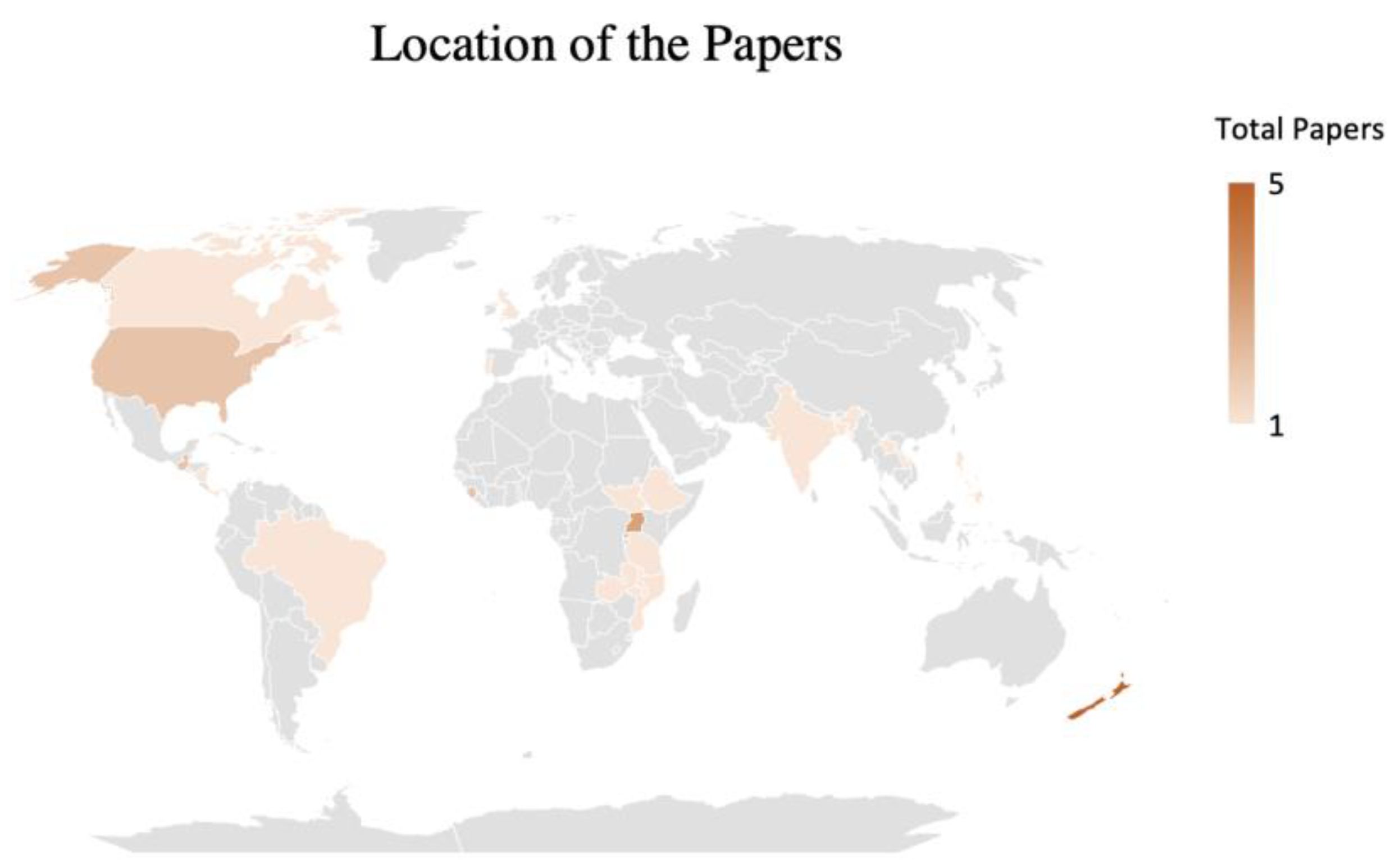
In addition to the studies centered around African countries, other papers were concentrated in Central/South American and Caribbean countries such as Nicaragua [33], Guatemala [35,37], Haiti [39], Panama [47], and Brazil [55]. Furthermore, developed countries with western medicine, who incorporated POCUS into their medical institutions in Portugal [46], United States [48,56], New Zealand (rural hospitals) [50,51,52,53,54], United Kingdom [23], Newfoundland and Labrador, were analyzed [58]. More specifically, all of the New Zealand-based papers that were reviewed selected five to six rural hospitals to examine, many of which were dispersed to represent various communities within the rural areas of New Zealand [53,54,55,56,57]. The rest of the studies contained settings from Asia in countries such as Bangladesh [44], Laos [41], the Philippines [45], and India [23]. The studies carried out in Bangladesh and Laos were from urban areas, with the Bangladesh study focusing on a large tertiary government hospital (Chittagong Medical College, Chittagong, Bangladesh), and the Laos study focusing on Vientiane, the capital of Laos [41,44]. The geographical distribution of the countries included in this study is visualized in Figure 2.
Overall, when analyzing the studies from developed countries, most of them compared the applications and outcomes of POCUS implementation between remote, rural areas to large, urban areas. In every country, most studies were conducted in hospitals or clinical settings, examining outcomes in emergency departments or intensive care units where patients presented with life-threatening conditions.

5. Types of Devices

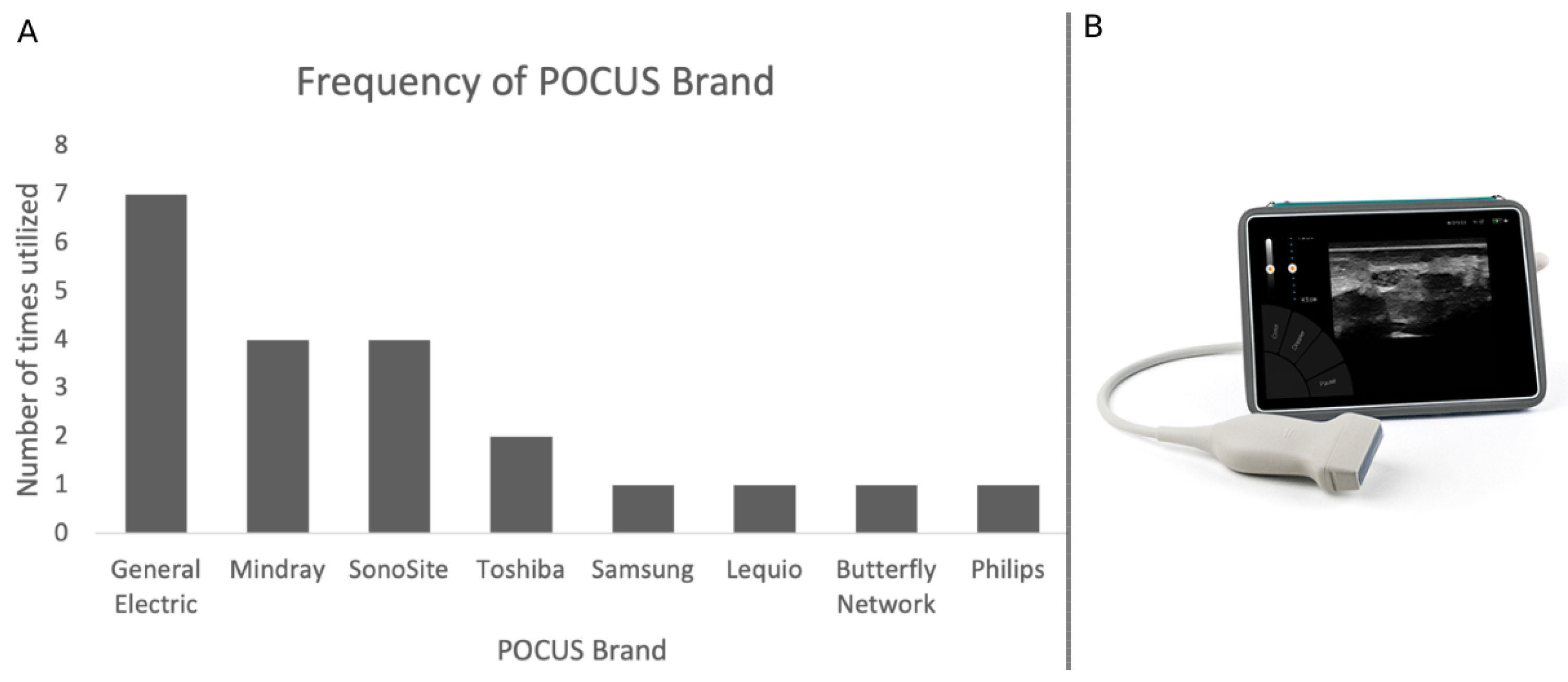
An extensive variety of POCUS devices were utilized in the selected literature. The most common brand across the papers analyzed was General Electric (7/32). Other brands included Mindray (4/32), SonoSite (4/32), Toshiba (2/32), and Phillips Lumify (1/32). The most common devices were the GE Vscan (3/32) and the SonoSite M-Turbo (3/32). Among the POCUS devices used, they were either handheld (4/32), cart-based (8/32), or laptop/tablet-based (9/32).
Some studies utilized more than one POCUS device during screenings, and several studies (13/32) did not mention the POCUS device used. Since many of these studies involved methods in which many hospitals were surveyed, it is difficult to standardize the specific device used due to various resource and budget constraints. Table 2 denotes the device(s) utilized (if any) in each study and their specifications. The frequency of POCUS device brands is depicted in Figure 3.

6. Clinical Applications of POCUS

Based on the studies reviewed, it is evident that the utilization of POCUS is crucial in addressing a broad range of health care issues and is an imperative tool in preventing negative health outcomes, especially in low and middle-income countries. The primary application of POCUS, as supported by most of the reviewed literature, focused on detecting abnormalities in obstetric/gynecologic, cardiac, and pulmonary care. Other examinations targeted the gallbladder, kidneys, gastrointestinal systems, genitourinary systems, and musculoskeletal systems. More specifically, several studies assessed the role and impact of ultrasound as an initial screening tool for individual patient management in various ailments such as tuberculosis, trauma, shock, malaria, dyspnea, among others. POCUS was also utilized to determine the presence of abscesses to support a presumptive diagnosis of melioidosis, pathological findings suggestive of HIV-associated tuberculosis, and the prevalence of early signs of acute respiratory distress syndrome in patients with malaria or sepsis [34,41,44].
Regarding obstetric/gynecologic care, some articles focused on using POCUS for antenatal and maternal care, specifically screening for pregnancy-related abnormalities including vaginal bleeding, pre-eclampsia/eclampsia, prepartum hemorrhage, and causes of fetal/maternal mortality such as fetal malpresentation, placental location, multiple pregnancies, etc. [23,36,42]. Many articles also highlighted the applications of POCUS for cardiac applications, where the commonly sought findings were pericardial effusion, chamber size, left ventricular function, and intravascular volume, inferior vena cava diameter, and jugular venous pressure [52,55].
In addition, POCUS was prevalent in emergency and trauma settings across various applications, utilizing FAST and Extended Focused Assessment with Sonography in Trauma (E-FAST) [46,48,57]. Epstein et al. studied the effectiveness of POCUS on patients in Uganda presenting with trauma casualties, shock, cardiorespiratory symptoms, and those undergoing invasive procedures [38].

7. Outcomes Measured

Across various studies, the outcome measures generally included the number and type of scans performed, the diagnostic results from the scans, and the extent of effectiveness of utilizing POCUS on clinical diagnosis and management, as shown in Table 2. Specifically, some articles focused on implementing training protocols for clinicians at their respective hospitals [35,36]. Upon deeper analysis, it was found that POCUS improves the accuracy and certainty of patient diagnoses in various clinical settings.
To come to this conclusion, a comprehensive review was carried out on the applications and effectiveness of POCUS in the context of each study. Most of the reviewed studies focused on analyzing antenatal and fetal monitoring. Dalmacion et al. posit the efficacy of handheld ultrasound technology in screening pregnancy-related abnormalities related to placental implantation, fetal malpresentation, irregular amniotic fluid volume, and increased fetal numbers [36]. It was also discovered that obstetric ultrasound imaging can help pregnant women in LRS decrease intrapartum complications, screen for risk factors such as vaginal bleeding, pre-eclampsia/eclampsia, and other high-risk pregnancies, and improve antenatal care [23,31,42]. Furthermore, it was found that by utilizing POCUS as a low-cost diagnostic procedure, maternal mortality and neonatal mortality were reduced to 0% and 36%, respectively [35]. Additionally, through the development of a computer-assisted POCUS protocol for pregnant women aimed at creating a dataset suitable for machine learning, Self et al. improved visualization of the cervix from 28% to 91% and classification of placental location from 82% to 94% [23].
Likewise, POCUS has made critical advancements in the diagnosis of many cardiac applications. Nixon et al. found that cardiac scans were the most commonly performed examinations (18% of total scans), while Pellegrini et al. found that the main applications of POCUS were ultrasound-guided central vein catheterization (49.4%) and beside echocardiographic assessment (33.9%) [52,55]. Overall, POCUS offered increased benefits in comparison to traditional physical exams since clinicians had better estimations of central venous pressure and the inferior vena cava measurements [50].
Although Barron et al. found that obstetric and cardiac applications are the most documented, their study focused on lung ultrasound, which was another commonly performed examination [33]. Shumbusho et al. notes an agreement among physicians on utilizing POCUS lung ultrasound as a reasonable alternative for chest X-rays as the sensitivity and specificity of resident lung ultrasound was 100% and 96%, respectively, compared to 48% and 100% for clinician-interpreted chest X-rays [59]. Additionally, lung ultrasounds were significantly faster to obtain than chest X-rays. Another study by Barron et al. found that POCUS examinations demonstrated much quicker response times, from request to performance, in contrast to the radiology department ultrasound. They noted that the average time to perform a POCUS examination was 6.0 min [33].
When examining the gastrointestinal system, POCUS devices assisted in accurately confirming the diagnosis in over three-fourths of cases. Additionally, using POCUS devices allowed physicians to correct their previous diagnoses when there were various non-specific presenting symptoms [36]. POCUS proved useful when used in other applications such as (1) detecting lung abnormalities in patients with malaria and sepsis to identify patients at risk of death, (2) identifying the presence of abscesses in the liver, spleen, and prostate to help identify risk factors for melioidosis, and (3) revealing pathological findings that were suggestive of tuberculosis in 27 out of 100 patients tested [34].

8. Discussion

This review aims to analyze the usage of point-of-care ultrasound (POCUS) in low-resource settings (LRS). Aside from being one of the limited reviews that assess the impact of POCUS on patient diagnosis, treatment, or management across various clinical fields in LRS, our main motivation resides in understanding the existing landscape to guide the potential for AI integration in POCUS applications.
This study specifically focused on the direct utilization of POCUS, excluding the assessment of novel training systems, proof of concept, and pilot studies. Each study targeted a unique combination of clinical applications and geographical considerations. POCUS usage and studies have been steadily increasing in developing countries, along with its integration into the medical school curriculum [24]. However, achieving the full integration of POCUS devices within LRS remains a challenge. A significant number of the studies analyzed in this review were completed in the African continent. Although other studies were performed in regions such as Central America or Asia, further research must be continued in these areas to fully elucidate the true benefits of POCUS in all LRS globally [23,41]. Furthermore, studies are often conducted in underdeveloped regions of developed countries due to the ease of access [52,53]. However, challenges such as accessibility and resource access found within an LRS are unable to be easily replicated in a remote area of a developed region [62].

8.1. Clinical Applications of POCUS in LRS

We examined the various clinical applications and pathologies in which POCUS has been utilized within LRS. The organ systems where POCUS was most utilized were the lungs or in preparation for general prenatal care [23]. For antenatal care in LRS, POCUS devices have been utilized to detect unique or crucial fetal markers such as heartbeats or the fetal head [36,42]. Studies have also been able to highlight the utilization of POCUS in LRS settings to identify pneumonia or diagnose cardiorespiratory symptoms [63,64,65]. For cardiac applications, POCUS devices have been used to evaluate aortic stenosis with anticipation of expanded utility to assess stenosis of other cardiac valves [66]. Direct training using a POCUS device allowed for increased diagnostic accuracy from younger medical students in comparison to traditional cardiac auscultation [67]. Additional case studies have also demonstrated the utility of POCUS devices not only for the cardiac system but also in the vasculature, whether it be for peripheral artery disease or aneurysms [68,69].
In the pulmonary space, POCUS devices have the potential to help elucidate various forms of pleural effusions by evaluating various features such as anechoic fluid, septations, fibrin strands, and the sonographic appearance of the pleural fluid [70,71].
We note that POCUS devices have the potential to be utilized in a much larger context within LRS, extending beyond the applications we discussed. In developed countries, these devices are heavily employed in general practice or even trauma settings to make instantaneous point-of-care decisions crucial to a patient’s life [46,57]. There was only a singular similar study in LRS that reported a 3% negative impact due to the use of POCUS imaging [51]. Nixon et al. specified that POCUS should not be used when other imaging modalities are indicated based on disease presentation; rather it should be used as an additional complimentary technique [51]. Regardless, each of these studies highly suggest the integration of such techniques into the trauma found in LRS could expedite treatment and improve patient care.
Due to its sheer value within acute conditions, POCUS devices show extreme promise in evaluating the retroperitoneal area, where both the pancreas and appendix are found, as well as bowel obstructions [72]. Although not specific to LRS, a review of various primary studies has shown extremely high sensitivity and specificity (>90%) for early appendicitis diagnosis using POCUS [72]. Similarly, POCUS devices have had nearly 100% diagnostic accuracy for evaluating pancreatic pathologies with great promise in early diagnosis of bowel obstructions with sensitivity ranges above 95% and high specificity as well [72,73]. Specifically, POCUS devices have been shown to promptly exact key discoveries such as bowel dilation, altered peristalsis, fluid accumulation, as well as wall thickening and collapsed colonic segments [74].
Another area of POCUS application is to evaluate renal pathology, specifically diagnosis of masses such as cysts and solid tumors. We did not find clearly defined POCUS applications for renal pathology in LRS. Similarly, liver fibrosis is another disease that is largely diagnosed through ultrasonography due to difficulties in retrieving a liver biopsy due to nearby vasculature [75]. POCUS devices are yet another form of ultrasonography that can help differentiate the difference in matrix stiffness between different areas of the liver [75]. Specifically, POCUS would allow for the identification of characteristics such as caudate lobe enlargement or additional streaks surrounding the hepatic portal branches [76].
On a related note, POCUS devices hold exceptional promise in the neurological realm for examining the optic nerve. They serve as a unique non-invasive method that not only detects damage to the optic nerve but also looks at the longstanding effects of increased intraocular pressure such as optic disc swelling [77]. Furthermore, both intraocular pressure and optic disc swelling are highly correlated with intracranial pressure, a crucial metric in managing neurological pathologies [78]. While these applications were not focused in LRS, these examples demonstrate the potential of POCUS to serve as a novel method for point-of-care and a non-invasive method for the detecting various pathologies, thus revolutionizing the state of bedside medicine in LRS.

8.2. Implementation of POCUS in LRS and Barriers

These applications in LRS may face various barriers that hinder the implementation of POCUS, particularly due to limited training and accurate on-site analysis and data extrapolation [29]. Limited training is an ongoing challenge that is actively being explored through training exercises and regiments or telemedicine. A previous study by Vinayak et al. was able to train midwives to utilize POCUS devices alongside mobile phones and tablets to capture high-enough resolution images to identify crucial features within images [79]. Similarly, Kwon et al. found that the majority of eighth graders were able to obtain adequate POCUS (FAST) images, even with minimal training, emphasizing the effectiveness of training and the ease of use of POCUS as an imaging modality [80]. Although the ability to teach the layperson is extremely helpful, these past methodologies rely on the continuous assurance that everyday citizens remember this training. An alternative for such a roadblock is the utilization of telemedicine. Despite limited studies within the realm of LRS POCUS, Wang et al. offered a unique setup revolving around augmented reality telemedicine to allow for the continuous ability for real-time training [81]. Regardless, there has been little research into the further development of ultrasound telemedicine applications [82]. Further research should be encouraged to transition these past studies towards POCUS modalities that can be implemented in low-resource settings.

8.3. Potentials and the Integration of AI-Enhanced POCUS in LRS

Due to the limited training and medical certification available in low-resource settings, diagnosing conditions using POCUS becomes difficult [83]. AI can play a crucial role in improving diagnoses through the integration of machine learning modalities with general ultrasound for pathology detection, feature extraction, and disease diagnosis [81,84]. For example, studies by Prabusankarlal et al. and Singh et al. utilized support vector machine (SVM) and artificial neural network (ANN) learning models, respectively, to diagnose breast masses with around 96% accuracy [85,86]. Similarly, the employment of ANNs and SVM models can help directly diagnose cirrhosis and fatty liver disease, amongst others, in the liver as well as lesion margin in the thyroid with extremely high accuracy [87,88,89]. Additionally, machine learning has the ability to augment current diagnostic procedures within ultrasound technology for commonly seen pathologies such as those found in the heart or lungs [89,90,91].
Despite these advances, there is a significant lack of studies focusing on AI applications in POCUS. While there are advancements in AI-enhanced POCUS and some studies exploring its use in LRS, none of these studies met the inclusion criteria for this review. Nonetheless, there are notable examples of POCUS and AI integration, particularly in the areas of COVID-19 and obstetrics. For instance, Cheema et al. developed a deep learning model that assisted sonographers in obtaining high-quality cardiac ultrasound images in the COVID-19 intensive care unit by utilizing a model trained on the hand movements of skilled cardiac sonographers [92]. Cheema et al. describe a ML-based algorithm that was used to estimate fetal gestational age estimation through POCUS [92]. Similarly, Pokaprakarn et al. developed an AI model that estimated gestational age with an accuracy that performed similarly to trained sonographers using low-cost ultrasound technology with data obtained from volunteers in Zambia and North Carolina [93]. Additionally, Kuroda et al. compared AI-POCUS with CT scans, and they found that AI-POCUS achieved a 94.5% accuracy in detecting CT-validated pneumonia [94]. Blaivas et al. analyzed six common DL image classification algorithms, and found that older and less complex CNN performed the best [95].
One major challenge in developing AI algorithms for POCUS lies in the limited availability of publicly available datasets, and the diversity of POCUS applications and ultrasound equipment makes it hard to standardize datasets [24]. To address this limitation, recent work has employed techniques such as CycleGAN to generate synthetic POCUS images, enabling the classification of breast cancer with a 95% confidence interval for AUC between 93.5 and 96.6 [96].
As AI integration continues to expand in healthcare, we anticipate that these models will likely be adapted for POCUS devices in preparation for their implementation in LRS. Through this comprehensive review, it is evident that POCUS holds significant advantages for healthcare delivery in various LRS across multiple clinical specialties. The integration of AI in POCUS presents a promising opportunity to overcome existing barriers and advance the applications within LRS.
Furthermore, AI-enhanced POCUS plays a crucial role in supporting the growth of telemedicine, a field that has been more increasingly adopted after the disruption of in-person hospital visits during the COVID-19 pandemic. It enables skilled clinicians to provide remote guidance and interpretation of ultrasound images, consequently facilitating remote monitoring and reducing the need for costly specialized visits. Specifically, the use of AI to help improve image quality, regardless of the user’s training level, yields diagnostically relevant images [97]. Additionally, by harnessing the power of augmented reality in combination with artificial intelligence, a remote teleguidance system can be designed to assist novice users with probe placement as well as image interpretation. Furthermore, AI can be used for direct quantification of crucial clinical metrics essential for diagnosis [98]. For instance, AI-enhanced POCUS can be used in cardiac applications for left ventricular ejection fraction, vena cava measurements, or blood velocity time integrals [98]. The system’s real-time image interpretation and analysis is immensely valuable for remote consultations and monitoring. Ultimately, AI-enhanced POCUS empowers healthcare providers in LRS to embrace telemedicine, allowing them to overcome training and geographical barriers and improve access to specialized care.

8.4. Future Perspectives and Research Opportunities

In the future, several strategies can be implemented to leverage AI and advance the state of POCUS in LRS. One approach is to develop training programs that provide personalized feedback to clinicians in LRS that can enhance their proficiency in performing POCUS examinations. Another important focus should be on the implementation of POCUS in local clinics, expanding access to this technology to a broader population. The adoption of implementation protocols similar to CALPOCUS can enhance ultrasound image quality and maximize diagnostic information in scans by providing automated image acquisition and real-time feedback on image quality and probe positioning. Such enhancements would contribute to the development of robust AI models capable of real-time analysis of POCUS data. Since it is not always feasible to collect large amounts of POCUS data across various clinical specialties, using complex deep learning algorithms to generate synthetic POCUS data as described in Karlsson et al. and understanding the differences between general ultrasound and POCUS images to perform image-to-image translations, as introduced in Jafari et al., shows great promise [99].
Although research specifically addressing the combined application of POCUS and AI in LRS remains limited, there is significant potential for growth in this field, and further research is warranted. Considering the existing evidence of POCUS and its associated benefits, it is evident that AI-enhanced POCUS has the potential to revolutionize healthcare delivery, bridge the healthcare disparity gap, and ensure quality care for underserved populations.

Author Contributions

All authors were in charge of the writing and reviewing. N.V. and M.G. were in charge of formalizing the draft. Authors N.V., M.G., A.G., S.N., N.C., K.G., S.P. (Shireen Pathak), K.V., C.S., T.B. and N.M. were in charge of the literature review process. S.P. (Saptarshi Purkayastha) and J.W.G. were in charge of reviewing, editing, and supervising the process. All authors have read and agreed to the published version of the manuscript.

Funding

J.W.G. and S.P. are funded by the US National Science Foundation, (grant number 1928481) from the Division of Electrical, Communication & Cyber Systems.

Institutional Review Board Statement

Not applicable.

Informed Consent Statement

Not applicable.

Data Availability Statement

No data were utilized or created as a result of this study.

Conflicts of Interest

The authors declare no conflict of interest. The funders had no role in the design of the study; in the collection, analyses, or interpretation of data; in the writing of the manuscript; or in the decision to publish the results.

References

  1. Newman, P.G.; Rozycki, G.S. The History of Ultrasound. Surg. Clin. N. Am. 1998, 78, 179–195. [Google Scholar] [CrossRef] [PubMed]
  2. Kim, D.-M.; Park, S.-K.; Park, S.-G. A Study on the Performance Evaluation Criteria and Methods of Abdominal Ultrasound Devices Based on International Standards. Safety 2021, 7, 31. [Google Scholar] [CrossRef]
  3. Carovac, A.; Smajlovic, F.; Junuzovic, D. Application of Ultrasound in Medicine. Acta Inform. Med. 2011, 19, 168. [Google Scholar] [CrossRef] [PubMed] [Green Version]
  4. Hertzberg, B.S.; Kliewer, M.A.; Bowie, J.D.; Carroll, B.A.; DeLong, D.H.; Gray, L.; Nelson, R.C. Physician Training Requirements in Sonography: How Many Cases Are Needed for Competence? Am. J. Roentgenol. 2000, 174, 1221–1227. [Google Scholar] [CrossRef]
  5. Gurara, D.; Klyuev, V.; Mwase, N.; Presbitero, A.; Xu, X.C.; Bannister, M.G.J. Trends and Challenges in Infrastructure Investment in Low-Income Developing Countries. IMF Work. Pap. 2017, 17, 1. [Google Scholar] [CrossRef] [Green Version]
  6. Le, M.-P.T.; Voigt, L.; Nathanson, R.; Maw, A.M.; Johnson, G.; Dancel, R.; Mathews, B.; Moreira, A.; Sauthoff, H.; Gelabert, C.; et al. Comparison of four handheld point-of-care ultrasound devices by expert users. Ultrasound J. 2022, 14, 27. [Google Scholar] [CrossRef]
  7. Marik, P.E.; Linde-Zwirble, W.T.; Bittner, E.A.; Sahatjian, J.; Hansell, D. Fluid administration in severe sepsis and septic shock, patterns and outcomes: An analysis of a large national database. Intensive Care Med. 2017, 43, 625–632. [Google Scholar] [CrossRef]
  8. Atkinson, P.; Bowra, J.; Milne, J.; Lewis, D.; Lambert, M.; Jarman, B.; Noble, V.E.; Lamprecht, H.; Harris, T.; Connolly, J.; et al. International Federation for Emergency Medicine Consensus Statement: Sonography in hypotension and cardiac arrest (SHoC): An international consensus on the use of point of care ultrasound for undifferentiated hypotension and during cardiac arrest. Can. J. Emerg. Med. 2017, 19, 459–470. [Google Scholar] [CrossRef] [Green Version]
  9. Long, B.; Alerhand, S.; Maliel, K.; Koyfman, A. Echocardiography in cardiac arrest: An emergency medicine review. Am. J. Emerg. Med. 2018, 36, 488–493. [Google Scholar] [CrossRef]
  10. Smallwood, N.; Dachsel, M. Point-of-care ultrasound (POCUS): Unnecessary gadgetry or evidence-based medicine? Clin. Med. 2018, 18, 219–224. [Google Scholar] [CrossRef] [Green Version]
  11. Seif, D.; Perera, P.; Mailhot, T.; Riley, D.; Mandavia, D. Bedside Ultrasound in Resuscitation and the Rapid Ultrasound in Shock Protocol. Crit. Care Res. Pract. 2012, 2012, 503254. [Google Scholar] [CrossRef] [PubMed] [Green Version]
  12. Bidner, A.; Bezak, E.; Parange, N. Evaluation of antenatal Point-of-Care Ultrasound (PoCUS) training: A systematic review. Med. Educ. Online 2022, 27, 2041366. [Google Scholar] [CrossRef] [PubMed]
  13. Meinen, R.D.; Bauer, A.S.; Devous, K.; Cowan, E. Point-of-care ultrasound use in umbilical line placement: A review. J. Perinatol. 2020, 40, 560–566. [Google Scholar] [CrossRef]
  14. Shaddock, L.; Smith, T. Potential for Use of Portable Ultrasound Devices in Rural and Remote Settings in Australia and Other Developed Countries: A Systematic Review. J. Multidiscip. Healthc. 2022, 15, 605–625. [Google Scholar] [CrossRef] [PubMed]
  15. Blanco, P.; Buendía, C.M. Point-of-care ultrasound in cardiopulmonary resuscitation: A concise review. J. Ultrasound 2017, 20, 193–198. [Google Scholar] [CrossRef] [PubMed]
  16. Andersen, C.A.; Holden, S.; Vela, J.; Rathleff, M.S.; Jensen, M.B. Point-of-Care Ultrasound in General Practice: A Systematic Review. Ann. Fam. Med. 2019, 17, 61–69. [Google Scholar] [CrossRef] [Green Version]
  17. Kok, B.; Wolthuis, D.; Bosch, F.; van der Hoeven, H.; Blans, M. POCUS in dyspnea, nontraumatic hypotension, and shock; a systematic review of existing evidence. Eur. J. Intern. Med. 2022, 106, 9–38. [Google Scholar] [CrossRef]
  18. World Health Organization. Increasing Access to Health Workers in Remote and Rural Areas through Improved Retention: Global Policy Recommendations; World Health Organization: Geneva, Switzerland, 2010; Volume 71. [Google Scholar]
  19. Micks, T.; Sue, K.; Rogers, P. Barriers to point-of-care ultrasound use in rural emergency departments. Can. J. Emerg. Med. 2016, 18, 475–479. [Google Scholar] [CrossRef] [Green Version]
  20. Hershman, M.; Hershman, M.; Blain, Y.; Moise, M.E.; Monchil, N.; Schmit, B.P. Experience with an ultrasound donation program in a low-income country. J. Glob. Radiol. 2021, 7, 1101. [Google Scholar] [CrossRef]
  21. Ienghong, K.; Cheung, L.W.; Tiamkao, S.; Bhudhisawasdi, V.; Apiratwarakul, K. Development and Remodeling of Point-of-Care Ultrasound Education for Emergency Medicine Residents in Resource Limited Countries during the COVID-19 Pandemic. Tomography 2021, 7, 721–733. [Google Scholar] [CrossRef]
  22. Sepulveda-Ortiz, V.; Warkentine, F.; Starr-Seal, R.; Rominger, A. The effectiveness of a longitudinal ultrasound curriculum for general pediatricians working in a Puerto Rican emergency department: A pilot study. Ultrasound J. 2020, 12, 20. [Google Scholar] [CrossRef] [PubMed] [Green Version]
  23. Self, A.; Chen, Q.; Desiraju, B.K.; Dhariwal, S.; Gleed, A.D.; Mishra, D.; Thiruvengadam, R.; Chandramohan, V.; Craik, R.; Wilden, E.; et al. Developing Clinical Artificial Intelligence for Obstetric Ultrasound to Improve Access in Underserved Regions: Protocol for a Computer-Assisted Low-Cost Point-of-Care UltraSound (CALOPUS) Study. JMIR Res. Protoc. 2022, 11, e37374. [Google Scholar] [CrossRef] [PubMed]
  24. Shokoohi, H.; LeSaux, M.A.; Roohani, Y.H.; Liteplo, A.; Huang, C.; Blaivas, M. Enhanced Point-of-Care Ultrasound Applications by Integrating Automated Feature-Learning Systems Using Deep Learning. J. Ultrasound Med. 2019, 38, 1887–1897. [Google Scholar] [CrossRef]
  25. Recker, F.; Höhne, E.; Damjanovic, D.; Schäfer, V.S. Ultrasound in Telemedicine: A Brief Overview. Appl. Sci. 2022, 12, 958. [Google Scholar] [CrossRef]
  26. Huang, Q.; Zhang, F.; Li, X. Machine Learning in Ultrasound Computer-Aided Diagnostic Systems: A Survey. BioMed Res. Int. 2018, 2018, 5137904. [Google Scholar] [CrossRef] [PubMed]
  27. Brattain, L.J.; Telfer, B.A.; Dhyani, M.; Grajo, J.R.; Samir, A.E. Machine learning for medical ultrasound: Status, methods, and future opportunities. Abdom. Radiol. 2018, 43, 786–799. [Google Scholar] [CrossRef]
  28. Doig, M.; Dizon, J.; Guerrero, K.; Parange, N. Exploring the availability and impact of antenatal point-of-care ultrasound services in rural and remote communities: A scoping review. Australas. J. Ultrasound Med. 2019, 22, 174–185. [Google Scholar] [CrossRef] [Green Version]
  29. Buonsenso, D.; De Rose, C. Implementation of lung ultrasound in low- to middle-income countries: A new challenge global health? Eur. J. Pediatr. 2022, 181, 447–452. [Google Scholar] [CrossRef]
  30. Baloescu, C.; Parhar, A.; Liu, R.; Wanjiku, G.W. Effect of Point-of-Care Ultrasound on Clinical Outcomes in Low-Resource Settings: A Systematic Review. Ultrasound Med. Biol. 2022, 48, 1711–1719. [Google Scholar] [CrossRef]
  31. Cherniak, W.; Anguyo, G.; Meaney, C.; Kong, L.Y.; Malhame, I.; Pace, R.; Sodhi, S.; Silverman, M. Effectiveness of advertising availability of prenatal ultrasound on uptake of antenatal care in rural Uganda: A cluster randomized trial. PLoS ONE 2017, 12, e0175440. [Google Scholar] [CrossRef] [Green Version]
  32. Baker, D.E.; Nolting, L.; Brown, H.A. Impact of point-of-care ultrasound on the diagnosis and treatment of patients in rural Uganda. Trop. Dr. 2021, 51, 291–296. [Google Scholar] [CrossRef] [PubMed]
  33. Barron, K.R.; Lai, J.C.; MenkinSmith, L.P.; Lee, J.S.; Humphrey, M.E.; Hall, J.W. Point-of-Care Ultrasound as Part of a Short-Term Medical Mission to Rural Nicaragua. South. Med. J. 2018, 111, 434–438. [Google Scholar] [CrossRef]
  34. Bobbio, F.; Di Gennaro, F.; Marotta, C.; Kok, J.; Akec, G.; Norbis, L.; Monno, L.; Saracino, A.; Mazzucco, W.; Lunardi, M. Focused ultrasound to diagnose HIV-associated tuberculosis (FASH) in the extremely resource-limited setting of South Sudan: A cross-sectional study. BMJ Open 2019, 9, e027179. [Google Scholar] [CrossRef] [PubMed] [Green Version]
  35. Milart, P.H.C.; Molina, C.A.D.; Prieto-Egido, I.; Martínez-Fernández, A. Use of a portable system with ultrasound and blood tests to improve prenatal controls in rural Guatemala. Reprod. Health 2016, 13, 110. [Google Scholar] [CrossRef] [PubMed] [Green Version]
  36. Dalmacion, G.V.; Reyles, R.T.; Habana, A.E.; Cruz, L.M.V.; Chua, M.C.; Ngo, A.T.; Tia-Jocson, M.J.; Baja, E.S. Handheld ultrasound to avert maternal and neonatal deaths in 2 regions of the Philippines: An iBuntis® intervention study. BMC Pregnancy Childbirth 2018, 18, 32. [Google Scholar] [CrossRef] [Green Version]
  37. Elsayes, A.; Rohren, S.; Islam, N.; Blair, K.; Silvestre, A.; Segura, D.; Dubinsky, T. Role of portable ultrasound during a short-term medical service trip to rural Guatemala: A collaborative mission of trainees and physicians. Rural. Remote Health 2021, 21, 6056. [Google Scholar] [CrossRef]
  38. Epstein, D.; Petersiel, N.; Klein, E.; Marcusohn, E.; Aviran, E.; Harel, R.; Azzam, Z.S.; Neuberger, A.; Fuchs, L. Pocket-size point-of-care ultrasound in rural Uganda—A unique opportunity “to see”, where no imaging facilities are available. Travel Med. Infect. Dis. 2018, 23, 87–93. [Google Scholar] [CrossRef]
  39. Gomes, D.J.; Kaufman, B.; Aluisio, A.R.; Kendall, S.; Thomas, V.; Bloem, C. Assessment of Acute Obstetrical Needs and the Potential Utility of Point-Of-Care Ultrasound in the North East Region of Haiti: A Cross-Sectional Study. Ann. Glob. Health 2020, 86, 72. [Google Scholar] [CrossRef]
  40. Haldeman, M.S.; Kunka, E.; Makasa, M.; Birkland, B. Resident perception on the impact of point-of-care ultrasound in clinical care at a family medicine training program in Zambia. Ultrasound J. 2022, 14, 18. [Google Scholar] [CrossRef]
  41. Huson, M.A.M.; Kling, K.; Chankongsin, S.; Phongluxa, K.; Keoluangkhot, V.; Newton, P.N.; Dance, D.; Heller, T.; Neumayr, A. Point-of-Care Ultrasound in the Diagnosis of Melioidosis in Laos. Am. J. Trop. Med. Hyg. 2020, 103, 675–678. [Google Scholar] [CrossRef]
  42. Kodaira, Y.; Pisani, L.; Boyle, S.; Olumide, S.; Orsi, M.; Adeniji, A.O.; Pisani, E.; Zanette, M.; Putoto, G.; Koroma, M.M. Reliability of ultrasound findings acquired with handheld apparatuses to inform urgent obstetric diagnosis in a high-volume resource-limited setting. Int. J. Gynecol. Obstet. 2021, 153, 280–286. [Google Scholar] [CrossRef] [PubMed]
  43. Lamorte, A.; Boero, E.; Crida, P.; Conteh, A.R.; Foletti, M.; Narcisi, P. The Sierra Leone Ultrasound Rainbow4Africa Project (SLURP): An observational study of ultrasound effectiveness in developing countries. Crit. Ultrasound J. 2016, 8, 14. [Google Scholar] [CrossRef] [Green Version]
  44. Leopold, S.J.; Ghose, A.; Plewes, K.A.; Mazumder, S.; Pisani, L.; Kingston, H.W.F.; Paul, S.; Barua, A.; Sattar, M.A.; Huson, M.A.M.; et al. Point-of-care lung ultrasound for the detection of pulmonary manifestations of malaria and sepsis: An observational study. PLoS ONE 2018, 13, e0204832. [Google Scholar] [CrossRef] [PubMed] [Green Version]
  45. Limani, F.; Dula, D.; Keeley, A.J.; Joekes, E.; Phiri, T.; Tembo, E.; Gadama, L.; Nnensa, V.; Jordan, S.; Mallewa, J.; et al. Diagnostic point-of-care ultrasound in medical inpatients at Queen Elizabeth Central Hospital, Malawi: An observational study of practice and evaluation of implementation. Trans. R. Soc. Trop. Med. Hyg. 2021, 115, 863–869. [Google Scholar] [CrossRef] [PubMed]
  46. Lobo, M.J.C.D.; Tavares, S.C.C.N.M.; de Almeida, R.P.P. Point of care prehospital ultrasound in Basic Emergency Services in Portugal. Health Sci. Rep. 2022, 5, e847. [Google Scholar] [CrossRef]
  47. Mazza, G.; Romo, C.M.; Torres, M.; Duffens, A.; Vyas, A.; Moran, K.; Livingston, J.; Gonzales, S.; Lahham, S.; Shniter, I.; et al. Assessment of clinical dehydration using point of care ultrasound for pediatric patients in rural Panama. World J. Emerg. Med. 2019, 10, 46. [Google Scholar] [CrossRef]
  48. Mengarelli, M.; Nepusz, A.; Kondrashova, T. A Comparison of Point-of-Care Ultrasonography Use in Rural Versus Urban Emergency Departments Throughout Missouri. Mo. Med. 2018, 115, 56–60. [Google Scholar]
  49. Nacarapa, E.; Munyangaju, I.; Osório, D.; Zindoga, P.; Mutaquiha, C.; Jose, B.; Macuacua, A.; Chongo, B.; de-Almeida, M.; Verdu, M.-E.; et al. Extrapulmonary tuberculosis mortality according to clinical and point of care ultrasound features in Mozambique. Sci. Rep. 2022, 12, 16675. [Google Scholar] [CrossRef]
  50. Nixon, G.; Blattner, K.; Finnie, W.; Lawrenson, R.; Kerse, N. Use of point-of-care ultrasound for the assessment of intravascular volume in five rural New Zealand hospitals. Can. J. Rural Med. 2019, 24, 109. [Google Scholar] [CrossRef]
  51. Nixon, G.; Blattner, K.; Koroheke-Rogers, M.; Muirhead, J.; Finnie, W.L.; Lawrenson, R.; Kerse, N. Point-of-care ultrasound in rural New Zealand: Safety, quality and impact on patient management. Aust. J. Rural Health 2018, 26, 342–349. [Google Scholar] [CrossRef]
  52. Nixon, G.; Blattner, K.; Muirhead, J.; Finnie, W.; Lawrenson, R.; Kerse, N. Scope of point-of-care ultrasound practice in rural New Zealand. J. Prim. Health Care 2018, 10, 224–236. [Google Scholar] [CrossRef] [PubMed]
  53. Nixon, G.; Blattner, K.; Muirhead, J.; Kerse, N. Rural point-of-care ultrasound of the kidney and bladder: Quality and effect on patient management. J. Prim. Health Care 2018, 10, 324–330. [Google Scholar] [CrossRef] [PubMed]
  54. Nixon, G.; Blattner, K.; Muirhead, J.; Kiuru, S.; Kerse, N. Point-of-care ultrasound for FAST and AAA in rural New Zealand: Quality and impact on patient care. Rural Remote Health 2019, 19, 5027. [Google Scholar] [CrossRef] [PubMed] [Green Version]
  55. Pellegrini, J.A.S.; Cordioli, R.L.; Grumann, A.C.B.; Ziegelmann, P.K.; Taniguchi, L.U. Point-of-care ultrasonography in Brazilian intensive care units: A national survey. Ann. Intensive Care 2018, 8, 50. [Google Scholar] [CrossRef] [PubMed] [Green Version]
  56. Peterman, N.J.; Yeo, E.; Kaptur, B.; Smith, E.J.; Christensen, A.; Huang, E.; Rasheed, M. Analysis of Rural Disparities in Ultrasound Access. Cureus 2022, 14, e25425. [Google Scholar] [CrossRef] [PubMed]
  57. Reynolds, T.A.; Amato, S.; Kulola, I.; Chen, C.-J.J.; Mfinanga, J.; Sawe, H.R. Impact of point-of-care ultrasound on clinical decision-making at an urban emergency department in Tanzania. PLoS ONE 2018, 13, e0194774. [Google Scholar] [CrossRef] [Green Version]
  58. Sheppard, G.; Devasahayam, A.; Campbell, C.; Najafizada, M.; Yi, Y.; Power, A. The prevalence and patterns of use of point-of-care ultrasound in Newfoundland and Labrador. Can. J. Rural Med. 2021, 26, 160. [Google Scholar] [CrossRef]
  59. Shumbusho, J.P.; Duanmu, Y.; Kim, S.H.; Bassett, I.V.; Boyer, E.W.; Ruutiainen, A.T.; Riviello, R.; Ntirenganya, F.; Henwood, P.C. Accuracy of Resident-Performed Point-of-Care Lung Ultrasound Examinations Versus Chest Radiography in Pneumothorax Follow-up after Tube Thoracostomy in Rwanda. J. Ultrasound Med. 2020, 39, 499–506. [Google Scholar] [CrossRef]
  60. Stachura, M.; Landes, M.; Aklilu, F.; Venugopal, R.; Hunchak, C.; Berman, S.; Maskalyk, J.; Sarrazin, J.; Kebede, T.; Azazh, A. Evaluation of a point-of-care ultrasound scan list in a resource-limited emergency centre in Addis Ababa Ethiopia. Afr. J. Emerg. Med. 2017, 7, 118–123. [Google Scholar] [CrossRef]
  61. Umuhire, O.F.; Henry, M.B.; Levine, A.C.; Cattermole, G.N.; Henwood, P. Impact of ultrasound on management for dyspnea presentations in a Rwandan emergency department. Ultrasound J. 2019, 11, 18. [Google Scholar] [CrossRef]
  62. Strasser, R. Rural health around the world: Challenges and solutions. Fam. Pract. 2003, 20, 457–463. [Google Scholar] [CrossRef] [PubMed] [Green Version]
  63. Kovell, L.C.; Ali, M.T.; Hays, A.G.; Metkus, T.S.; Madrazo, J.A.; Corretti, M.C.; Mayer, S.A.; Abraham, T.P.; Shapiro, E.P.; Mukherjee, M. Defining the Role of Point-of-Care Ultrasound in Cardiovascular Disease. Am. J. Cardiol. 2018, 122, 1443–1450. [Google Scholar] [CrossRef]
  64. Moulson, N.; Jaff, Z.; Wiltshire, V.; Taylor, T.; O’Connor, H.M.; Hopman, W.M.; Johri, A.M. Feasibility and Reliability of Nonexpert POCUS for Cardiovascular Preparticipation Screening of Varsity Athletes: The SHARP Protocol. Can. J. Cardiol. 2019, 35, 35–41. [Google Scholar] [CrossRef] [PubMed]
  65. Panebianco, N.; Baston, C.M.; Mehta, M.; Ferrari, V.A.; Jagasia, D.; Scherrer-Crosbie, M.; Adusumalli, S. Collaboration during Crisis: A Novel Point-of-Care Ultrasound Alliance among Emergency Medicine, Internal Medicine, and Cardiology in the COVID-19 Era. J. Am. Soc. Echocardiogr. 2021, 34, 325–326. [Google Scholar] [CrossRef]
  66. Bhagat, G.; Scheels, W. POCUS evaluation of symptomatic aortic stenosis. Vis. J. Emerg. Med. 2022, 29, 101569. [Google Scholar] [CrossRef]
  67. Zeller, L.; Fuchs, L.; Maman, T.; Fainguelernt, T.S.; Fainguelernt, I.; Barski, L.; Liel-Cohen, N.; Kobal, S.L. Pocket-sized Ultrasound versus Cardiac Auscultation in Diagnosing Cardiac Valve Pathologies: A Prospective Cohort Study. Int. J. Med. Stud. 2021, 9, 294–299. [Google Scholar] [CrossRef]
  68. Drake, A.; Dreyer, N.; Hoffer, M.; Boniface, K. Point-of-care Ultrasound for the Evaluation of Acute Arterial Pathology in the Emergency Department: A Case Series. Clin. Pract. Cases Emerg. Med. 2022, 6, 13–19. [Google Scholar] [CrossRef]
  69. Gupta, A.; Kindarara, D.M.; Chun, K.C.; Datta, S.; Anderson, R.C.; Irwin, Z.T.; Newton, E.A.; Lee, E.S. Accuracy of Point-of-Care Ultrasound in Follow Up Abdominal Aortic Aneurysm Imaging. Vasc. Endovasc. Surg. 2022, 56, 649–654. [Google Scholar] [CrossRef]
  70. Evans, P.T.; Zhang, R.S.; Cao, Y.; Breslin, S.; Panebianco, N.; Baston, C.M.; DiBardino, D.M. The Use of Thoracic Ultrasound to Predict Transudative and Exudative Pleural Effusion. Pocus J. 2021, 6, 97–102. [Google Scholar] [CrossRef]
  71. Soni, N.J.; Franco, R.; Velez, M.I.; Schnobrich, D.; Dancel, R.; Restrepo, M.I.; Mayo, P.H. Ultrasound in the diagnosis and management of pleural effusions: Ultrasound and Pleural Effusions. J. Hosp. Med. 2015, 10, 811–816. [Google Scholar] [CrossRef] [Green Version]
  72. Pourmand, A.; Dimbil, U.; Drake, A.; Shokoohi, H. The Accuracy of Point-of-Care Ultrasound in Detecting Small Bowel Obstruction in Emergency Department. Emerg. Med. Int. 2018, 2018, 3684081. [Google Scholar] [CrossRef] [PubMed]
  73. Ismaeel, S.M.; Day, N.J.; Earnshaw, D.; Lorains, J.W. Training requirements for point of care ultrasound in acute medicine. Acute Med. 2010, 9, 87–90. [Google Scholar] [CrossRef] [PubMed]
  74. Guttman, J.; Stone, M.B.; Kimberly, H.H.; Rempell, J.S. Point-of-care ultrasonography for the diagnosis of small bowel obstruction in the emergency department. Can. J. Emerg. Med. 2015, 17, 206–209. [Google Scholar] [CrossRef] [Green Version]
  75. Zhang, Y.N.; Fowler, K.J.; Ozturk, A.; Potu, C.K.; Louie, A.L.; Montes, V.; Henderson, W.C.; Wang, K.; Andre, M.P.; Samir, A.E.; et al. Liver fibrosis imaging: A clinical review of ultrasound and magnetic resonance elastography. J. Magn. Reson. Imaging 2020, 51, 25–42. [Google Scholar] [CrossRef]
  76. Bélard, S.; Joekes, E.; Tamarozzi, F.; Heller, T.; Bustinduy, A.L.; Kuhn, W.; Brunetti, E.; Wallrauch, C.; Grobusch, M.P. Point-of-Care Ultrasound Assessment of Tropical Infectious Diseases—A Review of Applications and Perspectives. Am. J. Trop. Med. Hyg. 2016, 94, 8–21. [Google Scholar] [CrossRef] [PubMed]
  77. Abbinante, G.; Vitiello, L.; Coppola, A.; Salerno, G.; Gagliardi, V.; Pellegrino, A. Optic Nerve Ultrasound Evaluation in Children: A Review. Diagnostics 2023, 13, 535. [Google Scholar] [CrossRef]
  78. Tessaro, M.O.; Friedman, N.; Al-Sani, F.; Gauthey, M.; Maguire, B.; Davis, A. Pediatric point-of-care ultrasound of optic disc elevation for increased intracranial pressure: A pilot study. Am. J. Emerg. Med. 2021, 49, 18–23. [Google Scholar] [CrossRef]
  79. Vinayak, S.; Brownie, S. Collaborative task-sharing to enhance the Point-Of-Care Ultrasound (POCUS) access among expectant women in Kenya: The role of midwife sonographers. J. Interprofessional Care 2018, 32, 641–644. [Google Scholar] [CrossRef] [Green Version]
  80. Kwon, A.S.; Lahham, S.; Fox, J.C. Can an 8th grade student learn point of care ultrasound? World J. Emerg. Med. 2019, 10, 109. [Google Scholar] [CrossRef] [Green Version]
  81. Wang, S.; Parsons, M.; Stone-McLean, J.; Rogers, P.; Boyd, S.; Hoover, K.; Meruvia-Pastor, O.; Gong, M.; Smith, A. Augmented Reality as a Telemedicine Platform for Remote Procedural Training. Sensors 2017, 17, 2294. [Google Scholar] [CrossRef]
  82. Kolbe, N.; Killu, K.; Coba, V.; Neri, L.; Garcia, K.M.; McCulloch, M.; Spreafico, A.; Dulchavsky, S. Point of care ultrasound (POCUS) telemedicine project in rural Nicaragua and its impact on patient management. J. Ultrasound 2015, 18, 179–185. [Google Scholar] [CrossRef] [PubMed] [Green Version]
  83. Shen, Y.-C.; Sarkar, N.; Hsia, R.Y. Structural Inequities for Historically Underserved Communities in the Adoption of Stroke Certification in the United States. JAMA Neurol. 2022, 79, 777. [Google Scholar] [CrossRef] [PubMed]
  84. Übeylı, E.D.; Güler, İ. Feature extraction from Doppler ultrasound signals for automated diagnostic systems. Comput. Biol. Med. 2005, 35, 735–764. [Google Scholar] [CrossRef] [PubMed]
  85. Singh, B.K.; Verma, K.; Thoke, A.S. Fuzzy cluster based neural network classifier for classifying breast tumors in ultrasound images. Expert Syst. Appl. 2016, 66, 114–123. [Google Scholar] [CrossRef]
  86. Prabusankarlal, K.M.; Thirumoorthy, P.; Manavalan, R. Assessment of combined textural and morphological features for diagnosis of breast masses in ultrasound. Hum.-Centric Comput. Inf. Sci. 2015, 5, 12. [Google Scholar] [CrossRef] [Green Version]
  87. Virmani, J.; Kumar, V.; Kalra, N.; Khandelwal, N. SVM-Based Characterization of Liver Ultrasound Images Using Wavelet Packet Texture Descriptors. J. Digit. Imaging 2013, 26, 530–543. [Google Scholar] [CrossRef] [PubMed] [Green Version]
  88. Ma, J.; Wu, F.; Jiang, T.; Zhu, J.; Kong, D. Cascade convolutional neural networks for automatic detection of thyroid nodules in ultrasound images. Med. Phys. 2017, 44, 1678–1691. [Google Scholar] [CrossRef]
  89. Gao, X.; Li, W.; Loomes, M.; Wang, L. A fused deep learning architecture for viewpoint classification of echocardiography. Inf. Fusion 2017, 36, 103–113. [Google Scholar] [CrossRef]
  90. Brattain, L.J.; Telfer, B.A.; Liteplo, A.S.; Noble, V.E. Automated B-Line Scoring on Thoracic Sonography. J. Ultrasound Med. 2013, 32, 2185–2190. [Google Scholar] [CrossRef]
  91. Veeramani, S.K.; Muthusamy, E. Detection of abnormalities in ultrasound lung image using multi-level RVM classification. J. Matern.-Fetal Neonatal Med. 2015, 29, 1844–1852. [Google Scholar] [CrossRef]
  92. Cheema, B.S.; Walter, J.; Narang, A.; Thomas, J.D. Artificial Intelligence–Enabled POCUS in the COVID-19 ICU. JACC Case Rep. 2021, 3, 258–263. [Google Scholar] [CrossRef] [PubMed]
  93. Pokaprakarn, T.; Prieto, J.C.; Price, J.T.; Kasaro, M.P.; Sindano, N.; Shah, H.R.; Peterson, M.; Akapelwa, M.M.; Kapilya, F.M.; Sebastião, Y.V.; et al. AI Estimation of Gestational Age from Blind Ultrasound Sweeps in Low-Resource Settings. NEJM Evid. 2022, 1, EVIDoa2100058. [Google Scholar] [CrossRef] [PubMed]
  94. Kuroda, Y.; Kaneko, T.; Yoshikawa, H.; Uchiyama, S.; Nagata, Y.; Matsushita, Y.; Hiki, M.; Minamino, T.; Takahashi, K.; Daida, H.; et al. Artificial intelligence-based point-of-care lung ultrasound for screening COVID-19 pneumoniae: Comparison with CT scans. PLoS ONE 2023, 18, e0281127. [Google Scholar] [CrossRef] [PubMed]
  95. Blaivas, M.; Blaivas, L. Are All Deep Learning Architectures Alike for Point-of-Care Ultrasound?: Evidence From a Cardiac Image Classification Model Suggests Otherwise. J. Ultrasound Med. 2020, 39, 1187–1194. [Google Scholar] [CrossRef]
  96. Karlsson, J.; Arvidsson, I.; Sahlin, F.; Åström, K.; Overgaard, N.C.; Lång, K.; Heyden, A. Classification of point-of-care ultrasound in breast imaging using deep learning. In Proceedings of the Medical Imaging 2023: Computer-Aided Diagnosis, San Diego, CA, USA, 19–23 February 2023; Iftekharuddin, K.M., Chen, W., Eds.; SPIE: San Diego, CA, USA, 2023; p. 51. [Google Scholar]
  97. Baribeau, Y.; Sharkey, A.; Chaudhary, O.; Krumm, S.; Fatima, H.; Mahmood, F.; Matyal, R. Handheld Point-of-Care Ultrasound Probes: The New Generation of POCUS. J. Cardiothorac. Vasc. Anesth. 2020, 34, 3139–3145. [Google Scholar] [CrossRef]
  98. Gohar, E.; Herling, A.; Mazuz, M.; Tsaban, G.; Gat, T.; Kobal, S.; Fuchs, L. Artificial Intelligence (AI) versus POCUS Expert: A Validation Study of Three Automatic AI-Based, Real-Time, Hemodynamic Echocardiographic Assessment Tools. J. Clin. Med. 2023, 12, 1352. [Google Scholar] [CrossRef]
  99. Jafari, M.H.; Girgis, H.; Van Woudenberg, N.; Moulson, N.; Luong, C.; Fung, A.; Balthazaar, S.; Jue, J.; Tsang, M.; Nair, P.; et al. Cardiac point-of-care to cart-based ultrasound translation using constrained CycleGAN. Int. J. Comput. Assist. Radiol. Surg. 2020, 15, 877–886. [Google Scholar] [CrossRef]
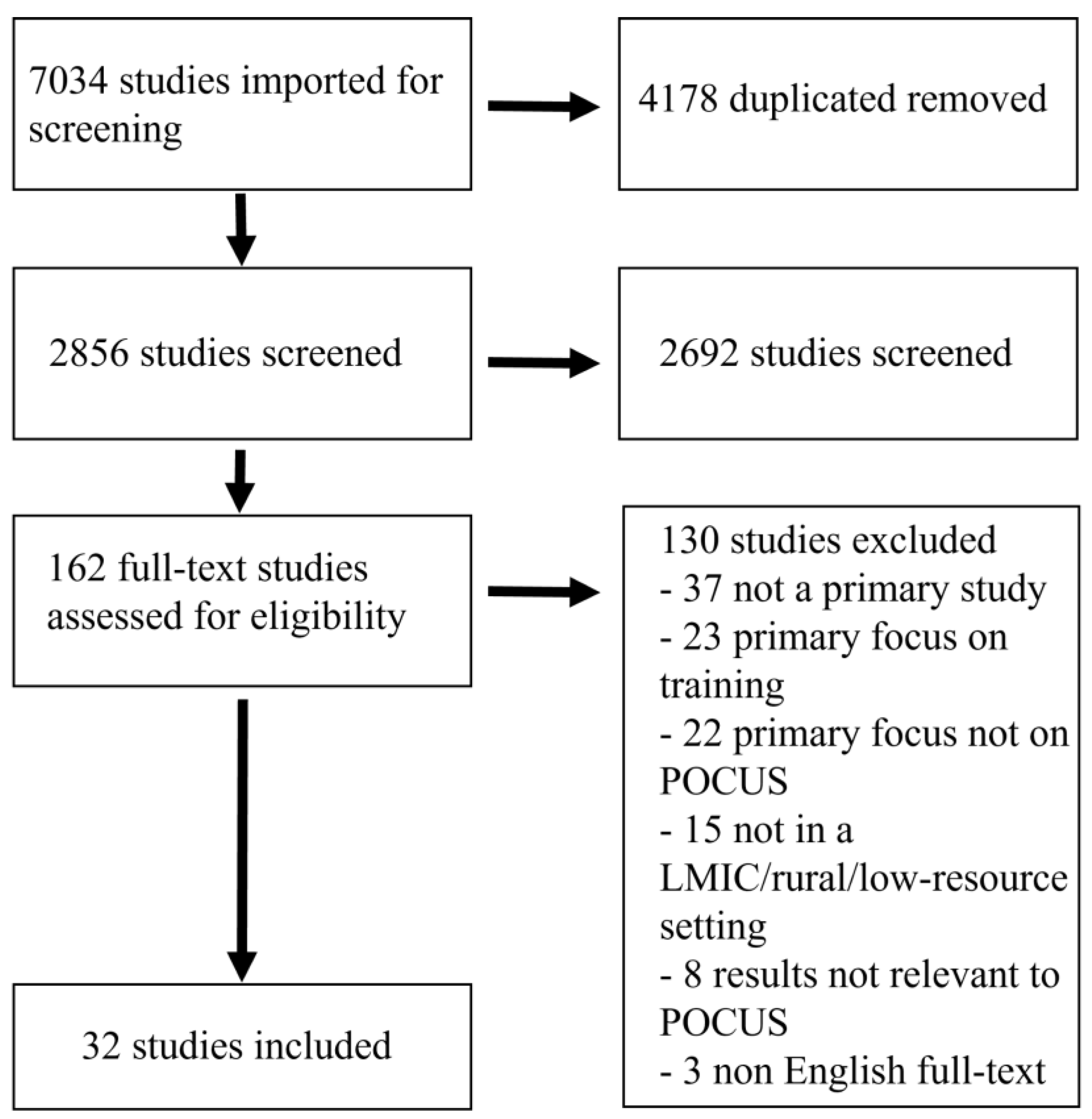
Figure 1. PRISMA flow diagram.
Applsci 13 08427 g001
Figure 2. Geographical distribution of studies conducted.
Applsci 13 08427 g002
Figure 3. (A) Histogram of the brands of the utilized POCUS devices (B) a visualization of a generic POCUS device.
Applsci 13 08427 g003
Table 1. Inclusion and exclusion criteria used in this scoping review.
Inclusion CriteriaExclusion Criteria
  • Point-of-care ultrasound used
  • Handheld/ portable/ mobile ultrasound used
  • Low-resource setting
  • Primary study
  • Non-portable ultrasound
  • Pre-2016
  • Non-English
  • Non-human
  • Not a rural setting
  • Study type
    -
    Non-primary studies
    -
    Proof-of-concept papers
    -
    Pilot studies
    -
    Primary focus on training
Table 2. A summary of the data extraction results of studies included in this scoping review.
Author (s); Journal; YearPopulation in Study (Criteria)Study Type and PurposeGeographical
Context
ResultsDevice(s)
Utilized
Baker et al., Tropical Doctor, 2021
[32]
144 patientsCross-sectional study
  • Impact of POCUS on diagnosis and treatment
Uganda, LMIC, 20 mobile clinics from local churches and schools in 6 separate rural locations, Masindi region in Western Uganda
  • In 73% of patients, findings either confirmed (50%) or changed (23%) diagnosis.
  • In 53% of patients, the ultrasound findings changed the treatment ment plan.
  • POCUS impacted patient care positively in clinics in rural Uganda by improving diagnostic capabilities and influencing treatment plans
3 Philips Lumify ultrasound probes attached to a Samsung Galaxy tablet with preloaded Lumify software;
Phased: −4 to 1 MHz
Linear: −12 to 4 MHz
Curvilinear: −5 to 2 MHz
Barron et al., Southern Medical Journal, 2018 [33]All patients who received POCUS in standard clinical practice during an Short Term Medical Mission (STMM)Observational prospective study
How POCUS would change medical management
Nicaragua (Sébaco), LMIC, rural Nicaraguan clinic
  • Average time to perform a POCUS examination was 6.0 min
  • Incorporating POCUS by trained physicians in an LRS often changed management
2 General Electric Vscans.
Single low frequency probe: (1.7–3.8 MHz)
Dual low and high frequency probe: (Phased array––1.7–3.8 MHz; Linear array––3.3–8.0 MHz)
Bobbio et al., BMJ Open, 2018
[34]
100 HIV-positive patients registered for antiretroviral treatmentCross-sectional study
  • Use of POCUS in diagnosing HIV infections in South Sudan
South Sudan, LMIC, Voluntary Counselling and Testing Centre (VCT) of Yirol Hospital
  • Focused Assessment with Sonography for HIV-associated tuberculosis (FASH) results supported tuberculosis treatment indication for 16/21 patients.
  • For 8/16 patients, treatment was based exclusively on FASH findings
2 Mindray portable black US scanners
Convex probe: 3.5 MHz
Linear probe: 7 MHz
Cherniak et al., PLoS ONE, 2017 [31]All women who were currently aware of being pregnant and presented to ANCNon-blinded cluster-randomized controlled trial
  • General effectiveness of POCUS in rural area
Uganda, LMIC, rural communities in southwestern Uganda, Kabale District
  • Portable obstetric ultrasound combined with radio messaging can serve as an important tool to rapidly increase uptake of ANC services in rural disadvantaged women
Device not specified
Milart et al., Reproductive Health, 2016
[35]
Pregnant women intervention group decided by the Health DirectorateObservational study
  • The effectiveness of POCUS on prenatal control in rural area
Guatemala, LMIC, Rural areas of the districts of Senahu, Campur, and Carcha, in Alta Verapaz Department
  • There were no maternal deaths reported in the intervention group
  • Five maternal deaths in the control group
  • 64% reduction for neonatal mortality
  • 37% prevalence of anemia
Device not specified
Dalmacion et al., BMC Pregnancy and Childbirth, 2018 [36]Pregnant women who were not allergic to gel, did not have concurrent medical or surgical conditions, and provided informed consentsCross-sectional study
  • Use of POCUS to prevent maternal/neonatal deaths
Philippines, LMIC, Parañaque city, the urban study site, and Tagum city, the rural study site
  • 146/460 (31.7%) showed abnormal ultrasound readings consisting of 17 placental implantation abnormalities, 123 fetal malpresentation, 3 twins, and 3 amniotic fluid volume abnormalities
  • Use of handheld ultrasound could have possibly averted 29 (6.3%) maternal deaths and 14.6% neonatal deaths at delivery time
General electric Vscan with dual probe: (Phased array––1.7–3.8 MHz; Linear array––3.3–8.0 MHz);

Utilized General Electric Logic 5 Premium as reference standard
Elsayes et al., Rural Remore Health, 2021 [37]All adult patients over the age of 18 at the mobile clinic in Antigua, GuatemalaRetrospective observational study
  • Usefulness of portable ultrasound imaging during a medical service trip to rural Guatemala
Guatemala, LMIC, a mobile clinic in Antigua, Guatemala
  • For 13/24 (54%) patients, ultrasound exams altered their medical management
  • For 11/24 (46%), exams confirmed the pre-test suspected diagnoses
Device not specified
Epstein et al., Travel Medicine and infectious disease, 2018 [38]101 patients at Kiboga hospitalMedical management
  • Effectiveness of POCUS in the rural area performed by physicians with only basic training
Uganda, LMIC, Kiboga hospital (general governmental hospital), Kampala, Uganda, Central Uganda
  • 16 out of 23 patients (70%) had positive findings
  • In 20 of them (87%), management was changed
General Electrical Vscan with a single low-frequency probe (1.7–3.8 MHz)
Gomes et al., Annals of Global Health, 2020 [39]Women presenting to the obstetrical departments of Fort Liberte Hospital and Centre Medicosocial de OuanamintheCross-sectional study
  • Evaluation of acute obstetrical needs and the potential role for POCUS programming in the North East region of Haiti
Haiti, LMIC, Obstetrical departments of two Ministry of Public Health and Population (MSPP)-affiliated public hospitals
  • POCUS may have a role in early identification of pathologies that need a cesarean section, resulting in improved outcomes and decreased complications
  • POCUS may also be a beneficial diagnostic adjunct to acute care in Haiti as it allows for rapid imaging and identification of complications
Device not specified
Haldeman et al., The Ultrasound Journal, 2022 [40]Residents enrolled in the UNZA Family Medicine programSingle-center, survey-based, prospective study
  • How POCUS would change medical management
Zambia, LMIC, Chilenje hospital
  • POCUS assisted most frequently by helping to determine a diagnosis in 51.4% (188/366) of patient encounters and in 44.7% (188/421) of assisting instances
3 Butterfly IQ handheld ultrasound devices (1–10 MHz)
Huson et al., The American Journal of Tropical Medicine and Hygiene, 2020 [41]Patients with fever at Mahosot HospitalProspective observational study
  • Effectiveness of POCUS in diagnosing melioidosis
Laos, LMIC, Mahosot Hospital, Vientiane Laos
  • Eleven (61%) patients with melioidosis had an abscess at one or more sites compared with one (1%) patient with an alternative diagnosis
  • The positive and negative predictive value of finding one or more abscesses in patients with melioidosis in the patient population was 92% and 93% respectively
Mindray portable black-and-white ultrasound;
Convex probe: 3.5 MHz
Linear probe: 7.5 MHz
Kodaira et al., International Journal of Gynecology and Obstetrics, 2020 [42]Pregnant women at Princess Christian Maternity HospitalSingle-center prospective observational study
  • Evaluating the reliability of obstetric handheld smartphone-based point-of-care ultrasound (POCUS) in a low-resource high-volume setting
Sierra Leone, LMIC, high-volume, low-resource hospital, Western Area of Sierra Leone
  • Mean aggregated diagnostic accuracy was 95.5%
  • Highest accuracy was reported for detecting free fluid collection in the abdominal cavity (100%)
  • Detection of low-lying placenta/placenta previa was least reliable
Hand-held smartphone-based ultrasound devices;
Lequio US-304 convex probe (3–5 MHz)
Mindray SD-10 (3–5 MHz convex probe and 7–10 MHz trans-vaginal probe)
Lamorte et al., Crit Ultrasound Journal, 2016 [43]105 patients re-
quested for ultrasound examination by the caring physician
Prospective, observational study
  • Effectiveness of POCUS in rural area
Sierra Leone, LMIC, Holy Spirit Hospital, Makeni, Bombali district
  • Within 194 diagnoses formulated, there was a mean of 1.85 diagnoses per patient
  • US also introduced 53 new diagnoses in 42 patients
Toshiba Memio 20
Leopold et al., PLoS ONE, 2018 [44]Patients enrolled in the general medical wards of Chittagong Medical CollegeProspective, observational study
  • Evaluating point-of-care lung ultrasound as a novel tool to determine the prevalence and early signs of ARDS in a low-resource setting among patients with severe malaria or sepsis
Bangladesh, LMIC, Large tertiary government hospital (Chittagong Medical College), Chittagong, Bangladesh
  • In a total of 102 patients, 71 patients had sepsis, 31 had malaria
  • 44 had normal lung ultrasound findings and were associated with 7% case fatality
  • ARDS was detected in 10 patients and associated with 90% case fatality
General Healthcare Vivid-I portable ultrasound;
Convex probe: 5 MHz
Limani et al., Transactions of The Royal Society of Tropical Medicine and Hygiene, 2020 [45]Patients over 16 who had received an ultrasound examinationProspective observational study
  • Evaluating uptake and impact of POCUS following the introduction of a training program
Malawi, LMIC, Queen Elizabeth Central Hospital, Blantyre, Malawi, tertiary referral center for the southern region
  • 133 of 267 ultrasound examinations were POCUS
  • For POCUS examinations, the time from request to examination was shorter compared to radiology department ultrasound
POCUS: General Electrical V-scan,
Mindray DP 30
Radiology department ultrasound: Mindray DC 30, General Electric vivid Q-i
Lobo et al., Health Science Reports, 2022 [46]Voluson Ultrasound Equipment, Toshiba Némio XG ultrasound data across 2 emergency centersCross sectional, observational and longitudinal study
  • Observing the impact of the diagnostic process and therapeutic approach
Portugal, Developed Country, two remote locations in Portugal (SUB N, SUB S), basic emergency services (SUB)
  • 289 (29.7%) were classified as normal, 628 (64.6%) were classified as abnormal, and 55 (5.7%) were considered inconclusive
  • 58% had a local resolution, 24% were referred to a hospital emergency service, and 18% were referred to ambulatory care
General Electric Voluson SN: Convex and Linear probes;
Toshiba Nemio XG ultrasound: Convex
Mazza et al., World J Emergency Medicine, 2019 [47]Subjects from the age of 11 months to 13 years, with risk of dehydrationProspective, observational study
  • Determining if medical students can detect pediatric dehydration using ultrasound on patients in rural Panama
Panama, LMIC, Bocas del Toro region of rural Panama, Floating Doctors clinics
  • 24 patients were clinically diagnosed with dehydration and 35 were classified to have normal hydration status.
Mindray M7 portable ultrasound machine with single phased array probe
Mengarelli et al., Missouri Medicine, 2018 [48]Hospitals that were in the records of the Missouri Department of Health and Senior ServicesSurvey
  • Advantages of POCUS in rural vs urban area
United States, Developed Country, Missouri, hospitals with emergency departments, large, medium, and small hospitals
  • Large-sized hospitals used POCUS significantly more than medium-sized or small-sized hospitals
  • Data suggested a shortage of this technology in rural environments
Device not specified
Nacarapa et al., Nature Scientific Reports, 2022 [49]Patients over 15 years from the CHC dataset with ultrasound findings of extrapulmonary TB ManifestationProspective, observational study
  • Determining the utility of POCUS in the diagnosis of EPTB and evaluate the in-hospital mortality
Mozambique, LMIC, rural Chókwè district, Mozambique, southern Gaza province, hospital
  • POCUS provides a simple, feasible, and affordable intervention in low-resource settings like Mozambique
Samsung SonoAce R3 with a C2–4/20 convex probe (1–10 MHz frequency)
Nixon et al., Canadian Journal of Rural Medicine, 2019 [50]28 rural generalist physicians trained by the Royal NZ college of General PractitionersSubgroup analysis
  • Effectiveness of POCUS in rural area
New Zealand, Developed Country, Five New Zealand rural hospitals
  • POCUS of inferior vena cava and jugular venous pressure are easily learned techniques and are more reliable than physical examination
  • POCUS frequently altered a physician’s impression of a patient’s intravascular volume compared to that based on physical examination alone
Device not specified
Nixon et al., Australian Journal of Rural Health, 2018 [51]All generalist doctors practicing ultrasound in study hospitalsCross-sectional descriptive study
  • Evaluating the safety, quality and impact of POCUS on patient management when performed by rural generalist doctors
New Zealand, Developed Country, Six rural small hospitals serving a range of communities in rural New Zealand
  • 4% overall reduction in patients needing admission to a hospital or transfer to an urban base hospital
  • 71% of POCUS scans had a positive overall impact on patient care
  • 3% of scans had the potential for patient harm
Device not specified
Nixon et al., Journal of Primary Health Care, 2018 [52]All the generalist rural doctors practicing POCUS in the 6 rural hospitalsMixed-methods descriptive study
  • Describing the scope of POCUS being practiced by rural generalist hospital doctors
New Zealand, Developed Country, Rural New Zealand, six rural hospitals
  • Wide variety of applications found: Cardiac + Circulatory (38%), Gallbladder (13%), Kidney (11%), Trauma (FAST) (7%), Bladder (6%), and Lungs (5%)
  • There was a significant increase in diagnostic accuracy and certainty in the practice
Device not specified
Nixon et al., Journal of Primary Health Care, 2018 [53]28 doctors in 6 New Zealand rural hospitalsSubgroup analysis
  • Determining the quality, safety, and effect of POCUS of kidney and bladder on patient care in rural New Zealand
New Zealand, Developed Country, Rural New Zealand, 6 rural hospitals
  • POCUS was utilized as a test for urinary retention and hydronephrosis
  • For rural doctors, it was a straightforward technology, improved diagnostic certainty, increased discharges, and overall had a positive effect on patient care
Device not specified
Nixon et al., Rural and Remote Health, 2019
[54]
28 physicians in 6 New Zealand rural hospitalsSubgroup analysis
  • Assess the extent that FAST altered diagnostic certainty and patient disposition
New Zealand, Developed Country, Rural New Zealand, 6 geographically dispersed rural hospitals
  • POCUS AAA is a reliable test to rule out ruptured abdominal aortic aneurysm
  • FAST scan can rule in solid organ injury
Device not specified
Pellegrini et al., Annals of Intensive Care, 2018 [55]1533 Brazilian intensivistsNational survey
  • Investigating current practice and education regarding POCUS among Brazilian intensivists
Brazil, LMIC, Intensive Care Units (ICU), all regions of Brazil
  • Main applications were central vein catheterization (49.4%) and echocardiograms (33.9%)
Device not specified
Peterman et al., Cureus, 2022 [56]3011 public datasets on a county levelGeospatial Analysis
  • Determining the usage of POCUS within the US and the differences between rural and urban area
United States, Developed Country, rural and metropolitan counties
  • POCUS access statistics—rural counties (38.84%) and metropolitan (74.19%)
Device not specified
Reynolds et al., PLoS ONE, 2018 [57]Patients receiving POCUS at Muhimbili National Hospital’s Emergency Medical Department (MNH EMD)Prospective descriptive cross-sectional study
  • Analyzing the utilization and impact of POCUS on clinical decision-making at an urban emergency department
Tanzania, LMIC, Urban emergency department in Dar es Salaam, Tanzania, Muhimbili National Hospital
  • POCUS had wide implications in urgent care, emergency medicine and trauma in LRS
  • Significant increase in clinical decision-making ability
SonoSite mTurbo
Self et al., JMIR Research Protocols, 2022 [23]Pregnant women in the 2 hospitalsProspective study
  • Improving pregnancy and risk assessment for women in underserved regions using POCUS
United Kingdom (Developed Country), India (LMIC), Group for Advanced Research on BirtH outcomes–Department of Biotechnology India Initiative (GARBH-Ini) cohort, John Radcliffe Hospital (Oxford)
  • Utilization of obstetric POCUS videos to develop various machine learning algorithms that improve diagnostic care in obstetric risk management
General Electric Voluson E8 with curvilinear probes: C2–9 (3–9 MHz) and C1–5 (2–5 MHz)
Sheppard et al., Canadian Journal of Rural Medicine, 2021 [58]10 physicians (3 females, 5 rural) participated in the interviewsMixed-methods cross-sectional study
  • Determining the prevalence of POCUS devices in NL and to characterize the patterns of POCUS use amongst physicians in NL
Canada (Developed Country), Newfoundland and Labrador, Urban (4) and rural geographic (19) locations
  • POCUS had benefits such as expedited investigations, decreased radiation and increased patient satisfaction
  • Barriers to POCUS use were lack of training, time, devices, image storing software, difficulty generating and interpreting images and patient body habitus
Device not specified
Shumbusho et al., Journal of ultrasound in medicine, 2019 [59]Patients 5 years and older with PTX managed by chest tubes at CHUKProspective, observational study
  • Evaluating the accuracy and timeliness of resident-performed point-of-care lung ultrasound for the follow-up of pneumothorax after tube thoracostomy
Rwanda, LMIC, Rwandan referral hospital (University Teaching Hospital of Kigali (CHUK))
  • Lung ultrasound speed and performance was significantly enhanced while also increased sensitivity and downstream interpretation within low-income settings
SonoSite M-Turbo
Stachura et al., African Journal of Emergency Medicine, 2017 [60]118 patients with clinical indications for POCUSProspective observational study
  • Identifying high impact POCUS scans most relevant to practice in an Ethiopian EC
Ethiopia, LMIC, Tikur Anbessa Specialized Hospital EC in Addis Ababa, urban, low-resource, academic EC in Ethiopia
  • POCUS provided clinically relevant information for patient management (polytrauma, undifferentiated shock and undifferentiated dyspnea) in urban, low-resource academic EC in Ethiopia
SonoSite MicroMaxx and Full Digital Laptop Ultrasound Scanner (RUS-9000F) with 3.5 MHz curved array probe; Last two weeks of the study, there was a SeeMore USB ultrasound system with two probes: abdominal (GP3.5/5.0 MHz) and SP7.5/24.0 MHz high frequency probe
Umuhire et al., Ultrasound Journal, 2019 [61]Adult participants presenting with dyspnea to an urban Rwandan emergency departmentProspective, observational study
  • Impact of POCUS on management
Rwanda, LMIC, Emergency Department at University Teaching Hospital of Kigali (UTH-K) in Rwanda
  • The physician diagnostic accuracy increased from 34.7% to 88.8% pre and post-ultrasound
  • Clinicians’ confidence in the primary diagnosis changed from a mean of 3.5 to a mean of 4.7 (Likert scale 0–5)
SonoSite M-Turbo
Disclaimer/Publisher’s Note: The statements, opinions and data contained in all publications are solely those of the individual author(s) and contributor(s) and not of MDPI and/or the editor(s). MDPI and/or the editor(s) disclaim responsibility for any injury to people or property resulting from any ideas, methods, instructions or products referred to in the content.