Article

Influence of the Polymer and Solvent Variables on the Nanoencapsulation of the Flavonoid Quercetin: Preliminary Study Based on Eudragit® Polymers

by
Joel H. Elizondo-Luevano
1,2,
Rocío Castro-Ríos
3,
Roberto Parra-Saldívar
1,2,
Horacio Larqué-García
4,5,
Marsela Garza-Tapia
3,
Elda M. Melchor-Martínez
1,2,* and
Abelardo Chávez-Montes
4,*
1
School of Engineering and Sciences, Tecnologico de Monterrey, Monterrey 64849, Mexico
2
Institute of Advanced Materials for Sustainable Manufacturing, Tecnologico de Monterrey, Monterrey 64849, Mexico
3
Department of Analytical Chemistry, Faculty of Medicine, Universidad Autónoma de Nuevo León (UANL), Monterrey 64460, Mexico
4
Department of Chemistry, Faculty of Biological Sciences, Universidad Autónoma de Nuevo León (UANL), San Nicolás de los Garza 66455, Mexico
5
Department of Pharmaceutical Chemistry, Faculty of Pharmacy, Universidad de Salamanca (USAL), Campus Miguel de Unamuno s/n, 37007 Salamanca, Spain
*
Authors to whom correspondence should be addressed.
Appl. Sci. 2023, 13(13), 7816; https://doi.org/10.3390/app13137816
Submission received: 28 April 2023 / Revised: 20 June 2023 / Accepted: 29 June 2023 / Published: 3 July 2023

Abstract

:

Featured Application

A methodology was developed for the efficient nanoencapsulation of non-water-soluble biomolecules, such as the flavonoid quercetin, through the nanoprecipitation technique and using Eudragit® polymers for pharmaceutical use. These data are intended to provide an approach for the future administration of bioactive compounds for pharmaceutical uses.

Abstract

Eudragit® polymers have proven their potential as a means to control the release of aqueous insoluble drugs in various delivery systems as polymer nanoparticles (PNPs). The size (S) and polydispersity index (PDI) of PNPs are crucial factors for their interaction with biological systems from a pharmaceutical standpoint. This study aimed to determine the impact of the volumes of the organic phase (OP) and aqueous phase (AP), as well as the polymer amount (PA), on the size and PDI of PNPs prepared using the nanoprecipitation method for encapsulating quercetin (Qr). The study also evaluated the toxic effects of PNPs on human erythrocytes. The PNPs were prepared using preformed polymers derived from methacrylic acid and polyvinyl alcohol (PVA) as a surfactant. The nanoprecipitation technique enabled the production of particles smaller than 200 nm with a PDI lower than 0.2, and the study established the significant impact (p < 0.05) of the three variables related to the polymers and solvents. The selected PNPs contained 5 mg of Qr and 50 mg of Eudragit polymers (1:10 w/w Eudragit® EPO, E100, L100, and Eudragit L100-55) and diverse concentrations of PVA. The study found that including PVA in the AP increased the Qr encapsulation by up to 98%. The hemolytic potential of Eudragit® PNPs and Qr was assessed in human erythrocytes, with no significant cytotoxic activity observed (p < 0.001) compared with the control. In conclusion, via the nanoprecipitation technique, preparing PNPs with defined and homogeneous S to entrap the flavonol Qr efficiently is possible.

1. Introduction

Polymer nanoparticles (PNPs) or nanocarriers have been widely researched and studied in the pharmaceutical field as active ingredient delivery systems offering a promising future since this type of carrier improves stability, bioavailability, and drug targeting due to the significant characteristics of PNPs, including size (S), which varies from 100 to 500 nm. These characteristics facilitate the entry and improve the interaction with biological substrates, giving them several advantages, such as a large contact surface, a capacity of bio-direction to specific organs or tissues, absorption of the drug in the target tissue, reduced adverse effects, decreased dosage, and internalization by cells, bacteria, or parasites [1,2]. These pharmaceutical carriers are generally made of natural or synthetic biodegradable polymers and form structures with diverse chemical natures where the drug can be encapsulated, adhered to, or absorbed [3]. To date, there are studies of NP-based formulations made from polymers derived from methacrylic acid, where different types of drugs are encapsulated; these formulations have been shown to incorporate a wide variety of active ingredients with an encapsulation efficiency between 60% and 90%, being efficient when applied via different routes (intravenous or respiratory), both in vitro and in vivo, showing low toxicity; hence, these types of drug carriers and nanoformulations represent an excellent option for the treatment of different diseases [4,5]. Drug encapsulation into PNPs increases the stability of active substances and protects sensitive substances from chemical changes, pH, and ultraviolet light. Encapsulation improves the drug’s specificity, efficacy, and tolerability [6].
Eudragit® is a polymer that has demonstrated its potential as a coating material and matrix former in various drug delivery systems to control drug release. By enabling site-specific drug delivery, it can enhance the bioavailability of drugs and reduce associated side-effects or toxicity, particularly for anticancer drugs [7]. Eudragit® is a versatile range of polymers composed of methacrylic acid and methyl methacrylate, representing anionic copolymers commonly used to stabilize and improve the bioavailability of various substances. These polymers can protect drugs that are susceptible to breakdown in an acidic environment, such as the stomach, enabling their release only at a pH level above 6.0 in the intestine [8]; these anionic copolymers are made of methacrylic acid and methyl methacrylate and are widely used for multiple PNP formulations, constituting tiny particles that vary from 1 to 300 nm. Eudragit® has been successfully used to enhance the therapeutic effects and bioavailability of various substances, including berberine, curcumin, quercetin, laccases, insulin, and pantoprazole [2,6,8,9,10]. These polymers effectively encapsulate hydrophobic molecules such as the flavonol quercetin, which is insoluble in aqueous solutions and can hinder absorption [10].
Quercetin, a natural metabolite known for its strong antioxidant activity, is commonly used as a nutritional supplement and treatment for various conditions such as diabetes, obesity, circulatory dysfunction, inflammation, and mood disorders [11]; moreover, flavonoids are present in a variety of medicinal plants [12,13,14]. Additionally, flavonols such as quercetin effectively inhibit bacterial growth and possess antiparasitic properties [11,15,16]. However, quercetin’s efficacy is limited due to its high hydrophobicity, instability in physiological media, poor gastrointestinal absorption, and extensive xenobiotic metabolism in the liver and intestine [10]. PNPs as a delivery system are proposed in this research to overcome these limitations, as they can improve quercetin’s bioavailability, protect it against degradation, and prevent its premature release [6].
Therefore, the present study focuses on two aspects. The first is determining the main parameters that influence the S (nm) and PDI of PNPs performed by methacrylic acid-derived polymers (Eudragit® polymers) via the nanoprecipitation technique (Graphical abstract). Three variables were evaluated: the effect of the organic phase ratio (OP in mL), the effect of the aqueous phase (AP in mL), and the impact of polymer amount (PA in mg). This allowed us to determine the best formulation in which the nanoparticle populations were homogeneous, before proceeding with the encapsulation of the Qr. The cytotoxic activity of the Eudragit® formulations at different concentrations was also evaluated; this assay on human erythrocytes was performed to determine that Eudragit® NPs do not present toxicity for future application. The second aspect was the variation in encapsulation of Qr using the parameters of the most effective formulation while adding polyvinyl alcohol (PVA) in different concentrations.

2. Materials and Methods

2.1. Ethical Statement

The institutional research ethics committee of the Universidad Autónoma de Nuevo León approved all procedures, which were conducted following the Declaration of Helsinki on biomedical research with human subjects. The ethics committee of the UANL approved (Reg. CI-06-19-2023) the study involving human erythrocytes, and informed consent was obtained from healthy donors following the Official Mexican Technical Standard NOM-253-SSA1-2012.

2.2. Chemicals and Reagents

Clariant México provided polyvinyl alcohol (PVA, Mowiol® R°4-88, CAS No: 9002-89-5; Mw~31,000), while Helm México donated polymers derived from methacrylate, including Eudragit® EPO, E100, L100, and L100-55. The standard-grade Quercetin (Qr) was procured from Sigma-Aldrich® (Merck KGaA, Darmstadt, Germany). Acetone and methanol (MeOH), used in this study, were of analytical grade.

2.3. Polymeric Nanoparticles Formulation

PNPs were prepared via the nanoprecipitation method [2]. Different Eudragit® polymers were used (Figure 1), EPO, E100, and L100 were dissolved in absolute MeOH, and L100-55 was dissolved in acetone by sonication applying three 3 min sonication cycles followed by a 1 min rest period (Ultrasonic Branson 2510MT, Merck KGaA, Darmstadt, Germany) [9]. An OP, which contained the polymer dissolved and was miscible with water, was injected into an AP under constant magnetic stirring (500 rpm). The diffusion of the OP was carried out via injection into the AP, which favored the aggregation of the nanoparticle-forming polymer. The PNP suspensions obtained were agitated using a digital laboratory stirrer (Eurostar Power-B Ika® -Werke, GmbH & CO. KG, Staufen, Germany) at 2000 rpm for 3 min, and then evaporated under reduced pressure with a Laborota-4003 rotary evaporator (Heidolph Instruments GmbH & Co. KG, Schwabach, Germany) at room temperature to remove the solvents. The physical characterization of the PNPs was then continued.

2.3.1. Eudragit® PNP Synthesis

The variables evaluated in this study were the following (Table 1): variation of the volume of OP over AP and constant polymer amount (PA) (12 mL + 50 mg, respectively), a variation of the volume of AP over OP and constant PA (12 mL + 50 mg, respectively), and variation of the PA over constant OP and AP volumes (12 mL each). The amounts tested were as follows: for OP and AP, the volumes of 5, 8, 10, 12, 15, 18, and 20 mL; for PA, the amounts of 5, 10, 25, 50, 75, and 100 mg of each polymer. The nano-emulsions were homogenized as previously described. Each batch was made in triplicate.

2.3.2. Quercetin Encapsulation into PNPs

For this purpose, according to the results of the previous test in which the proportions of OP, AP, and PA were varied, the formulation with the best conditions in terms of S and homogeneity was selected, which was 12 mL of OP (MeOH or acetone), 50 mg of PA, and 12 mL of AP (Table 2). For the encapsulation of the Qr, different treatments were evaluated. First, 5 mg of Qr and 50 mg of polymer were dissolved in the OP; then, the mixture was homogenized by sonication (three 3 min sonication cycles followed by a 1 min resting period). After that, the Qr and polymer solution was injected into 12 mL of an aqueous solution (at this point, changes were made in the composition of the AP, which consisted of adding different concentrations of PVA) which contained PVA (0.0%, 0.5%, 1.0%, 2.0%, 4.0%, and 8.0% w/v), under constant magnetic stirring (2000 rpm/20 min). The diffusion of OP in the PVA solution favored the suspension of the PNPs. The suspensions obtained were agitated at 2000 rpm for 3 min as previously described, and then subjected to rotaevaporation to remove the solvent; each batch of the formulations was stored in glass flasks and protected from light. Finally, the characterization was performed. Each batch was carried out in triplicate.

2.4. Encapsulation Efficiency

To measure the Qr immobilized in the PNPs, the formulations were centrifuged at 25,500 rpm/4 °C for 4 h. The encapsulated Qr in the PNPs and the nonencapsulated Qr in the supernatants were estimated. The quantitative analysis of Qr was performed compared with a calibration curve of Qr in a UV/Vis spectrophotometer (GENESYS 10S, Thermo Scientific Inc., Waltham, MA, USA). The encapsulation efficiency (% EE) was calculated as follows:
E E   % = Q R I Q R S Q R I × 100 ,
where QrI corresponds to the initial quercetin, and QrS corresponds to the remnant Qr in the supernatant [20].

2.5. Size and Morphology of Polymer Nanoparticles

The S in nanometers (nm) and PDI of the PNP formulations and Qr-PNPs were measured using photonic correlation spectroscopy in a Zetasizer Nano-ZS90 (Malvern Instruments Ltd., Malvern, UK). An aliquot from each batch was diluted in Milli-Q water before analysis. The morphology of the PNPs was examined using a scanning electron microscope (In-line FE-SEM SU8000, Hitachi, Ltd., Chiyoda, Tokyo, Japan) at 2 kV. The samples were diluted at a ratio of 1:100, mounted, and then dried in metal studs.

2.6. Hemolytic Activity Test

The hemolytic activity was assessed using the hemolysis test [15]. For the evaluation of the hemolysis, the red cell suspension was incubated with different concentrations of the polymers or Qr from 100 to 1000 μg/mL in microcentrifuge tubes (Eppendorf SE, Hamburg, Germany) (30 min/37 °C) protected from light, labeled as treatments (Tr). As a blank, a solution of erythrocytes without treatment was used, and the positive control consisted of erythrocytes without treatment with distilled water to produce osmotic hemolysis [21]. Once the incubation time elapsed, all treatments were centrifuged at 12,000 rpm for 3 min at 4 °C. First, 200 µL of each supernatant was placed in a 96-well flat-bottomed microplate (Corning Incorporated, Costar®, Glendale, AZ, USA). The percentage (%)hemolysis was determined by the optical density (OD) at 540 nm [12,22]. The readings were recorded as the ODs obtained for each treatment (OD540nm treatment). The hemolysis was computed as follows:
H e m o l y s i s   % = O D 540 n m   T r e a t m e n t O D 540 n m   B l a n k O D 540 n m   P o s i t i v e   c o n t r o l × 100 .

2.7. Statistical Analysis

The Statistical Package for the Social Sciences (SPSS) software, version 24.0 (IBM Inc. Armonk, NY, USA), was used for data analysis. All experiments were conducted in triplicate in at least three different trials. The mean ± standard deviation (SD, n = 3, p < 0.05) was used to present the results. A two-way ANOVA was used to evaluate the statistical significance of the S and PDI of PNPs in different combinations. A one-way ANOVA was used to evaluate the Qr-PNPs and hemolytic activity. Tukey’s post hoc HSD test was applied to determine the statistical difference between treatments (p < 0.05).

3. Results

3.1. Polymeric Nanoparticle Preparation

The particle size (S) of four different polymers used in pharmaceuticals derived from methacrylic acid was measured using the solvent displacement technique (nanoprecipitation) under varying preparation conditions (Figure 2). In addition, in the Supplementary Materials (Table S1), a table with the corresponding results of each formulation and the different concentrations evaluated is provided.
Initially, the volume of the organic phase was varied while keeping the volume of the aqueous phase and the amount of polymer constant (12 mL of AP + 50 mg of PA, respectively). Figure 2A (a) demonstrates that, regardless of the polymer used, the particle S decreased with little or no influence in the initial increments of the OP volume (from 5 mL to 18 mL). When the effect of this variable on the PDI of the EPO and E100 polymers was observed, there was a slight decrease as the volume of the OP used increased. The trend was significantly more evident for the L100 and L100-55 polymers, with a minimum of dispersion in size observed at the center of the range evaluated and increasing extremes at very small or large volumes of the organic phase (p < 0.05).
Afterward, the OP volume was held constant at 12 mL to investigate the impact of varying the volume of the AP while maintaining a fixed polymer mass of 50 mg. Figure 2B (b) illustrates that this variable had only a slight effect on E100, L100, and L100-55 in the first increments. It was observed that increasing the volume of the AP led to a decrease in particle S. A similar effect was observed for the EPO polymer, but only in the initial increases in the volume of the AP, with a more pronounced impact on decreasing the S. The effect of this variable on PNP size homogeneity was unclear, except for the EPO polymer, which displayed a decrease in the PDI as the volume of the AP increased.
The influence of PA ranging from 5 to 100 mg on particle S was assessed using constant volumes of 12 mL for each phase. Figure 2A (c) reveals that the E100 and L100-55 polymers tended to form larger particles as the PA increased. Conversely, EPO and L100 exhibited a slight decrease in S initially (p < 0.001), followed by an increase in particle S with increasing polymer mass (p < 0.001). The significant impact of this variable on the homogeneity of S in the PNPs for each formulation is depicted in Figure 2B (c) (p < 0.001). For EPO and L100-55 polymers, there was only a clear relationship between the two variables, albeit in the opposite direction. As the polymer mass increased in EPO formulations, the polydispersity index decreased significantly (p < 0.001), whereas, for the L100-55 polymer, it increased.

3.2. Qr-Loaded PNPs

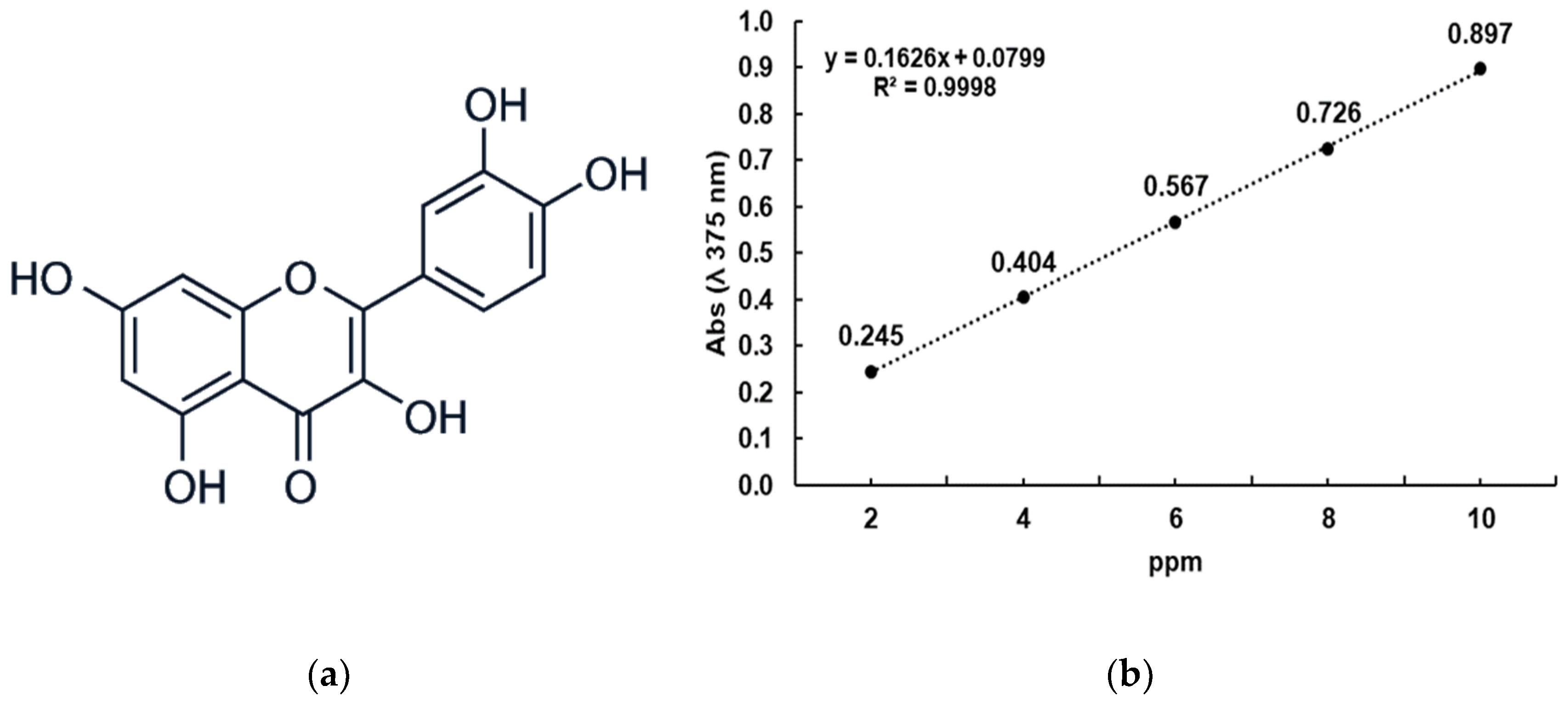
The elaboration of Qr-loaded PNPs was performed using the previously mentioned nanoprecipitation technique, and the encapsulation efficiency was determined by comparing it with the quercetin calibration curve at 375 nm (Figure 3). The size and PDI were measured by photon correlation spectroscopy in a Zetasizer.
Table 3 indicates the influence of PVA on the percentage Qr encapsulation into PNPs and its effect on the mean particle S and PDI. Remarkably, the four polymers presented a decrease in particle S and PDI as the PVA concentration increased; on the other hand, as the PVA concentration increased, the percentage Qr encapsulation increased into the PNPs. In all cases, the nanoencapsulation of Qr was possible without PVA; however, the encapsulation percentages were low. The PVA in the AP enhanced the encapsulation of Qr and helped maintain more homogeneous PNPs populations.

3.3. Morphology of Polymer Nanoparticles

Using a scanning electron microscope (SEM), the morphology of the selected formulation of Eudragit®-PNPs was determined for each polymer (12 mL OP, 12 mL AP, and 50 mg PA). When measured by photon correlation spectroscopy, the selected PNP nanoformulations showed homogeneous characteristics in size and PDI. It can be appreciated that the selected formulates showed homogeneous sizes with circular and spherical morphology (Figure 4).

3.4. Determination of the Hemolytic Activity of the PNPs

We determined the toxicity of Eudragit®-PNPs and Qr via hemolysis test. First, the formulations were mixed with PBS to obtain various concentrations, which were then added to erythrocytes for evaluation. The absorbance of the supernatant was measured at 540 nm. The results revealed that none of the four polymers and Qr at different concentrations showed any significant toxicity (p < 0.001 compared to positive control). As expected, the positive control (distilled water) exhibited 100% hemolysis. The degree of hemolytic activity in erythrocytes was expressed as a percentage of hemolysis and summarized in Table 4.

4. Discussion

The nanoprecipitation technique, also known as solvent displacement, enables the formation of nanocapsules and nanospheres [23]. This method requires two miscible solvents, typically an organic solvent and an aqueous solution. The polymer and drug are usually soluble in only one of the solvents, typically the organic phase (OP), while they are insoluble in water or an aqueous medium (AP) [24]. When the organic polymer solution is added to the nonsolvent, typically by injecting it into the aqueous phase, the polymer undergoes rapid desolvation, causing its precipitation once the organic solvent diffuses into the dispersion medium, leading to immediate drug entrapment [25].
Despite the simple process for preparing PNPs using the nanoprecipitation technique, complex interfacial hydrodynamic phenomena are involved. The origin of the PNP formation mechanism can be attributed to interfacial turbulence or spontaneous agitation of the interface between two immiscible liquid phases, which involve flow, diffusion, and surface processes [26]. The Marangoni effect is responsible for the rapid formation of PNPs. This effect establishes that the turbulences occurring at the interface between the solvent and the nonsolvent are due to various accumulated phenomena, including variations in diffusion, movement at the interface, and longitudinal variations in interfacial tension [27]. Although several factors have been identified to promote interfacial turbulence, they have yet to be widely described [28].
This study aimed to investigate the impact of various variables in the nanoprecipitation method on particle size and size homogeneity (PDI) of nanoparticles formulated using preformed methacrylic acid-based polymers (Eudragit®).
Figure 2A (a) indicates that increasing the OP led to a reduction in particle S until a certain point, beyond which there is little or no effect on particle S. This can be attributed to a decrease in the concentration of polymer chains per droplet formed in more dilute solutions. However, the influence of this variable becomes negligible with a further increase in OP volume, resulting in a significant increase in particle size (p < 0.05). This suggests that the droplet S increased due to a decline in the efficiency of OP diffusion into the AP. The ease of diffusion is affected by the concentration gradient, which decreases with increasing OP concentration in the AP. Consequently, larger droplets may form, leading to larger particles. In Figure 2B (a), an increase in PDI can be observed with an increase in OP volume in three of the polymers tested. This could be due to hindered diffusion leading to increased turbulence in the system.
Among the various precipitation techniques, nanoprecipitation by adding liquid and solvent is the most widely used. In this process, a solvent (such as acetone, ethanol, or MeOH), preferably a water-miscible solvent (in which the drug and the polymer have adequate solubility), is mixed with an antisolvent (usually water) [29] by applying mixing forces (sonication, agitation, and temperature), as it has been observed that the addition of some external factors (ultrasonic waves, high revolutions in mixing, or increases in temperature) usually results in a smaller particle size or a narrower particle size distribution [30,31]. The smaller particle size obtained by these methods results from faster mixing, affecting the nucleation stage or stopping particle growth [30]. Thus, we assume that the decrease in size with increasing OP and for certain polymers, AP is given in the first place via sonication to dissolve the polymers in the corresponding solvent, but mainly by applying high mixing forces [31].
In Figure 2B (b), it can be observed that particle size decreased only in the initial increments of PA volume variation. Droplet size is directly related to particle size in emulsion-based or pseudo-emulsion-based methods such as nanoprecipitation [32]. Therefore, it is possible that an increase in AP volume resulted in a decrease in droplet size. This could be because diffusion is facilitated by a higher concentration gradient, which typically decreases as OP is incorporated into AP, leading to dilution of the OP in AP. However, for three of the tested polymers, the influence of this variable quickly ended for the volumes tested. This indicates that the volume portion of the diffusion zone, known as the Marangoni effect zone [27], possibly has a defined volume for each system and cannot be expanded further despite increasing AP volume. On the other hand, the effect of this variable on particle size homogeneity needs further clarification. As discussed above, a successful formulation of stable and efficient nanocarriers requires the preparation of homogeneous (monodisperse) populations of a given S; however, it is difficult to control the particle size distribution without considering the composition of the nanocarriers and the nature of the solvents and cosolvents used during their preparation [33]. Figure 2A (c) shows that, when the amount of polymer was evaluated, larger particles were formed as the PA increased; this phenomenon may be due to the degree of supersaturation in the system, which is the ratio of the concentration of the solute in that solution at that particular condition. Therefore, the solubility profile of the solute in different solvent/antisolvent combinations at different conditions such as different temperatures must be known in advance to calculate the degree of supersaturation [30]; in our study, it was at room temperature. Any condition that increases the concentration of the solute or drug in solution or reduces the solubility of the solute (via an increase in antisolvent or decrease in temperature) increases the degree of supersaturation. Thus, if the degree of supersaturation is low and reached slowly (in the case of poor mixing), the nucleation rate is slow, and the system takes longer, which favors particle growth over nucleation [30]. However, high supersaturation also increases the mass transfer gradient and can increase particle size [29]. Thus, the effect of supersaturation on final particle size depends solely on how supersaturation is achieved; this can be modified by altering some parameters of the antisolvent phase (volume and temperature) and/or solvent phase (volume and concentration) [34]. This behavior suggests that, during the turbulence caused by the diffusion of the OP in the AP in our system, the tiny droplets formed may contain a more significant number of polymer chains than the particles formed, leading to a larger size.
On the other hand, Figure 2B (c) does not show a clear trend between an increase in the amount of polymer used and the homogeneity of size. This indicates that this variable did not have a significant influence that surpassed the combinations of implicit phenomena during the formation of the NP, which would have allowed for suggesting conditions to obtain a homogeneous S a priori. Instead, it suggests that the changes caused by increases in the amount of polymer mass (PA) are unique to each system and need to be studied on a case-by-case basis.
In terms of the polymer used, two structurally related groups were examined, as illustrated in Figure 1. The first group consisted of Eudragit® E100 and EPO polymers, which share the same monomer and have molecular weights of approximately 47,000 g/mol. In contrast, the second group comprised the structurally similar L100 and L00-55 polymers, with molecular weights around 125,000 g/mol [8,17]. The performance of these materials was similar within each group across the different variables tested. Regardless of the variable analyzed, the Eudragit® E100 and EPO polymers resulted in larger particles. This implies that molecular size was not the primary factor, as these polymers had lower molecular weights than L100 and L00-55 polymers. However, their polymeric chains had more extensive branching, resulting in a greater steric effect when compressed by aggregation during nanoparticle formation [35].
The key challenge of nanoprecipitation is identifying the critical parameters or variables for the system to establish a functional diffusion zone, typically referred to as the Ouzo region, where nanoparticles will form, as also noted by other authors [36]. However, the successful production of PNPs is limited to a narrow range within the Ouzo region, beyond which the formation of microparticles or polymer aggregates occurs [36]. Therefore, it was possible to develop PNPs with Eudragit polymers with homogeneous PDI, size, and round morphology, as shown in Figure 4, allowinh the development of nanocapsules with quercetin (Qr).
Qr is a natural flavonol widely studied to possess a wide range of physiological benefits in humans, including antioxidant, anticancer, antiamoebic, antiparasitic, and antiviral activities [37,38,39]; however, the clinical use of quercetin is limited due to its poor oral bioavailability nonaqueous solubility, as well as degradation in the physiological alkaline pH of the intestine. Thus, encapsulation has been used in several investigations [40,41]. There are several articles where Qr is encapsulated, demonstrating the advantages of encapsulation in terms of chemical stability and against photodegradation under ultraviolet light irradiation, thus increasing its antioxidant capacity [42], which indicates that Qr in NPs may be useful for improving the bioavailability. The literature provides some instances where the fundamental parameters suffice for generating particles with the desired characteristics [43]. However, there are situations where additional variables are necessary to establish a functional diffusion zone. Despite its simplicity and suitability as the preferred technique for particle preparation, the literature demonstrates that nanoprecipitation is not exempt from evaluations of its critical parameters, as these are related to the diffusion zone and still require further exploration in such studies [44]. Using nanoparticles with Eudragit® polymers is a new strategy for the targeted delivery of flavonoids such as luteolin and Qr, which favors their function in target organs such as the colon [45,46]. Furthermore, it has been observed that they do not cause significant changes in the organs after administration of the NPs [45].
To produce polymeric nanoparticles with a defined S and PDI, it is necessary to modify various experimental variables in the method employed for their preparation. There are several techniques for manufacturing nanoparticles, including evaporation of the emulsion, diffusion of the emulsion, and solvent displacement from preformed polymers. Among these methods, nanoprecipitation (solvent displacement) is a preferred one-step process for producing monodisperse NPs within a size range of approximately 50–300 nm [47]. This method is reproducible, fast, and cost-effective. However, despite its many advantages, there is still a need to fully understand the impact of the implicit variables involved in nanoparticle formation [48]. In addition, the use of surfactants has been reported to help decrease the average particle S in nanoformulations [49], such as Qr-PNPs.
For a pharmaceutical drug to be useful, it must possess bioactive properties and exhibit a noncytotoxic profile [50,51]. Erythrocytes have been used as a model system by several researchers to determine the interaction of drugs with mammalian membranes; the erythrocyte model has been commonly used in the elaboration of toxicity profiles since it provides a direct indication of the toxicity of formulated either injectable or administered by another route such as oral [52]. Hemolysis results from the destruction of the erythrocyte caused by the lysis of the lipid bilayer of the membrane; the lysis of erythrocytes can cause anemia, an increase in plasma hemoglobin that causes nephrotoxicity and vasomotor instability [53]. The four polymers and Qr did not show significant cytotoxicity, and the hemolytic activity was <0.1% (Table 2) in all the treatments; when compared to the positive control, there was a high significance (p < 0.001). Therefore, the hemolytic activity of less than 1% obtained for the four polymers indicates nontoxicity [36] for the red cell membrane [50], which favors subsequent studies with these polymers and flavonol.

5. Conclusions

In conclusion, the results of the formation of Eudragit® polymer nanoparticles via the nanoprecipitation method confirmed that S, PDI, and homogeneity are directly related to the fundamental variables of the technique. This comparative study allowed us to choose, in a simple way, the appropriate combination for the formulation of NPs with a defined particle size in the range of 70 to 230 nm and to reproduce it using methacrylic acid-derived polymers with highly homogeneous particle size populations. The PVA in the aqueous phase increased the encapsulation rate of Qr in the PNPs; thus, in this research, a methodology for the adequate encapsulation of flavonoids such as Qr was achieved, as well as the formation of homogeneous PNPs with spherical morphology.
A methodology was developed for the efficient nanoencapsulation of non-water-soluble biomolecules, such as the flavonoid quercetin, employing the nanoprecipitation technique and using Eudragit® polymers for pharmaceutical use. These data are intended to provide an approach for the future administration of this type of molecule for various uses, such as pharmaceuticals. As a perspective, the results encourage future studies involving the use of nanoencapsulation of products for their administration and show the need for further studies to evaluate their toxicity in mammalian cells.

Supplementary Materials

The following supporting information can be downloaded at https://www.mdpi.com/article/10.3390/app13137816/s1: Table S1. The average size of nanoparticles (PNPs) and polydispersity index (PDI) prepared by the nanoprecipitation technique in different concentrations of the organic phase (OP), aqueous phase (AP), and polymer amount (PA) using different Eudragit® polymers; informed consent form; Institutional Review Board Approval CI-06-19-2023.

Author Contributions

Conceptualization, R.C.-R.; methodology, H.L.-G. and M.G.-T.; validation, R.C.-R.; formal analysis and investigation, J.H.E.-L.; data curation, H.L.-G.; writing—original draft preparation, J.H.E.-L.; writing—review and editing, J.H.E.-L., E.M.M.-M. and A.C.-M.; visualization, M.G.-T.; supervision, R.P.-S. and A.C.-M.; project administration, M.G.-T. and A.C.-M.; funding acquisition, E.M.M.-M. and R.P.-S. All authors have read and agreed to the published version of the manuscript.

Funding

The authors thank the Tecnologico de Monterrey for their support in the APC of this research.

Institutional Review Board Statement

All procedures were approved by the institutional research ethics committee and performed by the recommendations of the Declaration of Helsinki on biomedical research involving human subjects.

Informed Consent Statement

The study with human erythrocytes was conducted according to and approved by the Ethics Committee of the UANL, registration number CI-06-19-2023. The letter of Informed Consent for donors of human biological sample material and the Institutional Review Board Approval are attached as Supplementary Materials.

Data Availability Statement

Not applicable.

Acknowledgments

We are grateful to Tecnologico de Monterrey for financial support for the publication of this research. The authors appreciate the support from USAL for the literature services.

Conflicts of Interest

The authors declare no conflict of interest.

References

  1. Chan, H.-K. Nanodrug particles and nanoformulations for drug delivery. Adv. Drug Deliv. Rev. 2011, 63, 405. [Google Scholar] [CrossRef]
  2. Pacheco-Ordaz, A.; Sánchez-García, E.; Quintanilla-Licea, R.; Bazaldúa-Rodríguez, A.F.; Pérez-Hernández, R.A.; Hernández-García, M.E.; Báez-González, J.G.; Castro-Ríos, R.; Elizondo-Luévano, J.H.; Chávez-Montes, A. Amoebicidal and trichomonicidal capacity of polymeric nanoparticles loaded with extracts of the plants Curcuma longa (Zingiberaceae) and Berberis vulgaris (Berberidaceae). Rev. Biol. Trop. 2022, 70, 319–331. [Google Scholar] [CrossRef]
  3. Lee, B.K.; Yun, Y.; Park, K. PLA micro- and nano-particles. Adv. Drug Deliv. Rev. 2016, 107, 176–191. [Google Scholar] [CrossRef] [Green Version]
  4. Heya, M.S.; Cordero-Díaz, A.; Galindo-Rodríguez, S.A.; Verde-Star, M.J.; Sánchez-García, E.; Villarreal-Villarreal, J.P.; Guillén-Meléndez, G.A. Overcoming tumor and mucosal barriers through active-loaded nanocarriers: Nanoparticles and exosomes. Appl. Nanosci. 2023, 13, 4485–4495. [Google Scholar] [CrossRef]
  5. Asimuddin, M.; Shaik, M.R.; Adil, S.F.; Siddiqui, M.R.H.; Alwarthan, A.; Jamil, K.; Khan, M. Azadirachta indica based biosynthesis of silver nanoparticles and evaluation of their antibacterial and cytotoxic effects. J. King Saud Univ.-Sci. 2020, 32, 648–656. [Google Scholar] [CrossRef]
  6. Cetin, M.; Atila, A.; Kadioglu, Y. Formulation and In vitro Characterization of Eudragit® L100 and Eudragit® L100-PLGA Nanoparticles Containing Diclofenac Sodium. AAPS PharmSciTech 2010, 11, 1250–1256. [Google Scholar] [CrossRef] [Green Version]
  7. Kriplani, P.; Guarve, K. Eudragit, a Nifty Polymer for Anticancer Preparations: A Patent Review. Recent Pat. Anticancer Drug Discov. 2022, 17, 92–101. [Google Scholar] [CrossRef]
  8. Patra, C.N.; Priya, R.; Swain, S.; Kumar Jena, G.; Panigrahi, K.C.; Ghose, D. Pharmaceutical significance of Eudragit: A review. Future J. Pharm. Sci. 2017, 3, 33–45. [Google Scholar] [CrossRef]
  9. Chong-Cerda, R.; Levin, L.; Castro-Ríos, R.; Hernández-Luna, C.E.; González-Horta, A.; Gutiérrez-Soto, G.; Chávez-Montes, A. Nanoencapsulated Laccases Obtained by Double-Emulsion Technique. Effects on Enzyme Activity pH-Dependence and Stability. Catalysts 2020, 10, 1085. [Google Scholar] [CrossRef]
  10. Caddeo, C.; Gabriele, M.; Fernàndez-Busquets, X.; Valenti, D.; Fadda, A.M.; Pucci, L.; Manconi, M. Antioxidant activity of quercetin in Eudragit-coated liposomes for intestinal delivery. Int. J. Pharm. 2019, 565, 64–69. [Google Scholar] [CrossRef]
  11. D’Andrea, G. Quercetin: A flavonol with multifaceted therapeutic applications? Fitoterapia 2015, 106, 256–271. [Google Scholar] [CrossRef]
  12. Rodríguez-Garza, N.E.; Quintanilla-Licea, R.; Romo-Sáenz, C.I.; Elizondo-Luevano, J.H.; Tamez-Guerra, P.; Rodríguez-Padilla, C.; Gomez-Flores, R. In Vitro Biological Activity and Lymphoma Cell Growth Inhibition by Selected Mexican Medicinal Plants. Life 2023, 13, 958. [Google Scholar] [CrossRef]
  13. Cárdenas Garza, G.R.; Elizondo Luévano, J.H.; Bazaldúa Rodríguez, A.F.; Chávez Montes, A.; Pérez Hernández, R.A.; Martínez Delgado, A.J.; López Villarreal, S.M.; Rodríguez Rodríguez, J.; Sánchez Casas, R.M.; Castillo Velázquez, U.; et al. Benefits of Cardamom (Elettaria cardamomum (L.) Maton) and Turmeric (Curcuma longa L.) Extracts for Their Applications as Natural Anti-Inflammatory Adjuvants. Plants 2021, 10, 1908. [Google Scholar] [CrossRef]
  14. Elizondo-Luévano, J.H.; Castro-Ríos, R.; Sánchez-García, E.; Hernández-García, M.E.; Vargas-Villarreal, J.; Rodríguez-Luis, O.E.; Chávez-Montes, A. In Vitro Study of Antiamoebic Activity of Methanol Extracts of Argemone mexicana on Trophozoites of Entamoeba histolytica HM1-IMSS. Can. J. Infect. Dis. Med. Microbiol. 2018, 2018, 7453787. [Google Scholar] [CrossRef] [Green Version]
  15. Elizondo-Luévano, J.H.; Hernández-García, M.E.; Pérez-Narváez, O.A.; Castro-Ríos, R.; Chávez-Montes, A. Berberina, curcumina y quercetina como potenciales agentes con capacidad antiparasitaria. Rev. Biol. Trop. 2020, 68, 1241–1249. [Google Scholar] [CrossRef]
  16. Tienda-Vázquez, M.A.; Melchor-Martínez, E.M.; Elizondo-Luévano, J.H.; Parra-Saldívar, R.; Lara-Ortiz, J.S.; Luna-Sosa, B.; Scheckhuber, C.Q. Antidiabetic Plants for the Treatment of Type 2 Diabetes Mellitus and Associated Bacterial Infections. Processes 2023, 11, 1299. [Google Scholar] [CrossRef]
  17. Ji, Y.; Lemberg, M.; Prudic, A.; Paus, R.; Sadowski, G. Modeling and analysis of dissolution of paracetamol/Eudragit® formulations. Chem. Eng. Res. Des. 2017, 121, 22–31. [Google Scholar] [CrossRef]
  18. Sester, C.; Ofridam, F.; Lebaz, N.; Gagnière, E.; Mangin, D.; Elaissari, A. pH-Sensitive methacrylic acid–methyl methacrylate copolymer Eudragit L100 and dimethylaminoethyl methacrylate, butyl methacrylate, and methyl methacrylate tri-copolymer Eudragit E100. Polym. Adv. Technol. 2020, 31, 440–450. [Google Scholar] [CrossRef]
  19. Ofridam, F.; Lebaz, N.; Gagnière, É.; Mangin, D.; Elaissari, A. Effect of secondary polymer on self-precipitation of pH-sensitive polymethylmethacrylate derivatives Eudragit E100 and Eudragit L100. Polym. Adv. Technol. 2020, 31, 1270–1279. [Google Scholar] [CrossRef]
  20. Rodríguez-Luis, O.; Verde-Star, J.; González-Horta, A.; Báez-González, G.; Castro-Ríos, R.; Sánchez-García, E.; Chávez-Montes, A. Preparation of polymer nanoparticles loaded with Syzygium aromaticum essential oil: An oral potential application. Bol. Latinoam. Caribe Plantas Med. Aromát. 2020, 19, 65–76. [Google Scholar]
  21. Elizondo-Luevano, J.H.; Verde-Star, J.; González-Horta, A.; Castro-Ríos, R.; Hernández-García, M.E.; Chávez-Montes, A. In Vitro Effect of Methanolic Extract of Argemone mexicana against Trichomonas vaginalis. Korean J. Parasitol. 2020, 58, 135–145. [Google Scholar] [CrossRef] [PubMed]
  22. Elizondo-Luévano, J.H.; Gomez-Flores, R.; Verde-Star, M.J.; Tamez-Guerra, P.; Romo-Sáenz, C.I.; Chávez-Montes, A.; Rodríguez-Garza, N.E.; Quintanilla-Licea, R. In Vitro Cytotoxic Activity of Methanol Extracts of Selected Medicinal Plants Traditionally Used in Mexico against Human Hepatocellular Carcinoma. Plants 2022, 11, 2862. [Google Scholar] [CrossRef] [PubMed]
  23. Fessi, H.; Puisieux, F.; Devissaguet, J.P.; Ammoury, N.; Benita, S. Nanocapsule formation by interfacial polymer deposition following solvent displacement. Int. J. Pharm. 1989, 55, R1–R4. [Google Scholar] [CrossRef]
  24. Urbán-Morlán, Z. Preparation of Ethyl Cellulose Nanoparticles by Solvent-Displacement Using the Conventional Method and a Recirculation System. J. Mex. Chem. Soc. 2017, 59, 173–180. [Google Scholar] [CrossRef]
  25. Bilati, U.; Allémann, E.; Doelker, E. Development of a nanoprecipitation method intended for the entrapment of hydrophilic drugs into nanoparticles. Eur. J. Pharm. Sci. 2005, 24, 67–75. [Google Scholar] [CrossRef]
  26. D’Addio, S.M.; Prud’homme, R.K. Controlling drug nanoparticle formation by rapid precipitation. Adv. Drug Deliv. Rev. 2011, 63, 417–426. [Google Scholar] [CrossRef]
  27. Wang, Y.; Li, P.; Tran, T.T.D.; Zhang, J.; Kong, L. Manufacturing techniques and surface engineering of polymer based nanoparticles for targeted drug delivery to cancer. Nanomaterials 2016, 6, 26. [Google Scholar] [CrossRef] [Green Version]
  28. Das, S.; Suresh, P.K.; Desmukh, R. Design of Eudragit RL 100 nanoparticles by nanoprecipitation method for ocular drug delivery. Nanomed. Nanotechnol. Biol. Med. 2010, 6, 318–323. [Google Scholar] [CrossRef]
  29. Mersmann, A. Crystallization and precipitation. Chem. Eng. Process. Process Intensif. 1999, 38, 345–353. [Google Scholar] [CrossRef]
  30. Sinha, B.; Müller, R.H.; Möschwitzer, J.P. Bottom-up approaches for preparing drug nanocrystals: Formulations and factors affecting particle size. Int. J. Pharm. 2013, 453, 126–141. [Google Scholar] [CrossRef]
  31. Murnane, D.; Marriott, C.; Martin, G.P. Developing an environmentally benign process for the production of microparticles: Amphiphilic crystallization. Eur. J. Pharm. Biopharm. 2008, 69, 72–82. [Google Scholar] [CrossRef]
  32. Shrimal, P.; Jadeja, G.; Patel, S. A review on novel methodologies for drug nanoparticle preparation: Microfluidic approach. Chem. Eng. Res. Des. 2020, 153, 728–756. [Google Scholar] [CrossRef]
  33. Danaei, M.; Dehghankhold, M.; Ataei, S.; Hasanzadeh Davarani, F.; Javanmard, R.; Dokhani, A.; Khorasani, S.; Mozafari, M. Impact of Particle Size and Polydispersity Index on the Clinical Applications of Lipidic Nanocarrier Systems. Pharmaceutics 2018, 10, 57. [Google Scholar] [CrossRef] [PubMed] [Green Version]
  34. Horn, D.; Rieger, J. Organic Nanoparticles in the Aqueous Phase-Theory, Experiment, and Use. Angew. Chem. Int. Ed. Engl. 2001, 40, 4330–4361. [Google Scholar] [CrossRef] [PubMed]
  35. Dillen, K.; Vandervoort, J.; Van den Mooter, G.; Ludwig, A. Evaluation of ciprofloxacin-loaded Eudragit® RS100 or RL100/PLGA nanoparticles. Int. J. Pharm. 2006, 314, 72–82. [Google Scholar] [CrossRef]
  36. Beck-Broichsitter, M.; Rytting, E.; Lebhardt, T.; Wang, X.; Kissel, T. Preparation of nanoparticles by solvent displacement for drug delivery: A shift in the “ouzo region” upon drug loading. Eur. J. Pharm. Sci. 2010, 41, 244–253. [Google Scholar] [CrossRef]
  37. Elizondo-Luévano, J.H.; Pérez-Narváez, O.A.; Sánchez-García, E.; Castro-Ríos, R.; Hernández-García, M.E.; Chávez-Montes, A. In-Vitro Effect of Kalanchoe daigremontiana and Its Main Component, Quercetin against Entamoeba histolytica and Trichomonas vaginalis. Iran. J. Parasitol. 2021, 16, 394–401. [Google Scholar] [CrossRef]
  38. Firuzi, O.; Lacanna, A.; Petrucci, R.; Marrosu, G.; Saso, L. Evaluation of the antioxidant activity of flavonoids by “ferric reducing antioxidant power” assay and cyclic voltammetry. Biochim. Biophys. Acta-Gen. Subj. 2005, 1721, 174–184. [Google Scholar] [CrossRef]
  39. Gašić, U.; Ćirić, I.; Pejčić, T.; Radenković, D.; Djordjević, V.; Radulović, S.; Tešić, Ž. Polyphenols as Possible Agents for Pancreatic Diseases. Antioxidants 2020, 9, 547. [Google Scholar] [CrossRef]
  40. Zheng, Y.; Haworth, I.S.; Zuo, Z.; Chow, M.S.S.; Chow, A.H.L. Physicochemical and Structural Characterization of Quercetin-β-Cyclodextrin Complexes. J. Pharm. Sci. 2005, 94, 1079–1089. [Google Scholar] [CrossRef]
  41. Zhang, Y.; Yang, Y.; Tang, K.; Hu, X.; Zou, G. Physicochemical characterization and antioxidant activity of quercetin-loaded chitosan nanoparticles. J. Appl. Polym. Sci. 2008, 107, 891–897. [Google Scholar] [CrossRef]
  42. Patel, A.R.; Heussen, P.C.M.; Hazekamp, J.; Drost, E.; Velikov, K.P. Quercetin loaded biopolymeric colloidal particles prepared by simultaneous precipitation of quercetin with hydrophobic protein in aqueous medium. Food Chem. 2012, 133, 423–429. [Google Scholar] [CrossRef] [PubMed]
  43. Budhian, A.; Siegel, S.J.; Winey, K.I. Haloperidol-loaded PLGA nanoparticles: Systematic study of particle size and drug content. Int. J. Pharm. 2007, 336, 367–375. [Google Scholar] [CrossRef]
  44. Rathod, L.V.; Kapadia, R.; Sawant, K.K. A novel nanoparticles impregnated ocular insert for enhanced bioavailability to posterior segment of eye: In vitro, in vivo and stability studies. Mater. Sci. Eng. C 2017, 71, 529–540. [Google Scholar] [CrossRef] [PubMed]
  45. Liu, X.; Zhang, M.; Tian, Y.; Liu, R.; Wang, Y.; Guo, F.; Gong, Y.; Yan, M. Development, Characterization, and Investigation of In Vivo Targeted Delivery Efficacy of Luteolin-Loaded, Eudragit S100-Coated mPEG-PLGA Nanoparticles. AAPS PharmSciTech 2022, 23, 100. [Google Scholar] [CrossRef]
  46. Jat, R.; Jain, S.; Singh, S.K.; Gupta, R. Formulation and In vitro—In vivo Evaluation of Quercetin Loaded Eudragit S100 Microspheres. Asian J. Pharm. 2018, 12, 31–37. [Google Scholar]
  47. Vauthier, C.; Bouchemal, K. Methods for the Preparation and Manufacture of Polymeric Nanoparticles. Pharm. Res. 2009, 26, 1025–1058. [Google Scholar] [CrossRef]
  48. Date, A.A.; Jain, A.; Khachane, P.; Nagarsenker, M.S. Organic solvent-free approach to single step fabrication of Eudragit nanoparticles using Labrasol. Pharmazie 2010, 65, 733–736. [Google Scholar] [CrossRef]
  49. Wang, C.; Cui, B.; Guo, L.; Wang, A.; Zhao, X.; Wang, Y.; Sun, C.; Zeng, Z.; Zhi, H.; Chen, H.; et al. Fabrication and Evaluation of Lambda-Cyhalothrin Nanosuspension by One-Step Melt Emulsification Technique. Nanomaterials 2019, 9, 145. [Google Scholar] [CrossRef] [Green Version]
  50. Elizondo-Luévano, J.H.; Rodríguez-Garza, N.E.; Bazaldúa-Rodríguez, A.F.; Romo-Sáenz, C.I.; Tamez-Guerra, P.; Verde-Star, M.J.; Gomez-Flores, R.; Quintanilla-Licea, R. Cytotoxic, Anti-Hemolytic, and Antioxidant Activities of Ruta chalepensis L. (Rutaceae) Extract, Fractions, and Isolated Compounds. Plants 2023, 12, 2203. [Google Scholar] [CrossRef]
  51. De La Cruz-Jiménez, L.; Hernández-Torres, M.A.; Monroy-García, I.N.; Rivas-Morales, C.; Verde-Star, M.J.; Gonzalez-Villasana, V.; Viveros-Valdez, E. Biological Activities of Seven Medicinal Plants Used in Chiapas, Mexico. Plants 2022, 11, 1790. [Google Scholar] [CrossRef] [PubMed]
  52. Mombeshora, M.; Mukanganyama, S. Antibacterial activities, proposed mode of action and cytotoxicity of leaf extracts from Triumfetta welwitschii against Pseudomonas aeruginosa. BMC Complement. Altern. Med. 2019, 19, 315. [Google Scholar] [CrossRef] [PubMed]
  53. Kalegaril, M.; Miguel, M.D.; Dias, J.D.F.G.; Lordelloz, A.L.L.; de Lima, C.P.; Miyazaki, C.M.S.; Zanin, S.M.W.; Verdam, M.C.D.S.; Miguel, O.G. Phytochemical constituents and preliminary toxicity evaluation of leaves from Rourea induta Planch. (Connaraceae). Braz. J. Pharm. Sci. 2011, 47, 635–642. [Google Scholar] [CrossRef] [Green Version]
Figure 1. Chemical structures of polymer monomers: (a) methacrylic acid; (b) EPO; (c) E100; (d) L100; (e) L100-55; (f) PVA [8,17,18,19]. Figures were collated and compared at https://pubchem.ncbi.nlm.nih.gov/; accessed on 25 April 2023.
Applsci 13 07816 g001aApplsci 13 07816 g001b
Figure 2. Measurements on the polymer formulations. Effect on (A) S (nm) and (B) PDI: (a) effect of OP volume on S (50 mg MP + 12 mL AP); (b) effect of AP volume on S (50 mg PA + 12 mL OP); (c) effect of PA on S (12 mL + OP 12 mL AP). The results are based on the average of three different experiments, with statistical significance indicated as * p < 0.05 and ** p < 0.001. The size and PDI were measured by photon correlation spectroscopy in a Zetasizer. OP: organic phase, AP: aqueous phase, PA: polymer amount.
Applsci 13 07816 g002
Figure 3. (a) Chemical structure of Qr (molecular weight: 302.23 g/mol; molecular formula C15H10O7; PubChem CID 5280343); (b) calibration curve of Qr at 375 nm (R2 = 0.999).
Applsci 13 07816 g003
Figure 4. Morphology of PNPs via SEM. Their spherical structures are shown: (A) EPO; (B) E100; (C) L100; (D) L100-55.
Applsci 13 07816 g004
Table 1. Variables and combinations for the development of Eudragit® PNPs.
Constant VariableCombinations
AP 12 mLOP volumes: 5, 8, 10, 12, 15, 18, and 20 mL
+
PA 50 mg
OP 12 mLAP volumes: 5, 8, 10, 12, 15, 18, and 20 mL
+
PA 50 mg
AP 12 mLPA volumes: 5, 10, 25, 50, 75, and 100 mg
+
OP 12 mL
OP: organic phase in mL, AP: aqueous phase in mL, PA: polymer amount in mg.
Table 2. Development of Qr-loaded PNPs.
Constant VariablesCombinations
AP 12 mLPVA content in AP: 0.0%, 0.5%, 1.0%, 2.0%, 4.0%, and 8.0% w/v
+
OP 12 mL
+
PA 50 mg
+
Qr 5 mg
Qr: quercetin in mg, OP: organic phase in mL, AP: aqueous phase in mL, PA: polymer amount in mg.
Table 3. Average S of Qr-PNPs, PDI, and percentage encapsulation values of Qr, prepared using the nanoprecipitation technique in different concentrations of polyvinyl alcohol.
Polymers% PVAS (nm)PDI% EE
EPO0.0150.7 ± 3.7d0.15 ± 0.01 d26.2 ± 0.8 a
0.5106.2 ± 1.4 c0.14 ± 0.01 d66.6 ± 2.0 b
1.0104.5 ± 2.2 c0.12 ± 0.02 c73.6 ± 2.4 b
2.080.7 ± 0.8 b0.07 ± 0.00 a83.4 ± 2.0 c
4.073.9 ± 2.5 b0.09 ± 0.01 b91.1 ± 3.1 cd
8.043.6 ± 1.9 a0.10 ± 0.01 c98.6 ± 1.8 d
ANOVA p0.001<0.001<0.001
E1000.0201.2 ± 4.3 d0.3 ± 0.02 e8.5 ± 2.1 a
0.594.5 ± 1.7 c0.13 ± 0.01 d81.1 ± 1.2 b
1.081.6 ± 3.0 b0.09 ± 0.00 c81.6 ± 3.3 b
2.076.3 ± 3.3 b 0.08 ± 0.01 bc87.2 ± 1.7 b
4.038.9 ± 2.0 a0.07 ± 0.00 b91.5 ± 1.8 bc
8.026.1 ± 1.6 a0.04 ± 0.00 a98.7 ± 3.0 c
ANOVA p<0.001<0.001<0.001
L1000.0186.5 ± 1.7 d0.23 ± 0.01 e23.9 ± 1.7 a
0.5101.5 ± 1.6 c0.19 ± 0.02 d48.7 ± 2.1 b
1.078.9 ± 2.1 b0.16 ± 0.02 c52.9 ± 2.7 b
2.078.1 ± 2.8 b0.16 ± 0.01 c53.0 ± 1.9 b
4.072.3 ± 3.5 b0.12 ± 0.02 b76.1 ± 1.6 c
8.052.3 ± 2.7 a0.08 ± 0.01 a78.5 ± 3.4 c
ANOVA p<0.0010.001<0.001
L100-550.0222.8 ± 3.4 d0.39 ± 0.02 c11.2 ± 2.1 a
0.5192.5 ± 1.7 c0.15 ± 0.01 b82.4 ± 2.4 b
1.0174.6 ± 5.4 b0.15 ± 0.02 b87.1 ± 1.6 bc
2.0168.2 ± 3.2 b0.15 ± 0.02 b88.9 ± 1.9 bc
4.053.1 ± 2.6 a0.13 ± 0.00 a89.1 ± 3.1 bc
8.051.6 ± 1.1 a0.13 ± 0.01 a96.0 ± 2.7 c
ANOVA p<0.001<0.001<0.001
The data are presented as the mean ± SD (p < 0.05). Different letters within the same column denote significant differences and were determined by Tukey’s test. Qr: quercetin; S: size in nm; PDI: polydispersity index; % EE: percentage encapsulation; PVA: polyvinyl alcohol.
Table 4. Hemolytic activity of Eudragit® polymers and quercetin at different concentrations.
μg/mLPolymersQrANOVA p
EPOE100L100L100-55
1000.01 ± 0.002 a,10.01 ± 0.00 a,10.03 ± 0.01 a,20.01 ± 0.00 a,10.01 ± 0.00 a,1<0.05
2000.02 ± 0.002 ab,20.01 ± 0.00 a,10.04 ± 0.01 ab,30.02 ± 0.01 b,20.01 ± 0.00 a,1<0.05
4000.02 ± 0.001 ab,10.02 ± 0.00 a,10.04 ± 0.01 ab,20.02 ± 0.01 b,10.11 ± 0.04 b,3<0.01
6000.02 ± 0.002 ab,10.02 ± 0.00 a,10.04 ± 0.01 ab,20.02 ± 0.00 b,10.20 ± 0.04 c,3<0.001
8000.02 ± 0.004 ab,10.02 ± 0.00 a,10.05 ± 0.01 b,20.02 ± 0.01 b,10.45 ± 0.12 d,3<0.001
10000.03 ± 0.003 b,10.02 ± 0.01 ab,10.05 ± 0.01 b,20.02 ± 0.01 b,10.68 ± 0.10 e,3<0.001
ANOVA p<0.001<0.05<0.001<0.01<0.001-
Values are shown as the mean ± SD (n = 3, p < 0.05) of the percentage red cell hemolysis. ANOVA p: Differences between concentrations of the same polymer or Qr; different letters within the same column are significantly different as determined by Tukey’s test. ANOVA p: Differences between the different polymers and Qr at the same concentration evaluated; different numbers within the same row are significantly different as determined by Tukey’s test.
Disclaimer/Publisher’s Note: The statements, opinions and data contained in all publications are solely those of the individual author(s) and contributor(s) and not of MDPI and/or the editor(s). MDPI and/or the editor(s) disclaim responsibility for any injury to people or property resulting from any ideas, methods, instructions or products referred to in the content.