Article

Peptide Mapping with LC-HRMS and In Silico Tools for Evaluation of Potential Allergenicity of Commercially Available Whey Protein Hydrolysates

1
Center of Food and Fermentation Technologies (TFTAK), Mäealuse 2/4, 12618 Tallinn, Estonia
2
Solbritt AS, Viljandi mnt 91e, 79514 Rapla, Estonia
*
Author to whom correspondence should be addressed.
Appl. Sci. 2023, 13(13), 7402; https://doi.org/10.3390/app13137402
Submission received: 24 February 2023 / Revised: 16 April 2023 / Accepted: 16 June 2023 / Published: 22 June 2023
(This article belongs to the Special Issue Food Biochemistry and Effects of Proteins and Peptides)

Abstract

:
Hypoallergenic formulas containing hydrolyzed protein are intended for use by infants to prevent cow’s milk protein allergy. The degree of hydrolysis of epitopic areas determines the residual allergenicity of whey protein hydrolysates (WPHs). However, only amino-acid-based infant formulas (IFs) are considered entirely nonallergenic. The aim of this study was to investigate four commercially available WPHs with different degree of hydrolysis (H1–H4) for potential allergenicity, by applying LC-HRMS analysis of peptides, and using in silico tools to search for the immunoglobulin (IgE)-binding allergenic epitopes from the Immune Epitope Database. Additionally, the molecular weight distribution of proteins and peptides in the WPHs was measured by SE-UPLC. Based on the peptide coverage and peptide-length distribution profiles, the WPHs showed different extents of hydrolysis: extensively (H1 and H2), partially (H3), and slightly hydrolyzed (H4). Altogether, numerous peptides related to 46 specific IgE-binding epitopes from β-lactoglobulin and α-lactalbumin were found in all the WPHs, regardless of their extent of hydrolysis. Sequence-based identification of the specific peptide composition, with an application of in silico tools, is a reliable approach for discovering the potential allergenicity of protein hydrolysates for IFs.

1. Introduction

Cow’s milk allergy (CMA) is a very common immunologically mediated reaction to cow’s milk proteins in infants and young children [1]. The major allergens in milk are α-lactalbumin (α-LA, MW = 14.2 kDa), β-lactoglobulin (β-LG, MW = 18.3 kDa), and caseins [2,3]. Hypoallergenic formulas are prescribed to non-breastfed infants who are allergic or intolerant to cow’s milk-based infant formula (IF) containing intact proteins. Enzymatically hydrolyzed caseins and whey proteins, which are broken down to shorter peptides and amino acids, have been successfully used in hypoallergenic IF, to prevent or reduce allergenic reactions caused by cow’s milk intact proteins [4,5]. Limited enzymatic hydrolysis of proteins can not only reduce allergenicity, but also enhance the functional properties of hydrolysates (solubility, emulsification, fat-binding, etc.) [6], and provide easy-to-digest peptides and essential amino acids for the developing child’s body.
Depending on the efficiency of the protein degradation process, hydrolysates with different degrees of hydrolysis can be produced [7]. Commercially available hypoallergenic formulas can be distinguished as three main types, based on the degree of hydrolysis and molecular weight (MW) distribution of the peptides: partially hydrolyzed (3–10 kDa), extensively hydrolyzed (<3 kDa) and free-amino-acid-based [8,9]. Although enzymatic treatment reduces protein allergenicity [10], the intact β-LG and the specific immunoglobulin E (IgE)-binding allergenic epitopes of different sizes may be still present in the hydrolysates, which can explain the allergic reaction of children toward IF [11,12,13]. It has been shown that only extensively hydrolyzed IF has significantly reduced cow’s milk protein-induced allergenic symptoms [14], and only pure amino acid formulas are considered to be nonallergenic [15]. For infants at a high risk of allergy, the extensively hydrolyzed formula is recommended to prevent CMA, as the partially hydrolyzed formula can still induce allergic reactions [15,16].
The size of peptides (the length and the MW) obtained after the hydrolysis of proteins is crucial to the assessment of residual allergenicity, and the development of optimal IF [17,18]. The IgE-binding allergenic epitope, which is able to bind with the corresponding Ig molecule, is usually 6–15 amino acids long [19]. Previous studies have shown that β-LG is widely covered by numerous allergenic epitopes, distributed regularly along the molecule, including those with the highest IgE-binding capacity, e.g., f25-40, f41-60, f75-86, f102-124, and f124-134, showing immunoreactivity toward the serum of patients with a milk allergy [20,21,22,23,24,25,26,27]. In addition, several IgE-binding epitopes have been identified in α-LA [20,24]. During the hydrolysis of proteins, the potential allergenic epitopes are cleaved, preventing the binding of IgE [5]. Indeed, not only the extent of hydrolysis, but also the proteolysis of the specific epitopic areas, is an important factor in reducing the allergenicity of proteins.
Numerous studies have investigated the reduction of the molecular weights of the peptides of enzymatically treated cow’s milk proteins, using sodium dodecyl sulfate-polyacrylamide gel electrophoresis [4,28,29]. Several works have measured the peptide profiles of milk protein hydrolysates, using reversed-phase high performance liquid chromatography, evaluating the disappearance of native proteins, and the occurrence of their degradation products, by the action of various enzymes [4,30]. Later research papers have also applied liquid chromatography–tandem mass spectrometry (LC-MS/MS) techniques to the identification of peptides in milk protein hydrolysates [31,32], during gastrointestinal proteolysis [27] and bacterial degradation of β-LG [33,34], on a lab scale, in order to locate certain IgE-binding epitopes related to these peptides. Moreover, in recent years, LC-MS/MS techniques have been applied to the analysis of allergens in milk and eggs [35,36], peanuts [35], soybeans [35], sesame [37], and peaches [38], in order to overcome the limitations of antibody-based methods (false-positive or -negative results, due to the cross reactivity of proteins, or structural modifications of proteins). Nevertheless, reliable data on the characterization of the peptide composition of commercially available whey protein hydrolysates (WPHs) in relation to their potential allergenicity, are not available.
The aim of this study was to provide an insight into the sequence-based characterization of the commercially available WPHs, applying liquid chromatography–high resolution mass spectrometry (LC-HRMS), and to investigate the potential IgE-binding epitopes related to identified peptides, using in silico tools.

2. Materials and Methods

2.1. Materials and Chemicals

The WPHs (H1–H4) were obtained from commercial sources. According to the specifications of the WPHs, the degree of hydrolysis was 25–30%, 10–16%, 9–15%, and 6.5%, and the average protein content was 87%, 77%, 80%, and 88% in H1, H2, H3, and H4, respectively.
Ultrapure water (18.2 MΩ × cm) was prepared with MilliQ® IQ 7000 equipped with LC-Pak (Merck KGaA, Darmstadt, Germany). Acetonitrile (MeCN; LiChrosolv, hypergrade for LC-MS), formic acid (LC-MS grade), sodium phosphate monobasic, ferritin type I (480 kDa), alpha-lactalbumin (α-LA, 14,146 Da), aprotinin (6512 Da), vitamin B12 (1355 Da), and L-glutathione (GSH, 307 Da) were acquired from Sigma-Aldrich (Darmstadt, Germany).

2.2. Size-Exclusion Ultra Performance Liquid Chromatography (SE-UPLC) of Proteins

The analysis of the molecular weights (MW) of protein fractions was performed by an Acquity UPLC system (Waters, MA, USA) equipped with an Acquity UPLC TUV detector (Waters, MA, USA). Instrument control and data acquisition were performed using Waters EmpowerTM 3 FR5 SR4 Build 3471 software. Separations were performed using a 125 Å, 1.7 µm, 4.6 mm × 150 mm Waters Acquity UPLC Protein Ethylene Bridged Hybrid (BEH) SEC Column (Waters, MA, USA), according to the Acquity UPLC Protein BEH SEC columns and standards manual.
The WPHs were resuspended in MilliQ water (5 g/L), centrifuged at 13,407× g for 10 min, diluted with MilliQ water (1:1), and filtered through a 0.2 μm PTFE filter (Merck Millipore Ltd., Darmstadt, Germany). Four molecular weight standards were dissolved in MilliQ water at an appropriate concentration (α-LA and aprotinin at 2.5 g/L, vitamin B12 at 0.25 g/L, GSH at 5 g/L). Standard ferritin was dissolved in 100 mM sodium phosphate buffer (5 g/L, pH 6.8), and diluted with the same buffer (1:4). Standards were mixed in equal volume, and filtered through a 0.2 μm PTFE filter before the injection. The sample and standard mix injection volume was 2.0 μL, and the flow rate was 0.3 mL/min; the absorbance was recorded at 220 nm. The mobile phase containing 100 mM sodium phosphate (pH 6.8) was used. The separation was performed at 30 °C for 15 min. The retention time of each standard obtained from the analysis was mapped against the corresponding logarithmic MW, to construct a calibration curve for setting the tentative MW boundaries for different protein fractions.

2.3. LC-HRMS Analysis

The WPHs were resuspended in deionized water (5 g/L), mixed with acetonitrile (MeCN) at 1:1 in an Eppendorf® tube (Eppendorf AG, Hamburg, Germany), and centrifuged at 13,407× g for 10 min. The supernatants were filtered through the ISOLUTE® PLD+ column, diluted with MilliQ water (1:9), vortexed for 15 s, and transferred into the autosampler vial.
Peptides were separated using an Acquity I-Class Plus UPLC system (Waters, MA, USA), with a 1 × 150 mm Waters Acquity UPLC HSS T3 column, and detected using a Vion IMS-QToF Mass Spectrometer (Waters, MA, USA), similarly to Arju et al. [39]. Spectra were recorded in HDMSe acquisition mode over the mass range 50–2000 m/z in a positive ionization mode.
For the identification of peptides, the sequences of nine bovine (Bos taurus) proteins were taken from the UniProt Database (https://www.uniprot.org (accessed on 10 May 2022)): α-LA (UniProtKB ID: P00711), β-lactoglobulin (β-LG, P02754), bovine serum albumin (P02769), lactotransferrin (P24627), complement C3 (Q2UVX4), apolipoprotein A (P15497), lactoperoxidase (P80025), fibrinogen alpha chain (P02672), and osteopontin (P31096). The peptide data were processed with Waters UNIFI 1.9.4 (3.1.0, Waters Corporation, Milford, MA, USA), using Peptide Map workflow, and filtered using the following selection criteria: mass error was between -5 and 5 ppm, and the number of matched first gen primary ions was ≥1. The visualization of the results obtained from UNIFI processing was performed using the in-house data analysis routines implemented in the Python programming language (Python Software Foundation, version 3.5, available at http://www.python.org (accessed on 2 June 2022)). The IgE-binding allergenic epitopes related to identified peptides were searched in the Immune Epitope Database (http://www.iedb.org (accessed on 11 April 2023) [40]) and Python script was used for mapping the epitopes on the protein sequences.

3. Results

3.1. Analysis of Protein Molecular Weight Distribution Using SE-UPLC

SE-UPLC analysis was performed to estimate the apparent MW distribution of protein fractions in the WPHs (Figure 1). The SE-UPLC elution profile was roughly divided into five fractions, including one high-MW fraction (>13,500 Da), two medium-MW fractions (3500–13,500 Da and 500–3500 Da), and two low-MW fractions (150–500 Da and <150 Da). Intact whey proteins of the WPHs can be seen in the high-MW fraction, whereas most of the short peptides obtained from LC-HRMS analysis belong to the 500–3500 Da fraction. Table 1 represents the relative distribution (% of the area) of MW of these protein fractions in the WPHs.
The analysis revealed that the H3 and H4 hydrolysates had a higher amount of proteins and large polypeptides (>13,500 Da), approximately 35% of the area of all fractions, than the H2 hydrolysate (6%). There were no α-LA and β-LG (14.2 kDa and 18.3 kDa, respectively) in the H1 hydrolysate (Table 1). Looking at the second fraction, there was more or less the same amount of peptides with the MW of 3500–13,500 Da in all the WPHs, with the larger share in the H3 sample (24%). A large amount of peptides with MW in the range of 500–3500 Da was observed in the H1 and H2 hydrolysates, comprising 54% and 43% of the area of all fractions, respectively. In H3 and H4, this fraction showed nearly two times lower relative area % than in the H1 and H2 hydrolysates. Approximately 16% of the area was covered by short peptides with MW in the range of 150–500 Da in the H1 and H2 hydrolysates, conversely to only 8% of area covered by this fraction in H3 and H4. The last fraction (MW < 150 Da), which represents low-MW molecules, accounted for 11% and 16% of the area in the H1 and H3, vs H2 and H4 hydrolysates, respectively, and will not be considered in further detail in this study (Figure 1, Table 1).

3.2. Determination of Peptides with LC-HRMS

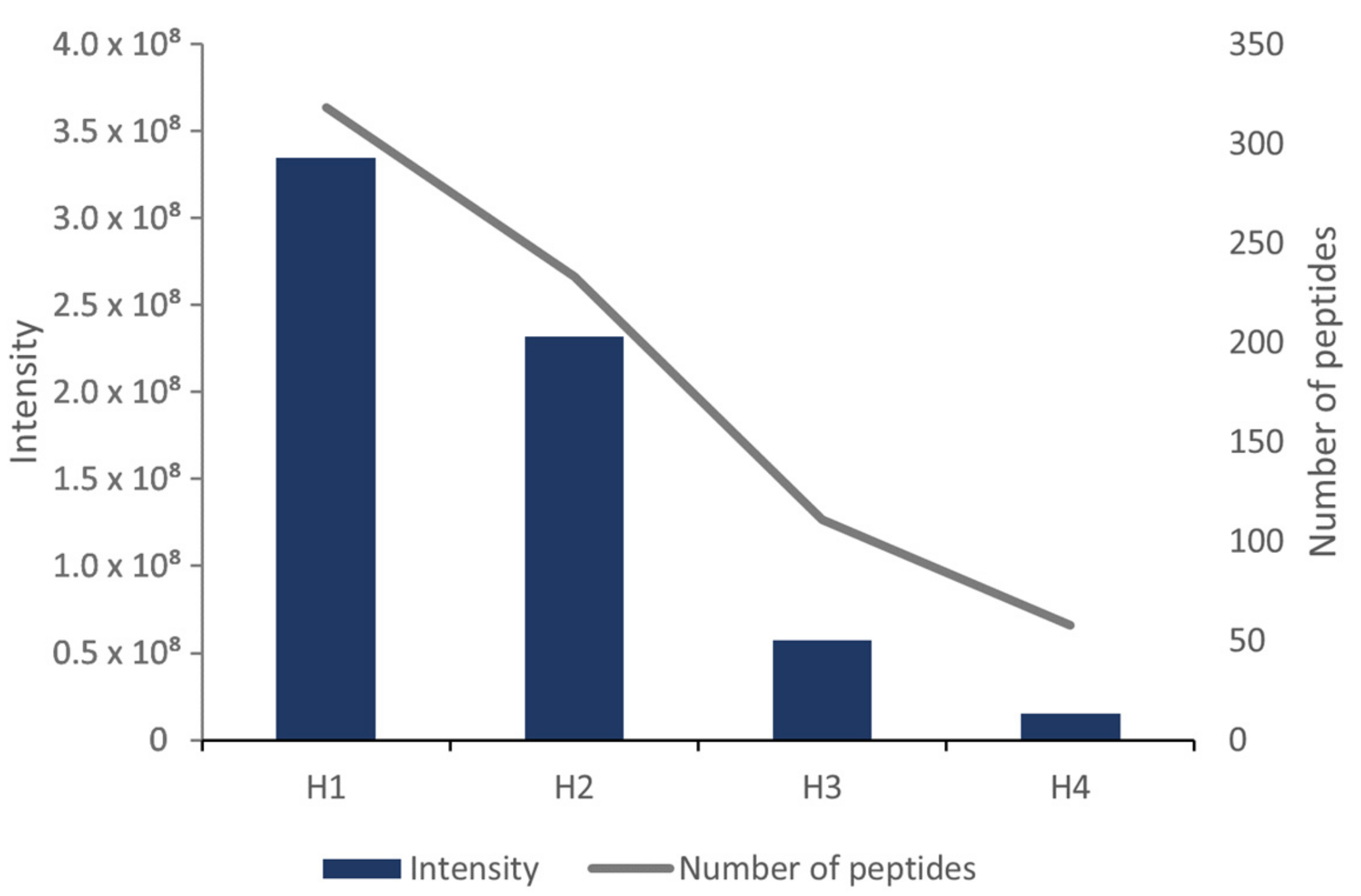
In addition to the robust analysis of the MW distribution of protein fractions, the analysis of peptides was carried out using LC-HRMS, to define the peptide composition more precisely, and to identify potential IgE-binding allergenic epitopes in the WPHs. Overall, 318, 233, 111, and 58 peptides were identified in the H1, H2, H3, and H4 hydrolysates, respectively (Figure 2). The summed intensity of all identified peptides was the highest in the sample with the highest degree of hydrolysis, H1, and it decreased gradually in the H2, H3, and H4 samples, accordingly (Figure 2).
The majority of the identified peptides were derived from the predominant milk whey proteins α-LA and β-LG (74.1–93.0% of the total intensity of peptides, depending on the sample). The exception to this observation was the hydrolysate H3, in which the peptides originating from α-LA accounted only for 1% of the total intensity of peptides (Table 2). Peptides originating from other whey proteins comprised up to 25.9% of the total intensity of peptides. These proteins included BSA and lactotransferrin, which can also induce milk allergy [2]. However, minor relative amounts of peptides originating from BSA and lactotransferrin were obtained, due to the low quantities of these proteins in milk. Therefore, the peptides from minor proteins will not be considered in further detail in this study.
The peptide length distribution showed that compared to the other hydrolysates, the H1 sample contained a higher amount of short (up to 4 amino acids long, approximately 450 Da) and average-length peptides (7–10 amino acids long, approximately 800–1100 Da) (Figure 3). In the H2 hydrolysate, the amount of identified peptides of 5–6 and 12–15 amino acids in length (approximately 1350–1650 Da) was higher than in the other hydrolysates.
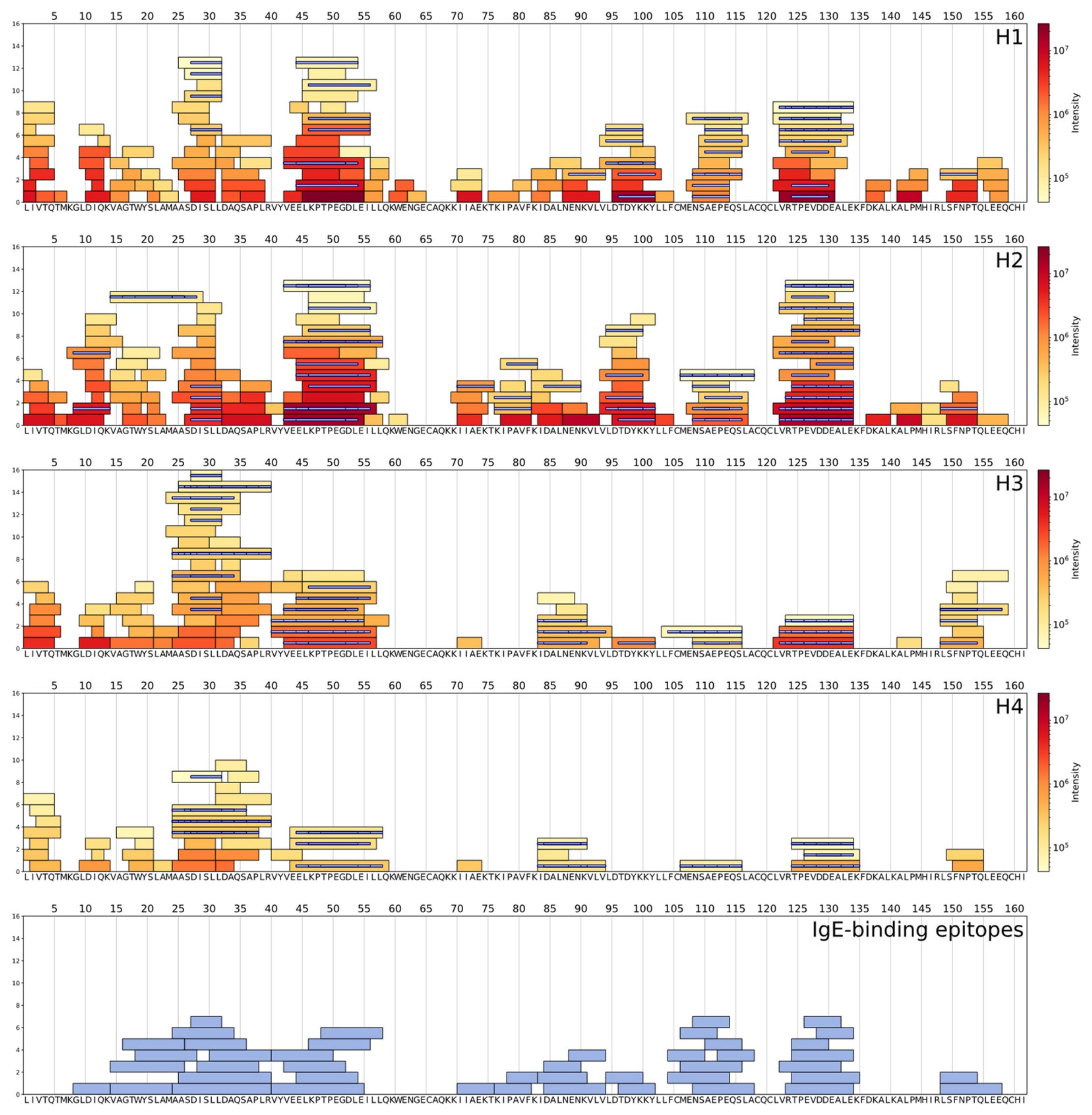
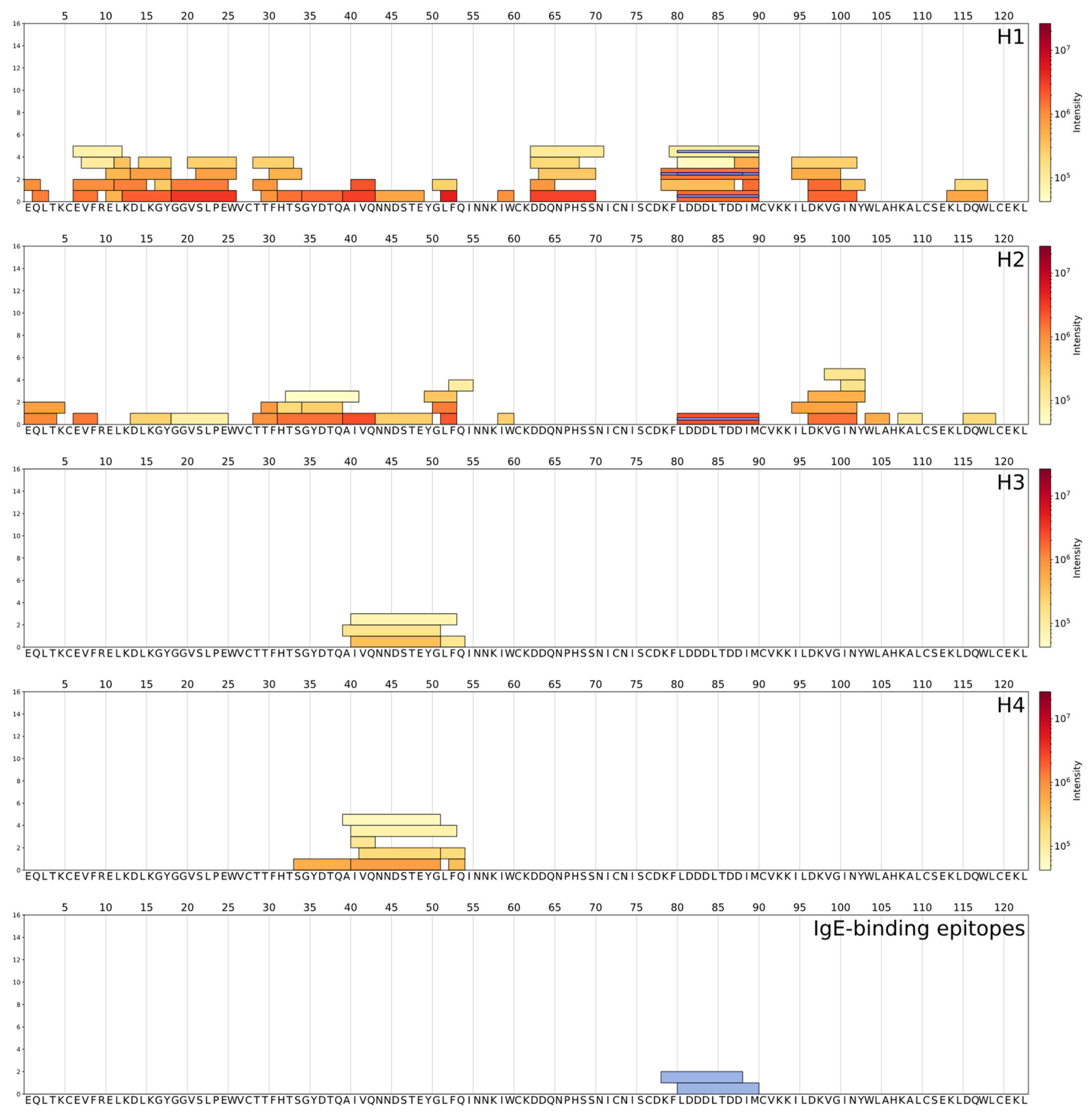
Peptide coverage profiles allow the visualization of hydrolysis by mapping all detected peptides on the corresponding protein sequence. Figure 4 shows the peptide coverage profiles of β-LG in the hydrolysate samples, obtained by the processing of LC-HRMS data, with the script written in Python. The Immune Epitope Database [40] was used for the search of the IgE-binding allergenic epitopes related to the identified peptides. Epitopes found in the WPHs were also mapped on the peptide coverage profiles of β-LG and α-LA proteins, using Python script (Python Software Foundation, version 3.5, available at http://www.python.org (accessed on 11 April 2023)) (Figure 4 and Figure 5).
In the H3 and H4 hydrolysates, most of the peptides originated from the N-terminal part of β-LG f1–60, whereas peptides from H1 and H2 were more evenly distributed along the whole protein sequence. In the case of α-LA, a few peptides identified in H4 and H3 originated from the central part of the α-LA sequence, while the coverage profiles of the H1 and H2 hydrolysates were covered by several peptides uniformly (Figure 5).
Overall, 78 epitopes from α-LA, and 209 IgE-binding allergenic epitopes from β-LG (T- and/or B-cell responses), are listed in the Immune Epitope Database [40]. In total, 44 different epitopes from β-LG were detected in the WPHs: 17 in H1, 30 in H2, 31 in H3, and 23 in H4 (Figure 4 and Figure 5, Table 3). Among the IgE-binding epitopes from α-LA found in the Immune Epitope Database, only two were found in the H1 and H2 hydrolysates. Altogether, 31 unique peptides from H1, 45 from H2, 27 from H3, and 13 from H4 were found to be related to these epitopes (Table 3).
In addition to the LC-HRMS and SE-UPLC analyses, the residual β-LG content in the WPHs was quantified in Romer Labs Diagnostic GmbH (Austria), using a commercial AgraQuant® enzyme-linked immunosorbent assay (ELISA) test kit, according to the manufacturer’s instructions. The amount of residual β-LG content was the lowest in the H1 hydrolysate (0.13 µg/g of WPH), the H2 sample contained a larger amount of β-LG compared to H1 (237 µg/g), and the most nonhydrolyzed samples showed a significantly larger β-LG content compared to the two previous samples (86,922 µg/g in H3 and 68,786 µg/g in H4) (Table 4).

4. Discussion

In this study, we provided a sequence-based characterization of commercial WPH peptide profiles, which enables the detection of specific peptides related to known IgE-binding allergenic epitopes listed in the Immune Epitope Database [40].
Overall, the results obtained from LC-HRMS (Figure 2, Figure 3, Figure 4 and Figure 5) were consistent with the findings from SE-UPLC, which showed a higher amount of low MW protein fractions (MW range of 500–3500 Da) in H1, followed by the H2, H3, and H4 hydrolysates (Table 1). This fraction was represented by the peptides identified using LC-HRMS, which also revealed a higher total intensity of peptides in H1 and H2, compared to H3 and H4. Furthermore, the results obtained in this study correspond with the information provided in the specification of the WPHs, where the degree of hydrolysis of the H1, H2, H3, and H4 hydrolysates was reported as 25–30%, 10–16%, 9–15%, and 6.5%, respectively. Several studies have shown a significant decrease in allergenicity in extensively hydrolyzed proteins, based on the binding capacity to IgE of sera from patients with CMA [27,47].
Nevertheless, the reduced allergenicity of hydrolysates does not depend only on the reduction in the size of peptides, but also on the content of the residual proteins [48]. The analyses showed that both the H3 and H4 hydrolysates contain a large amount of intact whey proteins (the fraction with MW > 13,500 Da) (Figure 1, Table 1). At the same time, the H3 and H4 hydrolysates contain a lower amount of β-LG-derived peptides, and few peptides from α-LA, compared to H1 and H2, indicating a lower degree of protein hydrolysis. The abovementioned intact protein fraction contains the major cow’s milk allergens, such as β-LG (MW 18,300 Da), α-LA (MW 14,200 Da), BSA (MW 66,400 Da), lactotransferrin (MW 76,200 Da), and Ig (MW > 150,000 Da), including a significant amount of IgE-binding epitopes, which can cause an allergenic reaction in infants with CMA [2]. The levels of residual β-LG in the WPHs measured using the ELISA test were in accordance with the MW distribution of protein fractions determined using SE-UPLC, and inversely proportional to the amount of peptides identified using LC-HRMS (Table 4). As expected, high levels of residual β-LG were measured in the H3 and H4 hydrolysates, with a lower extent of protein hydrolysis, and a lower amount of identified peptides.
To compare the amounts of β-LG in the WPHs measured using ELISA with cow’s milk and ready-for-consumption hypoallergenic IFs, the obtained values were recalculated to the amount of β-LG in ready-to-drink IF (Table 4). The estimated concentration of β-LG in ready-to-drink IFs, if prepared from the less proteolyzed H3 and H4 hydrolysates, was only 2–2.5 times lower than in mature cow’s milk, where β-LG concentration was approximately 4 × 10⁶ µg/L [11]. The calculated concentration of β-LG in ready-to-drink IFs produced from the H1 and H2 hydrolysates was significantly lower compared to cow’s milk. The results correspond with previous studies, showing that ready-to-drink partially hydrolyzed IF contained 31,200 µg/L, 48,180 µg/L, and 96,250 µg/L of β-LG, and extensively hydrolyzed IF—below 200 µg/L of β-LG [11,12]. Conventional IFs, containing nonhydrolyzed bovine milk proteins (both caseins and whey proteins) have shown similar residual β-LG values to ready-to-drink IFs based on the H3 and H4 hydrolysates (Table 4) with β-LG level of 1,320,000 µg/L [12].
A total of 287 IgE-binding allergenic epitopes (T- and/or B-cell responses) have been previously identified on bovine β-LG and α-LA sequences and included in the Immune Epitope Database [40]. Among these, several epitopes have been reported as major, which were recognized by the majority of IgE antibodies from human sera of patients with IgE-mediated milk allergy: f124-134 [20]; f97-108 [21]; f95-113 [22]; f1-8, f25-40, f41-60, f102-124, f149-162 [23]; f75-86, f127-146 [24]. The majority of IgE-binding epitopes from β-LG and α-LA range from 10 to 20 amino acids in length [40]. However, in this study, most of the identified epitopes were 5–15 amino acids long (Table 3). The more that peptides are hydrolyzed to smaller than 10-amino-acid-long fragments, the lesser the probability that they will bind with a specific IgE antibody to induce an allergenic reaction. Nevertheless, the results of the present study indicated that the presence of IgE-binding allergenic epitopes was not directly related to the extent of hydrolysis of the WPHs. As described earlier, 31, 45, 27, and 13 peptides from β-LG were found to be associated with 46 IgE-binding allergenic epitopes in the H1, H2, H3, and H4 hydrolysates, respectively (Figure 4 and Figure 5, Table 3). Furthermore, two major epitopes known to induce an IgE reaction, f25-40 (in H3 and H4) and f124-134 (in H1, H2, and H3) [20,23], were identified. The total intensity of peptides related to the epitopes comprised 17%, 39%, 24%, and 13% of all identified peptides in the H1, H2, H3, and H4 hydrolysates, respectively. Despite the fact that H1 did not contain intact α-LA and β-LG proteins, and was more hydrolyzed than the other WPHs, it still contained a significant proportion of peptides related to the IgE-binding epitopes.
Indeed, in this study, we demonstrated the absence of intact whey proteins in H1 (MW > 13,500 Da, Table 1) along with a higher content of α-LA- and β-LG-derived small peptides revealed using LC-HRMS (Figure 4 and Figure 5). The complete hydrolysis of intact whey proteins with trypsin was shown by Peñas et al. [4]. Also, it was shown that the application of high pressure was efficient to achieve complete hydrolysis of both major whey proteins by pepsin [47]. The inactivity of chymotrypsin against α-LA was revealed by Peñas et al. [4], who showed that the chymotrypsin used to produce WPHs mainly affects β-LG. α-LA seemed to be resistant to chymotrypsin activity [4], and this could be the case in the production of the H3 and H4 hydrolysates, where few α-LA related peptides were identified.
It is noteworthy that the enzyme specificity is very important in the hydrolysis of epitopes, and can decrease the WPH allergenicity. The cleavage sites with the production of peptides with amino acids Leu (L), Ile (I), and Met (M) at the C-terminal end were shown to be preferred by the enzymes chymotrypsin and elastase in β-LG [17]. These cleavages were found among the most frequent cleavage sites in the H1 and H3 hydrolysates (Figure 4), while trypsin was known to produce peptides with Lys (K) and Arg (R) at the C-terminal end [17]. Corresponding cleavage sites were identified among the abundant ones in the H2 and H4 hydrolysates (Figure 4).
The development of a hypoallergenic infant formula requires the selection of the hydrolysates with the appropriate extent of proteolysis. On one hand, it is important to achieve a sufficient degree of hydrolysis to remove the allergenic activity, but on the other hand, the proteins should not be totally hydrolyzed up to free amino acids, as this can result in bitterness and poor palatability of the hydrolysate- and amino acid-based IFs [49,50]. Moreover, some studies have shown that during protein digestion in the human intestine, short peptides (di- and tripeptides) are more rapidly absorbed in the blood through the peptide transport systems than free amino acids [17]. Hence, it is important not only to determine the allergenic potential of WPHs or Ifs, but also to achieve a more detailed comprehension of their specific peptide composition.

5. Conclusions

This study presented a sequence-based approach for the characterization of commercial WPHs, which in addition to the overall peptide composition and degree of hydrolysis, also enabled us to find known IgE-binding allergenic epitopes. Among the identified peptides widely scattered along the β-LG and the α-LA sequences, numerous peptides related to a total of 46 specific allergenic epitopes were found in all the WPHs, regardless of their extent of hydrolysis. LC-HRMS peptide mapping, combined with in silico tools, can be regarded as a fast and comprehensive alternative to classical methods for monitoring the differences in the potential allergenicity of WPHs. This approach can be applied to the science-based development and selection of hydrolysates with optimal peptide composition, to compose a safe formula for infants with, or at risk of developing, cow’s milk allergy.

Author Contributions

Conceptualization, A.T., T.K. and I.S.; methodology, A.T.; investigation, A.T. and M.-L.K.; resources, I.S. and M.T.; data curation, M.-L.K., T.K. and T.G.; writing—original draft preparation, A.T.; writing—review and editing, A.T., T.K., I.S. and M.-L.K.; visualization, A.T. and T.L.; supervision, M.-L.K. and T.K.; project administration, M.T. and T.G.; funding acquisition, T.G. All authors have read and agreed to the published version of the manuscript.

Funding

This research was funded by ERDF grant number 2014-2020.4.02.20-0247.

Institutional Review Board Statement

Not applicable.

Informed Consent Statement

Not applicable.

Data Availability Statement

Not applicable.

Acknowledgments

We express our sincere thanks to Georg Arju and Aleksandra Pahhomova for technical assistance with the LC-HRMS and SE-UPLC analyses.

Conflicts of Interest

The authors declare no conflict of interest.

References

  1. Knol, E.F.; de Jong, N.W.; Ulfman, L.H.; Tiemessen, M.M. Management of Cow’s Milk Allergy from an Immunological Perspective: What Are the Options? Nutrients 2019, 11, 2734. [Google Scholar] [CrossRef] [PubMed] [Green Version]
  2. Monaci, L.; Tregoat, V.; van Hengel, A.J.; Anklam, E. Milk allergens, their characteristics and their detection in food: A review. Eur. Food Res. Technol. 2006, 223, 149–179. [Google Scholar] [CrossRef]
  3. Sharma, S.; Kumar, P.; Betzel, C.; Singh, T.P. Structure and function of proteins involved in milk allergies. J. Chromatogr. B Biomed. Sci. Appl. 2001, 756, 183–187. [Google Scholar] [CrossRef] [PubMed]
  4. Peñas, E.; Préstamo, G.; Luisa Baeza, M.; Martínez-Molero, M.I.; Gomez, R. Effects of combined high pressure and enzymatic treatments on the hydrolysis and immunoreactivity of dairy whey proteins. Int. Dairy J. 2006, 16, 831–839. [Google Scholar] [CrossRef]
  5. Bu, G.; Luo, Y.; Chen, F.; Liu, K.; Zhu, T. Milk processing as a tool to reduce cow’s milk allergenicity: A mini-review. Dairy Sci. Technol. 2013, 93, 211–223. [Google Scholar] [CrossRef] [PubMed] [Green Version]
  6. Jeewanthi, R.K.C.; Lee, N.-K.; Paik, H.-D. Improved Functional Characteristics of Whey Protein Hydrolysates in Food Industry. Korean J. Food Sci. Anim. Resour. 2015, 35, 350–359. [Google Scholar] [CrossRef] [PubMed] [Green Version]
  7. Hays, T.; Wood, R.A. A Systematic Review of the Role of Hydrolyzed Infant Formulas in Allergy Prevention. Arch. Pediatr. Adolesc. Med. 2005, 159, 810. [Google Scholar] [CrossRef] [Green Version]
  8. Host, A.; Halken, S. Hypoallergenic formulas—When, to whom and how long: After more than 15 years we know the right indication! Allergy 2004, 59, 45–52. [Google Scholar] [CrossRef]
  9. Bøgh, K.L.; Barkholt, V.; Madsen, C.B. Characterization of the Immunogenicity and Allergenicity of Two Cow’s Milk Hydrolysates—A Study in Brown Norway Rats. Scand. J. Immunol. 2015, 81, 274–283. [Google Scholar] [CrossRef] [PubMed]
  10. Liang, X.; Wang, Z.; Yang, H.; Luo, X.; Sun, J.; Yang, M.; Shi, X.; Yue, X.; Zheng, Y. Evaluation of allergenicity of cow milk treated with enzymatic hydrolysis through a mouse model of allergy. J. Dairy Sci. 2022, 105, 1039–1050. [Google Scholar] [CrossRef]
  11. Mäkinen-Kiljunen, S.; Palosuo, T. A sensitive enzyme-linked immunosorbent assay for determination of bovine ?—Lactoglobulin in infant feeding formulas and in human milk. Allergy 1992, 47, 347–352. [Google Scholar] [CrossRef]
  12. Rosendal, A.; Barkholt, V. Detection of Potentially Allergenic Material in 12 Hydrolyzed Milk Formulas. J. Dairy Sci. 2000, 83, 2200–2210. [Google Scholar] [CrossRef] [PubMed]
  13. Puerta, A.; Diez-Masa, J.C.; de Frutos, M. Immunochromatographic determination of β-lactoglobulin and its antigenic peptides in hypoallergenic formulas. Int. Dairy J. 2006, 16, 406–414. [Google Scholar] [CrossRef] [Green Version]
  14. Oldæus, G.; Björkstén, B.; Jenmalm, M.; Kjellman, N.-I.M. Cow’s milk IgE and IgG antibody responses to cow’s milk formulas. Allergy 1999, 54, 352–357. [Google Scholar] [CrossRef] [PubMed]
  15. Bahna, S.L. Hypoallergenic formulas: Optimal choices for treatment versus prevention. Ann. Allergy Asthma Immunol. 2008, 101, 453–459. [Google Scholar] [CrossRef]
  16. Chan, Y.; Shek, L.; Aw, M.; Quak, S.; Lee, B. Use of hypoallergenic formula in the prevention of atopic disease among Asian children. J. Paediatr. Child Health 2002, 38, 84–88. [Google Scholar] [CrossRef] [PubMed]
  17. Siemensma, A.D.; Weijer, W.J.; Bak, H.J. The importance of peptide lengths in hypoallergenic infant formulae. Trends Food Sci. Technol. 1993, 4, 16–21. [Google Scholar] [CrossRef]
  18. Van Hoeyveld, E.M.; Escalona-Monge, M.; DE Swert, L.F.; Stevens, E.A. Allergenic and antigenic activity of peptide fragments in a whey hydrolysate formula: Allergenic and antigenic activity. Clin. Exp. Allergy 1998, 28, 1131–1137. [Google Scholar] [CrossRef]
  19. Businco, L.; Dreborg, S.; Einarsson, R.; Giampietro, P.G.; Hest, A.; Keller, K.M.; Strobel, S.; Wahn, U. Hydrolysed cow’s milk formulae: Allergenicity and use in treatment and prevention. An ESPACI position paper. Pediatr. Allergy Immunol. 1993, 4, 101–111. [Google Scholar] [CrossRef]
  20. Adams, S.L.; Barnett, D.; Walsh, B.J.; Pearce, R.J.; Hill, D.J.; Howden, M.E.H. Human IgE-binding synthetic peptides of bovine β-lactoglobulin and α-lactalbumin. In vitro cross-reactivity of the allergens. Immunol. Cell Biol. 1991, 69, 191–197. [Google Scholar] [CrossRef]
  21. Ball, G.; Shelton, M.J.; Walsh, B.J.; Hill, D.J.; Hosking, C.S.; Howden, M.E.H. A major continuous allergenic epitope of bovine beta-lactoglobulin recognized by human IgE binding. Clin. Htmlent Glyphamp Asciiamp Exp. Allergy 1994, 24, 758–764. [Google Scholar] [CrossRef] [PubMed]
  22. Heinzmann, A.; Blattmann, S.; Spuergin, P.; Forster, J.; Deichmann, K.A. The Recognition Pattern of Sequential B Cell Epitopes of Beta–Lactoglobulin Does Not Vary with the Clinical Manifestations of Cow’s Milk Allergy. Int. Arch. Allergy Immunol. 1999, 120, 280–286. [Google Scholar] [CrossRef] [PubMed]
  23. Sélo, I.; Clément, G.; Bernard, H.; Chatel, J.; Créminon, C.; Peltre, G.; Wal, J. Allergy to bovine β-lactoglobulin: Specificity of human IgE to tryptic peptides. Clin. Exp. Allergy 1999, 29, 1055–1063. [Google Scholar] [CrossRef] [PubMed]
  24. Järvinen, K.-M.; Chatchatee, P.; Bardina, L.; Beyer, K.; Sampson, H.A. IgE and IgG Binding Epitopes on α-Lactalbumin and β-Lactoglobulin in Cow’s Milk Allergy. Int. Arch. Allergy Immunol. 2001, 126, 111–118. [Google Scholar] [CrossRef]
  25. Cerecedo, I.; Zamora, J.; Shreffler, W.G.; Lin, J.; Bardina, L.; Dieguez, M.C.; Wang, J.; Muriel, A.; de la Hoz, B.; Sampson, H.A. Mapping of the IgE and IgG4 sequential epitopes of milk allergens with a peptide microarray–based immunoassay. J. Allergy Clin. Immunol. 2008, 122, 589–594. [Google Scholar] [CrossRef]
  26. Wang, J.; Lin, J.; Bardina, L.; Goldis, M.; Nowak-Węgrzyn, A.; Shreffler, W.G.; Sampson, H.A. Correlation of IgE/IgG4 milk epitopes and affinity of milk-specific IgE antibodies with different phenotypes of clinical milk allergy. J. Allergy Clin. Immunol. 2010, 125, 695–702.e6. [Google Scholar] [CrossRef] [Green Version]
  27. Benedé, S.; López-Expósito, I.; Giménez, G.; Grishina, G.; Bardina, L.; Sampson, H.A.; López-Fandiño, R.; Molina, E. Mapping of IgE epitopes in in vitro gastroduodenal digests of β-lactoglobulin produced with human and simulated fluids. Food Res. Int. 2014, 62, 1127–1133. [Google Scholar] [CrossRef]
  28. Oliveira, J.P.B.; Candreva, A.M.; Rizzo, G.; Ramos, M.V.; Oliveira, J.S.; Oliveira, H.D.; Ary, M.B.; Docena, G.; Freitas, C.D.T. Allergenicity reduction of cow’s milk proteins using latex peptidases. Food Chem. 2019, 284, 245–253. [Google Scholar] [CrossRef]
  29. Zhang, Q.; Chen, Q.-H.; He, G.-Q. Effect of ultrasonic-ionic liquid pretreatment on the hydrolysis degree and antigenicity of enzymatic hydrolysates from whey protein. Ultrason. Sonochem. 2020, 63, 104926. [Google Scholar] [CrossRef]
  30. Dąbrowska, A.; Bajzert, J.; Babij, K.; Szołtysik, M.; Stefaniak, T.; Willak-Janc, E.; Chrzanowska, J. Reduced IgE and IgG antigenic response to milk proteins hydrolysates obtained with the use of non-commercial serine protease from Yarrowia lipolytica. Food Chem. 2020, 302, 125350. [Google Scholar] [CrossRef]
  31. Lozano-Ojalvo, D.; Pérez-Rodríguez, L.; Pablos-Tanarro, A.; López-Fandiño, R.; Molina, E. Pepsin treatment of whey proteins under high pressure produces hypoallergenic hydrolysates. Innov. Food Sci. Emerg. Technol. 2017, 43, 154–162. [Google Scholar] [CrossRef]
  32. Ji, J.; Zhu, P.; Pi, F.; Sun, C.; Sun, J.; Jia, M.; Ying, C.; Zhang, Y.; Sun, X. Development of a liquid chromatography-tandem mass spectrometry method for simultaneous detection of the main milk allergens. Food Control 2017, 74, 79–88. [Google Scholar] [CrossRef]
  33. Pescuma, M.; Hébert, E.M.; Rabesona, H.; Drouet, M.; Choiset, Y.; Haertlé, T.; Mozzi, F.; de Valdez, G.F.; Chobert, J.-M. Proteolytic action of Lactobacillus delbrueckii subsp. bulgaricus CRL 656 reduces antigenic response to bovine β-lactoglobulin. Food Chem. 2011, 127, 487–492. [Google Scholar] [CrossRef] [PubMed]
  34. Zhao, Q.; Wang, Y.; Zhu, Z.; Zhao, Q.; Zhu, L.; Jiang, L. Efficient reduction of β-lactoglobulin allergenicity in milk using Clostridium tyrobutyricum Z816. Food Sci. Hum. Wellness 2023, 12, 809–816. [Google Scholar] [CrossRef]
  35. Planque, M.; Arnould, T.; Dieu, M.; Delahaut, P.; Renard, P.; Gillard, N. Advances in ultra-high performance liquid chromatography coupled to tandem mass spectrometry for sensitive detection of several food allergens in complex and processed foodstuffs. J. Chromatogr. A 2016, 1464, 115–123. [Google Scholar] [CrossRef]
  36. Fan, S.; Ma, J.; Liu, Z.; Ning, Y.; Cao, M.; Li, Q.; Zhang, Y. Determination of egg and milk allergen in food products by liquid chromatography-tandem mass spectrometry based on signature peptides and isotope-labeled internal standard. Food Sci. Hum. Wellness 2023, 12, 728–736. [Google Scholar] [CrossRef]
  37. Ma, X.; Li, H.; Zhang, J.; Huang, W.; Han, J.; Ge, Y.; Sun, J.; Chen, Y. Comprehensive quantification of sesame allergens in processed food using liquid chromatography-tandem mass spectrometry. Food Control 2020, 107, 106744. [Google Scholar] [CrossRef]
  38. Kang, W.; Zhang, J.; Li, H.; Yu, N.; Tang, R.; Sun, X.; Wei, L.; Sun, J.; Chen, Y. Quantification of major allergens in peach based on shotgun proteomics using liquid chromatography-tandem mass spectrometry. LWT 2022, 160, 113234. [Google Scholar] [CrossRef]
  39. Arju, G.; Berg, H.Y.; Lints, T.; Nisamedtinov, I. Methodology for Analysis of Peptide Consumption by Yeast during Fermentation of Enzymatic Protein Hydrolysate Supplemented Synthetic Medium Using UPLC-IMS-HRMS. Fermentation 2022, 8, 145. [Google Scholar] [CrossRef]
  40. Vita, R.; Mahajan, S.; Overton, J.A.; Dhanda, S.K.; Martini, S.; Cantrell, J.R.; Wheeler, D.K.; Sette, A.; Peters, B. The Immune Epitope Database (IEDB): 2018 update. Nucleic Acids Res. 2019, 47, D339–D343. [Google Scholar] [CrossRef] [Green Version]
  41. Järvinen, K.-M.; Beyer, K.; Vila, L.; Chatchatee, P.; Busse, P.J.; Sampson, H.A. B-cell epitopes as a screening instrument for persistent cow’s milk allergy. J. Allergy Clin. Immunol. 2002, 110, 293–297. [Google Scholar] [CrossRef]
  42. Hattori, M.; Ametani, A.; Katakura, Y.; Shimizu, M.; Kaminogawa, S. Unfolding/refolding studies on bovine beta-lactoglobulin with monoclonal antibodies as probes. Does a renatured protein completely refold? J. Biol. Chem. 1993, 268, 22414–22419. [Google Scholar] [CrossRef] [PubMed]
  43. Li, X.; Yuan, S.; He, S.; Gao, J.; Chen, H. Identification and characterization of the antigenic site (epitope) on bovine β-lactoglobulin: Common residues in linear and conformational epitopes. J. Sci. Food Agric. 2015, 95, 2916–2923. [Google Scholar] [CrossRef] [PubMed]
  44. Cong, Y.J.; Li, L.F. Identification of the critical amino acid residues of immunoglobulin E and immunoglobulin G epitopes in β-lactoglobulin by alanine scanninapplsci-13-07402g analysis. J. Dairy Sci. 2012, 95, 6307–6312. [Google Scholar] [CrossRef] [PubMed] [Green Version]
  45. Totsuka, M.; Ametani, A.; Kaminogawa, S. Fine mapping of T-cell determinants of bovine β-lactoglobulin. Cytotechnology 1997, 25, 101–113. [Google Scholar] [CrossRef]
  46. Gouw, J.W.; Jo, J.; Meulenbroek, L.A.P.M.; Heijjer, T.S.; Kremer, E.; Sandalova, E.; Knulst, A.C.; Jeurink, P.V.; Garssen, J.; Rijnierse, A.; et al. Identification of peptides with tolerogenic potential in a hydrolysed whey-based infant formula. Clin. Exp. Allergy 2018, 48, 1345–1353. [Google Scholar] [CrossRef] [Green Version]
  47. Chicón, R.; Belloque, J.; Alonso, E.; López-Fandiño, R. Antibody binding and functional properties of whey protein hydrolysates obtained under high pressure. Food Hydrocoll. 2009, 23, 593–599. [Google Scholar] [CrossRef]
  48. Svenning, C.; Brynhildsvold, J.; Molland, T.; Langsrud, T.; Elisabeth Vegarud, G. Antigenic response of whey proteins and genetic variants of β-lactoglobulin—The effect of proteolysis and processing. Int. Dairy J. 2000, 10, 699–711. [Google Scholar] [CrossRef]
  49. Leksrisompong, P.P.; Miracle, R.E.; Drake, M. Characterization of Flavor of Whey Protein Hydrolysates. J. Agric. Food Chem. 2010, 58, 6318–6327. [Google Scholar] [CrossRef]
  50. Miraglia Del Giudice, M.; D’Auria, E.; Peroni, D.; Palazzo, S.; Radaelli, G.; Comberiati, P.; Galdo, F.; Maiello, N.; Riva, E. Flavor, relative palatability and components of cow’s milk hydrolysed formulas and amino acid-based formula. Ital. J. Pediatr. 2015, 41, 42. [Google Scholar] [CrossRef] [Green Version]
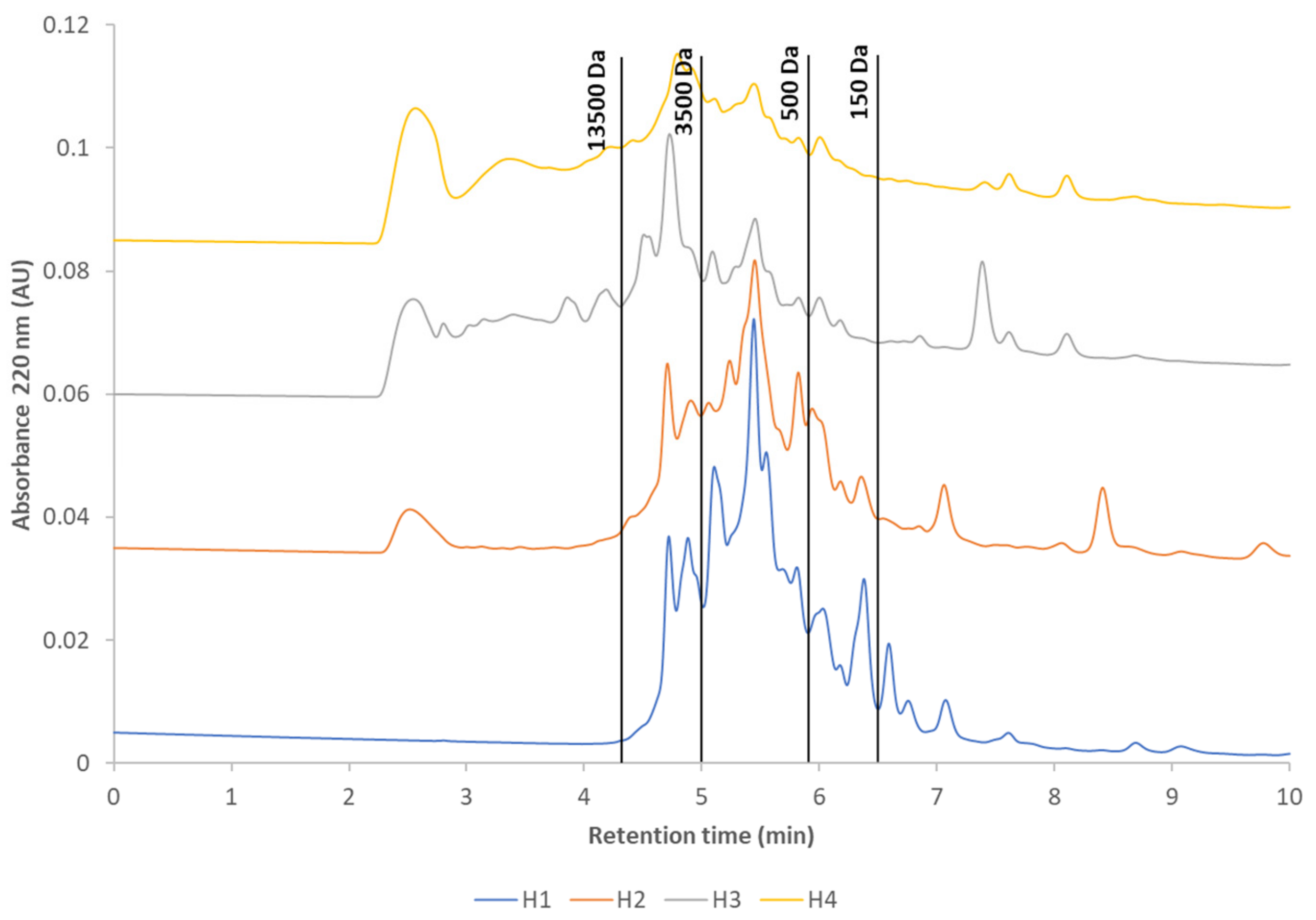
Figure 1. The SE-UPLC elution profiles of the WPHs. UV detection was at 220 nm. The blue line is H1, the orange line is H2, the gray line is H3, and the yellow line is H4. Using the standards, the calibration curve was constructed for setting the tentative MW boundaries for different protein fractions.
Applsci 13 07402 g001
Figure 2. Total number and summed intensity of all identified peptides in the H1, H2, H3, and H4 WPHs, determined using LC-HRMS analysis. The summed intensity of peptides was normalized against the average protein content of each sample.
Applsci 13 07402 g002
Figure 3. Distribution of the length of peptides identified in the H1, H2, H3, and H4 hydrolysates using LC-HRMS.
Applsci 13 07402 g003
Figure 4. β-LG derived peptides identified using LC-HRMS in the H1, H2, H3, and H4 hydrolysates. X-axis: the amino acid sequence of β-LG. Y-axis: the number of peptides. The intensity of peptides is shown in the scale on the right. Entire IgE-binding allergenic epitopes included in the sequences of identified peptides, and derived from the Immune Epitope Database, are marked with blue bars on the peptides of each hydrolysate, and mapped separately on the bottom pattern of β-LG sequence.
Applsci 13 07402 g004
Figure 5. α-LA derived peptides identified using LC-HRMS in the H1, H2, H3, and H4 hydrolysates. X-axis: the amino acid sequence of α-LA. Y-axis: the number of peptides. The intensity of peptides is shown in the scale on the right. Entire IgE-binding allergenic epitopes included in the sequences of identified peptides, and derived from the Immune Epitope Database, are marked with blue bars on the peptides of each hydrolysate, and mapped separately on the bottom pattern of α-LA sequence.
Applsci 13 07402 g005
Table 1. Molecular weight (MW) distribution of protein fractions in WPHs, determined using SE-UPLC.
% of Total Area (Mean ± SD)
MW, DaRT, Min *H1H2H3H4
>13,5000–4.40.0 ± 0.06.0 ± 0.435.4 ± 0.734.0 ± 1.0
3500–13,5004.4–5.017.8 ± 0.119.5 ± 0.224.4 ± 0.820.3 ± 0.0
500–35005.0–5.954.2 ± 0.242.8 ± 1.021.5 ± 0.221.1 ± 0.1
150–5005.9–6.516.9 ± 0.115.7 ± 1.17.5 ± 0.38.1 ± 0.1
<1506.5–15.011.1 ± 0.016.1 ± 0.311.2 ± 1.016.5 ± 0.8
* retention time.
Table 2. Distribution of peptides in WPHs (% of the total intensity of identified peptides).
% of the Total Intensity
ProteinH1H2H3H4
α-LA12.86.71.013.0
β-LG61.385.173.380.0
BSA4.91.71.80.9
Apolipoprotein1.50.83.91.3
Complement C35.32.16.34.4
Fibrinogen alpha1.50.52.00.0
Lactoperoxidase2.51.08.30.0
Lactotransferrin8.01.12.10.5
Osteopontin2.40.91.40.0
Table 3. Peptides from α-LA and β-LG identified in WPHs, and entire IgE-binding allergenic epitopes found from the Immune Epitope Database [40].
Protein (UniProt Database ID)Identified PeptidesWPHEpitope SequenceEpitope Location in Protein ChainImmune Epitope Database IDReferences
α-LA (P00711)f79-90H1KFLDDDLTDDf79-88115308[24]
f81-90
f80-90
f79-90
H1, H2
H1
H1
LDDDLTDDIMf81-90115340[24]
β-LG (P02754)f25-40H3, H4AASDISLLDAQSAPLRf25-40426[23]
f9-14
f8-14
H2
H2
GLDIQKf9-1420801[23]
f84-91
f84-93
f84-95
f84-94
H3, H4
H3
H3
H4
IDALNENKf84-9125535[23]
f46-56
f46-57
f47-57
f45-57
f43-56
f43-57
f43-58
f41-57
f44-57
f44-58
f44-59
H1, H2
H1, H2, H3
H2
H2, H3
H2
H2, H3
H2
H3
H4
H4
H4
KPTPEGDLEIf47-5632907[24,41]
f78-83H2PAVFKf79-8346987[23]
f123-134
f122-134
f124-134
H1, H2, H3
H1, H2, H3
H2, H3
RTPEVDDEALEf124-13456145[20]
f125-135H2, H4TPEVDDEALEKf125-13565565[22,23,42]
f41-55
f41-57
H3
H3
VYVEELKPTPf41-5072177[24]
f111-116
f110-116
f110-117
f109-116
f108-117
f109-117
f107-118
f104-116
f107-116
H1
H1
H1, H2
H1, H3
H1, H2
H2
H2
H3
H4
AEPEQSf111-11695218[21]
f83-90
f84-91
f84-93
f84-95
f84-94
H2
H3, H4
H3
H3
H4
DALNENf85-9095300[21]
f123-134
f122-134
f128-134
f127-134
f125-134
f125-135
f124-134
f127-135
H1, H2, H3
H1, H2, H3
H2
H2
H2, H4
H2, H4
H2, H3
H4
DDEALEf129-13495306[21]
f88-94
f84-95
f84-94
H1
H3
H4
ENKVLVf89-9495357[21]
f122-132
f123-133
f123-134
f122-134
f127-134
f125-134
f125-135
f124-134
f127-135
H1
H1
H1, H2, H3
H1, H2, H3
H2
H2, H4
H2, H4
H2, H3
H4
EVDDEAf127-13295369[21]
f104-116H3FCMENSf105-11095378[21]
f71-76H2IIAEKTf71-7695461[21]
f77-82
f76-82
H2
H2
KIPAVFf77-8295498[21,43]
f95-100
f94-100
f94-102
f94-101
H1, H2
H1, H2
H1, H2
H2
LDTDYKf95-10095545[21]
f149-154
f149-155
f149-159
H1, H2, H3
H3
H3
LSFNPTf149-15495574[21]
f107-118
f104-116
f107-116
H2
H3
H4
MENSAEf107-11295590[21]
f109-114
f108-114
f109-116
f108-117
f109-117
f107-118
f104-116
f107-116
H1, H2
H1
H1, H3
H1, H2
H2
H2
H3
H4
NSAEPEf109-11495638[21]
f107-118H2PEQSLAf113-11895652[21]
f96-102
f96-103
f94-102
H1, H2, H3
H1
H1, H2
TDYKKYf97-10295898[21]
f124-131
f123-131
f122-131
f122-132
f123-133
f123-134
f122-134
f125-134
f125-135
f124-134
H1, H2
H1, H2
H1, H2
H1
H1
H1, H2, H3
H1, H2, H3
H2, H4
H2, H4
H2, H3
TPEVDDf125-13095922[21]
f15-29H2VAGTWYSLAMAAf15-2695946[21]
f26-40
f25-40
H3
H3, H4
ASDISLLDAQSAPLRf26-4096113[44]
f41-55
f41-57
H3
H3
VYVEELKPTPEGDLEf41-5597098[45]
f25-35
f24-35
f25-40
f25-36
f25-38
H3
H3
H3, H4
H4
H4
AASDISLLDAf25-3498677[24]
f84-95
f84-94
H3
H4
DALNENKVLVf85-9498723[24]
f45-54
f45-55
f43-55
f45-57
f43-56
f43-57
f43-58
f41-55
f41-57
f44-57
f44-58
f44-59
H1
H1, H2
H1, H2, H3
H2, H3
H2
H2, H3
H2
H3
H3
H4
H4
H4
ELKPTPEGDLf45-5498760[24]
f104-116H3FCMENSAEPEf105-11498772[24]
f26-40
f25-40
f25-38
H3
H3, H4
H4
ISLLDAQSAPf29-3898836[24]
f26-40
f25-40
H3
H3, H4
LLDAQSAPLRf31-4098881[24]
f149-159H3LSFNPTQLEEf149-15898893[24]
f107-118
f104-116
f107-116
H2
H3
H4
MENSAEPEQSf107-11698902[24]
f107-118H2NSAEPEQSLAf109-11898919[24]
f26-40
f25-40
f25-36
f25-38
H3
H3, H4
H4
H4
SDISLLDAQSf27-3698997
f43-58
f44-58
f44-59
H2
H4
H4
TPEGDLEILLf49-5899028[24]
f43-55
f43-56
f43-57
f43-58
f41-55
f41-57
H1, H2, H3
H2
H2, H3
H2
H3
H3
VEELKPTPEGf43-5299036[24]
f122-132
f123-133
f123-134
f122-134
H1
H1
H1, H2, H3
H1, H2, H3
VRTPEVDDEAf123-13299049[24]
f15-29H2WYSLAMAASDf19-2899061[24]
f15-29H2GTWYSLAMAAf17-26115275[24]
f123-134
f122-134
f125-134
f125-135
f124-134
H1, H2, H3
H1, H2, H3
H2, H4
H2, H4
H2, H3
TPEVDDEALEf125-134115519[24]
f15-29H2VAGTWYSLAMf15-24115530[24]
f28-32
f27-32
f26-32
f25-32
f26-35
f25-35
f24-35
f26-40
f25-40
f25-36
f25-38
H1, H2, H3
H1, H2, H3
H1, H2, H3
H2, H3, H4
H3
H3
H3
H3
H3, H4
H4
H4
DISLLf28-32851640[46]
Table 4. β-LG content in WPHs, IF powder, and ready-to-drink IFs based on WPHs 1.
WPHµg/g (Mean ± SD)µg/g IF Powderµg/L in Ready-to-Drink IF
H10.13 ± 0.070.022.45
H2237 ± 118395049
H386,922 ± 43,46113,0381,786,252
H468,786 ± 34,39293801,285,042
1 Calculated concentration of β-LG in IF powder and ready-to-drink IFs based on the H1, H2, H3, and H4 hydrolysates, taking into account that for its preparation, 137 g/L of powder is needed according to the manufacturer’s instructions; the average protein content in powder IF is 12 g/100 g, according to the Commission Directive 2006/141/EC on IF and follow-on formulas; the average protein content in the WPHs is 87% (H1), 77% (H2), 80% (H3), and 88% (H4), according to the specification.
Disclaimer/Publisher’s Note: The statements, opinions and data contained in all publications are solely those of the individual author(s) and contributor(s) and not of MDPI and/or the editor(s). MDPI and/or the editor(s) disclaim responsibility for any injury to people or property resulting from any ideas, methods, instructions or products referred to in the content.