Article

Ameliorative Effect of Omega-3-Rich Fish Diet on the Neurotoxic Effects of Propionic Acid in a Rodent Model of Autism

by
Nouf Saad Alsaqer
1,2,
Doha M. Al-Nouri
1,*,
Ramesa Shafi Bhat
3,
Shaista Arzoo
1,*,
Laila Naif Al-Harbi
1,
Manal Abdulaziz Bin Obead
1,
Abdullah Yaseen Almubarak
4,
Hisham Alkhalidi
5,
Ahmad Almotairi
5 and
Afaf Kamal El-Din El-Ansary
6
1
Department of Food and Nutrition Sciences, College of Food and Agriculture Sciences, King Saud University, Riyadh 11495, Saudi Arabia
2
Clinics Department, Applied Medical Sciences College, King Saud University, Riyadh 11495, Saudi Arabia
3
Department of Biochemistry, College of Science, King Saud University, Riyadh 11495, Saudi Arabia
4
Experimental Surgery & Animal Laboratory, College of Medicine, King Saud University, Riyadh 11495, Saudi Arabia
5
Department of Pathology, College of Medicine, King Saud University, Riyadh 11495, Saudi Arabia
6
Central Research Laboratory, Female Centre for Medical Studies and Scientific Section, King Saud University, Riyadh 11495, Saudi Arabia
*
Authors to whom correspondence should be addressed.
Appl. Sci. 2023, 13(13), 7392; https://doi.org/10.3390/app13137392
Submission received: 7 March 2023 / Revised: 13 June 2023 / Accepted: 20 June 2023 / Published: 21 June 2023

Abstract

:
Despite the increased social and financial burden associated with autism spectrum disorder (ASD), no particular treatment for this illness has been identified. A detailed examination of prior trials conducted to treat autism revealed that nutrition intervention was commonly utilised as an additional method of therapy. Indeed, the early detection of nutritional deficiencies and metabolic problems, together with appropriate therapeutic measures, can be a cornerstone for enhancing the metabolic and behavioural abilities of individuals with autism. In this work, a propionic acid (PPA)-induced rodent model of ASD was fed Spangled emperor (Lethrinus nebuloses), Dusky grouper (Epinephelus marginatus), and Parrot Fish (Scaridae), which are locally named Hammour, Shour, and Hareed, respectively, in Saudi Arabia. The aim of this study was to investigate the effect of dietary intervention with three kinds of whole fish (Lethrinus nebuloses, Epinephelus marginatus, and Scaridae), as a rich source of ω-3 fatty acids, on selected biochemical markers (reduced glutathione (GSH), thiobarbituric acid reactive substances (TBARS), creatine kinase (CK), lactate dehydrogenase (LDH), dopamine) together with a histopathological examination of the cerebellum and hippocampus as neurotoxic features of propionic acid in a rodent model of autism. Briefly, our findings give preliminary evidence in favour of employing fish as a rich supply of omega-3 fatty acids to reduce the neurotoxic effect of a PPA-induced ASD in a rat model. It may be beneficial to provide an extra marine omega-3-rich diet for improving certain metabolic autistic features related to oxidative stress, energy metabolism, and brain neurotransmitters.

1. Introduction

Autism spectrum disorder (ASD) is characterised by a set of persistent symptoms that can range in severity from common and acute to significantly low or high performance [1,2]. ASD includes neurological developmental disorders, of which stages start throughout childhood and the development stage and last the entirety of a person’s lifespan. There may be a number of factors that affect the development of autism, although the precise cause of ASD is currently unknown. The condition is influenced by genetic [3,4,5], environmental [4,5], and gene expression abnormalities [6]. Whatever the cause, early identification and therapy offer opportunities for intervention and recovery [1]. Vital indicators of autism include oxidative stress, extreme glutamate toxicity, polyneuritis, weak fatty acid and energy metabolism, and excessive propionic acid. These indicators can all point to treatable abnormalities and provide a baseline that we can use to assist autistic people and lessen their symptoms [7].
The human body cannot synthesise α-linolenic acid (ALA) or linoleic acid (LA), also known as essential fatty acids, so they must be consumed in the diet. Plant oils are rich in an omega-3 fatty acid called α-linolenic acid, which is a metabolic precursor for eicosapentaenoic acid (EPA), docosapentaenoic acid (DPA), and docosahexaenoic acid (DHA) (which are found in fish and fish oils) in the human body [8]. Both omega-3 and omega-6 pathways utilise the same enzymes, and both of them have to compete for the same enzymes in order to produce their final products. The benefits of DHA depend on dietary intake because of the low conversion rates of ALA to EPA and DPA and little to no DHA synthesis [9]. Omega-3 PUFAs improve the symptoms of metabolic disorders and are beneficial for various chronic diseases, such as diabetes, insulin resistance, heart disease, and obesity. Diets high in n-3 PUFA has been found to be involved in increasing the expression of apolipoprotein A-I in lipid metabolism, S-adenosylmethionine synthase in one carbon metabolism, fructose-1, 6-bisphosphatase, ketohexokinase and 6-phosphogluconolactonase in carbohydrate metabolism, cytosolic malate dehydrogenase, GTP-specific succinyl CoA synthase beta subunit and ornithine aminotransferase in the citric acid cycle, and disulfide isomerase-A3 and lactoylglutathione lyase in protein synthesis. Apart from this, they reduce the expression of regucalcin in lipid metabolism, aldehyde dehydrogenase in protein synthesis, and adenosine kinase in one carbon metabolism [10].
Nearly all ASD cases are reported to have an omega-3 fatty acid deficiency, especially for children having low levels of total omega-3 fatty acids in their plasma [11]. Moreover, it has been revealed that the red blood cell membranes of pervasive developmental disorder patients have insufficient levels of eicosapentaenoic acid/docosahexaenoic acid (EPA/DHA) [12]. Amminger et al. [13] reported an improvement in stereotypy (the repetitive repetition of an act) and hyperactivity in autistic children when put on a DHA/EPEPA-rich diet. Long-chain omega-3 supplements have been proven to benefit not only autism but also some neurological conditions, such as dyspraxia, dyslexia, and aggressiveness [14]. Omega-3 long-chain polyunsaturated fatty acids are structural components of cells present in the central nervous system [15]. These fatty acids play a major role in development of neurons, synapses, and visual acuity and regulate gene expression levels [16,17]. They can normalise oxidative stress and anti-inflammatory pathways [18]. D-resolvins, protectins, and maresins, from DHA, are key lipid mediators in the resolution of inflammation [19], and neuroprotection D1, in particular, suppresses neuronal death [20]. Although EPA is found in neural cell membranes in lesser concentrations than DHA, it serves key physiological roles as a source of anti-inflammatory chemicals, including PGE3, LTB5, and E-resolvins [21,22].
Various studies in animals have investigated the consequences of polyunsaturated fatty acid (PUFA) deficiencies in neurodevelopment. Both in vitro and ex vivo studies with cultured mouse neurons have shown that PUFAs play an important role in dendritic growth and the synaptogenesis of neuronal cells [23].
Bazinet and Layé (2014) [24] have studied the effect of nutritional intervention and its role in inflammatory control. They reported that dietary insufficiency may be a risk factor for ASD. Omega-6 (ω-6) PUFAs, such as arachidonic acid (AA) and linoleic acid (LA), have been thought to be proinflammatory, whereas ω-3 PUFAs are thought to be anti-inflammatory and/or pro-resolving [25]. Since fish is a significant source of omega-3 fatty acids (n-3 FAs), which are found in the membranes of brain tissue as well [26,27], studies are exploring their nutritional value as a major preventive for neurodevelopmental disorders. Moreover, fishery products are advised as dietary sources because eating fish is linked to a decreased risk of vascular brain disease [28]. Dietary fish are found to decrease abnormalities in nerve fibres and enlarged fluid-filled spaces around blood vessels in the brain’s white matter. Fish eaters are likely to have much lower prevalence of vascular brain disease. Fish is rich in some essential nutrients or minerals, such as selenium, vitamin D, and iodine, which are essential for the growth and development of the brain [29]. According to a recent study on dietary fish intake, fish protein and oil feed can prevent age-related short-term memory loss in mice. The findings also showed that fish feed have physiological advantages for preserving axon shape in the hippocampus and lowering the risk of brain damage [30]. Several studies have demonstrated the role of omega-3 fatty acids in behaviour and reading skills [31,32]. Almost 30% of kids with autism are put on fish oil supplements. It has been observed that fish oil supplements can reduce hyperactive behaviour in autistic patients [33,34]. Figure 1 illustrates the involvement of dietary omega-3 fatty acids in various physiological functions related to the development and functioning of the brain and its relationship with a decreased risk of developing various mental disorders, especially autism.
The three different types of sea fish (Lethrinus nebuloses, Epinephelus marginatus, and Scaridae), which is widely consumed in the Kingdom of Saudi Arabia, have been used in the present study and were analysed for their fatty-acid profile prior to use. One strategy that could aid in figuring out the process by which autism arises in humans is animal modelling. Presently, we used the PPA-induced rat model of autism. Propionic acid (PPA) has frequently been documented to cause a range of oxidative stresses, altered neurotransmission, impaired energy metabolism, and behavioural abnormalities in rats, which are similar to ASD [35,36]. This dietary short-chain fatty acid is produced metabolically by enteric bacteria in the stomach and is a common food preservative. Blood–brain barriers (BBB) and the gut–blood barrier (GBB) are both easily crossed by PPA, allowing it to enter the central nervous system (CNS). It can penetrate cell membranes in the brain and build up inside of cells, causing intracellular acidification, which can change neurotransmitter release and, eventually, affect neuronal activity and communication [35,36]. All these findings motivated us to explore the ameliorative effect of an omega-3-rich fish (Lethrinus nebuloses, Epinephelus marginatus, and Scaridae) diet (whole fish diet as a nutritional intervention) on the neurotoxic effects of propionic acid in a rodent model of autism.

2. Materials and Methods

2.1. Materials

Fresh samples of three different species of sea fish, Spangled empero (Lethrinus nebuloses), D. Grouper (Epinephelus marginatus), and Parrot Fish (Scaridae), which are locally named Hammour, Shour, and Hareed, respectively, in Saudi Arabia, were purchased from a local market in Riyadh city. Sodium chloride (NaCl), metaphosphoric acid, and ethylene diamine tetra acetic acid (EDTA) were obtained from Loba Chemie (Mumbai, India), thiobarbituric acid (TBA) was obtained from Applichem (Darmstadt, Germany), and sodium citrate, disodium hydrogen phosphate, perchloric acid, and trichloroacetic acid were obtained from Sigma-Aldrich (Darmstadt, Germany).

2.2. Preparation of Fish Samples and Its Fatty Acid Composition

The head, skin, and bone of fish samples were all taken off. The bodies of the fish samples were blended and portioned in Petri dishes and stored at −80 °C for 24 h and then placed into the freeze-drying chamber (LABCONCO, Christ Alpha 1-2 LD plus, Kansas City, MO, USA) at a pressure of 20 Pa; the temperature of the heat shelf was set as −76 °C and 0.040 mbar, with the cold trap at −50 °C for 8 h. Fatty acid methyl esters (FAMEs) from freeze-dried fish were prepared using a fatty acid methylation kit from Sigma-Aldrich Company (catalogue number, MAK224), following the manufacturer’s instructions. Fatty acid compositions were determined using a gas chromatography (GC Clarus 500, Perkin Elmer, Shelton, WA, USA), and fatty acid concentrations were expressed as the weight percentage of PUFA (% wt) (Table 1).

2.3. Experimental Design

2.3.1. Animals

Forty weaning (3-weeks-old) Wister Albino male rat pups with an average weight of 70 g ± 20 g were obtained from the Experimental surgery and Animals laboratory, Faculty of Medicine, King Saud University. Rats were housed in polypropylene cages under standardised controlled conditions (temperature 24–25 °C, relative humidity of 55–45%, 12 h light/dark cycle). Food and tap water were offered ad libitum during the experimental period. All rats were fed a standard diet prepared based on Saudi Grains Organization (SAGO) for a one-week adaptation period (acclimatisation period). All three rats were placed in a separate cage for 7 days to become acclimatised, before being randomly divided into five groups of eight rats each as follows:
The control group received only phosphate buffer and was fed a standard diet; the PPA group received a neurotoxic dose of PPA (250 mg/kg body weight) [37] for the first three consecutive days and was put on a standard diet. The PPA + S. Empero group was administered the same dose of PPA and put on a standard diet mixed with Lethrinus nebuloses fish for 36 days. The PPA + D. Grouper and PPA + P. Fish groups were also administered the same dose of PPA and put on a standard diet mixed with Epinephelus marginatus (Shour) and Scaridae fish (Hareed), respectively, for 36 days. Freeze-dried whole fish was mixed with the standard diet at 12.5%.
By the end of the feeding trials, on day 37, rats were decapitated and brain samples were collected and immediately stored at −80 °C until use. The experimental procedure was pre-approved by the Scientific Research Ethical Committee of Bioethics in King Saud University (KSU), Ref Number: No: KSU-SE-19-101.

2.3.2. Biochemical Analysis

One half of the brain tissue was homogenised in bi-distilled water (1:10, w/v), centrifuged at 3000× g rpm, at 4 °C for 10 min, and stored at −80 °C until use for the following mentioned assays.

Reduced Glutathione (GSH)

The assay was carried out in the brain according to the method of Beutler et al. (1963) [38]. The method was based on the development of a relatively stable yellow colour when 5,5-dithiobis-2-nitrobenzoic acid (DTNB) is added to sulphahydryl compounds. The concentrations were expressed as ug/mL.

Assay of Lipid Peroxides

The extent of lipid peroxidation was determined by measuring the levels of the lipid peroxidation products, the thiobarbituric acid reactive substances (TBARS), mainly malondialdehyde (MDA), according to the thiobarbituric acid (TBA) test of Ruiz–Larrea et al. (1994) [39]. The concentrations were expressed as mol/mL.

Assay of Creatine Kinase (CK)

Using a spectrophotometric method, creatine kinase was determined using the CK National Saudi Company (NSC) kit for its simplicity, as a product of the NSC Human, Germany Kit obtained from the United Diagnostics Industry. The concentrations were expressed as U/L.

Assay of Lactate Dehydrogenase (LDH)

The quantitative determination of lactate dehydrogenase (LDH) in the brain homogenates was performed using the lactate-to-pyruvate kinetic method described by Todd (1974) [40], with the kit obtained from United Diagnostics Industry. The concentrations were expressed as U/L.

Assay of Dopamine

Dopamine (Cat. No: MBS725908) was measured using a competitive enzyme-linked immunosorbent assay ELISA kit (My BioSource Company, San Diego, CA, USA) following the manufacturer’s instructions. The concentrations were expressed as ng/mL.

Assay of Serotonin

Serotonin (5-HT) was measured (Cat. No: MBS725497) using a Sandwich ELISA kit (My BioSource Company, USA), following the manufacturer’s instructions. The concentrations were expressed as ng/mL.

2.3.3. Fatty Acid Profile of Brain Tissue

The other half of the brain tissue was freeze dried for the fatty acid profile. Fatty acid methyl esters (FAMEs) from freeze-dried brain samples of treated rats were prepared using a fatty acid methylation kit (Sigma-Aldrich Company, Catalogue Number MAK224) following the manufacturer’s instructions. FAMEs were separated via gas chromatography (GC Clarus 500, Perkin Elmer, Shelton, WA, USA), equipped with an OmegawaxTM 320 capillary column (30 m × 0.32 mm i.d × 0.25 µm film thickness, Supelco, Inc., Bellefonte, PA, USA) as follows: oven temperature of 200 °C; carrier gas, helium, 25 cm/s at 200 °C; flame ionisation detector (FID) at 260 °C; injection, 1 µL, split 100:1 at 250 °C. FAMEs (C14–C22) were identified based on a comparison with retention times of standard fatty acids (Supelco, Inc., Bellefonte, PA, USA). The PUFA-2, animal source, and fatty acid concentration were expressed as the weight percentage of each fatty acid in total fatty acids (% wt/wt).

2.3.4. Liver Histology

The brain of every rat was fixed in 10% neutral buffered formalin. Then, they underwent routine histopathological processing, embedding in paraffin, sectioning to “5 µm thickness”, and staining with haematoxylin and eosin. The slides were then examined using light microscope by a certified surgical pathologist.

2.3.5. Statistical Analysis

The data were expressed as the mean ± standard division (S.D). Normality was checked by using the Shapiro–Wilk test using the SPSS software. Comparisons between different groups were performed using a one-way analysis of variance (ANOVA) with multiple comparisons (Dunnett test) for parametric groups and using the Kruskal–Wallis Test with the Mann–Whitney Test as multiple comparisons test for nonparametric groups.
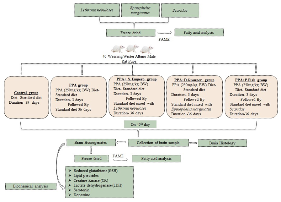
Figure 2 demonstrates the research design of the present study.

3. Results

The fatty acid profile analyses included the saturated, monounsaturated, and polyunsaturated fatty acids in control, PPA, PPA + Lethrinus nebuloses, PPA+ Epinephelus marginatus, and PPA + Scaridae in brain homogenates of study models via gas chromatography. Table 2 demonstrates a significant difference in saturated fatty acids, such as undecanoic acid (p = 0.061), lauric acid (p = 0.037), myristic acid (p = 0.044), palmitic acid (p = 0.046), stearic acid (p = 0.017), heneicosanoic acid (p = 0.023), and behenic acid (p = 0.068), between various groups. Similarly, a significant difference was observed between groups in gondoic acid (MUFA) (p = 0.013) and in some PUFAs, such as cis-5,8,11,14,17-eicosapentaenoic (p = 0.023) and cis 13,16-docasadienoic acid (p = 0.020)
While LDH did not show any significant variation among the five studied groups, Figure 3A demonstrates the non-significant increase in CK in PPA-treated rats as a rodent model of autism and the significant decrease in response to the three different nutritional interventions, with Lethrinus nebuloses and Epinephelus marginatus fish being more effective than Scaridae. Both kinds were reported to be significantly lower in CK compared to the PPA-rodent model of ASD, with a comparable level to the control group. The levels of GSH and lipid peroxides in all groups studied are shown in Figure 3B,C. While no significant changes were observed in the PPA-rodent model of ASD, in Lethrinus nebuloses- and Epinephelus marginatus-fed groups, compared to the control group, a significant decrease in glutathione in Scaridae-fed rats was observed (p < 0.001). Lipid peroxides recorded a significantly elevated level in the PPA-treated group (p < 0.05) and only a lower level in Lethrinus nebuloses rats. Figure 3D demonstrates the considerable changes in 5-HT in response to PPA treatment and fish extract feeding trials. Figure 3E demonstrates a remarkable decrease in dopamine (DA) in PPA-treated rats as a rodent model of autism and the significant rescue of DA levels in rats fed Epinephelus marginatus and Lethrinus nebuloses ω3 PUFA-rich fish extract, with only Epinephelus marginatus recording significantly higher dopamine compared to the PPA-group and control (p < 0.05).
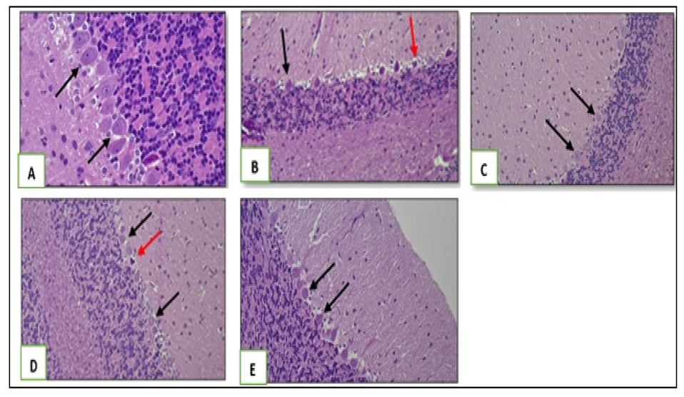
Thirty-five samples from the rats’ brains were examined. They included parts of the cerebrum, cerebellum, and hippocampus from each rat. PPA-induced changes in rats were predominantly observed in the cerebellum and hippocampus. Compared to the control group, the PPA group showed decreased size and density of Purkinje cells in the cerebellum. However, the PPA + Lethrinus nebuloses group, PPA + Epinephelus marginatus group, and PPA + Scaridae group showed an increase in the density and size of Purkinje cells (Figure 4).
Hippocampal changes (Figure 5) include decreased neuronal density in the CA4 (Cornu Ammonis) region of the PPA group compared to that in the control group. PPA+ Lethrinus nebuloses, PPA+ Epinephelus marginatus, and PPA+ Scaridae groups showed a slightly increased density of the neurons in the CA4 region compared to that in the PPA group.

4. Discussion

Diet and nutrition are extremely crucial aspects of everyone’s lives. They contribute to the development of a healthy body and a strong mind. It is commonly known that nutrient-dense foods can help the body clear toxins, build a strong immune system, control hunger, and avoid obesity [41]. Eating disorders are widespread in children with autism ASDs [42]. Their penchant for high-energy, low-nutrient foods can change their metabolism, creating an accumulation of reactive radicals and mental and physical deterioration. Although dieting and weight loss are already widespread in the general community, it has become difficult to raise awareness about diet, nutrition, and obesity among children with special needs. Despite their best efforts, parents of such children are frequently unable to control their children’s eating habits due to irritability and behavioural issues. Doctors and parents must now collaborate with nutritionists and dieticians to help these children eat healthy foods in order to stay fit and improve their quality of life [43,44].
Brain function, particularly neuronal activity, requires much energy in mammals [45].The most energy-intensive process in the central nervous system is neurotransmission. It is, specifically, the restoration of membrane potential by Na+/K+-exchanging ATPase following an action potential that consumes 50% of brain ATP [46]. The reversible phosphorylation of creatine by ATP is catalysed by creatine kinase. CK isoenzymes are specifically positioned in the cell at crucial locations of ATP consumption to efficiently regenerate ATP in situ via phosphocreatine, as well as at sites of ATP synthesis to build up a phosphocreatine pool. As a result, the CK/Pcr system is important for cellular energy buffering and transfer, especially in cells with high and fluctuating energy demands, such as neurons [47]. This enzyme is sensitive to oxidative stress and is assumed to be one of the key targets of oxidative modification in neurodegenerative disorders [48].
The present study’s considerable elevation of CK in the PPA-rodent model of ASD (Figure 3A) can find support in previous clinical studies by Al-Mosalem et al. (2009) and Poling et al. (2006) [49,50]. However, according to Frye et al. (2013) [51,52], only some of the ASD patients with mitochondrial dysfunction had elevated CK levels. The significantly lower CK reported in rats fed fish (Figure 3A) could find support in the previous study of [53] who showed that ω-3 was effective in lowering CK and LDH serum levels in healthy persons. Therefore, ω-3 should be prioritised as a recovery agent for exercise-induced muscle damage in therapies. Omega-3 fatty acid may reduce the severity of or enhance recovery from mild traumatic brain injury (mTBI) [54]. Based on this, they recommended fatty fish consumption, which would include the increased intake of ω-3 fatty acids, protein, vitamins, and minerals as more appropriate from the nutritional point of view than a recommendation for fish oil supplementation.
The slight increase in LDH in fish-fed groups could be attributed to a metabolic increase in glycolytic flux to replenish depleted ATP as a neurotoxic effect of PPA [36]. This contradicts a previous report [55], which found that ω-3 PUFA suppressed LDH expression as a brain glycolytic enzyme, but it may be supported by recent study by Akhigbe et al. (2021) [56], which found that ω-3 PUFA can help restore ischaemia/perfusion-induced damage by modulating lactate transport and metabolism.
Oxidative stress is the major etiological mechanism that contributes to the pathogenesis of ASD. While oxidative stress is promoted and supported by enzymatic and nonenzymatic mediators of the omega-6 family, oxidative stress is inhibited by enzymatic lipid mediators of omega-3 fatty acids [57]. The significant decrease in glutathione (Figure 3B) in Scaridae fish-fed rats could be attributed to heavy metal contamination, e.g., cadmium. The mechanism of toxicity of cadmium is linked to cadmium’s disruption of the cellular redox state and its structural similarities with divalent cations, such as zinc and calcium [58]. The redox activity of cadmium depletes antioxidants and glutathione, causes oxidative stress, enhances lipid peroxidation, and alters the lipid composition of membranes.
Generally speaking, lipid peroxidation is the process by which oxidants, such as free radicals, destroy lipids with carbon–carbon double bonds, particularly polyunsaturated fatty acids (PUFAs).The unexpected increase in lipid peroxides in the Epinephelus marginatus and Scaridae fish-fed rodent model of autism (Figure 3C) could be supported by Avramovic et al. (2012) [59], who discovered that additional ω-3 PUFA may increase lipid metabolism species, particularly lipid peroxides or MDA, but that this was accompanied by significantly higher anti-oxidant enzymes, such as superoxide dismutase SOD, and glutathione peroxidase, highlighting the advantages of a diet high in ω-3 PUFA in treating oxidative stress [60]. This may mean that, although not being antioxidants themselves, PUFAs have shown an anti-oxidative impact in circumstances when more conventional antioxidants, such vitamin E, have failed [61,62].
Given that dopamine (DA) is a key modulator of neuronal activity in some brain areas linked to ASD, there is a convincing reason to ask whether dysfunctional DA signalling could impact brain activity across a range of ASD-implicated brain assemblies. The formally suggested dopamine hypothesis of ASD [63] assumes that the functional dysregulation of DA pathways could contribute to the behavioural alterations that lead to an ASD clinical presentation. For example, aberrant mesolimbic DA signalling could diminish the reward value assigned to social stimuli, finally leading to impaired social interaction and/or communication skills. Abnormal nigrostriatal DA signalling could promote stereotyped or repetitive behaviours. Thus, the dopamine hypothesis of ASD, as currently stated, predicts that either hyper or hypo-dopaminergic signalling within certain areas could lead to or worsen ASD-related behavioural features [63]. This is consistent with the idea of an “inverted U” shape to describe the optimal level of DA receptor signalling, wherein too much or too little DA is detrimental to cognitive functions [64,65]. This can find support by the previous study by Healy-Stoffel and Levant (2018) [66], which reported the involvement of a ω-3 PUFA-rich diet in neurological disorders linked to dopamine systems, such as Parkinson’s disease (PD), schizophrenia, ASD, attention deficit hyperactivity disorder (ADHD), and depression. With the inverted U-shape DA hypothesis, the significant increase in DA is still within the recommended level, i.e., it is not too much higher compared to control levels. This suggested the beneficial effects of the reasonable increase in DA in rats fed ω-3 PUFA-rich fish (Figure 3E), which could find support in the study reporting that low levels of ω-3 PUFAs in the brain affect the brain dopamine systems and, when combined with appropriate genetic and other factors, increase the risk of developing these disorders and/or the severity of the disease. Moreover, it is accepted that increasing omega-3 fatty acids in the brain may reduce inflammatory cytokines, which may improve neurotransmitter function. There is also a reduction in dopamine and serotonin signalling with omega-3 deficiency in the brain [67]. This interpretation could find support in a recent study, which suggests that although the DA system is modified differently in different ASD rodent models, the intranasal administration of DA effectively corrects their behavioural phenotypes, which may present a hopeful treatment strategy for different types of ASD [68].
Figure 4 shows the neurotoxic effects of PPA in the induced rodent model, which include the strikingly reduced size and density of Purkinje cells in the cerebellum. This is supported by a prior study that found that, on average, the cross-sectional areas of Purkinje cells in autistic patients were 24% smaller than those in control people. In this study, two out of five autistic participants had mean Purkinje cell sizes that amounted to a size reduction of more than 50% [69] and also in PTEN knock-out mice as a rodent model of autism [70]. Interestingly there was a noticeable increase in the density and size of Purkinje cells in the PPA-induced rodent model fed the three kinds of fish (Lethrinus nebulosus, Epinephelus marginatus, and Scaridae) [70].
This could be related to the high omega-3 fatty acid content of the three types of fish tested. Our findings are supported by research that found considerable improvement in altered histopathology and oxidative stress in lead-intoxicated mice. They discovered that omega-3 fatty acids have neuroprotective properties [71]. Additional histopathological abnormalities were observed as the hippocampus showed decreased neuronal density in the CA4 (Cornu Ammonis) region of the PPA group compared to that in the control group. This is consistent with another study that found multiple synaptic alterations in the CA1 region of the hippocampus in a propionic acid mouse model of ASD, including the presence of some atypically enlarged presynaptic terminals with a reduced density of synaptic vesicles and short active zones [72]. The observed increase in the hippocampus neuronal density in the PPA-rodent model fed Lethrinus nebulosus, Epinephelus marginatus, and Scaridae could possibly be attributed to the fact that a higher omega-3 index is usually associated with greater hippocampal volumes, as a structure in the brain, which plays a significant role in learning and memory [72].

5. Conclusions

Our results provide some preliminary evidence in favour of using fish as a rich source of omega-3 fatty acid to amend the neurotoxic effects in a PPA-induced rodent model of ASD. Omega-3 can be used as an effective antioxidant, which can help to reduce oxidative stress as an etiological mechanism of autism. Based on the fact that oxidative stress is directly related to glutamate toxicity and neuroinflammation, which are another two etiological mechanisms of this disorder, this suggests that an omega-3-rich diet can be used as an early intervention strategy to avoid a clinical presentation or as a treatable approach for autism.

Author Contributions

Conceptualisation, D.M.A.-N. and A.K.E.-D.E.-A.; Methodology, N.S.A., H.A. and A.Y.A.; Software N.S.A.; Validation, D.M.A.-N., M.A.B.O., A.Y.A. and A.K.E.-D.E.-A.; Formal analysis, N.S.A., S.A., R.S.B., H.A., and A.A.; Investigation, N.S.A., S.A., R.S.B., H.A. and A.A.; Resources, N.S.A. and D.M.A.-N.; Data curation, N.S.A., R.S.B., S.A. and A.K.E.-D.E.-A.; Writing—original draft, N.S.A. and A.K.E.-D.E.-A.; Writing—review & editing, D.M.A.-N., S.A., L.N.A.-H., M.A.B.O. and R.S.B.; Visualisation, N.S.A., D.M.A.-N., L.N.A.-H. and A.K.E.-D.E.-A.; Supervision, D.M.A.-N., A.Y.A. and A.K.E.-D.E.-A.; Project administration, D.M.A.-N.; Funding acquisition, N.S.A. and D.M.A.-N. All authors have read and agreed to the published version of the manuscript.

Funding

This research was funded by Researchers Supporting Project Number (RSPD2023R805), King Saud University, Riyadh, Saudi Arabia.

Institutional Review Board Statement

All experimental procedures of this work were approved by and carried out in accordance with the guidelines of the Graduate Studies and Scientific Research Ethical Committee of Bioethics in King Saud University (KSU) Ref Number: No. KSU-SE-19-101. Our study was carried out in compliance with the ARRIVE guidelines.

Informed Consent Statement

Not applicable.

Data Availability Statement

The data presented in this study are available upon request from the corresponding author.

Acknowledgments

The authors extend thanks to the Researchers Supporting Project Number (RSPD2023R805), King Saud University, Riyadh Saudi Arabia.

Conflicts of Interest

The authors declare no conflict of interest.

References

  1. Kroncke, A.P.; Willard, M.; Huckabee, H. Assessment of Autism Spectrum Disorder: Critical Issues in Clinical, Forensic and School Settings, 1st ed.; Springe: Berlin/Heidelberg, Germany, 2016; 546p. [Google Scholar]
  2. Lord, C.; Elsabbagh, M.; Baird, G.; Veenstra-Vanderweele, J. Autism spectrum disorder. Lancet 2018, 392, 508–520. [Google Scholar] [CrossRef] [PubMed]
  3. Rylaarsdam, L.; Guemez-Gamboa, A. Genetic Causes and Modifiers of Autism Spectrum Disorder. Front. Cell. Neurosci. 2019, 13, 385. [Google Scholar] [CrossRef] [PubMed]
  4. Masini, E.; Loi, E.; Vega-Benedetti, A.F.; Carta, M.; Doneddu, G.; Fadda, R.; Zavattari, P. An Overview of the Main Genetic, Epigenetic and Environmental Factors Involved in Autism Spectrum Disorder Focusing on Synaptic Activity. Int. J. Mol. Sci. 2020, 21, 8290. [Google Scholar] [CrossRef]
  5. Almandil, N.B.; Alkuroud, D.N.; AbdulAzeez, S.; AlSulaiman, A.; Elaissari, A.; Borgio, J.F. Environmental and genetic factors in autism spectrum disorders: Special emphasis on data from Arabian studies. Int. J. Environ. Res. Public Health 2019, 16, 658. [Google Scholar] [CrossRef] [Green Version]
  6. Hughes, H.K.; Rowland, M.E.; Onore, C.E.; Rogers, S.; Ciernia, A.V.; Ashwood, P. Dysregulated gene expression associated with inflammatory and translation pathways in activated monocytes from children with autism spectrum disorder. Transl. Psychiatry 2022, 12, 39. [Google Scholar] [CrossRef] [PubMed]
  7. El-Ansary, A.K. Biomarker-Guided Nutritional Intervention to Treat Autism. Nov. Tech. Nutri. Food Sci. 2017, 1, NTNF.000501. [Google Scholar] [CrossRef]
  8. Ratnayake, W.M.; Galli, C. Fat and fatty acid terminology, methods of analysis and fat digestion and metabolism: A background review paper. Ann. Nutr. Metab. 2009, 55, 8–43. [Google Scholar] [CrossRef]
  9. Pawlosky, R.J.; Hibbeln, J.R.; Novotny, J.A.; Salem, N., Jr. Physiological compartmental analysis of alpha-linolenic acid metabolism in adult humans. J. Lipid Res. 2001, 42, 1257–1265. [Google Scholar] [CrossRef]
  10. Ahmed, A.A.; Balogun, K.A.; Bykova, N.V.; Cheema, S.K. Novel regulatory roles of omega-3 fatty acids in metabolic pathways: A proteomics approach. Nutr. Metab. 2014, 11, 6. [Google Scholar] [CrossRef] [Green Version]
  11. De la Torre-Aguilar, M.J.; Gomez-Fernandez, A.; Flores-Rojas, K.; Martin-Borreguero, P.; Mesa, M.D.; Perez-Navero, J.L.; Olivares, M.; Gil, A.; Gil-Campos, M. Docosahexaenoic and Eicosapentaenoic intervention modifies plasma and erythrocyte omega-3 fatty acid profiles but not the clinical course of children with autism spectrum disorder: A randomized control trial. Front. Nutr. 2022, 9, 790250. [Google Scholar] [CrossRef]
  12. Sala-Vila, A.; Satizabal, C.L.; Tintle, N.; Melo van Lent, D.; Vasan, R.S.; Beiser, A.S.; Seshadri, S.; Harris, W.S. Red Blood cell dha is inversely associated with risk of incident alzheimer’s disease and all-cause dementia: Framingham offspring study. Nutrients 2022, 14, 2408. [Google Scholar] [CrossRef] [PubMed]
  13. Amminger, G.P.; Berger, G.E.; Schafer, M.R.; Klier, C.; Friedrich, M.H.; Feucht, M. Omega-3 fatty acids supplementation in children with autism: A double-blind randomized, placebo-controlled pilot study. Biol. Psychiatry 2007, 61, 551–553. [Google Scholar] [CrossRef] [PubMed]
  14. DiNicolantonio, J.J.; O’Keefe, J.H. The importance of marine omega-3s for brain development and the prevention and treatment of behavior, mood, and other brain disorders. Nutrients 2020, 12, 2333. [Google Scholar] [CrossRef] [PubMed]
  15. Zhou, L.; Xiong, J.Y.; Chai, Y.Q.; Huang, L.; Tang, Z.Y.; Zhang, X.F.; Liu, B.; Zhang, J.T. Possible antidepressant mechanisms of omega-3 polyunsaturated fatty acids acting on the central nervous system. Front. Psychiatry 2022, 13, 933704. [Google Scholar] [CrossRef]
  16. Petermann, A.B.; Reyna-Jeldes, M.; Ortega, L.; Coddou, C.; Yévenes, G.E. Roles of the unsaturated fatty acid docosahexaenoic acid in the central nervous system: Molecular and cellular insights. Int. J. Mol. Sci. 2022, 23, 5390. [Google Scholar] [CrossRef]
  17. Schuchardt, J.P.; Huss, M.; Stauss-Grabo, M.; Hahn, A. significance of long-chain polyunsaturated fatty acids (PUFAs) for the development and behaviour of children. Eur. J. Pediatr. 2010, 169, 149–164. [Google Scholar] [CrossRef]
  18. Agostoni, C.; Nobile, M.; Ciappolino, V.; Delvecchio, G.; Tesei, A.; Turolo, S.; Crippa, A.; Mazzocchi, A.; Altamura, C.A.; Brambilla, P. The role of omega-3 fatty acids in developmental psychopathology: A systematic review on early psychosis, autism, and ADHD. Int. J. Mol. Sci. 2017, 18, 2608. [Google Scholar] [CrossRef] [Green Version]
  19. Chiang, N.; Serha, C.N. Specialized pro-resolving mediator network: An update on production and actions. Essays Biochem. 2020, 64, 443–462. [Google Scholar]
  20. Bazan, N.G. Neuroprotectin D1-mediated anti-inflammatory and survival signaling in stroke, retinal degenerations, and Alzheimer’s disease. J. Lipid Res. 2009, 50, S400–S405. [Google Scholar] [CrossRef] [Green Version]
  21. Serhan, C.N.; Petasis, N.A. Resolvins and protectins in inflammation resolution. Chem. Rev. 2011, 111, 5922–5943. [Google Scholar] [CrossRef] [Green Version]
  22. Panezai, J.; van Dyke, T. Polyunsaturated fatty acids and their immunomodulatory actions in periodontal disease. Nutrients 2023, 15, 821. [Google Scholar] [CrossRef] [PubMed]
  23. van Elst, K.; Bruining, H.; Birtoli, B.; Terreaux, C.; Buitelaar, J.K.; Kas, M.J. Food for thought: Dietary changes in essential fatty acid ratios and the increase in autism spectrum disorders. Neurosci. Biobehav. Rev. 2014, 45, 369–378. [Google Scholar] [CrossRef] [PubMed]
  24. Bazinet, R.P.; Layé, S. Polyunsaturated fatty acids and their metabolites in brain function and disease. Nat. Rev. Neurosci. 2014, 15, 771–785. [Google Scholar] [CrossRef]
  25. Harris, W.S. The Omega-6:Omega-3 ratio: A critical appraisal and possible successor. Prostaglandins Leukot. Essent. Fatty Acids 2018, 132, 34–40. [Google Scholar] [CrossRef] [PubMed]
  26. Martínez-Cué, C.; Bartesaghi, R. Fatty Acids: A Safe tool for improving neurodevelopmental alterations in down syndrome? Nutrients 2022, 14, 2880. [Google Scholar] [CrossRef]
  27. Weiser, M.; Butt, C.; Mohajeri, M. Docosahexaenoic acid and cognition throughout the lifespan. Nutrients 2016, 8, 99. [Google Scholar] [CrossRef] [Green Version]
  28. Thomas, A.; Crivello, F.; Mazoyer, B.; Debette, S.; Tzourio, C.; Samieri, C. Fish Intake and mri burden of cerebrovascular disease in older adults. Neurology 2021, 97, e2213–e2222. [Google Scholar] [CrossRef]
  29. Avella-Garcia, C.B.; Julvez, J. Seafood intake and neurodevelopment: A systematic review. Curr. Envir. Health Rep. 2014, 1, 46–77. [Google Scholar] [CrossRef] [Green Version]
  30. Murakami, Y.; Hosomi, R.; Nishimoto, A.; Nishiyama, T.; Yoshida, M.; Fukunaga, K. Protective Effects of Fish (Alaska Pollock) Protein Intake against Short-Term Memory Decline in Senescence-Accelerated Mice. Nutrients 2022, 14, 4618. [Google Scholar] [CrossRef]
  31. Kirby, A.; Derbyshire, E. Omega-3/6 fatty acids and learning in children and young people: A review of randomised controlled trials published in the last 5 years. J. Nutr. Food Sci. 2018, 8, 1–10. [Google Scholar]
  32. Reimers, A.; Ljung, H. The emerging role of omega-3 fatty acids as a therapeutic option in neuropsychiatric disorders. Ther. Adv. Psychopharmacol. 2019, 9, 1–18. [Google Scholar] [CrossRef] [PubMed]
  33. Johnson, M.; Östlund, S.; Fransson, G.; Kadesjö, B.; Gillberg, C. Omega-3/omega-6 fatty acids for attention deficit hyperactivity disorder: A randomized placebo-controlled trial in children and adolescents. J. Atten. Disord. 2009, 12, 394–401. [Google Scholar] [CrossRef] [PubMed] [Green Version]
  34. Iqra; Sughra, K.; Ali, A.; Afzal, F.; Yousaf, M.J.; Khalid, W.; Rasul, H.F.; Aziz, Z.; Aqlan, F.M.; Al-Farga, A.; et al. Wheat-based gluten and its association with pathogenesis of celiac disease: A review. Int. J. Food Prop. 2023, 26, 511–525. [Google Scholar] [CrossRef]
  35. El-Ansary, A.K.; Bacha, A.B.; Kotb, M. Etiology of autistic features: The persisting neurotoxic Effects of Propionic Acid. J. Neuroinflamm. 2012, 9, 74. [Google Scholar] [CrossRef] [Green Version]
  36. Abuaish, S.; Al-Otaibi, N.M.; Aabed, K.; Abujamel, T.S.; Alzahrani, S.A.; Alotaibi, S.M.; Bhat, R.S.; Arzoo, S.; El-Ansary, A. The role of sex-differentiated variations in stress hormones, antioxidants, and neuroimmune responses in relation to social interaction impairment in a rodent model of autism. Metab. Brain Dis. 2021, 36, 1369–1379. [Google Scholar] [CrossRef]
  37. Alsubaiei, S.R.M.; Alfawaz, H.A.; Almubarak, A.Y.; Alabdali, N.A.; Ben Bacha, A.; El-Ansary, A. Independent and combined effects of probiotics and prebiotics as supplements or food-rich diets on a propionic-acid-induced rodent model of autism spectrum disorder. Metabolites 2023, 13, 50. [Google Scholar] [CrossRef]
  38. Beutler, E. Improved Method for the Determination of Blood Glutathione. J. Lab. Clin. Med. 1963, 61, 882–888. [Google Scholar]
  39. Ruiz-Larrea, M.B.; Leal, A.M.; Liza, M.; Lacort, M.; de Groot, H. Antioxidant effects of estradiol and 2-hydroxyestradiol on iron-induced lipid peroxidation of rat liver microsomes. Steroids 1994, 59, 383–388. [Google Scholar]
  40. Todd, J.C.; Sanford, A.H.; Davidsohn, I.; Henry, J.B. Clinical Diagnosis and Management by Laboratory Methods; Saunders: Philadelphia, PA, USA, 1979. [Google Scholar]
  41. D’Angelo, S.; Motti, M.L.; Meccariello, R. ω-3 and ω-6 Polyunsaturated Fatty Acids, Obesity and Cancer. Nutrients 2020, 12, 2751. [Google Scholar] [CrossRef]
  42. Numata, N.; Nakagawa, A.; Yoshioka, K.; Isomura, K.; Matsuzawa, D.; Setsu, R.; Nakazato, M.; Shimizu, E. Associations between Autism Spectrum Disorder and Eating Disorders with and without Self-Induced Vomiting: An Empirical Study. J. Eat. Disord. 2021, 9, 1–9. [Google Scholar] [CrossRef]
  43. Ho, H.H.; Eaves, L.C.; Peabody, D. Nutrient intake and obesity in children with autism. Focus Autism Dev. Disabil. 1997, 12, 187–192. [Google Scholar] [CrossRef]
  44. Williams, K.E.; Hendy, H.; Knecht, S. Parent feeding practices and child variables associated with childhood feeding problems. J. Dev. Phys. Disabil. 2008, 20, 231–242. [Google Scholar] [CrossRef]
  45. Zhang, Z.; Chen, W.; Zhao, Y.; Yang, Y. Spatiotemporal Imaging of cellular energy metabolism with genetically-encoded fluorescent sensors in brain. Neurosci. Bull. 2018, 34, 875–886. [Google Scholar] [CrossRef] [PubMed]
  46. Pifferi, F.; Cunnane, S.C.; Guesnet, P. Evidence of the role of omega-3 polyunsaturated fatty acids in brain glucose metabolism. Nutrients 2020, 12, 1382. [Google Scholar] [CrossRef] [PubMed]
  47. Andres, R.; Meiler, F.; Huber, A.; Perez-Bouza, A.; Seiler, R.; Wallimann, T.; Widmer, H.; Schlattner, U. Human Fetal CNS Tissue Expresses Kreatine Kinases and the Creatine Transporter. FENS 2002, 1, 208. [Google Scholar]
  48. Feng, S.; Xu, Z.; Yan, Y.B. Blocking creatine kinase refolding by trace amounts of copper ions. J. Inorg. Biochem. 2008, 102, 928–935. [Google Scholar] [CrossRef]
  49. Al-Mosalem, O.; El-Ansary, A.; Attas, O.; Al-Ayadhi, L. Metabolic biomarkers related to energy metabolism in Saudi autistic children. Clin. Biochem. 2009, 42, 949–957. [Google Scholar] [CrossRef]
  50. Poling, J.S.; Frye, R.E.; Shoffner, J.; Zimmerman, A.W. Developmental Regression and Mitochondrial Dysfunction in a Child with Autism. J. Child Neurol. 2006, 21, 170–172. [Google Scholar] [CrossRef] [Green Version]
  51. Frye, R.; Delatorre, R.; Taylor, H.; Slattery, J.; Melnyk, S.; Chowdhury, N.; James, S. Redox Metabolism Abnormalities in Autistic Children Associated with Mitochondrial Disease. Transl. Psychiatry 2013, 3, e273. [Google Scholar] [CrossRef] [Green Version]
  52. Frye, R.E.; Melnyk, S.; MacFabe, D.F. Unique acyl-carnitine profiles are potential biomarkers for acquired mitochondrial disease in autism spectrum disorder. Transl. Psychiatry 2013, 3, e220. [Google Scholar] [CrossRef] [Green Version]
  53. Xin, G.; Eshaghi, H. Effect of omega-3 fatty acids supplementation on indirect blood markers of exercise-induced muscle damage: Systematic review and meta-analysis of randomized controlled trials. Food Sci. Nutr. 2021, 9, 6429–6442. [Google Scholar] [CrossRef]
  54. Rawson, E.S.; Miles, M.P.; Larson-Meyer, D.E. Dietary Supplements for health, adaptation, and recovery in athletes. Int. J. Sport Nutr. Exerc. Metab. 2018, 28, 188–199. [Google Scholar] [CrossRef] [Green Version]
  55. Dentin, R.; Benhamed, F.; Pégorier, J.-P.; Foufelle, F.; Viollet, B.; Vaulont, S.; Girard, J.; Postic, C. Polyunsaturated fatty acids suppress glycolytic and lipogenic genes through the inhibition of ChREBP nuclear protein translocation. J. Clin. Investig. 2005, 115, 2843–2854. [Google Scholar] [CrossRef] [PubMed] [Green Version]
  56. Akhigbe, R.; Hamed, M.; Odetayo, A.; Akhigbe, T.; Ajayi, A.; Ajibogun, F. Omega-3 fatty acid rescues ischaemia/perfusion-induced testicular and sperm damage via modulation of lactate transport and xanthine oxidase/uric acid signaling. Biomed. Pharmacother. 2021, 142, 111975. [Google Scholar] [CrossRef] [PubMed]
  57. Farooqui, A.A.; Farooqui, T. Prevention of oxidative stress by omega-3 fatty acids in the brain. In Omega-3 Fatty Acids; Springer: Berlin/Heidelberg, Germany, 2016; pp. 239–249. [Google Scholar]
  58. Pinot, F.; Kreps, S.E.; Bachelet, M.; Hainaut, P.; Bakonyi, M.; Polla, B.S. Cadmium in the Environment: Sources, Mechanisms of Biotoxicity, and Biomarkers. Rev. Environ. Health 2000, 15, 299–324. [Google Scholar] [CrossRef]
  59. Avramovic, N.; Dragutinovic, V.; Krstic, D.; Colovic, M.; Trbovic, A.; de Luka, S.; Milovanovic, I.; Popovic, T. The Effects of omega 3 fatty acid supplementation on brain tissue oxidative status in aged wistar rats. Hippokratia 2012, 16, 241. [Google Scholar]
  60. Lee, H.-J.; Kim, H.-K.; Choi, H. Different sources of omega fatty acids at the fixed ratio of p/s affect glutathione dependent enzymes in rat hepatocarcinogenesis. Korean J. Nutr. 2003, 36, 785–792. [Google Scholar]
  61. Arnal, E.; Miranda, M.; Barcia, J.; Bosch-Morell, F.; Romero, F. Lutein and docosahexaenoic acid prevent cortex lipid peroxidation in streptozotocin-induced diabetic rat cerebral cortex. Neuroscience 2010, 166, 271–278. [Google Scholar] [CrossRef] [PubMed]
  62. Shelton, R.J.; Velavan, P.; Nikitin, N.P.; Coletta, A.P.; Clark, A.L.; Rigby, A.S.; Freemantle, N.; Cleland, J.G. Clinical trials update from the American Heart Association Meeting: ACORN-CSD, Primary Care Trial of Chronic Disease Management, PEACE, CREATE, SHIELD, A-HeFT, GEMINI, Vitamin E Meta-analysis, ESCAPE, CARP, and SCD-HeFT Cost-effectiveness Study. Eur. J. Heart Fail. 2005, 7, 127–135. [Google Scholar] [CrossRef] [Green Version]
  63. Pavăl, D. A Dopamine hypothesis of autism spectrum disorder. Dev. Neurosci. 2017, 39, 355–360. [Google Scholar] [CrossRef]
  64. Arnsten, A.F. Catecholamine regulation of the prefrontal cortex. J. Psychopharmacol. 1997, 11, 151–162. [Google Scholar] [CrossRef]
  65. Cools, R.; D’Esposito, M. Inverted-u–shaped dopamine actions on human working memory and cognitive control. Biol. Psychiatry 2011, 69, e113–e125. [Google Scholar] [CrossRef] [Green Version]
  66. Healy-Stoffel, M.; Levant, B. N-3 (omega-3) fatty acids: Effects on brain dopamine systems and potential role in the etiology and treatment of neuropsychiatric disorders. CNS Neurol. Disord. Drug Targets 2018, 17, 216–232. [Google Scholar] [CrossRef]
  67. Logan, A.C. Neurobehavioral aspects of omega-3 fatty acids: Possible mechanisms and therapeutic value in major depression. Altern. Med. Rev. 2003, 8, 410–425. [Google Scholar] [PubMed]
  68. Chao, O.Y.; Pathak, S.S.; Zhang, H.; Dunaway, N.; Li, J.-S.; Mattern, C.; Nikolaus, S.; Huston, J.P.; Yang, Y.-M. Altered dopaminergic pathways and therapeutic effects of intranasal dopamine in two distinct mouse models of autism. Mol. Brain 2020, 13, 111. [Google Scholar] [CrossRef]
  69. Fatemi, S.H.; Halt, A.R.; Realmuto, G.; Earle, J.; Kist, D.A.; Thuras, P.; Merz, A. Purkinje cell size is reduced in cerebellum of patients with autism. Cell. Mol. Neurobiol. 2002, 22, 71–75. [Google Scholar]
  70. Cupolillo, D.; Hoxha, E.; Faralli, A.; De Luca, A.; Rossi, F.; Tempia, F.; Carulli, D. Autistic-Like Traits and Cerebellar Dysfunction in Purkinje Cell PTEN Knock-Out Mice. Neuropsychopharmacology 2016, 41, 1457–1466. [Google Scholar] [CrossRef] [PubMed] [Green Version]
  71. Singh, P.K.; Singh, M.K.; Yadav, R.S.; Nath, R.; Mehrotra, A.; Rawat, A.; Dixit, R.K. Omega-3 fatty acid attenuates oxidative stress in cerebral cortex, cerebellum, and hippocampus tissue and improves neurobehavioral activity in chronic lead-induced neurotoxicity. Nutr. Neurosci. 2019, 22, 83–97. [Google Scholar] [CrossRef]
  72. Lobzhanidze, G.; Japaridze, N.; Lordkipanidze, T.; Rzayev, F.; MacFabe, D.; Zhvania, M. Behavioural and brain ultrastructural changes following the systemic administration of propionic acid in adolescent male rats. Further development of a rodent model of autism. Int. J. Dev. Neurosci. 2020, 80, 139–156. [Google Scholar] [CrossRef] [PubMed] [Green Version]
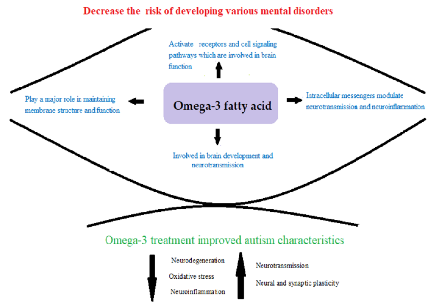
Figure 1. Figure illustrating the involvement of dietary omega-3 fatty acids in various physiological functions related to the development and functioning of the brain and its relationship with a decreased risk of developing various mental disorders, especially autism.
Applsci 13 07392 g001
Figure 2. Research design of the ameliorative effect of dietary fish omega-3s on modifying the neurotoxic effects of propionic acid-induced autism.
Applsci 13 07392 g002
Figure 3. (AE) Comparison of the different measured variables among control, PPA treatment as a rodent model of ASD, Spangled empero (Lethrinus nebuloses), D. Grouper (Epinephelus marginatus), and Parrot Fish (Scaridae) fed as nutritionally treated model of ASD (** p ≤ 0.01, * p ≤ 0.05).
Applsci 13 07392 g003
Figure 4. H&E-stained cerebellum sections with arrows highlighting the Purkinje layer in the cerebellum. (A): The control group sections show back-to-back large cells (black arrows) with abundant cytoplasm, round vesicular nuclei, and prominent nucleoli (H&E, X400). (B): In the PPA-group, the Purkinje layer shows a paucity of Purkinje cells (black arrow) with scattered cellular degenerative changes (red arrow), such as smaller and shrinking cells with dark basophilic staining (H&E, 200). (C): The PPA + Lethrinus nebuloses group shows a reduction in Purkinje cell density but still higher density than the PPA-group (black arrows, H&E, X200). (D): A rat from the PPA+ Epinephelus marginatus group shows a higher density of Purkinje cells (black arrow) than the PP-group and the occasional degeneration of scattered Purkinje cells (red arrow, H&E, X200). (E): PPA + Scaridae group also shows a higher density of the Purkinje cells (black arrows) than the PPA-group (H&E, X200).
Applsci 13 07392 g004
Figure 5. H&E-stained sections of the hippocampus proper (CA1–CA4) and the dentate gyrus. (A): A sample from the control-group hippocampus tissue exhibits densely packed cellular pyramidal neurons (black arrows in the CA4 region) with no degenerative changes (H&E, X400). (B): The PPA-group shows decreased density of the pyramidal neuron layer (black arrows) with degenerative cellular changes in the granule cell layer of the dentate gyrus (red arrows, H&E, X100). (CE): The PPA + Lethrines nebuloses, PPA + Epinephelus marginatus, and PPA + Scaridae groups show an increase in the density of pyramidal neurons (black arrows) in CA4 region of the hippocampus compared to that in the PPA-group. No degenerative changes were detected in the granule cell layer of the dentate gyrus (red arrow, H&E X400).
Applsci 13 07392 g005
Table 1. Polyunsaturated fatty acid profiles of Lethrinus nebuloses, Epinephelus marginatus, and Scaridae.
Fatty AcidsPPA +
Lethrinus
nebuloses
PPA +
Epinephelus
marginatus
PPA +
Scaridae
p-Value
Linoleic acid (C18:2)0.414 ± 0.0670.452 ± 0.1210.503 ± 0.1210.428
α-Linolenic acid (C18:3)0.362 ± 0.1290.208 ± 0.0200.280 ± 0.1160.243
Arachidonic acid (C20:4)0.28 ± 0.047 ac2.172 ± 0.101 bc0.644 ± 0.141 ab0.001
Eicosapentaenoic acid (C20:5)0.28 ± 0.0470.415 ± 0.0390.280 ± 0.1130.105
Docosatetraenoic acid (C22:4)22.870 ± 0.207 ac16.601 ± 0.217 bc2.547 ± 0.199 ab0.001
Docosahexaenoic acid (C22:6)0.155 ± 0.135 a24.657 ± 2.839 bc0.979 ± 0.195 a0.001
Kruskal–Wallis Test among all groups with multiple comparisons (Mann–Whitney Test) within the entire groups for each parameter. a Describes a significant difference between the group and the Lethrinus nebuloses group at a significant level (0.05). b Describes a significant difference between the group and the Epinephelus marginatus group at a significant level (0.05). c Describes a significant difference between the group and the Scaridae group at a significant level (0.05).
Table 2. Fatty acid, omega-3, and omega-6 precursor profiles of brain homogenates in the study model.
Fatty AcidsControlPPAPPA +
Lethrinus nebuloses
PPA +
Epinephelus marginatus
PPA +
Scaridae
p-Value
Saturated fatty acids (SFA)
 Caprylic acid (C8:0)0.007 ± 0.010.003 ± 0.0030.074 ± 0.18 b0.073 ± 0.15 b0.039 ± 0.0960.284
 Undecanoic acid (C11:0)0.015 ± 0.030.031 ± 0.04 a0.002 ± 0.002 b0.098 ± 0.150.051 ± 0.0950.061
 Lauric acid (C12:0)0.005 ± 0.010.015 ± 0.02 a0.027 ± 0.050.133 ± 0.14 ab0.049 ± 0.0960.037
 Tridecanoic acid (C13:0)0.023 ± 0.040.026 ± 0.020.013 ± 0.0080.047 ± 0.070.056 ± 0.0930.641
 Myristic acid (C14:0)1.391 ± 3.063.942 ± 3.52 a2.57 ± 1.62 a2.375 ± 2.67 a1.062 ± 1.888 b0.044
 Pentadecanoic acid (C15: 0) 0.428 ± 0.560.687 ± 0.460.602 ± 0.320.556 ± 0.470.533 ± 0.4830.904
 Palmitic acid (C16:0)33.243 ± 9.9739.184 ± 4.2644.471± 4.23 ab40.291 ± 6.7342.36 ± 5.241 a0.046
 Heptadecanoic acid (C17:0)0.717 ± 0.310.749 ± 0.251.026 ± 0.510.759 ± 0.240.644 ± 0.1690.607
 Stearic acid (C18:0)0.578 ± 0.280.557 ± 0.130.613 ± 0.180.350 ± 0.15 b0.755 ± 0.17 b0.017
 Arachidic acid (C20:0) 0.369 ± 0.230.317 ± 0.210.524 ± 0.541.781 ± 3.580.2 ± 0.160.581
 Heneicosanoic acid (C21:0)0.162 ± 0.110.048 ± 0.02 a0.104 ± 0.08 b0.029 ± 0.02 a0.069 ± 0.05 a0.023
 Behenic acid (C22:0)0.256 ± 0.100.370 ± 0.410.523 ± 0.47 a0.609 ± 0.810.156 ± 0.11 a0.068
 Tricosanic acid (C23:0)0.126 ± 0.070.080 ± 0.020.143 ± 0.100.167 ± 0.210.064 ± 0.050.161
 Lignoceric acid (C24:0)0.346 ± 0.220.297 ± 0.130.317 ± 0.160.361 ± 0.310.147 ± 0.140.275
Monounsaturated fatty acids (MUFA)
 Gondoic acid (C20:1)0.271 ± 0.161.867 ± 2.27 a1.687 ± 3.374.074 ± 3.56 a0.293 ± 0.334 b0.013
 Palmitoleic acid (C16: 1)1.623 ± 0.671.348 ± 0.671.708 ± 1.198.893± 11.27 ab2.997 ± 2.520.048
 cis 10-Heptadecanoic acid (C17: 1)10.669 ± 26.770.573 ± 0.190.829 ± 0.441.562 ± 2.375.023 ± 11.810.845
Polyunsaturated fatty acids (PUFA)
 Linoleic acid (C18:2)3.806 ± 2.5510.780 ± 8.076.035 ± 2.9911.848 ± 14.655.481 ± 3.640.517
 Methyl8,11,14-eicosatrienoate (C20:3)1.206 ± 0.635.204 ± 7.093.709 ± 4.882.067 ± 4.290.868 ± 0.290.185
 cis 11,14-Eicosadienoic acid (C20:4)1.159 ± 0.570.942 ± 0.531.227 ± 0.981.74 ± 1.791.357 ± 0.640.851
 Linolenic acid (C18:3)1.422 ± 0.484.488 ± 4.783.731 ± 2.915.827 ± 7.802.874 ± 2.170.300
 cis-5,8,11,14,17-Eicosapentaenoic (C20:5)19.472 ± 8.0914.018 ± 4.1815.792 ± 4.617.863 ± 4.29 ab17.26 ± 8.080.023
 cis 13,16-Docasadienoic acid (C22:2)22.704 ± 10.0514.475 ± 4.53 a14.275 ± 7.11 a8.497 ± 5.34 a18.694 ± 8.820.020
Kruskal–Wallis Test among all groups with multiple comparisons (Mann–Whitney Test) within the entire group for each parameter. a Describes a significant difference between the group and the control group at the significant level (0.05). b Describes a significant difference between the group and the PPA group at a significant level (0.05).
Disclaimer/Publisher’s Note: The statements, opinions and data contained in all publications are solely those of the individual author(s) and contributor(s) and not of MDPI and/or the editor(s). MDPI and/or the editor(s) disclaim responsibility for any injury to people or property resulting from any ideas, methods, instructions or products referred to in the content.