Article

Investigation into the Binding Site of (-)-Spirobrassinin for Herbicidal Activity Using Molecular Docking and Molecular Dynamics Simulations

1
College of Horticulture and Plant Protection, Inner Mongolia Agricultural University, Hohhot 010020, China
2
Bayannaoer Center of Cultivated Land Quality Monitoring and Protection, Bayannaoer 015400, China
3
Ulanqab Institute of Agricultural and Animal Husbandry Sciences, Ulanqab 012000, China
*
Author to whom correspondence should be addressed.
These authors contributed equally to this work.
Appl. Sci. 2023, 13(12), 7287; https://doi.org/10.3390/app13127287
Submission received: 5 June 2023 / Revised: 13 June 2023 / Accepted: 15 June 2023 / Published: 19 June 2023

Abstract

:
(-)-Spirobrassinin, a glucosinolate compound from Brassicaceae plants, has shown inhibitory effects on weeds. However, its specific target sites are not well explored. This study used molecular docking, molecular dynamics simulations, and biological experiments to investigate (-)-Spirobrassinin’s target sites. The inhibitory effects of (-)-Spirobrassinin were observed on various enzymes crucial for plant metabolic pathways, including dihydroxyacid dehydrogenase, 4-hydroxyphenylpyruvate dioxygenase, protoporphyrinogen oxidase, and acetolactate synthase. Additionally, it disrupts the metabolism of vital phytohormones, namely abscisic acid and gibberellin. Molecular dynamics simulations revealed stable interactions between (-)-Spirobrassinin and specific residues (Phe270 and Phe261) of the photosystem II D1, involving electrostatic, hydrophobic, and van der Waals forces. This study provides valuable insights into (-)-Spirobrassinin’s mode of action, highlighting its potential as a natural herbicide from Brassicaceae plants.

1. Introduction

The use of herbicides has played a crucial role in effectively managing weed infestation, thereby facilitating agricultural progress [1]. Nevertheless, the escalating emergence of herbicide resistance in weeds poses significant challenges to their efficacy [2,3]. The repeated utilization of herbicides with similar modes of action often leads to the development of resistance in weeds. This phenomenon has resulted in the proliferation of herbicide-resistant weed species, which can cause substantial reductions in agricultural yields, ranging from 40% to 90% [4].
Considering the detrimental impacts of current synthetic herbicides and the formidable obstacles presented by resistant weeds, the replacement of harmful chemicals with promising natural products emerges as a viable long-term approach for weed management [5]. In this context, the utilization of allelochemicals obtained from plants presents a pathway towards sustainable agriculture. Allelochemicals refers to secondary metabolites produced by different plant parts, which have the ability to influence the growth and development of adjacent plants [6].
Allelochemicals consist of phenolic acids, flavonoids, alkaloids, steroids, and fatty acids, among other compounds [7]. These substances exert toxicity on weeds by disrupting their photosynthetic system, respiratory system, cell division, and lignin content [8]. A sulfur-containing compound called (-)-Spirobrassinin has been isolated from broccoli, and it is unique to the Brassicaceae family (Figure 1). (-)-Spirobrassinin has demonstrated inhibitory effects on the growth of Panicum miliaceum L. and Amaranthus retroflexus L. (Figures S2 and S3). This compound exhibits potential for development as a novel plant-based herbicide. However, the mode of action and specific target site of (-)-Spirobrassinin have not been investigated yet.
Currently, herbicides available in the market act through various modes of action. One of the common targets of herbicides is the enzyme acetolactate synthase (ALS), which is responsible for catalyzing the formation of acetohydroxybutyrate and acetolactate [9]. Another target is dihydroxyacid dehydratase (DHAD), which catalyzes the α,β-dehydration reaction in the biosynthetic pathway of branched-chain amino acids, producing α-keto acids that serve as precursors for leucine, isoleucine, and valine [10,11]. Inhibitors of hydroxyphenylpyruvate dioxygenase (HPPD) target the photosynthesis process by interfering with the conversion of hydroxyphenylpyruvate to homogentisate. This disruption affects the synthesis of tocopherols and plastoquinones, ultimately impacting the biosynthesis of carotenoids in the target organisms and leading to plant death [12]. Protochlorophyllide oxidase (PPO), found in both mitochondria and chloroplasts, is another important enzyme involved in tetrapyrrole biosynthesis, specifically in the synthesis of protoheme and chlorophyll [13,14]. Herbicide targets also include components of photosystem II (PSBD1), where they compete with plastoquinone for binding sites, disrupt the electron transport chain, and interfere with light reactions [15]. Furthermore, there are targets related to plant growth regulation, including binding sites for auxins (TIR1) [16], jasmonic acid (COLI-ASK1) [17], abscisic acid (PLY2) [18], Strigolactone (D14-D3-ASK1) [19] and gibberellins (GIDI) [20].
In this study, we employed molecular docking, virtual screening, biological validation experiments, and molecular dynamics simulations to investigate the mode of action and binding sites of (-)-Spirobrassinin. The primary objective was to establish a theoretical basis for the potential development of (-)-Spirobrassinin as a natural product herbicide.

2. Materials and Methods

2.1. Molecular Docking

The pdb format files for herbicide targets were retrieved from the protein structure database via PDB (https://www.rcsb.org/, accessed on 29 January 2023). Same as following highlights. ALS (PDB code: 1OZF), DHAD (PDB code: 5ZE4), PPO (PDB code: 3I6D), acetyl CoA carboxylase (ACC, PDB code: 3PGQ), PSBD1(PDB code: 7CZL), and HPPD (PDB code: 6J63), and plant growth regulatory targets: PLY2 (PDB code: 3KDI), decreased apical dominance 2 (DAD2, PDB code: 6O5J), GIDI (PDB code: 3ED1), D14-D3-ASK1 (PDB code: 5HZG), COLI-ASK1(PDB code: 3OGM), and TIR1 (PDB code: 2P1Q) within the download (-)-Spirobrassinin structure in the sdf format from the PubChem database (https://pubchem.ncbi./, accessed on 29 January 2023). The AutoDock tool was used to add data on essential hydrogen atoms, and the Autogrid program was used to generate affinity (grid) maps of X:58 Y:60 Z:62 Å grid points with a spacing of 0.375 Å. The calculation of the van der Waals and electrostatic terms was carried out by the AutoDock parameter set and distance-dependent dielectric function, respectively [1]. The Lamarckian genetic algorithm (LGA) and the Solis and Wets local search method were used to generate the docking [2,3].

2.2. Quantification of Phytohormones Content

L. sativa var. seedlings were frozen in liquid nitrogen, ground into powder, and extracted with 1 mL methanol/water/formic acid (15:4:1, v/v/v). The analytical conditions were as follows: LC: C18 (100 mm × 2.1 mm, 1.8 µm); (A) water, and (B) acetonitrile. ESI-MS/MS conditions: AB 6500+QTRAP® LC-MS/MS system, equipped with an ESI Turbo Ion-Spray interface. The integrated peak area ratios of sample detected were substituted into the linear equation of the standard curve and calculated to obtain the phytohormones content.

2.3. Docking Results Verification Experiments

A double antibody one-step sandwich enzyme-linked immunosorbent assay (ELISA) was used. To the precovered wells with enzymes (ALS, DHAD, HPPD, and PPO) anti-bodies, specimens, standards, and horseradish peroxidase labelled detection antibodies were added sequentially, incubated, and washed thoroughly. The wells were developed with the substrate 3,3′,5,5′-Tetramethylbenzidine (TMB), which was converted to blue using peroxidase and to the final yellow using acid. TMB was converted to blue using peroxidase and to a final yellow color by the action of acid. The shade of color was positively correlated with enzymes present in the sample. The absorbance (OD) was measured at 450 nm using an enzymes marker to calculate the sample activity.

2.4. Cultivation and Bioassay of C. vulgaris

To determine the inhibitory effect of (-)-Spirobrassinin on C. vulgaris, C. vulgaris was inoculated into 50 mL of Aquatic No.4 medium. The C. vulgaris was pre-cultivated under the conditions of a light intensity of 5000 l× and agitation at 100 rpm for 7 days to promote rapid cell growth. The pre-cultured algal suspension was then inoculated into centrifuge tubes, with each tube containing 2 mL, to achieve an initial cell density of 8 × 10⁵ cells/mL. (-)-Spirobrassinin was introduced into the system to create six concentration gradients, while a blank control without (-)-Spirobrassinin was maintained. The system was incubated for 4 days. Aquatic No.4 medium was used as the reference solution. The cells were counted using a hemocytometer under a microscope, and the absorbance at 680 nm was measured. A growth curve was constructed, and the inhibition rate was calculated.

2.5. Preparation of PSBD1 and (-)-Spirobrassinin Structure

UCSF Chimera was used to remove water molecules and irrelevant atoms, retaining only PSBD1 structure. The AMBER14SB force field was utilized to calculate the atomic charges of PSBD1. The H++3 online tool was employed to calculate and assign the pKa values of amino acids under neutral conditions (pH = 7.0) [4]. The structure of (-)-Spirobrassinin was generated in three dimensions using the open-source cheminformatics software package RDKit 2019.03. Conformational sampling was performed, and the MMFF94 force field was used to optimize the conformations, yielding low-energy conformers. The AM1-BCC partial charges were assigned using UCSF Chimera [21,22,23].

2.6. Molecular Dynamics Simulations

In order to investigate the stable binding mode between PSBD1 and (-)-Spirobrassinin, molecular dynamics simulations were performed using the open-source software package Gromacs 5.1.5. The simulation system was set up in a periodic box with a closed environment. The temperature was set to 289.15 K, pH was set to 7.0, and the pressure was set to 1 bar (equivalent to atmospheric pressure). The simulation system employed periodic boundary conditions, with PSBD1 at the center and a minimum distance of 0.1 nm between PSBD1 edges and the box edges. The receptor structure was converted to a GROMACS-readable file using the pdb2gmx tool, with the AMBERff14SB force field parameters applied. The coenzyme molecule NADH and the substrate (-)-Spirobrassinin were converted to itp format topology files recognized by GROMACS using the AmberTools toolset, and the ligand atoms were treated with the GAFF force field. TIP3P water molecules were added to simulate the water environment, and the system’s charge was balanced with NaCl solvent. After the initial system construction, the energy of the system was minimized using the steepest descent method for all atoms. Following that, constrained molecular dynamics simulations were conducted for 1000 ps in the NVT ensemble (constant number of particles, volume, and temperature) with the protein position restrained, followed by 1000 ps in the NPT ensemble (constant number of particles, pressure, and temperature). After NVT and NPT equilibration, both the wild-type and mutant systems were subjected to 50 ns of production molecular dynamics simulations, with a simulation time step of 2 fs. Covalent bond lengths were constrained using the linear constraint solver algorithm, and long-range electrostatic interactions were handled using the Particle Mesh Ewald method (PME) [5]. The contact atom analysis was performed using VMD 1.9.3 [6], while the analysis of interaction types was conducted using PyMOL 2.04 [7]. Upon completion of all simulations, analysis was performed using the gmx module to calculate parameters such as radius of gyration (Rg), hydrophobic contacts, root mean square deviation (RMSD), and root mean square fluctuation (RMSF) [24,25,26,27].

2.7. Binding Free Energy Calculation

In this study, the binding free energy ΔG bind between (-)-Spirobrassinin and PSBD1 was calculated using the molecular mechanics/generalized Born and surface area (MM/GBSA) approach. The AmberTools-integrated MMPBSA.Py program was used to compute the binding energy according to the following formula [28,29]:
∆Gbind = ∆H − T∆S ≈ ΔGsolv + ∆GGAS − T∆S
∆GGAS = ∆Eint + ΔEvdw + ΔEele
ΔGsolv = ∆Esurf + ∆EGB
where ΔGGAS represents the difference in dynamic energy in vacuum before and after the binding of the receptor and ligand, further divided into Eint, Evdw, and Eelec. Eint denotes the energy change of bonds, angles, and dihedrals; Evdw represents the change in van der Waals energy before and after binding; and Eele corresponds to the variation in electrostatic interactions. ΔGsolv represents the solvent effect and can be divided into the polar term ΔEGB and the nonpolar term ΔEsurf. The calculation of ΔEGB is relatively complex and time-consuming, thus, the APBS program is employed for this calculation. ΔEsurf is obtained by calculating the solvent-accessible surface area.

2.8. Statistical Analysis

Data were determined using the F-test or Levene’s test. Statistical significance was evaluated using a two-tailed t-test (for all two group comparisons) or one-way analysis of variance (ANOVA) followed by Tukey’s and an LSD test (for multi-group comparisons). Data were presented as mean ± standard error (SE) and the p-value < 0.05 was considered statistically significant.

3. Results

3.1. Molecular Docking of (-)-Spirobrassinin with Herbicidal Target Sites and Plant Growth Regulator Target Sites

Using Autodock for screening the binding sites of (-)-Spirobrassinin (Figure S1), we observed binding energies below −5 kcal/mol between (-)-Spirobrassinin and several herbicidal targets, including DHAD, ALS, HPPD, PPO, and PSBD1. According to Gaillard, binding energies below −5 kcal/mol are considered indicative of good docking results, suggesting a stable interaction between (-)-Spirobrassinin and these targets [30]. Furthermore, (-)-Spirobrassinin exhibited binding affinities below −5 kcal/mol with GIDI and PLY2, resulting in the formation of stable structures (Table 1).

3.2. The Effects of (-)-Spirobrassinin on HPPD, ALS, DHAD, PPO, and Phytohormones

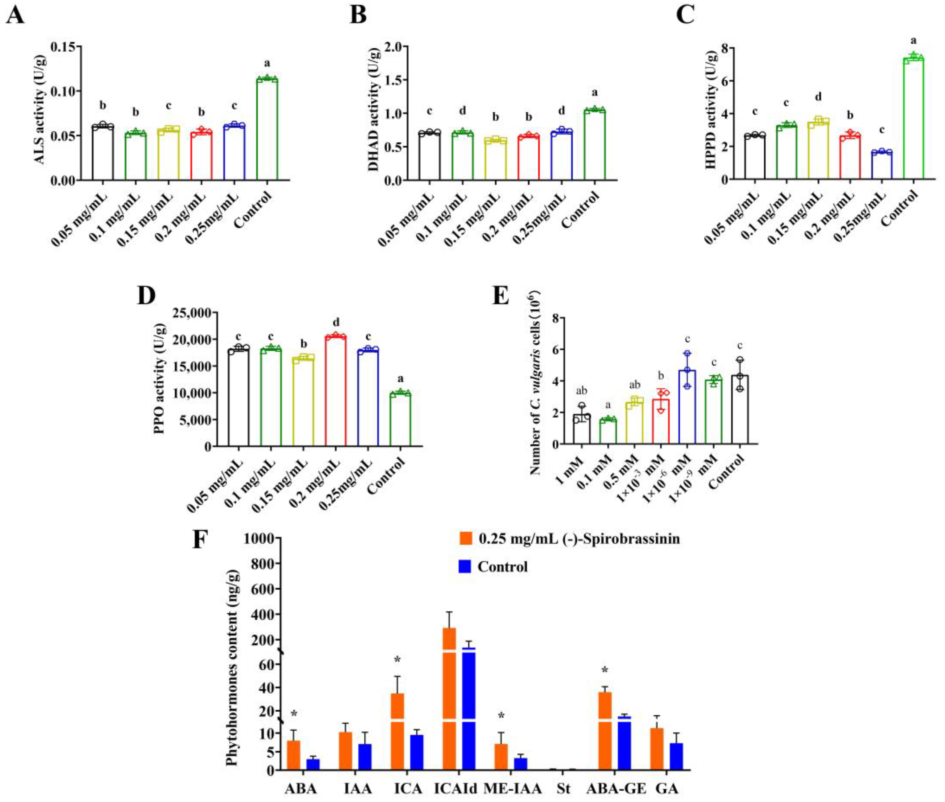
We assessed the activities of HPPD, ALS, DHAD, and PPO enzymes in L. sativa Linn., which served as the receptor plant. Intriguingly, upon treatment with (-)-Spirobrassinin, we observed a significant reduction in the activities of ALS, DHAD, and HPPD, even at a concentration as low as 0.05 mg/mL (Figure 2A–C). Particularly noteworthy was the substantial enhancement of PPO activity (Figure 2D). It is important to highlight that HPPD and PPO are crucial enzymes involved in the synthesis of photosynthetic pigments. We investigated the effects of (-)-Spirobrassinin on the reproduction of C. vulgaris. As the concentration of (-)-Spirobrassinin increased, the reproduction of C. vulgaris was significantly inhibited. At concentrations of 1 mM and 0.1 mM, the cell count of C. vulgaris was significantly suppressed (Figure 2E). Moreover, employing LC-MS analysis, we observed alterations in phytohormones content, specifically significant increases in abscisic acid (ABA) and gibberellins (GA) (Figure 2F). These findings are consistent with the docking results obtained for GIDI and PLY2, providing additional support for their relevance in the mode of action of (-)-Spirobrassinin.

3.3. The Stable Binding Mode between (-)-Spirobrassinin and PSBD1

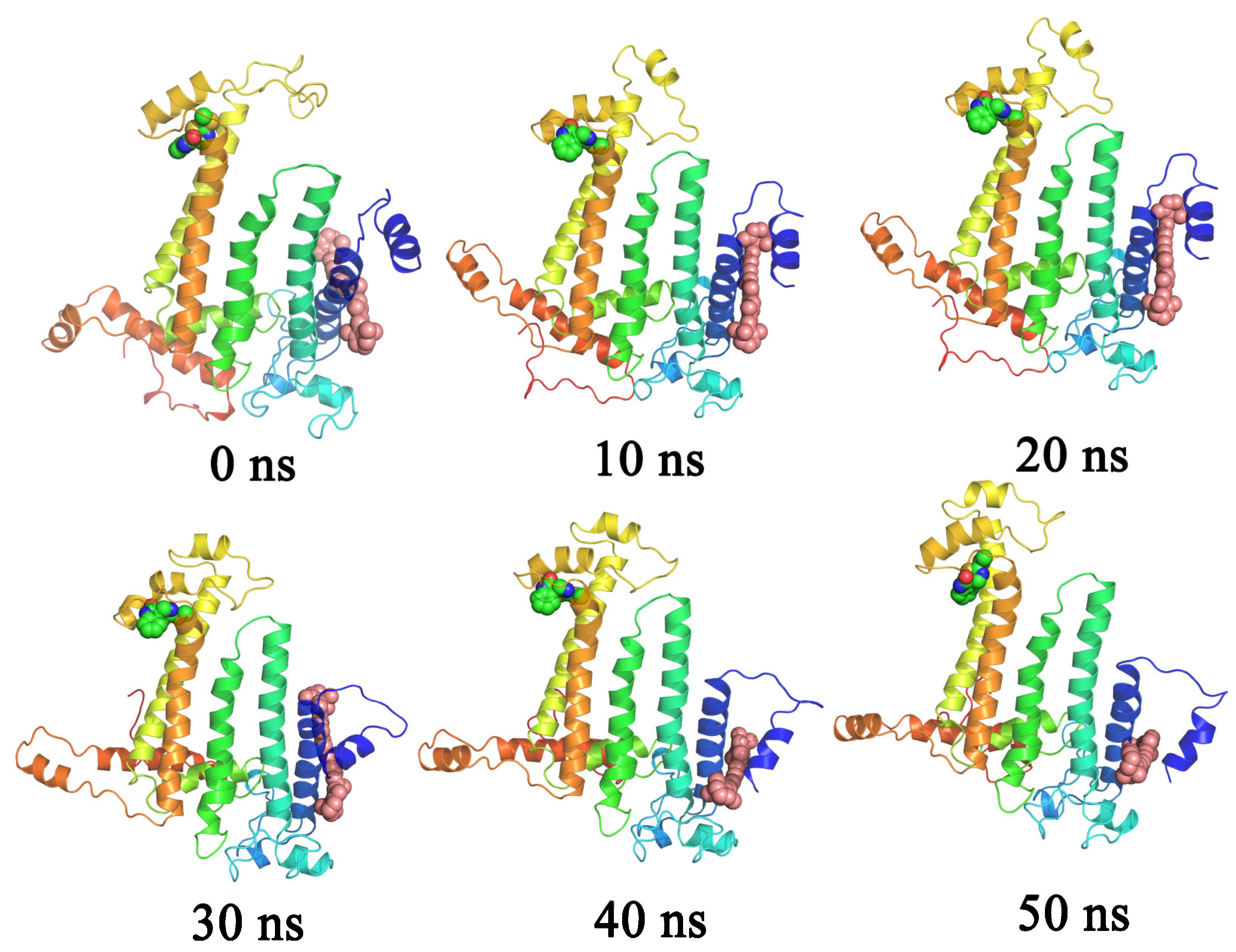
A 50 ns molecular dynamics simulation was conducted to examine the stable binding mode between (-)-Spirobrassinin and PSBD1. Snapshots were taken at various time points during the simulation (Figure 3). The analysis demonstrated that (-)-Spirobrassinin consistently maintained its binding to PSBD1 throughout the entire 50 ns simulation, with no observed dissociation events. Furthermore, no significant alterations were observed in the binding site of (-)-Spirobrassinin.

3.4. Stability of the System during the Simulation Process

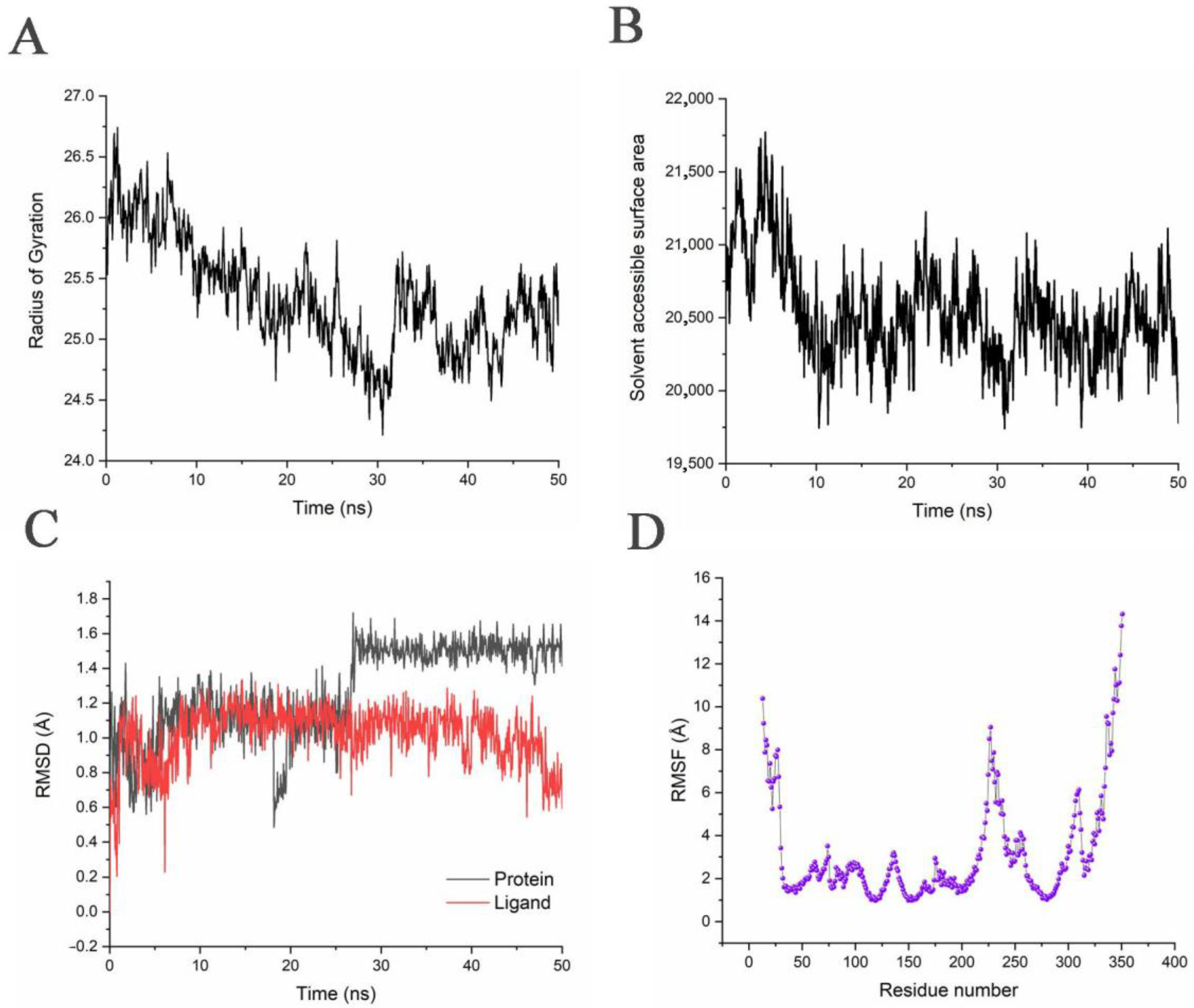
In this study, we calculated the root mean square deviation (RMSD) values to assess the stability of the system and the deviation of different molecules within the system throughout the simulation. The RMSD values provide insights into the equilibrium state of the system and indicate whether it has reached stability. The analysis demonstrates that the RMSD value of the PSBD1 remains relatively constant at around 1.5 Å after 30 ns of simulation, indicating system equilibrium. Moreover, the overall RMSD values are below 2 Å, indicating minimal structural changes occurring in the system. The red curve represents the variation of (-)-Spirobrassinin’s binding site relative to its initial position, showing slight fluctuations. This suggests that the binding site of (-)-Spirobrassinin undergoes subtle adjustments. However, the overall value remains below 1.4 Å, suggesting minimal differences in the position of the binding site (Figure 4A). The radius of gyration (Rg) is a parameter used to assess the compactness of a protein structure, with lower values indicating a more compact structure and higher values indicating a more extended or loose structure. An increase in protein compactness is generally associated with improved stability. Our analysis demonstrates that the compactness of the PSBD1 increases upon binding of the (-)-Spirobrassinin, as evidenced by a decrease in Rg values throughout the simulation period. This trend continues until approximately 30 ns, after which the change in compactness becomes more moderate (Figure 4B).
The variation in the solvent-accessible surface area (SASA) of the protein provides insights into its structural changes and reflects the degree of burial of hydrophobic surfaces. A higher SASA value indicates a larger exposed area of the PSBD1 with fewer buried hydrophobic surfaces, while a smaller SASA value suggests a more compact PSBD1 structure with more buried hydrophobic surfaces. Our analysis indicates that the SASA of the PSBD1 decreases during the simulation period, indicating structural compaction. Specifically, at the beginning of the simulation, the SASA value was 20,756.5996 Å2, and upon reaching equilibrium, the SASA value decreased to 19,779.24741 Å2 when the (-)-Spirobrassinin was bound (Figure 4C).
The root mean square fluctuation (RMSF) is a measure of the fluctuations exhibited by each amino acid in a protein structure. A higher RMSF value signifies greater flexibility of the amino acid, whereas a lower RMSF value indicates reduced flexibility. This value can provide insights into the stability of the PSBD1 to some extent (Figure 4D and Table 2).

3.5. Binding Mode of the (-)-Spirobrassinin with the PSBD1

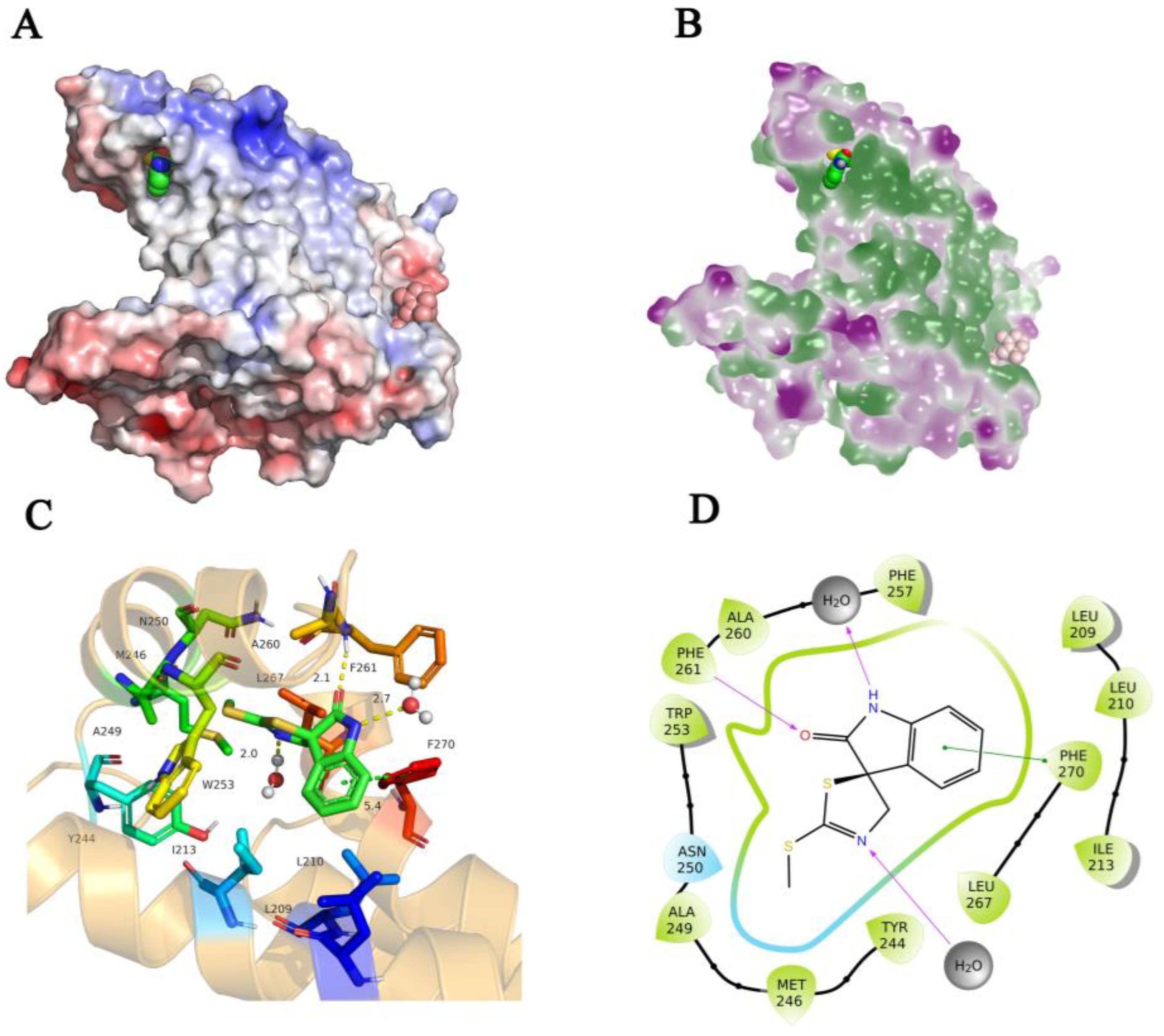
The hydrophobic and hydrophilic surface characteristics, as well as the electrostatic potential surface, were examined to understand the interaction between the (-)-Spirobrassinin and the PSBD1. Our findings indicate that (-)-Spirobrassinin predominantly binds to the hydrophobic grooves of PSBD1. Additionally, the binding of (-)-Spirobrassinin occurs at the interface between the positively and negatively charged regions of PSBD1, influenced by the charge properties of (-)-Spirobrassinin itself (Figure 5A,B).
During the equilibrium simulations, we conducted a detailed analysis of the specific interactions between (-)-Spirobrassinin and PSBD1. Our analysis revealed the presence of hydrogen bonding, hydrophobic interactions, and van der Waals contacts between (-)-Spirobrassinin and PSBD1, as well as hydrogen bonding interactions with water molecules in the simulation system. Specifically, the benzene ring of (-)-Spirobrassinin formed a π-π stacking interaction with the phenylalanine residue Phe270, with a distance of 5.4 Å. The carbonyl group of (-)-Spirobrassinin formed a hydrogen bond with the backbone amide of Phe261, with a distance of 2.1 Å. The amino group on the ring of (-)-Spirobrassinin formed a hydrogen bond with a water molecule at a distance of 2.7 Å, and the unsaturated nitrogen formed a hydrogen bond with another water molecule at a distance of 2.0 Å. Furthermore, (-)-Spirobrassinin exhibited hydrophobic interactions with nearby hydrophobic amino acids, including Leu209, Ile213, Phe257, Ala260, Trp253, Met246, and others (Figure 5C,D).

3.6. The Analysis of the (-)-Spirobrassinin-PSBD1 Contacts Revealed the Types and Quantities of Amino Acids Involved

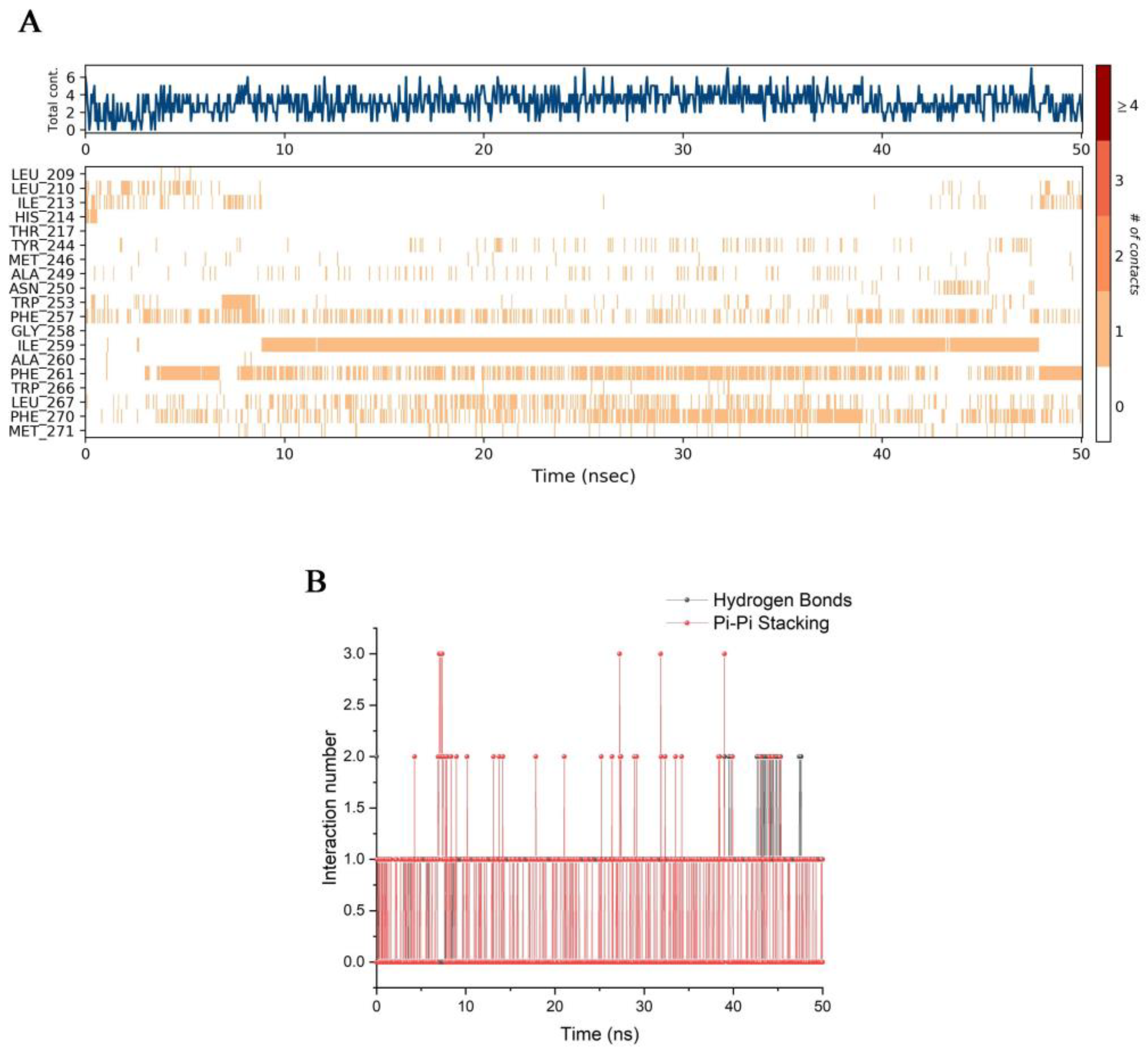
The analysis revealed that the main interactions between (-)-Spirobrassinin and PSBD1 were hydrogen bonding and π-π stacking. Hydrophobic interactions were not quantified but were determined based on the types of amino acids involved. The yellow color indicates the presence of contact, while the blue color indicates the number of interacting amino acid residues (Figure 6A). A consistent presence of yellow blocks throughout the 50 ns simulation for an amino acid suggests continuous interaction with (-)-Spirobrassinin. For example, Ile259 exhibited continuous blocks from 10 ns to 48 ns, indicating persistent hydrophobic interactions since this amino acid does not participate in hydrogen bonding. Similar analyses were performed for other amino acids. Notably, Phe261 and Phe270 showed a high frequency of continuous color blocks, indicating their critical roles in the interaction (Figure 6B).

3.7. Binding Free Energy between the (-)-Spirobrassinin and PSBD1

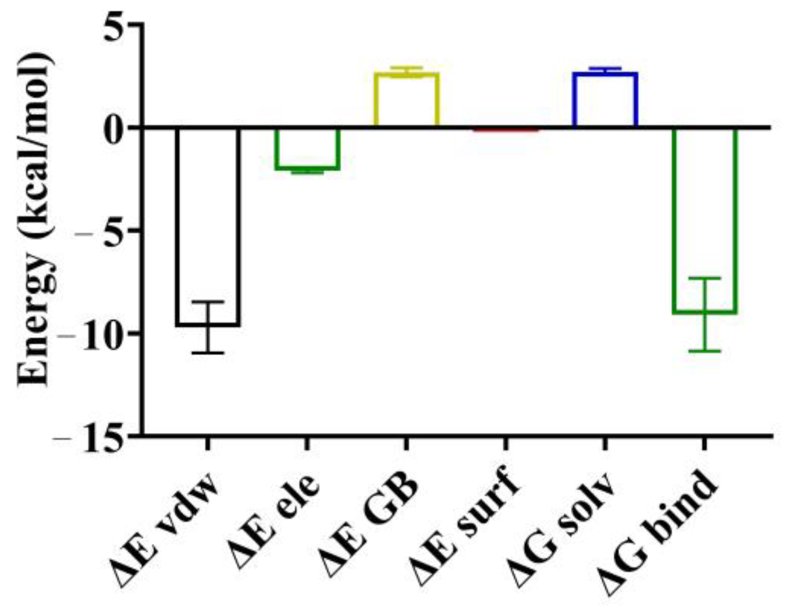
The MM/GBSA scores revealed that the binding free energy, ΔGbind, between (-)-Spirobrassinin and PSBD1 was determined to be −9.09 kcal/mol. In terms of energy contributions, the binding was primarily facilitated by van der Waals forces, including hydrogen bonding and hydrophobic interactions, as well as electrostatic interactions such as π-π stacking interactions. The van der Waals energy contribution, ΔEvdw, was calculated to be −9.71 kcal/mol, while the electrostatic energy contribution, ΔEele, was −2.09 kcal/mol. Conversely, the solvent free energy, ΔGsolv, was positive at 2.7 kcal/mol, comprising both the polar solvent free energy, ΔEGB, and nonpolar solvent free energy, ΔEsurf (Figure 7).

4. Discussion

(-)-Spirobrassinin is a unique secondary metabolite found exclusively in plants belonging to the Brassicaceae family. It has been investigated for its potential medical applications in cancer treatment, as it has demonstrated inhibitory effects on cancer cell proliferation [31]. Brassicaceae plants are known to be rich sources of natural products and have the potential to provide natural herbicides [32]. Certain isothiocyanates derived from Brassicaceae plants have been shown to inhibit the germination and growth of other plants [33]. In general, allelochemicals exert their toxic effects on plants by targeting processes such as photosynthesis, respiration, and amino acid biosynthesis in receptor plants [34]. However, the specific mode of action and binding sites of (-)-Spirobrassinin have not been reported thus far.
In this study, we assessed the binding energies of (-)-Spirobrassinin to the receptors, and these energies were found to be below −5 kcal/mol. To validate these findings, verification experiments were conducted using L. sativa Linn. as the receptor plant for these target proteins. ELISA tests were performed, confirming the reliability of the molecular docking results. The increase in PPO activity may indicate a resistance or adaptive phenomenon. This is similar to weeds developing herbicide resistance, which is a plant’s adaptive ability to specific chemicals, resulting in previously effective herbicides no longer being effective [35,36]. Moreover, the balance of phytohormones in plants was disrupted, as indicated by the accumulation of abscisic acid and gibberellins. These disruptions resulted in phenotypic changes, such as root shortening [37]. The abnormal increase in the content of abscisic acid and gibberellins may be attributed to the interference of (-)-Spirobrassinin as exogenous compounds, which disrupt the normal binding of abscisic acid and gibberellins to their target proteins [38]. This disruption leads to an abnormal increase in the levels of abscisic acid and gibberellins. On the other hand, gibberellins play a vital role in regulating plant germination and flowering processes, promoting bud elongation, and facilitating the transition to flowering [39]. The increase in gibberellin content may also be a self-regulatory response of plants to cope with external compound stress [40].
These findings provide evidence that supports the credibility of the molecular docking results, indicating their reliability. Traditional methods of searching for active sites in small molecules can be time-consuming and resource intensive. However, by employing molecular docking and computer-based calculations, we can predict and validate binding sites more efficiently. This approach significantly reduces the reliance on natural product resources and minimizes the time and cost involved, thereby overcoming challenges associated with the synthesis and low yield of natural products. We need to acknowledge the limitations of this method, as it is restricted to proteins with known crystal structures. However, Fpocket successfully identified the binding pocket of α-amylase for acarbose [8]. In the case of (-)-Spirobrassinin, further studies on its mode of action using innovative methods can expedite its development and application.
In the final part of our study, we investigated the potential of (-)-Spirobrassinin as a photosynthetic inhibitor-type herbicide. Due to the significant time and resource requirements for synthesis and experimental testing, we focused on computer simulations and growth inhibition assays using C. vulgaris. We performed a docking of (-)-Spirobrassinin to the active site of PSBD1 using the highest-resolution protein structure available, specifically the protein structure of Thermostichus vulcanus photosystem II with properly positioned quinones [41]. Throughout the 50 ns simulation, the RMSD values remained around 1.5 Å, indicating a stable and favorable binding of (-)-Spirobrassinin as a potential molecule for inhibiting photosystem II. Docking was considered successful when the RMSD value was below 2 Å [9].
(-)-Spirobrassinin exhibits inhibitory or stimulatory effects on the protein activity of herbicide targets, while also influencing phytohormonal balance, suggesting that (-)-Spirobrassinin acts as an allelochemical that affects various stages of plant growth. Molecular dynamics simulations have demonstrated the stable binding of (-)-Spirobrassinin to PSBD1, thus, establishing it as a potential candidate molecule for photosystem II inhibiting herbicides and for providing ‘green herbicides’ for the development of sustainable agriculture.

Supplementary Materials

The following supporting information can be downloaded at: https://www.mdpi.com/article/10.3390/app13127287/s1, Figure S1: Visualization of molecular docking of (-)-Spirobrassinin with plant growth-regulating target proteins and herbicide target proteins.; Figure S2: The inhibitory effect of (-)-Spirobrassinin on Panicum miliaceum L. and Amaranthus retroflexus L.; Figure S3: The inhibitory effect of (-)-Spirobrassinin on Panicum miliaceum L. and Amaranthus retroflexus L.

Author Contributions

Conceptualization, Y.W. and H.Z.; methodology, Y.W.; software, W.L.; resources, B.D.; data curation, D.W.; writing—original draft preparation, Y.W.; writing—review and editing, Y.W.; visualization, X.J.; supervision, Q.Z.; funding acquisition, H.Z. All authors have read and agreed to the published version of the manuscript.

Funding

This research was funded by the China Agriculture Research System (CARS-07-C-3).

Institutional Review Board Statement

Not applicable.

Informed Consent Statement

Not applicable.

Data Availability Statement

Data available within the article or its Supplementary Materials.

Acknowledgments

The authors acknowledge partial support of China Agriculture Research System.

Conflicts of Interest

The authors declare no conflict of interest.

References

  1. Day, M.; Witt, A.; Winston, R. Weed biological control in low- and middle-income countries. Curr. Opin. Insect Sci. 2020, 38, 92–98. [Google Scholar] [CrossRef]
  2. Hasanuzzaman, M.; Mohsin, S.M.; Bhuyan, M.H.M.B.; Bhuiyan, T.F.; Anee, T.I.; Masud, A.A.C.; Nahar, K. Chapter 3—Phytotoxicity, environmental and health hazards of herbicides: Challenges and ways forward. In Agrochemicals Detection, Treatment and Remediation; Prasad, M.N.V., Ed.; Butterworth-Heinemann: Oxford, UK, 2020; pp. 55–99. [Google Scholar]
  3. Qu, T.; Du, X.; Peng, Y.; Guo, W.; Zhao, C.; Losapio, G. Invasive species allelopathy decreases plant growth and soil microbial activity. PLoS ONE 2021, 16, e0246685. [Google Scholar] [CrossRef]
  4. Khaledi, R.; Fayaz, F.; Kahrizi, D.; Talebi, R. PCR-based identification of point mutation mediating acetolactate synthase-inhibiting herbicide resistance in weed wild mustard (Sinapis arvensis). Mol. Biol. Rep. 2019, 46, 5113–5121. [Google Scholar] [CrossRef]
  5. Das, C.; Dey, A.; Bandyopadhyay, A. Allelochemicals: An Emerging Tool for Weed Management. In Evidence Based Validation of Traditional Medicines; Springer: Berlin/Heidelberg, Germany, 2021; pp. 249–259. [Google Scholar]
  6. Palanivel, H.; Tilaye, G.; Belliathan, S.; Benor, S.; Gebrie, S.; Murugesan, K. Allelochemicals as Natural Herbicides for Sustainable Agriculture to Promote a Cleaner Environment. In Strategies and Tools for Pollutant Mitigation; Springer: Berlin/Heidelberg, Germany, 2021; pp. 93–116. [Google Scholar]
  7. Khamare, Y.; Chen, J.; Marble, S.C. Allelopathy and its application as a weed management tool: A review. Front. Plant Sci. 2022, 13, 1034649. [Google Scholar] [CrossRef]
  8. Duke, S.O.; Dayan, F.E.; Rimando, A.M.; Schrader, K.K.; Aliotta, G.; Oliva, A.; Romagni, J.G. Chemicals from nature for weed management. Weed Sci. 2002, 50, 138–151. [Google Scholar] [CrossRef]
  9. Heap, I. Global perspective of herbicide-resistant weeds. Pest Manag. Sci. 2014, 70, 1306–1315. [Google Scholar] [CrossRef]
  10. Flint, D.H.; Emptage, M.H. Dihydroxy acid dehydratase from spinach contains a [2Fe-2S] cluster. J. Biol. Chem. 1988, 263, 3558–3564. [Google Scholar] [CrossRef]
  11. Flint, D.H.; Emptage, M.H.; Finnegan, M.G.; Fu, W.; Johnson, M.K. The role and properties of the iron-sulfur cluster in Escherichia coli dihydroxy-acid dehydratase. J. Biol. Chem. 1993, 268, 14732–14742. [Google Scholar] [CrossRef] [PubMed]
  12. The inhibitory activity of natural products on plant p-hydroxyphenylpyruvate dioxygenase. J. Phytochem. 2002, 60, 281–288. [CrossRef] [PubMed]
  13. Hao, G.F.; Zuo, Y.; Yang, S.G.; Chen, Q.; Zhang, Y.; Yin, C.Y.; Niu, C.W.; Xi, Z.; Yang, G.F. Computational Discovery of Potent and Bioselective Protoporphyrinogen IX Oxidase Inhibitor via Fragment Deconstruction Analysis. J. Agric. Food Chem. 2017, 65, 5581–5588. [Google Scholar] [CrossRef] [PubMed]
  14. Hao, G.F.; Tan, Y.; Yang, S.G.; Wang, Z.F.; Zhan, C.G.; Xi, Z.; Yang, G.F. Computational and experimental insights into the mechanism of substrate recognition and feedback inhibition of protoporphyrinogen oxidase. PLoS ONE 2013, 8, e69198. [Google Scholar] [CrossRef] [Green Version]
  15. Anwar, S.; Naseem, S.; Ali, Z. Biochemical analysis, photosynthetic gene (psbA) down-regulation, and in silico receptor prediction in weeds in response to exogenous application of phenolic acids and their analogs. PLoS ONE 2023, 18, e0277146. [Google Scholar] [CrossRef] [PubMed]
  16. Qi, L.; Kwiatkowski, M.; Chen, H.; Hoermayer, L.; Sinclair, S.; Zou, M.; Del Genio, C.I.; Kubeš, M.F.; Napier, R.; Jaworski, K.; et al. Adenylate cyclase activity of TIR1/AFB auxin receptors in plants. Nature 2022, 611, 133–138. [Google Scholar] [CrossRef]
  17. Sheard, L.B.; Tan, X.; Mao, H.; Withers, J.; Ben-Nissan, G.; Hinds, T.R.; Kobayashi, Y.; Hsu, F.F.; Sharon, M.; Browse, J.; et al. Jasmonate perception by inositol-phosphate-potentiated COI1-JAZ co-receptor. Nature 2010, 468, 400–405. [Google Scholar] [CrossRef] [PubMed] [Green Version]
  18. Okamoto, M.; Peterson, F.C.; Defries, A.; Park, S.Y.; Endo, A.; Nambara, E.; Volkman, B.F.; Cutler, S.R. Activation of dimeric ABA receptors elicits guard cell closure, ABA-regulated gene expression, and drought tolerance. Proc. Natl. Acad. Sci. USA 2013, 110, 12132–12137. [Google Scholar] [CrossRef] [PubMed] [Green Version]
  19. Hao, Q.; Yin, P.; Li, W.; Wang, L.; Yan, C.; Lin, Z.; Wu, J.Z.; Wang, J.; Yan, S.F.; Yan, N. The Molecular Basis of ABA-Independent Inhibition of PP2Cs by a Subclass of PYL Proteins. Mol. Cell 2011, 42, 662–672. [Google Scholar] [CrossRef] [PubMed]
  20. Shimada, A.; Ueguchi-Tanaka, M.; Nakatsu, T.; Nakajima, M.; Naoe, Y.; Ohmiya, H.; Kato, H.; Matsuoka, M. Structural basis for gibberellin recognition by its receptor GID1. Nature 2008, 456, 520–523. [Google Scholar] [CrossRef]
  21. Jakalian, A.; Jack, D.B.; Bayly, C.I. Fast, efficient generation of high-quality atomic charges. AM1-BCC model: II. Parameterization and validation. J. Comput. Chem. 2002, 23, 1623–1641. [Google Scholar] [CrossRef]
  22. Anandakrishnan, R.; Aguilar, B.; Onufriev, A.V. H++ 3.0: Automating pK prediction and the preparation of biomolecular structures for atomistic molecular modeling and simulations. Nucleic Acids Res. 2012, 40, W537–W541. [Google Scholar] [CrossRef] [Green Version]
  23. Pettersen, E.F.; Goddard, T.D.; Huang, C.C.; Couch, G.S.; Greenblatt, D.M.; Meng, E.C.; Ferrin, T.E. UCSF Chimera—A visualization system for exploratory research and analysis. J. Comput. Chem. 2004, 25, 1605–1612. [Google Scholar] [CrossRef] [Green Version]
  24. Van Der Spoel, D.; Lindahl, E.; Hess, B.; Groenhof, G.; Mark, A.E.; Berendsen, H.J. GROMACS: Fast, flexible, and free. J. Comput. Chem. 2005, 26, 1701–1718. [Google Scholar] [CrossRef] [PubMed]
  25. Maffucci, I.; Contini, A. An Updated Test of AMBER Force Fields and Implicit Solvent Models in Predicting the Secondary Structure of Helical, β-Hairpin, and Intrinsically Disordered Peptides. J. Chem. Theory Comput. 2016, 12, 714–727. [Google Scholar] [CrossRef] [PubMed]
  26. Case, D.A.; Cheatham, T.E., 3rd; Darden, T.; Gohlke, H.; Luo, R.; Merz, K.M., Jr.; Onufriev, A.; Simmerling, C.; Wang, B.; Woods, R.J. The Amber biomolecular simulation programs. J. Comput. Chem. 2005, 26, 1668–1688. [Google Scholar] [CrossRef] [PubMed] [Green Version]
  27. Sprenger, K.G.; Jaeger, V.W.; Pfaendtner, J. The general AMBER force field (GAFF) can accurately predict thermodynamic and transport properties of many ionic liquids. J. Phys. Chem. B 2015, 119, 5882–5895. [Google Scholar] [CrossRef]
  28. Genheden, S.; Ryde, U. The MM/PBSA and MM/GBSA methods to estimate ligand-binding affinities. Expert Opin. Drug Discov. 2015, 10, 449–461. [Google Scholar] [CrossRef]
  29. Miller, B.R., 3rd; McGee, T.D., Jr.; Swails, J.M.; Homeyer, N.; Gohlke, H.; Roitberg, A.E. MMPBSA.py: An Efficient Program for End-State Free Energy Calculations. J. Chem. Theory Comput. 2012, 8, 3314–3321. [Google Scholar] [CrossRef]
  30. Gaillard, T. Evaluation of AutoDock and AutoDock Vina on the CASF-2013 Benchmark. J. Chem. Inf. Model. 2018, 58, 1697–1706. [Google Scholar] [CrossRef]
  31. Martina Chripkova, F.Z.a.J.M. Antiproliferative Effect of Indole Phytoalexins. Molecules 2016, 21, 1626. [Google Scholar] [CrossRef] [Green Version]
  32. Jan Petersen, R.B. Frank Walker, and Karl Hurle, Weed Suppression by Release of Isothiocyanates from Turnip-Rape Mulch. J. Agron. 2001, 93, 37–43. [Google Scholar] [CrossRef] [Green Version]
  33. Cheng, F.; Cheng, Z. Research Progress on the use of Plant Allelopathy in Agriculture and the Physiological and Ecological Mechanisms of Allelopathy. Front. Plant Sci. 2015, 6, 1020. [Google Scholar] [CrossRef] [Green Version]
  34. Wang, Y.; Xiong, Y.; Garcia, E.A.L.; Wang, Y.; Butch, C.J. Drug Chemical Space as a Guide for New Herbicide Development: A Cheminformatic Analysis. J. Agric. Food Chem. 2022, 70, 9625–9636. [Google Scholar] [CrossRef]
  35. Brzezowski, P.; Ksas, B.; Havaux, M.; Grimm, B.; Chazaux, M.; Peltier, G.; Johnson, X.; Alric, J. The function of protoporphyrinogen IX oxidase in chlorophyll biosynthesis requires oxidised plastoquinone in Chlamydomonas reinhardtii. Commun. Biol. 2019, 2, 159. [Google Scholar] [CrossRef] [Green Version]
  36. Koch, M.; Breithaupt, C.; Kiefersauer, R.; Freigang, J.; Huber, R.; Messerschmidt, A. Crystal structure of protoporphyrinogen IX oxidase: A key enzyme in haem and chlorophyll biosynthesis. EMBO J. 2004, 23, 1720–1728. [Google Scholar] [CrossRef] [Green Version]
  37. Steenackers, W.; Cesarino, I.; Klíma, P.; Quareshy, M.; Vanholme, R.; Corneillie, S.; Kumpf, R.P.; Van de Wouwer, D.; Ljung, K.; Goeminne, G.; et al. The Allelochemical MDCA Inhibits Lignification and Affects Auxin Homeostasis. Plant. Physiol. 2016, 172, 874–888. [Google Scholar] [CrossRef] [Green Version]
  38. Fujii, H.; Chinnusamy, V.; Rodrigues, A.; Rubio, S.; Antoni, R.; Park, S.Y.; Cutler, S.R.; Sheen, J.; Rodriguez, P.L.; Zhu, J.K. In vitro reconstitution of an abscisic acid signalling pathway. Nature 2009, 462, 660–664. [Google Scholar] [CrossRef] [Green Version]
  39. McCarty, D. Genetic Control and Integration of Maturation and Germination Pathways in Seed Development. Annu. Rev. Plant. Physiol. Plant. Mol. Biol. 2003, 46, 71–93. [Google Scholar] [CrossRef]
  40. Liao, Z.; Yu, H.; Duan, J.; Yuan, K.; Yu, C.; Meng, X.; Kou, L.; Chen, M.; Jing, Y.; Liu, G.; et al. SLR1 inhibits MOC1 degradation to coordinate tiller number and plant height in rice. Nat. Commun. 2019, 10, 2738. [Google Scholar] [CrossRef] [PubMed] [Green Version]
  41. Battaglino, B.; Grinzato, A.; Pagliano, C. Binding Properties of Photosynthetic Herbicides with the Q(B) Site of the D1 Protein in Plant Photosystem II: A Combined Functional and Molecular Docking Study. Plants 2021, 10, 1501. [Google Scholar] [CrossRef] [PubMed]
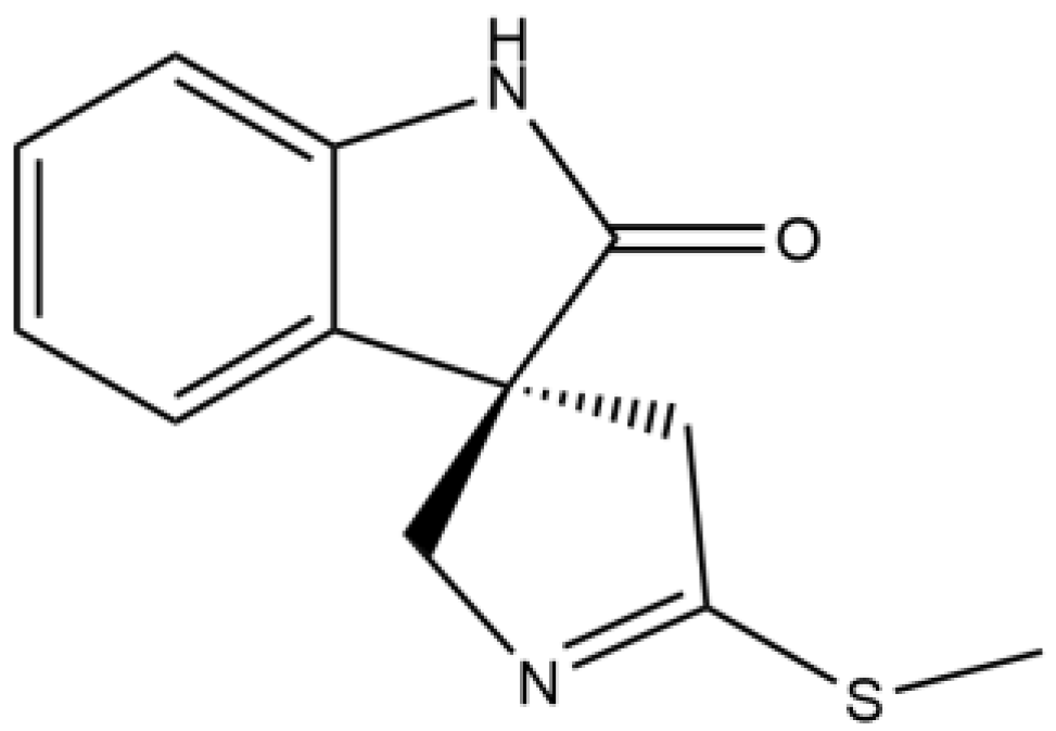
Figure 1. Chemical structures of (-)-Spirobrassinin.
Applsci 13 07287 g001
Figure 2. Effects of (-)-Spirobrassinin on the enzymatic activities of different target sites. * Indicates significant differences relative to the control (p < 0.05) and each bar graph is the mean ± standard error. Lowercase letters indicate significance (p < 0.05). The different symbols and colors in the columns indicate the enzymatic activities data of L. sativa Linn. seedlings after (-)-Spirobrassinin treatments. (A), Influence of (-)-Spirobrassinin on ALS activity; (B), influence of (-)-Spirobrassinin on DHAD activity; (C), influence of (-)-Spirobrassinin on HPPD activity; (D), influence of (-)-Spirobrassinin on PPO activity; (E), impact of (-)-Spirobrassinin on C. vulgaris growth; and (F), influence of (-)-Spirobrassinin on phytohormones content in L. sativa Linn. ABA: abscisic acid; IAA: indole-3-acetic acid; ICA: indole-3-carboxaldehyde; ICAId: indole-3-carboxaldehyde; ME-IAA: Methyl indole-3-acetate; St: strigolactones; ABA-GE: ABA-glucosyl ester; and GA: gibberellin.
Applsci 13 07287 g002
Figure 3. Snapshots from the 50 ns molecular dynamics simulation showing the (-)-Spirobrassinin (shown in green) bound to the PSBD1 (represented by ribbon visualization). (-)-Spirobrassinin and PSBD1 are represented by spheres and cartoon representations, respectively.
Applsci 13 07287 g003
Figure 4. Stability during simulation period. (A), The trend of the PSBD1’s radius of gyration (Rg) values during the simulation period. (B), The trend of the PSBD1’s solvent accessible surface area (SASA) values during the simulation period. (C), The trend of RMSD values for the molecules in the system during the simulation period. (D), The statistical analysis of the root mean square fluctuation (RMSF) of each amino acid in the PSBD1 during the simulation period.
Applsci 13 07287 g004
Figure 5. Analysis of the binding mode between the (-)-Spirobrassinin (green) and the PSBD1 during stable simulations. (A) Electrostatic potential surface of the PSBD1, with red indicating negatively charged regions and blue indicating positively charged regions; (B), distribution of hydrophobic and hydrophilic regions in the PSBD1, with green representing hydrophobic regions and magenta representing hydrophilic regions; (C), 3D interaction diagram, with hydrogen bonds shown as yellow dashed lines and the corresponding bond distances displayed. π-π stacking interactions represented by green dashed lines; and (D) 2D interaction diagram, with green representing hydrophobic amino acids, cyan representing polar amino acids, and gray representing the water molecules. Hydrogen bonds depicted as arrows and π-π stacking interactions shown as green lines.
Applsci 13 07287 g005
Figure 6. Analysis of amino acid contacts. (A), (-)-Spirobrassinin interaction with amino acids in PSBD1 and (B), the types and quantities of interactions between (-)-Spirobrassinin and PSBD1.
Applsci 13 07287 g006
Figure 7. Statistical analysis of the binding free energy between the (-)-Spirobrassinin and the PSBD1; each bar graph was the mean ± standard error. The binding free energies are predictions from MM/GBSA calculations.
Applsci 13 07287 g007
Table 1. The binding energy of (-)-Spirobrassinin to herbicidal target sites and plant growth regulator target sites.
Herbicidal Target Sites/Plant Growth Regulator Target SitesBinding Energy (Kcal/mol)
ALS−5.22
DHAD−5.56
HPPD−5.89
PPO−6.43
ACC−4.27
PSBD1−7.3
DAD2−4.97
GIDI−5.94
TIR1−4.49
D14−D3−ASK111.93
COLI−ASK1−3.92
PLY2−6.15
Note: The binding energies are predictions from Autodock docking calculations.
Table 2. Flexible RMSF (Root Mean Square Fluctuation) of Amino Acid Residues.
Residue NameRMSF(Å)
ALA14.468
ASN14.033
GLU12.360
GLY12.329
ARG11.412
PRO11.190
VAL10.923
HIS10.758
GLU10.574
GLU10.338
LEU10.268
GLY10.096
ASN9.928
PRO9.707
GLU9.427
ASP8.934
PHE8.914
TRP8.766
ARG8.653
VAL8.521
ILE8.452
Disclaimer/Publisher’s Note: The statements, opinions and data contained in all publications are solely those of the individual author(s) and contributor(s) and not of MDPI and/or the editor(s). MDPI and/or the editor(s) disclaim responsibility for any injury to people or property resulting from any ideas, methods, instructions or products referred to in the content.