Article

Hand Joint Hypermobility among Dental Students—A Cross-Sectional Study

1
College of Dental Medicine—Illinois, Midwestern University, Downers Grove, IL 60515, USA
2
Occupational Therapy, Midwestern University, Downers Grove, IL 60515, USA
3
College of Biomedical Sciences, Midwestern University, Downers Grove, IL 60515, USA
4
College of Dental Medicine—Arizona, Midwestern University, Glendale, AZ 85308, USA
*
Author to whom correspondence should be addressed.
Appl. Sci. 2023, 13(12), 7158; https://doi.org/10.3390/app13127158
Submission received: 17 May 2023 / Revised: 14 June 2023 / Accepted: 14 June 2023 / Published: 15 June 2023
(This article belongs to the Special Issue Advances in Reconstruction Dentistry)

Abstract

:
(1) Background: Joint hypermobility refers to joint movement beyond normal range. Dentistry requires fine motor skills and manual dexterity; thus, the presence of hand joint hypermobility requires early assessment and intervention. The aim of this cross-sectional study was to assess the prevalence of hand joint hypermobility among first-year dental students and investigate correlations between the severity of their hypermobility and muscular strength. (2) Methods: First-year dental students were evaluated by an occupational therapy team composed of certified hand therapists. The extension of distal interphalangeal, proximal interphalangeal, and metacarpophalangeal finger joints was assessed with bilateral testing of digit hypermobility (with a cut-off of >20° for hypermobility) and classified as high, moderate, or low risk according to number/types of joints involved. Muscular strength was assessed by comparing grip strength, lateral pinch, and three-point pinch to norm values. Right- vs. left-handed individuals, and females vs. males were compared with an independent t-test; the relationships between the variables were investigated using the Pearson correlation. (3) Results: Out of 141 dental students (24.6 ± 3.0, 70% females, 87% right-handed), 15% and 16% were classified as having a high and moderate risk of hypermobility, respectively, with no sex difference. Right-handed students scored significantly higher in the right lateral pinch than left-handed students (18.2 ± 4.8 vs. 13.2 ± 4.1, p = 0.045). A higher severity of hypermobility was associated with bilateral lower grip and pinch strength (p < 0.05). (4) Conclusions: One third of the first-year dental students exhibited moderate to severe hand joint hypermobility. Moreover, those identified as having moderate to severe hand joint hypermobility showed a decreased grip strength.

1. Introduction

Joint hypermobility is defined as movement of the joint beyond the normal range of motion [1,2]. Several causes contribute to its etiopathogenesis. One of the primary reasons is attributed to abnormalities in collagen, a protein responsible for the flexibility and strength of joints, ligaments, and tendons [1,3]. Collagen dysfunction can arise from genetic mutations affecting the genes responsible for the structure, composition, or function of connective tissues [3,4]. Syndromes such as Ehlers–Danlos syndrome (EDS) and Marfan syndrome are examples of collagen disorders in which joint hypermobility serves as a primary manifestation [3]. Hormonal changes, such as those occurring during pregnancy or the menstrual cycle, can also contribute to joint hypermobility status [5]. Additionally, joint hypermobility may result from previous joint injuries that caused damage to ligaments or the joint capsule, such as sprains or dislocations [5]. Muscle imbalance can further contribute to joint hypermobility if the supporting muscles are unable to provide sufficient stability [5].
The list of the negative consequences of having joint hypermobility is extensive. First of all, the most prominent effect is joint instability, which increases an individual’s susceptibility to subluxations, as well as partial or complete dislocations with a subsequent impairment of their quality of life [6]. Other signs and symptoms are chronic musculoskeletal pain, fatigue, muscle weakness, and joint degeneration [4,6]. Additionally, the severity of hypermobility can increase the likelihood of connective tissue injuries and damage, leading to conditions such as sprains, strains, and tendonitis [6].
A recent classification has categorized joint hypermobility as generalized (i.e., multiple large joints involved, including axial joints, as well as upper and lower limbs), localized (i.e., between one and five joints involved), and peripheral (i.e., limited to the hands and/or feet) [6]. Epidemiological studies on the general population have revealed a prevalence ranging from 2 to 57% [7]. These numbers tend to increase when specific populations are considered, such as young children, individuals with EDS, specific ethnic groups (i.e., Asian, Middle Eastern, or African descent), females, and dancers, among others. Nevertheless, this condition is often underdiagnosed and undertreated, especially in certain patient populations [5]. For example, a study conducted on a university cohort reported that up to 75% of 335 students exhibited some form of joint hypermobility [4]. Studies conducted on medical, physical therapy, and music students revealed a prevalence of generalized joint hypermobility of approximately 13–27% [4,8,9], often accompanied by musculoskeletal complaints and psychological distress [4,10]. While several studies have investigated the prevalence of generalized joint hypermobility, specific estimates for the prevalence of localized and peripheral joint hypermobility are limited [11].
When the affected individuals, such as dental care providers, require fine motor skills and manual dexterity to perform daily working activities, their functional performance may be impaired to varying degrees. Dental professionals rely on their stability, strength, and tactile senses to achieve acceptable clinical outcomes [8]. Unfortunately, individuals with peripheral joint hypermobility have been found to have lower proprioception [12,13], which may result in inappropriate pressure, further hyperflexion of supporting muscles, and an increased risk of musculoskeletal symptoms and injuries [14,15,16,17,18,19,20]. Despite a significant negative impact on these individuals’ daily working activities, limited data are available on peripheral joint hypermobility in dental professionals. To the best of our knowledge, only one study has been conducted among dental students investigating the prevalence of this condition [4], which further demonstrates the limited assessment and therefore the undertreatment of peripheral joint hypermobility within dental school settings.
The potential implications on clinical performance and the urge for the early recognition of this condition led to a collaboration between the Occupational Therapy (OT) and Dentistry departments of the College of Dental Medicine—Illinois (CDMI) at Midwestern University. Accordingly, first-year dental students are now routinely assessed by an OT team at the beginning of their program. By doing so, students that exhibit concerning degrees of hand joint hypermobility can receive adequate and prompt interventions.
Therefore, the aim of this study was twofold: (1) to assess the prevalence of peripheral joint hypermobility among first-year dental students, and (2) to investigate the correlation between the severity of their hypermobility and muscle strength. As the first aim was exploratory in nature, no a priori hypothesis was formulated. As for the secondary aim, it was hypothesized that individuals with higher degrees of hypermobility exhibit a lower level of muscle strength.

2. Materials and Methods

This was an observational cross-sectional study conducted on first-year dental students enrolled at Midwestern University (Downers Grove, IL, USA). The study was approved by the Institutional Review Board of the Office of Research of Midwestern University (20CIRB-IL 23004).

2.1. Eligibility Criteria

Inclusion criteria were subjects (1) who were enrolled in their first year of dental school at CDMI of MWU between 2022 and 2023; (2) who had no previously diagnosed systemic conditions that manifested as hypermobility (such as Ehlers–Danlos syndrome or generalized hypermobility syndrome); (3) who were between 18 and 30 years old; and (4) who understood English. Exclusion criteria were subjects (1) who were enrolled in any other year of dental school at CDMI of MWU, different from their first year; (2) who did not attend the in-person assessment performed by the OT team; and (3) who were not within the age range of 18–30 years.

2.2. Study Procedure

First-year dental students were evaluated by a team constituted of an OT faculty and OT students during 2 h orientation at the simulation lab clinic. Prior to the assessment, the OT students were calibrated by an OT faculty member/Certified Hand Therapist (α > 0.90).

2.3. Assessment of Outcome Measures

All the outcome measures described below were collected by OT students into the Hand Screening by Occupational Therapy for Dental Practice assessment, which was individually completed for each participant. The assessment sheet included the following domains:
Demographics: Participants self-reported their age (years), biological sex (male, female), and hand dominance (right, left, ambidextrous).
Hand joint hypermobility: A digit hypermobility test was conducted on both hands by assessing the extension of distal (DIP), proximal (PIP), and metacarpophalangeal (MP) joints of each finger (thumb, index, middle, ring, and small finger). The digit hypermobility test consisted of pushing each finger (DIP and PIP joints) against a table to test for hypermobility and one’s ability to stabilize joints. An extension of >20° was set as cut-off for hypermobility, as supported by other studies [21]. The hypermobility was classified as follows:
  • at high risk, if hyperextension of multiple joints was observed, with particular emphasis on specific fingers used as fulcrum during dental procedures. Specifically, participants were considered at high risk if hypermobility was observed in the thumb IP, in the index DIP and in the middle DIP fingers, or in the ring PIP/DIP and in the small PIP/DIP fingers.
  • at medium risk, if the participants’ hypermobility involved joints which are not strictly considered as important for finger rest;
  • at low risk, if joints appeared stable with little to no hypermobility (i.e., <20 degrees observed).
Figure 1 presents a detailed summary of this classification.
Muscle strength: Grip, lateral pinch, and 3-point pinch strength were assessed using an annually calibrated isokinetic dynamometer (Jaymar® Dynamometer, AliMed Inc., Dedham, MA, USA) and a pinch meter. The values obtained for each individual were then compared with norm guidelines established according to biological sex (see Table 1 for details). Grip and pinch strength were evaluated for both hands at one time point. Between each test conducted on the same hand, a 5 second rest was given to the subjects to avoid any confounding factors related to fatigue [22]. Before moving to the opposite hand, a 2 min rest was provided [22]. Next, wrist extension strength was evaluated using the Manual Muscle Testing scale which uses a 1–5 scoring system. Wrist extension strength ranged between 1 and 5, with higher values (from 4 to 5) corresponding to normal strength and lower values (below 3) corresponding to reduced strength.

2.4. Statistical Analysis

Prior to conducting the statistical analysis, all the variables were investigated for missing data. In cases in which there were missing data, these were removed from the analysis on a pairwise basis. When outliers were identified, analyses were run with and without outliers. However, if removing the outliers did not change the results, the outliers were kept in the analysis to maximize its power. First, Shapiro–Wilk test was used to confirm the normal distribution of the data collected. As no violation of skewness was found, all the data were analyzed with parametric test. Descriptive statistics was used to present the data as mean, standard deviation, range, and median, as appropriate.
To test Aim 1 (e.g., assessing the prevalence of peripheral joint hypermobility), frequency distribution was computed for all categories of low, moderate, and high risk of hypermobility. Next, independent t-test was used to compare right- vs. left-handed participants as well as females vs. males in the prevalence of peripheral joint hypermobility.
To test Aim 2 (e.g., assessing the correlation between severity of hypermobility and muscle strength), right- and left-handed participants were first compared in terms of muscle strength. Next, values obtained by females and males were compared with the corresponding norm values, and the difference between the muscle strength and the norm values was compared across each sex. Next, correlations between severity of hypermobility (as continuous variable) and muscle strength were investigated with linear correlation. Correlation between severity of hypermobility and demographics was calculated with Pearson correlation.
For all analyses, p-values were set at <0.05. Data were analyzed with SPSS (IBM SPSS Statistics Macintosh, Version 27.000, IBM Corp., Armonk, NY, USA).

3. Results

A total of 141 first-year dental students (70% females, with a mean age of 24.6 ± 3.0 years) were included in the analysis. This constituted the totality of the class, as all students were present on the day of the assessment. The vast majority of them were right-handed (87.2%), followed by left-handed (7.8%) and ambidextrous (0.7%). The remaining 4.3% of them had missing data for this domain.

3.1. Aim 1: Prevalence of Hand Joint Hypermobility

Fifteen percent of the 141 students were classified as being at a high risk of hypermobility, and 16% of them were classified as being at a moderate risk of hypermobility (Figure 2). The remaining 69% exhibited a low risk of hypermobility.
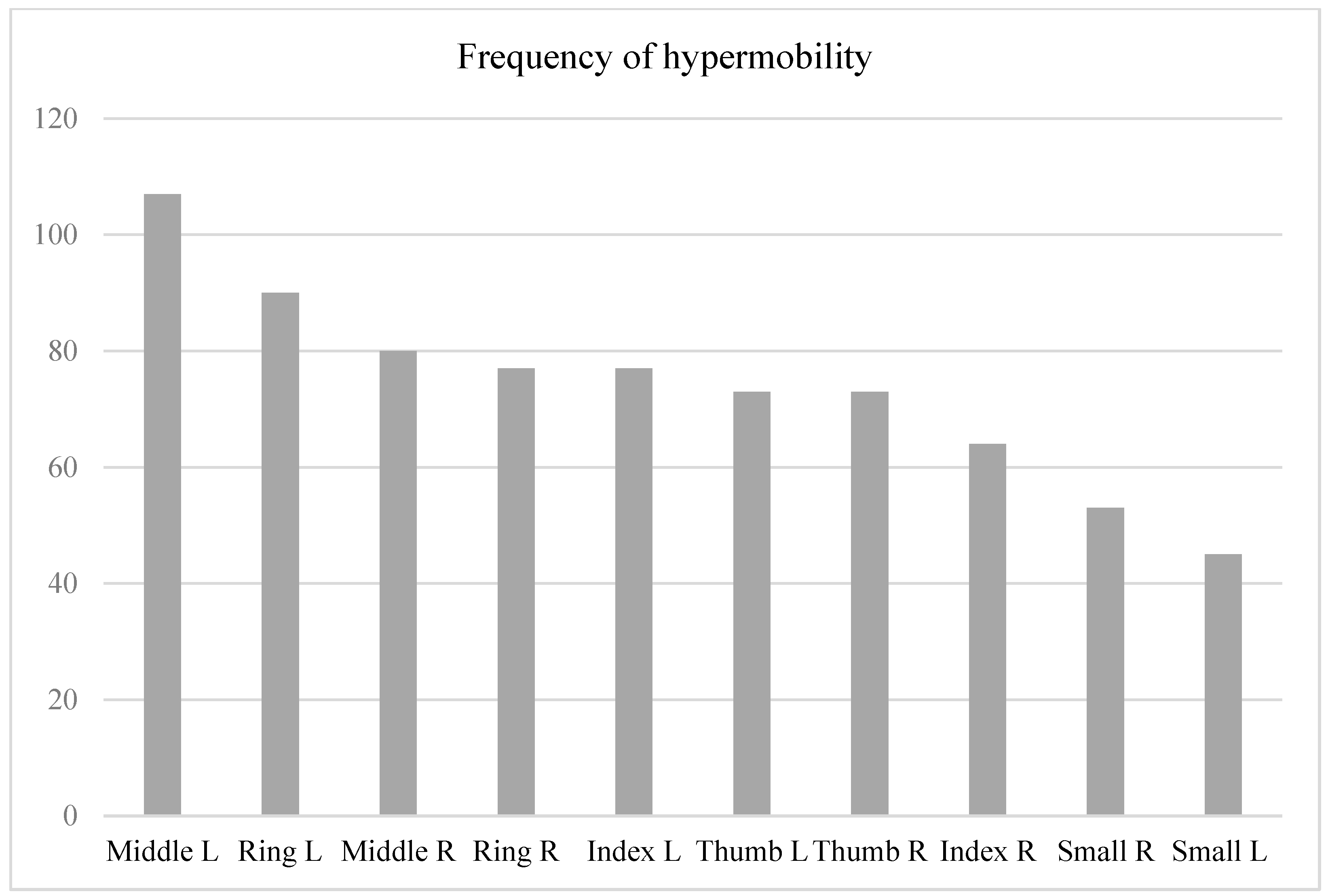
The fingers that were more significantly affected by hypermobility were the left middle finger, followed by the left ring finger, right middle finger, and right ring finger. The least affected in our student cohort were the right and left small fingers, as well as the right index finger. Figure 3 displays the frequency distribution of observed hypermobility according to finger and hand side.
No statistically significant difference was observed in the participants’ risk of hypermobility between females and males (p > 0.05). As there was only one ambidextrous participant, this subject was excluded from the comparison between right- and left-handed individuals. There was no difference in the participants’ risk of hypermobility between right- and left-handed subjects (p > 0.05).

3.2. Aim 2: Correlation between Hypermobility and Muscular Strength

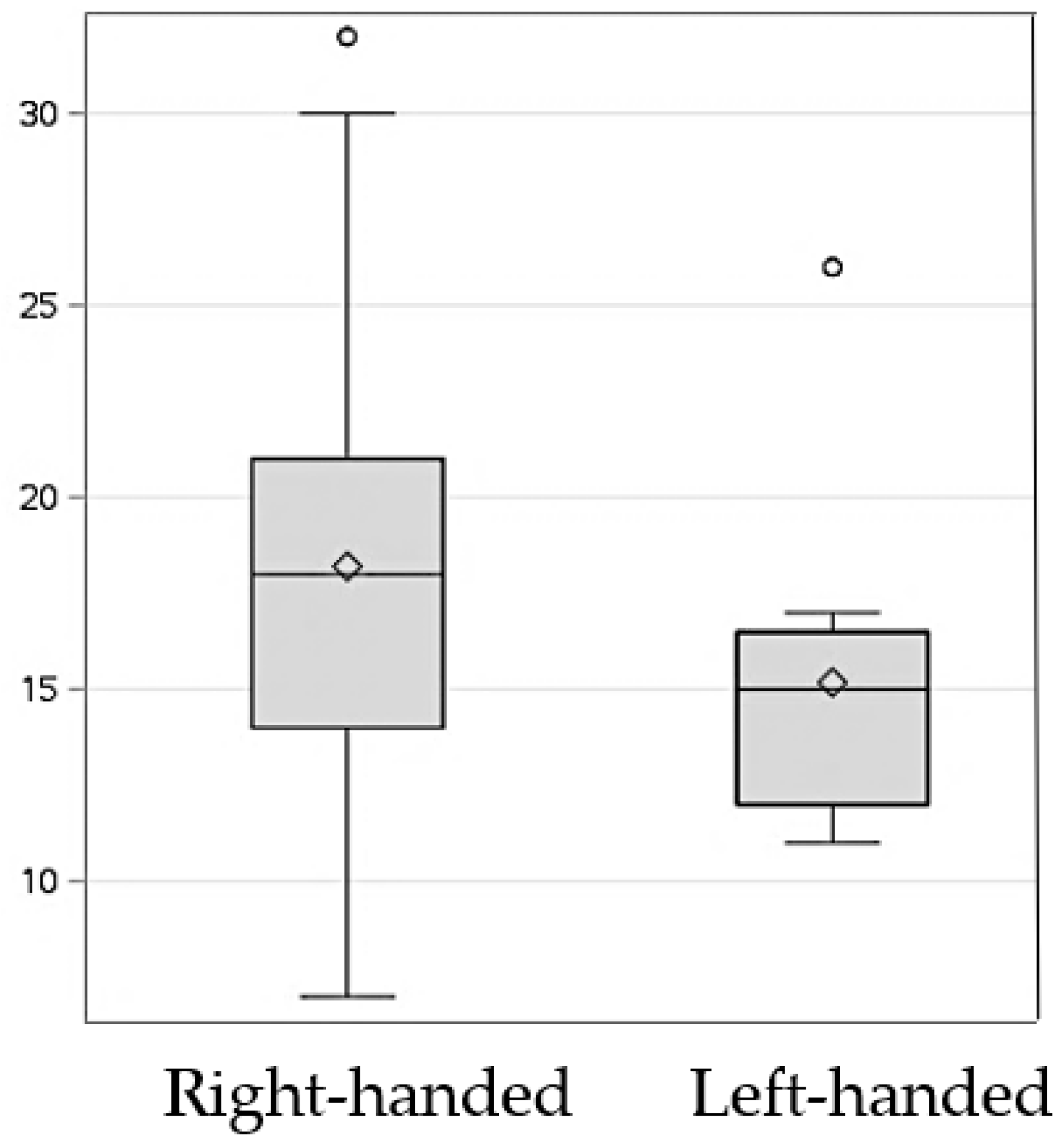
The male students had significantly higher right grip strength (36.9 ± 27.7 vs. 28.4 ± 20.3, t = 1.988, p = 0.049, 95% CI 0.04317–16.80384) and left grip strength (30.5 ± 23.3 vs. 22.9 ± 19.6, t = 1.974, p = 0.050, 95% CI −1.09295, 17.93996) than those of the females. The right-handed students exhibited scored significantly higher in right lateral pinch than left-handed students (18.2 ± 4.8 vs. 15.2 ± 4.1, t = 2.019, p = 0.045, 95% CI 0.616, 5.9813, Figure 4). The scores for right and left wrist extension did not differ between sex (p = 0.506 and p = 0.245, respectively) and ranged between 4.97 ± 0.2 and 5.00 ± 0.0.
A significant correlation of hypermobility was observed between the right and left hands of the same subject (r (141) = 0.808, p = 0.000). A higher severity of hypermobility was associated with bilateral lower grip and pinch strength (Table 2). The severity of hypermobility was not correlated with age (r (127) = 0.018, p = 0.839) nor with sex (r (129) = 0.109, p = 0.220).

4. Discussion

This retrospective study aimed at assessing the prevalence of peripheral hand joint hypermobility among first-year dental students at a single university and correlating hypermobility severity with muscular strength. Our analysis revealed that as many as 30% of the students exhibited a moderate to severe risk of hypermobility, which was correlated to a lower score for muscular hand strength. The findings of this study corroborated the importance of early assessment with a multidisciplinary collaboration between OT, certified hand therapy, and dentistry.

4.1. High Prevalence of Hand Joint Hypermobility among a Cohort of Dental Students

The prevalence of generalized joint hypermobility in the literature is 2–65%. This broad range may be due to different age groups and ethnicities of the included individuals, as well as various scoring systems and cut-off criteria [11,23,24]. The majority of these studies adopted the Beighton [21,25] or the Brighton criteria [26], with prevalence rates in university students ranging from 20% when the Brighton scale was utilized [27] to 26% with a cut-off Beighton score of ≥5 [27] and approximately 40% with a cut-off Beighton score of ≥4 [28]. Specifically, studies that investigated the prevalence of localized joint hypermobility among university students have identified rates ranging between 22% and 58% [4,8,9]. However, one of these studies specifically involved volunteers from physiotherapy and medical courses, which could lead to overrepresentation and potentially result in an overestimation of the prevalence of hypermobility [8]. Another study was conducted on individuals attending a music school [9], a category that has been speculated to have a higher risk [29]. Conversely, a single study assessed the prevalence of peripheral hand joint hypermobility in a similar sample population [4]. This research was conducted on 319 dental students across five years of dental school, divided into different ethnic and age groups for an analysis [4]. The findings of this study revealed that Taiwanese individuals displayed a higher prevalence of peripheral joint hypermobility, with 29.0% of them exhibiting a severe form while 25.8% exhibited a moderate form. On the other hand, Arab students were the second most common group to present with moderate hypermobility, which was observed in 40.0% of them. It is important to note that this investigation utilized different assessment tools to identify hypermobility compared to our present study, namely the Beighton scale with a cut-off of 4 and the Brighton criteria with a minimum of two major criteria or one major and two minor criteria. As a result, this study examined hyperextension of the elbow, knee, trunk, and palms of the hands resting on the floor, which were not evaluated in our study. Additionally, this study did not explicitly exclude subjects diagnosed with a generalized hypermobility syndrome. Consequently, the results obtained from this study may not be directly comparable with our findings.
The current study revealed a worrisome prevalence rate of peripheral hand joint hypermobility among a cohort of first-year dental students. As many as 15% and 16% of them had a high and moderate risk of hand joint hypermobility, respectively. This is a concerning number, considering that a higher degree of hypermobility has been associated with the development of musculoskeletal complaints in longitudinal and cross-sectional studies [30,31]. This risk factor further contributes to the already elevated prevalence of musculoskeletal disorders among dental professionals, which accounts for as high as 98% [32,33,34,35].
One other potential musculoskeletal condition is the development of osteoarthritis. Complaints of osteoarthritis in one’s hands have been specifically associated with an increased laxity at the thumb MP joint [36,37]. In our study, a greater hypermobility at the thumb joints was detected in a minority of the students. Conversely, the greatest degree of hypermobility was most commonly observed in the middle and ring fingers of both hands. This is in contrast to other studies that have identified the small finger and the thumb as the fingers with more severe hypermobility [7,38]. However, these studies were not conducted on a dental student population. Our finding of the middle and ring finger joints being the most affected is noteworthy for the field of dentistry, considering that these fingers constitute the finger rest for the majority of the hand grip position during dental procedures (e.g., pen grasp and modified pen grasp) [39]. Consequently, students may naturally adjust their hand position to one that is more stable, comfortable, and efficient to overcome their hypermobility. Yet, an inappropriate instrument grasp has been associated to compromised clinical performance, the use of increased force and pressure, and reduced control and stability [40].
Finally, contrary to other studies in the literature that have suggested a higher prevalence of hypermobility in females [8,9,27,40,41], the present investigation did not reveal any sex predominance. This is in accordance with other studies conducted in a similar population of university students [4,23].

4.2. Hypermobility Is Associated with Lower Muscle Strength

Our results suggested that individuals with higher finger hypermobility exhibited lower muscle strength, namely reduced bilateral lower grip and pinch strength. This corroborated the findings of other investigations that have revealed strength and proprioception deficits in patients with hypermobility [15,42,43]. The main reason for this appears to be attributed to a significant tendon laxity, rendering them unable to effectively transmit the power generated by the muscles [44]. Interestingly, a study found that males, but not females, with hypermobility showed lower muscle strength [22]. Future studies should be designed to confirm our findings, as to the best of our knowledge, this is the first study that specifically assesses a correlation between muscle strength and hypermobility in participants’ hands. Moreover, for the present study, three measures of muscle strength were selected (e.g., grip pinch, lateral pinch, and three-point pinch strength). Future research should incorporate other assessment measures, such as palmar pinch strength, power grip strength, key grip strength, finger abduction/adduction strength, or finger flexion/extension strength, among others.
It is important to note that the negative association between the severity of hypermobility and muscle strength did not take into consideration potential confounding factors derived from one’s age, body mass index (BMI), and/or physical activity level. Age is a crucial factor as muscle strength tends to naturally decline with increasing age, attributable to a 3–8% loss of muscle mass per decade after the age of 30 [45] and reduced muscle function [46]. As such, failing to adjust for age may lead to an inaccurate overestimation of the relationship between these variables. However, the age range within our sample population was limited, spanning from 21 to 27 years. Given that previous studies have consistently reported a notable decline in muscle strength beyond the age of 30 [45], it is unlikely that this confounding factor significantly impacted our findings. Moreover, the results of another study have questioned the existence of a relationship between age and hypermobility [47]. However, that study focused specifically on 14-year-old adolescents, which may not accurately reflect possible associations observed in older age groups. A second confounding factor is the body mass index (BMI). BMI can influence muscle strength in that higher BMI values are often associated with increased body weight and potentially more muscle mass [48]. This has also been confirmed in studies specifically examining handgrip strength [49]. Although in this study the students’ BMI was not assessed, it should also be noted that BMI alone does not provide a complete picture of body composition, and other factors such as the muscle-to-fat ratio can vary among individuals with the same BMI [50]. Finally, physical activity levels can significantly affect muscle strength. Individuals who engage in higher levels of physical activity tend to exhibit overall stronger muscles, regardless of their joint hypermobility status. Various studies have specifically examined the impact of structured exercise programs on muscle strength in people with generalized joint hypermobility. These investigations have shown increased muscle strength in specific areas, such as knee extensor muscle torque [51], shoulder strength [52], and trunk muscle endurance [53]. Nevertheless, a recent randomized controlled trial involving women with joint hypermobility failed to demonstrate any improvement in muscle mass and muscle strength in knee extensors [54]. These differences in findings may be attributed to variations in the studies’ assessments of strength, different resistance levels, and training frequency. Moreover, the fact that the aforementioned study exclusively included females could also contribute to the contradictory findings. It is worth noting that our study specifically excluded individuals with generalized joint hypermobility, unlike the subjects included in these previous studies. Hence, it is recommended that future investigations explore and account for physical activity level as a potential confounding factor to obtain a more comprehensive understanding of the relationship between these variables.

4.3. Future Directions

So far, the management of peripheral hand joint hypermobility has included the use of prefabricated finger orthoses to limit the hypermobility of the finger joints, thereby reducing pain and discomfort. However, to date, little is known on the uptake, long-term compliance, and effectiveness of these devices. In other words, for dental students who have been assessed and identified, is a finger orthosis a feasible and accepted strategy to be used while performing dental tasks? Similarly, if a finger orthosis is provided to an individual with hypermobility, what is the rate of compliance in the long term? Moreover, provided that the device is used, is it objectively effective in addressing their impaired functioning?
There are very few studies available in the literature that have attempted to answer these research questions. These investigations tested these devices by having participants perform validated tasks, such as a targeted box and block test, writing test, and test involving picking up coins [1,55,56]. Nevertheless, to the best of our knowledge, no study has investigated the effectiveness of these finger orthoses while performing dental tasks, nor have pain or discomfort been assessed while using these devices. From our clinical experience, the dental students that exhibit hypermobility express frustration and distress when they realize at the beginning of their dental career that a physical and not previously addressed impairment might lead to unsatisfied clinical outcomes. As such, future clinical studies are advocated to address these important gaps.
Moreover, it has been observed that an adjustment of the finger position naturally occurs in these individuals to limit finger bending due to their hypermobility. Therefore, personalized interventions are advocated at an early stage of dental students’ careers, ideally during the preclinical curriculum when proper ergonomics, instrumentation, and hand skills are developed.
Lastly, nothing is known about the actual comfort of the prefabricated devices available on the market. Recently, advances have been made with Computer-Aided Design and Computer-Aided Manufacturing (CAD-CAM) systems in fabricating custom-made devices. Potentially, this may increase the comfort of these devices. Our group of researchers is currently working on testing the feasibility and the effectiveness of CAD-CAM finger orthoses in a dental student population.

4.4. Strength and Limitations

This study highlighted the worrisome prevalence of young individuals with hand joint hypermobility among a cohort of first-year dental students. It adopted a grading system specifically designed for dental professionals and was able to identify the severity of the participants’ hypermobility, in contrast to the dichotomous pattern of other validated criteria [10,57]. It also reinforced the need for multidisciplinary assessments, followed by appropriate interventions, conducted by occupational therapy and/or hand specialists.
This study also had some important limitations. First, its cross-sectional nature reflected only one time point, and nothing can be ascertained on the progression of hand joint hypermobility. Studies in the literature have suggested a progressive decrease in hypermobility and hyperlaxity over time. As such, the current investigation can only be generalized to young individuals belonging to the same age group (i.e., the range between 21 and 27 years). To overcome the limitations derived from our cross-sectional design, we plan to conduct a standardized assessment of hand joint hypermobility on incoming classes of first-year dental students over an extended period. By implementing this repeated longitudinal assessment, possible trends and patterns of hypermobility may emerge over time and will contribute to more robust and generalizable findings. Furthermore, repeating the assessment on the same subjects over time will provide valuable insights into the natural progression of hypermobility, particularly within this specific population. Longitudinal studies also have the potential to identify risk factors that contribute to the development or progression of hand joint hypermobility among dental students. Factors such as ergonomics, workload, and specific dental procedures can be examined to understand their potential influence on the severity of hypermobility.
Moreover, the relatively small sample size of the study (n = 140) limits the generalizability and external validity of our findings. However, this assessment has now been incorporated in the dental curriculum for first-year dental students. Consequently, future studies employing a longitudinal design and standardized assessment tool will be able to validate these findings. In addition, our study relied on self-reported data on the participants’ dominant hand, which limits the reliability of the findings. To enhance future study designs, it is recommended to validate self-reported data through means such as medical records or a clinical assessment, in which the students’ actual performance on a manikin is observed. Next, the results of this study can only report the extent of hypermobility with no speculation on pain symptomatology. Future studies are needed to investigate pain and discomfort in individuals with peripheral joint hypermobility while performing functional dental tasks. Moreover, the present investigation utilized a hand screening assessment developed by an OT to assess dental students. As such, comparisons with other available studies in the literature that used different scoring systems (e.g., the Beighton score, Brighton criteria, or Horan Joint Mobility Index) may not be accurate. The hand screening assessment was adopted in our study because it was considered more pertinent for our cohort of dental students, as the finger joints, rather than elbow, knee, and trunk, are particularly crucial for daily performance tasks. In addition, even by adopting a standardized scoring system, the results would have been difficult to compare across the studies, due to the several recommended cut-off scores [58]. Moreover, the aim of this study was to assess the prevalence of joint hypermobility limited to the hand; therefore, a more comprehensive assessment was not needed.
This study did not assess the contribution of ethnicity to the presence of hand joint hypermobility, which is known to represent a non-modifiable risk factor [4,7]. However, the cohort included in this study was constituted predominantly by white individuals, and, as such, a comparison between different ethnicities would have not achieved enough power for inferential statistics. Lastly, although a significant correlation was found between the severity of hypermobility and reduced muscle strength, the results were not controlled for possible confounding factors such as BMI, pain symptoms, or participation in sports [22]. Future studies adjusting for these variables are advocated.

5. Conclusions

The results of this study showed that approximately one-third of a cohort of first-year dental students exhibited moderate to severe hand joint hypermobility. Moreover, individuals with higher degrees of joint hypermobility were revealed to have decreased grip strength. Ultimately, these findings suggest that hand joint hypermobility may have significant implications for certain professions that require fine-motor skills, such as dentistry. This study also demonstrated the importance of early assessment as well as interprofessional and multidisciplinary collaboration within the predoctoral dental field.

Author Contributions

Conceptualization, D.L.; methodology, D.L.; software, L.S.; formal analysis, L.S.; data curation, N.K. and E.W.-P.; writing—original draft preparation, L.S.; writing—review and editing, L.S., M.A. and D.L.; supervision, J.C.M. All authors have read and agreed to the published version of the manuscript.

Funding

This research received no external funding.

Institutional Review Board Statement

The study was conducted in accordance with the Declaration of Helsinki and approved by the Institutional Review Board of Midwestern University (protocol code CIRB IL—23004 of 1 March 2023).

Informed Consent Statement

Informed consent was obtained from all subjects involved in the study.

Data Availability Statement

Data are available at request to the corresponding author.

Conflicts of Interest

The authors declare no conflict of interest.

References

  1. Jensen, A.-M.; Andersen, J.Q.; Quisth, L.; Ramstrand, N. Finger orthoses for management of joint hypermobility disorders: Relative effects on hand function and cognitive load. Prosthet. Orthot. Int. 2020, 45, 36–45. [Google Scholar] [CrossRef] [PubMed]
  2. Boyle, K.L.; Witt, P.; Riegger-Krugh, C. Intrarater and interrater reliaility of the Brighton and Horan Joint Mobility Index. J. Athl. Train. 2003, 38, 281–285. [Google Scholar] [PubMed]
  3. Kobayasi, T. Dermal elastic fibres in the inherited hypermobile disorders. J. Dermatol. Sci. 2006, 41, 175–185. [Google Scholar] [CrossRef] [PubMed]
  4. Szalewska, M.; Lupa, M.; Pochroń, N.; Pudzianowski, S.; Szalewski, L.; Bożyk, A.; Borowicz, J. Prevalence of Joint Hypermobility Syndrome Amongst Dental Students of the Medical University of Lublin. Pol. J. Public Health 2014, 125, 73–76. [Google Scholar] [CrossRef] [Green Version]
  5. Grahame, R.; Halkim, A.J. Hypermobility. Curr. Opin. Rheumatol. 2008, 20, 106–110. [Google Scholar] [CrossRef]
  6. Castori, M.; Tinkle, B.; Levy, H.; Grahame, R.; Malfait, F.; Hakim, A. A framework for the classification of joint hypermobility and related conditions. Am. J. Med. Genet. Part C Semin. Med Genet. 2017, 174, 148–157. [Google Scholar] [CrossRef] [Green Version]
  7. Remvig, L.; Jensen, D.V.; Ward, R.C. Epidemiology of general joint hypermobility and basis for the proposed criteria for benign joint hypermobility syndrome: Review of the literature. J. Rheumatol. 2007, 34, 804–809. [Google Scholar]
  8. Antonio, D.H.; Magalhães, C.S. Survey on joint hypermobility in university students aged 18–25 years old. Adv. Rheumatol. 2018, 58, 3. [Google Scholar] [CrossRef]
  9. Larsson, L.-G.; Baum, J.; Mudholkar, G.S. Hypermobility: Features and differential incidence between the sexes. Arthritis Rheum. 1987, 30, 1426–1430. [Google Scholar] [CrossRef]
  10. Kirk, J.A.; Ansell, B.M.; Bywaters, E.G. The hypermobility syndrome: Musculoskeletal complaints associated with generalized joint hypermobility. Ann. Rheum. Dis. 1967, 26, 419–425. [Google Scholar] [CrossRef] [Green Version]
  11. Ituen, O.A.; Anieto, E.M.; Ferguson, G.; Duysens, J.; Smits-Engelsman, B. Prevalence and Demographic Distribution of Hypermobility in a Random Group of School-Aged Children in Nigeria. Healthcare 2023, 11, 1092. [Google Scholar] [CrossRef] [PubMed]
  12. Clayton, H.A.; Jones, S.A.H.; Henriques, D.Y.P. Proprioceptive precision is impaired in Ehlers–Danlos syndrome. Springerplus 2015, 4, 1–8. [Google Scholar] [CrossRef] [PubMed] [Green Version]
  13. Cooper, R.A.; Brems, J.J. The inferior capsular-shift procedure for multidirectional instability of the shoulder. J. Bone Jt. Surg. 1992, 74, 1516–1521. [Google Scholar] [CrossRef]
  14. Finsterbush, A.; Poground, H. The hypermobility syndrome: Musculoskeeltal complaints in 100 consecutive cases of generalized joint hypermobility. Clin. Orthop. Relat. Res. 1982, 168, 124–127. [Google Scholar] [CrossRef]
  15. Wolf, J.M.; Cameron, K.L.; Owens, B.D. Impact of Joint Laxity and Hypermobility on the Musculoskeletal System. J. Am. Acad. Orthop. Surg. 2011, 19, 463–471. [Google Scholar] [CrossRef] [PubMed]
  16. Scollinge, R.; Simmonds, J. Hypermobility, injury rate and rehabilitation in a professional football squad: A preliminary study. Phys. Ther. Sport 2009, 10, 91–96. [Google Scholar] [CrossRef]
  17. Stewart, D.R.; Burden, S. Does generalised limgamentous laxity increase seasonal incidence of injuries in male first division club rugby players? Br. J. Sports Med. 2004, 38, 457–460. [Google Scholar] [CrossRef] [Green Version]
  18. Rozen, T.D.; Roth, J.M.; Denenberg, N. Cervical spine joint hypermobility: A possible predisposing factor for new dailty persistent headache. Cephalagia 2006, 26, 1182–1185. [Google Scholar] [CrossRef]
  19. Soderman, K.; Alfredson, H.; Pietila, T.; Werner, S. Risk factors for leg injures in female soccer players: A propsetive investigation during one out-door season. Knee Surg. Sports Traumatol. Arthrisc. 2001, 9, 313–321. [Google Scholar] [CrossRef]
  20. Neilson, D.; Martin, V.T. Joint Hypermobility and Headache: Understanding the Glue That Binds the Two Together—Part 1. Headache 2014, 54, 1393–1402. [Google Scholar] [CrossRef]
  21. Smits-Engelsman, B.; Klerks, M.; Kirby, A. Beighton Score: A Valid Measure for Generalized Hypermobility in Children. J. Pediatr. 2011, 158, 119–123.e4. [Google Scholar] [CrossRef] [PubMed]
  22. Jindal, P.; Narayan, A.; Ganesan, S.; MacDermid, J.C. Muscle strength differences in healthy young adults with and without generalized joint hypermobility: A cross-sectional study. BMC Sports Sci. Med. Rehabil. 2016, 8, 12. [Google Scholar] [CrossRef] [PubMed] [Green Version]
  23. Reuter, P.R.; Fichthorn, K.R. Prevalence of generalized joint hypermobility, musculoskeletal injuries, and chronic musculoskeletal pain among American university students. PeerJ 2019, 7, e7625. [Google Scholar] [CrossRef]
  24. Jansson, A.; Saartok, T.; Werner, S.; Renström, P. General joint laxity in 1845 Swedish school children of different ages: Age- and gender-specific distributions. Acta Paediatr. 2004, 93, 1202–1206. [Google Scholar] [CrossRef] [PubMed]
  25. Beighton, P.; Horan, F. Orthopaedic aspects of the Ehlers-Danlos syndrome. J. Bone Jt. Surg. 1969, 51, 444–453. [Google Scholar] [CrossRef]
  26. Juul-Kristensen, B.; Rogind, H.; Jensen, D.V.; Remvig, L. Inter-examiner reproducibility of tests and criteria for generalized joint hypermobility and benign joint hypermobility syndrome. Rheumatology 2007, 46, 1835–1841. [Google Scholar] [CrossRef] [PubMed] [Green Version]
  27. Russek, L.N.; Errico, D.M. Prevalence, injury rate and, symptom frequency in generalized joint laxity and joint hypermobility syndrome in a “healthy” college population. Clin. Rheumatol. 2016, 35, 1029–1039. [Google Scholar] [CrossRef]
  28. Baeza-Velasco, C.; Gely-Nargeot, M.-C.; Vilarrasa, A.B.; Fenetrier, C.; Bravo, J.F. Association between psychopathological factors and joint hypermobility syndrome in a group of undergraduates from a French university. Int. J. Psychiatry Med. 2011, 41, 187–201. [Google Scholar] [CrossRef]
  29. Artigues-Cano, I.; Bird, H.A. Hypermobility and Proprioception in the Finger Joints of Flautists. Am. J. Clin. Oncol. 2014, 20, 203–208. [Google Scholar] [CrossRef]
  30. Leone, V.; Tornese, G.; Zerial, M.; Locatelli, C.; Ciambra, R.; Bensa, M.; Pocecco, M. Joint hypermobility and its relationship to musculoskeletal pain in schoolchildren: A cross-sectional study. Arch. Dis. Child. 2009, 94, 627–632. [Google Scholar] [CrossRef]
  31. Sperotto, F.; Balzarin, M.; Parolin, M.; Monteforte, N.; Vittadello, F.; Zulian, F. Joint hypermobility, growing pain and obesity are mutually exclusive as causes of musculoskeletal pain in schoolchildren. Clin. Exp. Rheumatol. 2014, 32, 131–136. [Google Scholar] [CrossRef] [PubMed]
  32. Lietz, J.; Kozak, A.; Nienhaus, A. Prevalence and occupational risk factors of musculoskeletal diseases and pain among dental professionals in Western countries: A systematic literature review and meta-analysis. PLoS ONE 2018, 13, e0208628. [Google Scholar] [CrossRef] [PubMed] [Green Version]
  33. Ohlendorf, D.; Naser, A.; Haas, Y.; Haenel, J.; Fraeulin, L.; Holzgreve, F.; Erbe, C.; Betz, W.; Wanke, E.M.; Brueggmann, D.; et al. Prevalence of Musculoskeletal Disorders among Dentists and Dental Students in Germany. Int. J. Environ. Res. Public Health 2020, 17, 8740. [Google Scholar] [CrossRef] [PubMed]
  34. Samoladas, E.; Barmpagianni, C.; Papadopoulos, D.V.; Gelalis, I.D. Lower back and neck pain among dentistry students: A cross-sectional study in dentistry students in Northern Greece. Eur. J. Orthop. Surg. Traumatol. 2018, 28, 1261–1267. [Google Scholar] [CrossRef] [PubMed]
  35. Thornton, L.J.; Barr, A.E.; Stuart-Buttle, C.; Gaughan, J.P.; Wilson, E.R.; Jackson, A.D.; Wyszynski, T.C.; Smarkola, C. Perceived musculoskeletal symptoms among dental students in the clinic work environment. Ergonomics 2008, 51, 573–586. [Google Scholar] [CrossRef]
  36. Freedman, D.M.; Eaton, R.G.; Glickel, S. Long-term results of volar ligament reconstruction for symptomatic basal joint laxity. J. Hand Surg. 2000, 25, 297–304. [Google Scholar] [CrossRef]
  37. Jónsson, H.; Vatysdóttire, S.T. Hypermobility features in patients with hand osteoarthritis. Osteoarhtitis Cartil. 1995, 3, 1–5. [Google Scholar] [CrossRef] [Green Version]
  38. Kwon, J.-W.; Lee, W.-J.; Park, S.-B.; Kim, M.J.; Jang, S.H.; Choi, C.K. Generalized Joint Hypermobility in Healthy Female Koreans: Prevalence and Age-Related Differences. Ann. Rehabil. Med. 2013, 37, 832–838. [Google Scholar] [CrossRef] [Green Version]
  39. Canakci, V.; Orbak, R.; Tezel, A.; Canakci, C.F. Influence of different periodontal curette grips on the outcome of mechanical non-surgical therapy. Int. Dent. J. 2003, 53, 153–158. [Google Scholar] [CrossRef]
  40. DeCoster, L.C.; Vailas, J.C.; Lindsay, R.H.; Williams, G.R. Prevalence and Features of Joint Hypermobility among Adolescent Athletes. Arch. Pediatr. Adolesc. Med. 1997, 151, 989–992. [Google Scholar] [CrossRef]
  41. Didia, B.C.; Dapper, D.V.; Boboye, S.B. Joint hypermobility syndrome among undergraduate students. East Afr. Med. J. 2002, 79, 80–81. [Google Scholar] [CrossRef] [PubMed] [Green Version]
  42. Sahin, N.; Baskent, A.; Ugurlu, H.; Berker, E. Isokinetic evaluation of knee extensor/flexor muscule strength in patients with hypermobility syndrome. Rheumatol. Int. 2008, 28, 643–648. [Google Scholar] [CrossRef] [PubMed]
  43. Kumar, B.; Lenert, P. Joint Hypermobility Syndrome: Recognizing a Commonly Overlooked Cause of Chronic Pain. Am. J. Med. 2017, 130, 640–647. [Google Scholar] [CrossRef] [PubMed] [Green Version]
  44. Rombaut, L.; Malfait, F.; De Wandele, I.; Taes, Y.; Thijs, Y.; De Paepe, A.; Calders, P. Muscle mass, muscle strength, functional performance, and physical impairment in women with the hypermobility type of Ehlers-Danlos syndrome. Arthritis Care Res. 2012, 64, 1584–1592. [Google Scholar] [CrossRef]
  45. Volpi, E.; Nazemi, R.; Fujita, S. Muscle tissue changes with aging. Curr. Opin. Clin. Nutr. Metab. Care 2004, 7, 405–410. [Google Scholar] [CrossRef] [Green Version]
  46. Wilkinson, D.J.; Piasecki, M.; Atherton, P.J. The age-related loss of skeletal muscle mass and function: Measurement and physiology of muscle fiber atrophy and muscle fiber loss in humans. Ageing Res. Rev. 2018, 47, 123–132. [Google Scholar] [CrossRef]
  47. Clinch, J.; Deere, K.; Sayers, A.; Palmer, S.; Riddoch, C.; Tobias, J.H.; Clark, E.M. Epidemiogloy of generalized joint laxity (hypermobility) in fourteen-year-old children from the UK: A population-based evaluation. Arthritis Rheum. 2011, 64, 2819–2827. [Google Scholar] [CrossRef]
  48. Pasdar, Y.; Darbandi, M.; Mirtaher, E.; Rezaeian, S.; Najafi, F.; Hamzeh, B. Associations between Muscle Strength with Different Measures of Obesity and Lipid Profiles in Men and Women: Results from RaNCD Cohort Study. Clin. Nutr. Res. 2019, 8, 148–158. [Google Scholar] [CrossRef] [Green Version]
  49. Jeong, S.-M.; Choi, S.; Kim, K.; Kim, S.M.; Kim, S.; Park, S.M. Association among handgrip strength, body mass index and decline in cognitive function among the elderly women. BMC Geriatr. 2018, 18, 225. [Google Scholar] [CrossRef]
  50. Humphreys, S. The unethical use of BMI in contemporary general practice. Br. J. Gen. Pract. 2010, 60, 696–697. [Google Scholar] [CrossRef] [Green Version]
  51. To, M.; Alexander, C.M. Are people with joint hypermobility syndrome slow to strengthen? Arch. Phys. Med. Rehabil. 2019, 100, 1243–1250. [Google Scholar] [CrossRef] [PubMed]
  52. Liaghat, B.; Skou, S.T.; Jørgensen, U.; Sondergaard, J.; Søgaard, K.; Juul-Kristensen, B. Heavy shoulder strengthening exercise in people with hypermobility spectrum disorder (HSD) and long-lasting shoulder symptoms: A feasibility study. Pilot Feasibility Stud. 2020, 6, 97. [Google Scholar] [CrossRef] [PubMed]
  53. Celenay, S.T.; Ozer, K.D. Effects of spinal stabilization exercises in women with benign joint hypermobility syndrome: A randomized controlled trial. Rheumatol. Int. 2017, 37, 1461–1468. [Google Scholar] [CrossRef] [PubMed]
  54. Luder, G.; Aeberli, D.; Mebes, C.M.; Haupt-Bertschy, B.; Baeyens, J.-P.; Verra, M.L. Effect of resistance training on muscle properties and function in women with generalized joint hypermobility: A single-blind pragmatic randomized controlled trial. BMC Sports Sci. Med. Rehabil. 2021, 13, 10. [Google Scholar] [CrossRef]
  55. Desrosiers, J.; Bravo, G.; Hébert, R.; Dutil, É.; Mercier, L. Validation of the Box and Block Test as a measure of dexterity of elderly people: Realibility, validity, and norms studies. Arch. Phys. Med. Rehabil. 1994, 75, 751–755. [Google Scholar] [CrossRef]
  56. Singh, H.P.; Dias, J.J.; Thompson, J.R. Timed Sollerman hand function test for analysis of hand function in normal volunteers. J. Hand Surg. 2014, 40, 298–309. [Google Scholar] [CrossRef]
  57. Singh, H.; McKay, M.; Baldwin, J.; Nicholson, L.; Chan, C.; Burns, J.; Hiller, C.E. Beighton scores and cut-offs across the lifespan: Cross-sectional study of an Australian population. Rheumatology 2017, 56, 1857–1864. [Google Scholar] [CrossRef] [Green Version]
  58. Jelsma, L.D.; Geuze, R.H.; Klerks, M.H.; Niemeijer, A.S.; Smits-Engelsman, B.C. The relationship between joint mobility and motor performance in children with and without the diagnosis of developmental coordination disorder. BMC Pediatr. 2013, 13, 35. [Google Scholar] [CrossRef] [Green Version]
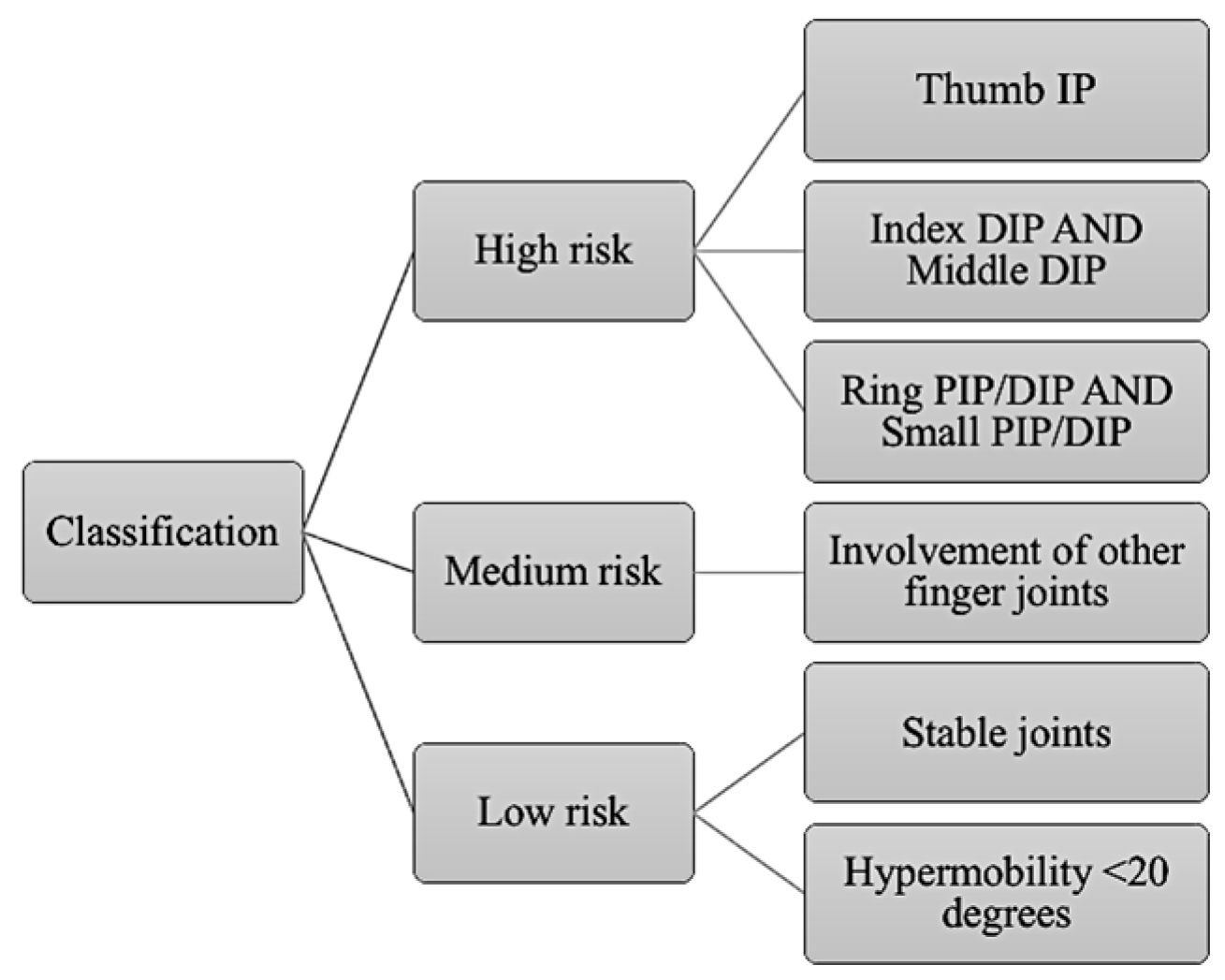
Figure 1. Classification of risk of hypermobility modified according to dental students’ functional tasks.
Applsci 13 07158 g001
Figure 2. Frequency distribution of risk of hand joint hypermobility.
Applsci 13 07158 g002
Figure 3. Frequency distribution of joint hypermobility according to each finger (L: left; R: right).
Applsci 13 07158 g003
Figure 4. The box plot displays a statistically significant difference in right lateral pinch between right- and left-handed students.
Applsci 13 07158 g004
Table 1. Norm guidelines according to biological sex used to assess muscular strength.
OutcomeNorm Guidelines
MalesFemales
Grip Strength7145
Lateral Pinch Strength1913
3-Point Pinch Strength1511
Table 2. Correlation between muscle strength and severity of hypermobility in the right hand (a) and in the left hand (b). * denotes p values < 0.05.
(a) Right hand
2.3.4.5.
1. Severity of hypermobility0.030 *0.0710.007 *0.875
2. Grip strength10.000 *0.000 *0.574
3. Lateral pinch 10.000 *0.671
4. Three-point pinch 10.155
5. Wrist extension 1
(b) Left hand
2.3.4.5.
1. Severity of hypermobility0.021 *0.008 *0.015 *0.301
2. Grip strength10.000 *0.000 *0.602
3. Lateral pinch 10.000 *0.860
4. Three-point pinch 10.795
5. Wrist extension 1
Disclaimer/Publisher’s Note: The statements, opinions and data contained in all publications are solely those of the individual author(s) and contributor(s) and not of MDPI and/or the editor(s). MDPI and/or the editor(s) disclaim responsibility for any injury to people or property resulting from any ideas, methods, instructions or products referred to in the content.