Communication

Inactivation of Cercospora lactucae-sativa through Application of Non-Thermal Atmospheric Pressure Gliding Arc, Tesla Coil and Dielectric Barrier Discharge Plasmas

by
Salit Supakitthanakorn
1,*,
On-Uma Ruangwong
2 and
Dheerawan Boonyawan
3,*
1
Department of Plant Pathology, Faculty of Agriculture at Kamphaeng Sean, Kasetsart University, Kamphaeng Sean Campus, Nakhon Pathom 73140, Thailand
2
Division of Plant Pathology, Department of Entomology and Plant Pathology, Faculty of Agriculture, Chiang Mai University, Chiang Mai 50200, Thailand
3
Plasma and Beam Physics Research Facility, Faculty of Science, Chiang Mai University, Chiang Mai 50200, Thailand
*
Authors to whom correspondence should be addressed.
Appl. Sci. 2023, 13(11), 6643; https://doi.org/10.3390/app13116643
Submission received: 15 April 2023 / Revised: 27 May 2023 / Accepted: 29 May 2023 / Published: 30 May 2023
(This article belongs to the Special Issue Advances in Pest Treatment and Plant Protection)

Abstract

:
Cercospora leaf spot disease is a serious problem for lettuce cultivation worldwide. Cercospora lactucae-sativa, the causative agent of leaf spot disease on lettuce, was treated with non-thermal atmospheric pressure gliding arc (GA), tesla coil (TC) and dielectric barrier discharge (DBD) plasmas for the in vitro fungal inactivation of both mycelial growth and conidial germination. The fungus was exposed to the three plasmas individually for 5, 10, 15 and 20 min. The results showed that DBD plasma inactivated fungal growth during all exposure periods, and the highest inhibitory effect was caused by exposure to DBD plasma for 20 min, at 93.33% inhibition. The germination of fungal conidia was completely inactivated after exposure to all three non-thermal plasmas for 5 min, as observed 4 and 24 h after incubation. The pathogenesis of C. lactucae-sativa on lettuce after plasma treatments for 5 min was examined by spraying an inoculation of the treated conidia on lettuce. The results showed that all three plasmas reduced the disease incidence and severity compared to the non-treated control. Therefore, the non-thermal atmospheric pressure GA, TC and DBD plasmas have antifungal potential for the inactivation of C. lactucae-sativa, making them an interesting novel technology for plant disease control.

1. Introduction

Lettuce (Lactuca sativa L.) is one of the most economically important vegetable plants worldwide, and it is the most popular salad vegetable in Thailand [1]. The cultivation and production of lettuce are often obstructed by a leaf spot disease caused by Cercospora lactucae-sativa [1,2]. Leaf spot disease is a common and destructive disease of lettuce that occurs mostly during the rainy season and leads to high yield losses of up to 80% [3]. Infection begins on the lower, older leaves and progresses to the younger leaves through the penetration of the fungal hyphae via leaf stomata. The lesions are light to dark brown and further characterized by light ash to white-colored spots and, later, holes in the leaves, a symptom unique to C. lactucae-sativa on lettuce [4]. C. lactucae-sativae is a slow-growing hemibiotrophic fungus with a narrow host range restricted to brassicaceae plants [4,5].
To control leaf spot disease, fungicides are routinely used. However, fungicide usage has harmful effects and leaves residues on plants, humans and the environment [6]. Recently, non-thermal plasma (NTP) technology usage has expanded to new biological areas of application for the inactivation of harmful microorganisms, the preparation of ready-to-eat food and biofilm degradation, or in healthcare, the treatment of cancer cells, the initiation of apoptosis, prion inactivation, the prevention of nosocomial infections and the therapy of infected wounds [7,8].
Plasma is an ionized gas, referred to as the fourth state of matter [7]. Plasma is a neutrally charged collection of ions, electrons, photons and atoms in their fundamental or excited states [9]. The plasma state can be produced by applying a high electric voltage to gas. Then, reactive species consisting of reactive oxygen species (ROS), reactive nitrogen species (RNS), charged species, UV radiation, free electrons and an electric field are generated in the plasma state, and these components can greatly affect biological processes [10].
Plasmas can be classified according to their temperature into two large groups: thermal plasmas and cold plasmas [11]. More specifically, low-temperature plasma can be distinguished into thermal plasma and non-thermal plasma (NTP) based on thermodynamic equilibrium [7]. NTP can be generated by different discharge configurators, such as atmospheric uniform glow, corona, dielectric barrier discharge, gliding arc, micro- hollow cathode, plasma jet and needle, by passing gases through electric fields [12]. The lethality of NTP depends on various processing variables, including input power (voltage and frequency), treatment time, gas type, flow rate and exposure mode. Higher frequencies and input voltages have been related to greater inactivation efficacy of NTP [13].
Gliding arc (GA) plasma is a hybrid plasma in which discharge takes place and is carried out between two divergent horn-like electrodes. Gliding arc plasma is produced as a low-impedance heat arc discharge at the narrow gap; it is then prolonged by the gas flow and quenched into a non-thermal plasma. The plasma cools down at the wide gap because no further discharge can take place. Accordingly, the primary plasma column disappears, and the travel cycle for the new plasma column is repeated [14].
Tesla coil (TC) is a self-resonance, self-breakdown discharge. The sharp electrode end generates a series of brief, transient pulses of electrical discharge to the surrounding gas; this is a short-lived phenomenon with a continuous cycle. Typically, it is much more efficient in gas dissociation with transient plasmas than with homogeneous plasmas [15].
Dielectric barrier discharge (DBD) plasma at atmospheric pressure is generally a nonuniform filamentary discharge. The formation and development of electric streamers arise from separated local sites of the dielectric barrier. Each streamer represents a so-called microdischarge, which is distributed in the gap and is unstable, moving around erratically [16].
NTPs in atmospheric pressure have been used for agricultural applications such as decontamination or inactivation of microorganisms in seeds, postharvest products, the plant tissue culture process, pork cuts and chicken meat, and the decontamination of pesticide residues [17,18,19,20,21]. The antimicrobial activity of NTP is associated with the generation of high concentrations of ROS during the discharge process; these ROS consist of superoxide (O2·), hydroxyl (OH) and hydrogen peroxide (H2O2) which are toxic to microbial cells and cause oxidation of their macromolecules, thereby resulting in cell death [22]. In this study, we applied and compared three non-thermal plasmas, GA, TC and DBD, for the inactivation of C. lactucae-sativa in laboratory conditions and observed the resulting fungal pathogenicity after plasma treatments.

2. Materials and Methods

2.1. Source of the Fungus

C. lactucae-sativa (GenBank accession no. OQ566288) was isolated from infected green oak lettuce collected from the cultivation area in Chiang Mai Province, Northern Thailand, using a single-spore isolation method on a potato dextrose agar (PDA). To identify fungal species, fungal DNA was extracted from mycelia using a Genomic DNA Isolation Kit (blood/cultured cell/fungus) (column-based) (Bio-Helix, New Taipei City, Taiwan) according to the manufacturer’s instructions. The polymerase chain reaction (PCR) technique was used to amplify the transition elongation factor 1-alpha (TEF-1α) gene [23] using 2X PCR SuperMix (Bio-Helix, New Taipei City, Taiwan). The PCR product was purified using a PCR Clean Up and Gel Extraction Kit (Bio-Helix, New Taipei City, Taiwan) according to the manufacturer’s instructions. The nucleotide sequence was directly analyzed using fluorescent dye terminator sequencing on ABI PrismTM 3730xI DNA sequencers (Applied Biosystems, Foster City, CA, USA). The obtained sequence was analyzed and aligned using the Basic Local Alignment Search Tool (BLAST) and deposited into the GenBank database.
To induce fungal sporulation, the mycelial discs were inoculated by placing them on lettuce leaves, followed by incubation in the growth chamber at 25 °C, 80% relative humidity (RH), and a photoperiod of 12 h in the light and 12 h in the dark for 14 days. The conidia were collected from the inoculated leaves by scraping the conidia produced on the leaves into autoclaved distilled water and counting the conidial concentration using a haemacytometer.

2.2. Plasma Device and Properties

Ambient air reactive oxygen/nitrogen species (RONS) were generated using GA, TC and DBD plasmas in a semi-closure frame, as shown in Figure 1.
The GA and TC plasmas were individually driven by a 20 W RF self-resonance power supply. The driver can supply a sinusoidal AC output with a peak voltage (Vp) of 6–10 kV at a frequency of 700–900 kHz. Meanwhile, the DBD plasma was driven by a continuous 18 W AC high-voltage source of 7.6 kVpp with a repetition rate of 18.2 kHz. The planar mesh-powered electrode was configured on an alumina ceramic sheet of a 1.0 mm thickness with lateral dimensions of 5.3 cm × 11.3 cm.
Thus, the ambient air molecules (N2 and O2) were ionized and dissociated by these strong electric fields, which induced the RONS with high-energy electrons in the plasma phase. The ionization and dissociation equation could be found in the reaction below (Equation (1)). However, the direct electron impact dissociation of N2 is much slower than that of O2, and so the major intermediate product is the atomic oxygen O. The electron collision with N2 leads preferentially to the formation of various excited N2* molecules, while the generation of RONS follows in the plasma propagation phase (Equations (2)–(7)) [24]. In humid air, electron impact can also produce atomic oxygen (O) and OH through dissociative excitation (Equation (8)). Both O and OH are transient chemical species that have very strong oxidative properties compared to neutral oxidant molecules such as ozone.
e + N2 → e + N2* (N2 (C) or N2 (B)),
N2* + O2 → N2 + O + O,
O + O2 + M → O3 + M,
O + O2 → NO + O,
O + N2 → NO + N,
NO + O +M → NO2 + M,
O3 + NO → O2 + NO2,
e + H2O → e + H + OH.

2.3. Inactivation of Mycelial Growth

The spread plate method was applied for conducting in vitro inactivation instead of using mycelial discs due to the very slow growth rate of C. lactucae-sativa [4]. A total of 5 mycelial discs were added to a 15 mL centrifuge tube containing 10 mL of autoclaved distilled water; they were mixed by vortexing and then incubated at room temperature (25 ± 3 °C) overnight to increase fungal mass. The spread plate method was applied using 200 µL of fungal suspension, and the PDA plates were exposed to GA, TC and DBD plasmas for 5, 10, 15 and 20 min, after which the plates were incubated at room temperature (25 ± 3 °C) for 7 days. Each treatment contained three replicates, and the experiment was repeated 3 times. The diameter of the clear zone was recorded. The percent inhibition of mycelial growth was calculated according to the modified formula of [25] (Equation (9)), as follows:
Inhibition   of   mycelial   growth   ( % ) = d c d t d c   ×   100 ,
where dc is the colony diameter of the non-treated control (9 cm) and dt is the value of the fungal growing area (9 cm clear zone (cm)) in each plasma-treated case.

2.4. Inactivation of Conidial Germination

To study the effect of GA, TC and DBD plasmas on the inactivation of conidial germination, the conidia of C. lactucae-sativa were collected from inoculated lettuce leaves and adjusted to a concentration of 106 conidia/mL using a hemacytometer under a compound microscope. Some 100 µL of conidial suspension was dropped onto a slide culture before exposure to the GA, TC and DBD plasmas for 5, 10, 15 and 20 min. The slide culture was incubated at room temperature (25 ± 3 °C). Conidial germination was observed at 4 and 24 h after incubation. The non-treated conidia were used as a positive control. Each treatment contained three replicates (20 conidia/replicate), and the experiment was repeated three times. The percent inhibition of conidial germination was calculated according to the formula of [26] (Equation (10)), as follows:
Inhibition   of   conidial   germination   ( % ) = g c g t g c   ×   100 ,  
where gc is the number of germinated conidia in the non-treated control and gt is the number of germinated conidia in each plasma-treated case.

2.5. Inactivation of Fungal Pathogenesis after Plasma Treatments

To evaluate the antifungal efficacy of non-thermal atmospheric pressure GA, TC and DBD plasmas on the inactivation of C. lactucae-sativa pathogenesis, conidia were prepared, as previously described in Section 2.4, and exposed to all plasmas for 5 min. Green oak lettuce (Lactuca sativa) was grown in a 5-inch pot containing approximately 200 g peat moss as a growing medium. The plasma-treated conidia were sprayed on all the lettuce leaves at the age of 14 days (5 mL/plant). The plants were covered with plastic bags and kept in a growth chamber at 25 °C, 80% RH, and underwent a photoperiod of 12 h in the light and 12 h in the dark for 14 days. Each treatment contained nine replications (plants). The disease incidence was recorded and calculated according to [27] (Equation (11)), as follows:
Disease   incidence   ( % ) = N u m b e r   o f   i n f e c t e d   p l a n t s N u m b e r   o f   t o t a l   p l a n t s   a c c e s s e d   ×   100 .
Disease severity was assessed after 14 days of inoculation using the scale reported by [27] as follows: 0 = no spots; 1 = 1–20% spots; 2 = 21–40% spots; 3 = 41–60% spots; 4 = 61–80% spots; and 5 = 81–100% spots. The disease severity index was then determined using the formula from [27] (Equation (12)), as follows:
Disease   severity   index   ( % ) = S c a l e × A m o u n t s   o f   p l a n t s   i n   e a c h   t r e a t m e n t M a x i m u m   s c a l e × T o t a l   p l a n t s   a c c e s s e d   ×   100 .

2.6. Statistical Analysis

All data in the present study were analyzed using Statistix version 8 (Analytical Software, Tallahassee, FL, USA). Data were analyzed using a one-way analysis of variance (ANOVA) followed by a least significant difference (LSD) test (p < 0.05). Each treatment had three biological replicates. All data were expressed as means ± standard deviation (SD).

3. Results

3.1. Plasma Device and Properties

A 3-in-1 plasma device, composed of GA, TC and DBD plasmas, was devised (Figure 1a–d). Optical emission spectra with an acquisition time of 100 ms were used to identify the aforementioned species inside the plasmas, GA, TC and DBD, respectively. Groups of NO A–X were found in the ultraviolet range between 200 and 300 nm and OH at 308.98 nm, N2 at 336.61 nm, 357.17 nm (second positive band) and 379.99 nm (first negative band). Comparing the types of plasma, TC produced the highest concentration of air plasma radicals followed by a gliding arc and a dielectric barrier discharge. The difference in the intensity of plasma-generated species is clearly notable in the GA and TC over the DBD as the first two types are point sources, whereas DBD is a planar source (Figure 1e).

3.2. Inactivation of Mycelial Growth

All three NTPs inhibited fungal growth by different percentages. DBD plasma exposure for 20 min showed the highest fungal inhibition, with 93.33%, followed by exposure for 15, 10 and 5 min, at 72.59, 49.26 and 27.41%, respectively (Table 1 and Figure 2). GA plasma exposure for 20, 15 and 10 min inhibited fungal growth by 45.19, 40.47 and 33.70%, respectively, but exposure for only 5 min could not inhibit fungal growth (Table 1 and Figure 2). The lowest fungal inhibition was found to result from TC plasma treatments, with 13.33% inhibition resulting from exposure for 20 min; however, exposure for less than 20 min could not inhibit fungal growth (Table 1 and Figure 2).

3.3. Inactivation of Conidial Germination

The conidial germination of C. lactucae-sativa was observed at 4 and 24 h after treatment with GA, TC and DBD plasma. The results showed that the conidial germination of C. lactucae-sativa was completely inactivated (100% inhibition) after exposure to all plasmas for 5 min compared to the control in which germ tubes germinated from the conidia (Figure 3). However, the morphological characteristics of the conidia in the treatment groups and the control group were not different.

3.4. Inactivation of Fungal Pathogenesis after Plasma Treatments

After treatment with non-thermal atmospheric pressure GA, TC and DBD plasma for 5 min, the conidia of C. lactucae-sativa were sprayed on lettuce leaves for estimation of fungal pathogenesis. The results showed that the disease incidence of all the plasma-treated cases was zero, whereas the disease incidence of the non-treated control was 100% (Figure S1). The disease severity of all the plasma-treated cases was zero, whereas the disease severity of the non-treated control was scale 3. The disease severity index of all the plasma-treated cases was zero, whereas the disease severity index of the non-treated control was 25.00% (Figure S1). As a result, exposure to the three NTPs for 5 min showed complete inactivation of the pathogenesis of C. lactucae-sativa via conidial inoculation on lettuce.

4. Discussion

C. lactucae-sativa is an airborne plant pathogen that can infect dozens of plants, and brassicaceae plants are one of its main targets [4,5]. The fungus is difficult to eliminate and can be resistant to fungicides due to its extensive reliance on fungicides [28]. Moreover, in organic farming, the incidence and severity of leaf spot disease on lettuce are regularly observed to result in a reduction in product quality and quantity [2]. Cercospora leaf spot disease has important economic implications around the world and has caused major damage to lettuce cultivation in Thailand [1]. Therefore, the application of NTP to inactivate fungal growth and control leaf spot disease is an interesting proposition.
NTP at atmospheric pressure is composed of several reactive species such as O, OH and NO2, which account for chemical and physical changes in biological materials [29]. Therefore, RONS are considered to play an important role in the decontamination and inactivation of microorganisms [30].
The mechanisms of NTP that are involved in the inactivation of microorganisms are still elusive and inconsistent due to complex plasma and microbial systems. However, thus far, there are two main categories of mechanisms: biological and physical [31]. In biological mechanisms, UV irradiation generated by NTP causes damage to cells, lipid peroxidation, protein modulation and apoptosis induction [32]. Physical mechanisms involve electrostatic disruption and electroporation [13,33].
NTPs have been applied to inhibit, inactivate and eliminate many important fungal phytopathogens such as Athelia rolfsii which causes southern blight disease on lettuce [34]; Aspergillus niger, which contaminates palm fruits [35]; Fusarium graminicearum, which causes Fusarium head blight in wheat [36]; and Colletotrichum gloeosporioides, which causes anthracnose disease in chili [18]. The results of the application of NTP demonstrate the complete inactivation of fungal growth (in both mycelial and conidial germination) and the inactivation of fungal pathogenesis [14].
In this study, the inactivation of mycelial growth differed depending on the type of plasmas. DBD plasma showed the strongest fungal inhibition due to its wide discharge plate, which increased the area of plasma available to interact with the fungus [33]. In contrast, GA and TC plasmas were plasma jet types, in which the plasma was generated through a single plasma dispenser [12]. The fungal mycelia that were not exposed to plasma (the non-treated control) could grow regularly. Therefore, DBD plasma was considered the most effective and capable non-thermal atmospheric pressure plasma for the inactivation of the mycelial growth of C. lactucae-sativa. However, non-thermal atmospheric pressure GA, TC and DBD plasmas inactivated the conidial germination of C. lactucae-sativa completely after exposure for 5 min. Accordingly, fungal pathogenicity was examined by spraying conidia, which were exposed to all three plasmas for 5 min, on lettuce leaves. The results showed that the disease incidence, severity and severity index were completely inactivated compared to the non-treated control. Ebrahimi et al. [37] used air plasma surface barrier discharge (SBD) on inactivation of conidia germination of Aspergillus niger and showed that after exposure for 2 min, the conidia of A. niger were completely inactivated. it was concluded that the dominant active species produced in the plasma were nitrogen species analyzed by optical emission spectroscopy (OES). Thus, in this study, the dominant active species was nitrogen species which might be the major factor in fungal inactivation.
NTPs are an interesting novel technology for plant disease control. They can be applied to directly inactivate plant pathogens; however, the effects of NTPs on plants’ physiology, biochemistry, growth and development will be further investigated. The conditions of NTP applications including input power, treatment time, flow rate and exposure mode must be evaluated in order to elucidate their effects on plants. Nevertheless, there is a lot of encouraging information about the positive effects of NTPs on plants, as a promising priming agent that enhances their growth parameters and affects processes that are a part of their reaction to stress [38]. In conclusion, to inactivate C. lactucae-sativa which causes leaf spot disease on lettuce, non-thermal atmospheric pressure GA, TC and DBD plasmas can be applied due to their antifungal activity.

Supplementary Materials

The following supporting information can be downloaded at: https://www.mdpi.com/article/10.3390/app13116643/s1, Figure S1: Pathogenesis of Cercospora lactucae-sativa after exposure of conidia to gliding arc, tesla coil and dielectric barrier discharge plasma for 5 min.

Author Contributions

Conceptualization, S.S.; methodology, S.S. and D.B.; validation, S.S., O.-U.R. and D.B.; formal analysis, S.S. and D.B.; investigation, S.S., O.-U.R. and D.B.; resources, O.-U.R. and D.B.; data curation, S.S.; writing—original draft preparation, S.S. and D.B.; writing—review and editing, S.S., O.-U.R. and D.B.; visualization, S.S., O.-U.R. and D.B.; supervision, S.S., O.-U.R. and D.B.; funding acquisition, D.B. All authors have read and agreed to the published version of the manuscript.

Funding

This research project was supported by the Fundamental Fund 2022, Chiang Mai University, grant number FF65/117. The authors would like to thank Choncharoen Sawangrat and Miss Wimada Srisuwan from the Agriculture and Bio Plasma Technology Center (ABPlas), Science and Technology Park, Chiang Mai University for providing the laboratory materials and facilitating the research.

Institutional Review Board Statement

Not applicable.

Informed Consent Statement

Not applicable.

Data Availability Statement

Not applicable.

Conflicts of Interest

The authors declare no conflict of interest.

References

  1. Srimai, K.; Akarapisarn, A. Bacillus subtilis LBF02 as Biocontrol Agent against Leaf Spot Diseases Caused by Cercospora lactucae-sativae in Lettuce. J. Agric. Sci. 2014, 6, 151–158. [Google Scholar] [CrossRef]
  2. Koohakan, P.; Jeanaksorn, T.; Nuntagij, I. Major Diseases of Lettuce Grown by Commercial Nutrient Film Technique in Thailand. Curr. Appl. Sci. Technol. 2008, 8, 56–63. [Google Scholar]
  3. Liberato, J.R.; Stephens, P.M. Cercospora apii s. Lat. on Lettuce in Australia. Australas. Plant Pathol. 2006, 35, 379–381. [Google Scholar] [CrossRef]
  4. Thomas, A.; Saravanakumar, D. Effect of Host Extract on Growth and Sporulation of Cercospora lactucae-sativae. Aust. Plant Dis. Notes 2019, 14, 19. [Google Scholar] [CrossRef]
  5. To-anun, C.; Hidayat, I.; Meeboon, J. Genus Cercospora in Thailand: Taxonomy and Phylogeny (with a Dichotomous Key to Species). Plant Pathol. Quar. 2011, 1, 11–87. [Google Scholar] [CrossRef]
  6. Lamichhane, J.R.; Durr, C.; Schwanck, A.A.; Robin, M.H.; Sarthou, J.P.; Cellier, V.; Messean, A.; Aubertot, J.N. Integrated Management of Damping-Off Diseases: A Review. Agron. Sustain. Dev. 2017, 37, 10. [Google Scholar] [CrossRef]
  7. Scholtz, V.; Pazlarova, J.; Souskova, H.; Khun, J.; Julak, J. Nonthermal plasma—A tool for decontamination and disinfection. Biotechnol. Adv. 2015, 33, 1108–1119. [Google Scholar] [CrossRef]
  8. Zhang, J.J.; Kwon, T.; Kim, S.B.; Jeong, D.K. Plasma Farming: Non-Thermal Dielectric Barrier Discharge Plasma Technology for Improving the Growth of Soybean Sprouts and Chickens. Plasma 2018, 1, 25. [Google Scholar] [CrossRef]
  9. Misra, N.N.; Tiwari, B.K.; Raghavarao, K.S.M.S.; Cullen, P.J. Nonthermal Plasma Inactivation of Food-Borne Pathogens. Food Eng. Rev. 2011, 3, 159–170. [Google Scholar] [CrossRef]
  10. Adhikari, B.; Pangomm, K.; Veerana, M.; Mitra, S.; Park, G. Plant Disease Control by Non-Thermal Atmospheric-Pressure Plasma. Front. Plant Sci. 2020, 11, 77. [Google Scholar] [CrossRef]
  11. López, M.; Calvo, T.; Prieto, M.; Múgica-Vidal, R.; Muro-Fraguas, I.; Alba-Elías, F.; Alvarez-Ordóñez, A. A Review on Non-thermal Atmospheric Plasma for Food Preservation: Mode of Action, Determinants of Effectiveness, and Applications. Front. Microbiol. 2019, 10, 622. [Google Scholar] [CrossRef]
  12. Liao, X.; Liu, D.; Xiang, Q.; Ahn, J.; Chen, S.; Ye, X.; Ding, T. Inactivation Mechanisms of Non-Thermal Plasma on Microbes: A Review. Food Control 2017, 75, 83–91. [Google Scholar] [CrossRef]
  13. Conrads, H.; Schmidt, M. Plasma Generation and Plasma Sources. Plasma Sources Sci. Technol. 2000, 9, 441. [Google Scholar] [CrossRef]
  14. Darvish, F.; Sarkari, N.M.; Khani, M.; Eslami, E.; Shokri, B.; Mohseni, M.; Ebrahimi, M.; Alizadeh, M.; Dee, C.F. Direct Plasma Treatment Approach Based on Non-Thermal Gliding Arc for Surface Modification of Biaxially-Oriented Polypropylene with Post-Exposure Hydrophilicity Improvement and Minus Aging Effects. Appl. Surf. Sci. 2020, 509, 144–815. [Google Scholar] [CrossRef]
  15. Graves, D.B. Lessons from Tesla for Plasma Medicine. IEEE Trans. Radiat. Plasma Med. Sci. 2018, 2, 594–607. [Google Scholar] [CrossRef]
  16. Kogelschatz, U. Dielectric-Barrier Discharges: Their History, Discharge Physics, and Industrial Applications. Plasma Chem. Plasma Process. 2002, 23, 1–46. [Google Scholar] [CrossRef]
  17. Jiang, J.; Lu, Y.; Li, J.; Li, L.; He, X.; Shao, H.; Dong, Y. Effect of Seed Treatment by Cold Plasma on the Resistance of Tomato to Ralstonia solanacearum (bacterial wilt). PLoS ONE 2014, 9, e97753. [Google Scholar] [CrossRef]
  18. Sawangrat, C.; Phimolsiripol, Y.; Leksakul, K.; Thanapornpoonpong, S.-n.; Sojithamporn, P.; Lavilla, M.; Castagnini, J.M.; Barba, F.J.; Boonyawan, D. Application of Pinhole Plasma Jet Activated Water against Escherichia coli, Colletotrichum gloeosporioides, and Decontamination of Pesticide Residues on Chili (Capsicum annuum L.). Foods 2022, 11, 2859. [Google Scholar] [CrossRef]
  19. Boonyawan, D.; Lamasai, K.; Umongno, C.; Rattanatabtimtong, S.; Yu, L.D.; Kuensaen, C.; Maitip, J.; Thana, P. Surface Dielectric Barrier Discharge Plasma–Treated Pork Cut Parts: Bactericidal Efficacy and Physiochemical Characteristics. Heliyon 2022, 8, e10915. [Google Scholar] [CrossRef]
  20. Royintarat, T.; Choi, E.H.; Boonyawan, D.; Seesuriyachan, P.; Wattanutchariya, W. Chemical-Free and Synergistic Interaction of Ultrasound Combined with Plasma-Activated Water (PAW) to Enhance Microbial Inactivation in Chicken Meat and Skin. Sci. Rep. 2020, 10, 1559. [Google Scholar] [CrossRef]
  21. Kosumsupamala, K.; Thana, P.; Palee, N.; Lamasai, K.; Kuensaen, C.; Ngamjarurojana, A.; Yangkhamman, P.; Boonyawan, D. Air to H2-N2 Pulse Plasma Jet for In-Vitro Plant Tissue Culture Process: Source Characteristics. Plasma Chem. Plasma Process. 2022, 42, 535–559. [Google Scholar] [CrossRef]
  22. Torres, M.A.; Jones, D.G.; Dangl, J.L. Reactive Oxygen Species Signaling in Response to Pathogens. Plant Physiol. 2006, 141, 373–378. [Google Scholar] [CrossRef]
  23. Cruywagen, E.M.; Slippers, B.; Roux, J.; Wingfield, M.J. Phylogenetic Species Recognition and Hybridization in Lasiodiplodia: A Case Study on Species from Baobabs. Fungal biol. 2017, 121, 420–436. [Google Scholar] [CrossRef]
  24. Tarabova, B.; Lukes, P.; Janda, M.; Hensel, K.; Sikurova, L.; Machala, Z. Specificity of Detection Methods of Nitrites and Ozone in Aqueous Solutions Activated by Air Plasma. Plasma Process. Polym. 2018, 15, 1800030. [Google Scholar] [CrossRef]
  25. Kumar, V.; Tyagi, D. Antifungal Activity of Different Extracts of Bergeniastracheyi. Int. J. Curr. Microbiol. Appl. Sci. 2013, 2, 69–78. [Google Scholar]
  26. Chakraborty, M.; Mahmud, N.U.; Muzahid, A.N.M.; Rabby, S.M.F.; Islam, T. Oligomycins Inhibit Magnaporthe oryzae Triticum and Suppress Wheat Blast Disease. PLoS ONE 2020, 15, e0233665. [Google Scholar] [CrossRef]
  27. Promwe, A.; Intana, W. Trichoderma asperellum (NST-009): A Potential Native Antagonistic Fungus to Control Cercospora Leaf Spot and Promote the Growth of ‘Green Oak’ Lettuce (Lactuca sativa L.) Cultivated in the Commercial NFT Hydroponic System. Plant Prot. Sci. 2022, 58, 139–149. [Google Scholar] [CrossRef]
  28. Kumar, R.; Mazakova, J.; Ali, A.; Sur, V.P.; Sen, M.K.; Bolton, M.D.; Manasova, M.; Rysanek, P.; Zouhar, M. Characterization of the Molecular Mechanisms of Resistance Against DMI Fungicides in Cercospora beticola Populations from the Czech Republic. J. Fungi 2021, 7, 1062. [Google Scholar] [CrossRef]
  29. Dharini, M.; Jaspin, S.; Mahendran, R. Cold Plasma Reactive Species: Generation, Properties, and Interaction with Food Biomolecules. Food Chem. 2023, 45, 134746. [Google Scholar] [CrossRef]
  30. Moisan, M.; Barbeau, J.; Crevier, M.C.; Pelletier, J.; Philip, N.; Saoudi, B. Plasma Sterilization. Methods and Mechanisms. Pure Appl. Chem. 2022, 74, 349–358. [Google Scholar] [CrossRef]
  31. Polcic, P.; Machala, Z. Effects of Non-Thermal Plasma on Yeast Saccharomyces cerevisiae. Int. J. Mol. Sci. 2021, 22, 2247. [Google Scholar] [CrossRef]
  32. Volkov, A.G.; Xu, K.G.; Kolobov, V.I. Plasma-Generated Reactive Oxygen and Nitrogen Species Can Lead to Closure, Locking and Constriction of the Dionaea muscipula Ellis trap. J. R. Soc. Interface 2019, 16, 20180713. [Google Scholar] [CrossRef]
  33. Laroussi, M. Nonthermal Decontamination of Biological Media by Atmospheric pressure Plasmas: Review, Analysis, and Prospects. IEEE Trans. Plasma Sci. 2002, 30, 1409–1415. [Google Scholar] [CrossRef]
  34. Supakitthanakorn, S.; Ruangwong, O.-U.; Sawangrat, C.; Srisuwan, W.; Boonyawan, D. Potential of Nonthermal Atmospheric pressure Dielectric Barrier Discharge Plasma for Inhibition of Athelia Rolfsii Causing Southern Blight Disease in Lettuce. Agriculture 2023, 13, 167. [Google Scholar] [CrossRef]
  35. Lotfy, K.; Al-Qahtani, S.; Al-Harbi, N.; El-Absy, K.; Shulaybi, F.B.; Alali, S.; Mashtoly, T. Decontamination Potential of Date Palm Fruit via Non-Thermal Plasma Technique. Sci. Rep. 2022, 12, 17323. [Google Scholar] [CrossRef]
  36. Na, Y.H.; Park, G.; Choi, E.H.; Ehm, H.S. Effects of the Physical Parameters of a Microwave Plasma Jet on the Inactivation of Fungal Spores. Thin Solid Films 2013, 547, 125–131. [Google Scholar] [CrossRef]
  37. Ebrahimi, E.; Hosseini, S.I.; Samadlouie, H.R.; Mohammadhosseini, B.; Cullen, P.J. Surface Barrier Discharge Remote Plasma Inactivation of Aspergillus niger ATCC 10864 Spores for Packaged Crocus sativus. Food Bioprocess Technol. 2023. [Google Scholar] [CrossRef]
  38. Holubova, L.; Kyzek, S.; Durovcova, I.; Fabova, J.; Horvathova, E.; Sevcovicova, A.; Galova, E. Non-Thermal Plasma—A New Green Priming Agent for Plants? Int. J. Mol. Sci. 2020, 21, 9466. [Google Scholar] [CrossRef]
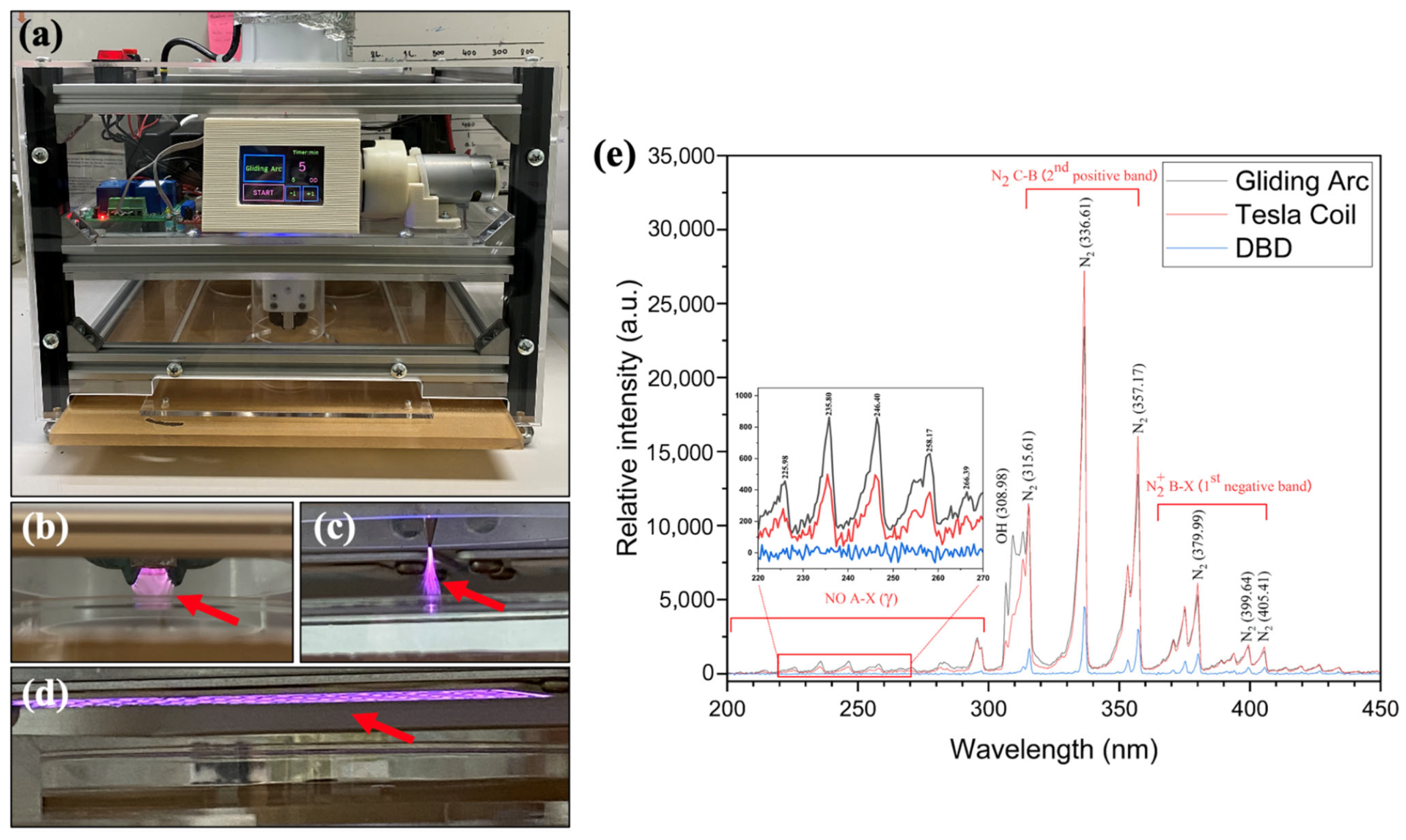
Figure 1. The 3-in-1 nonthermal atmospheric pressure plasma device. (a) Overall structure of the 3-in-1 plasma device, (b) gliding arc (GA) plasma, (c) tesla coil (TC) plasma, (d) dielectric barrier discharge (DBD) plasma and (e) optical emission spectra of the 3 plasma devices, GA, TC and DBD, in the range from 200 to 450 nm. Red arrows indicate the plasmas generated from different plasma dispensers.
Applsci 13 06643 g001
Figure 2. Antifungal efficacy of non-thermal atmospheric pressure gliding arc (GA), tesla coil (TC) and dielectric barrier discharge (DBD) plasmas on inhibition of Cercospora lactucae-sativa mycelial growth within different periods of exposure for 5, 10, 15 and 20 min, photographed at 7 days after incubation.
Applsci 13 06643 g002
Figure 3. Antifungal efficacy of non-thermal atmospheric pressure gliding arc (GA), tesla coil (TC) and dielectric barrier discharge (DBD) plasmas on the inactivation of Cercospora lactucae-sativa conidial germination after 5 min exposure to each plasma, observed 4 and 24 h after incubation. Conidial germination was completely inactivated in all plasma treatments. C = conidia; Gt = germ tube; Hp = hypha.
Applsci 13 06643 g003
Table 1. Percent inhibition of mycelial growth of Cercospora lactucae-sativa by non-thermal atmospheric pressure gliding arc (GA), tesla coil (TC) and dielectric barrier discharge (DBD) plasmas.
TreatmentMean of Percent Inhibition * ± SD
5 min10 min15 min20 min
Non-treated control0.00 ± 0.00 b **0.00 ± 0.00 c0.00 ± 0.00 c0.00 ± 0.00 d
Gliding arc (GA)0.00 ± 0.00 b33.70 ± 0.64 b40.74 ± 1.28 b45.19 ± 0.64 b
Tesla coil (TC)0.00 ± 0.00 b0.00 ± 0.00 c0.00 ± 0.00 c13.33 ± 0.11 c
Dielectric barrier discharge (DBD)27.41 ± 1.70 a49.26 ± 1.28 a72.59 ± 1.70 a93.33 ± 1.11 a
CV (%)12.373.423.752.24
p-value0.690.580.870.70
* Average from three replicates; SD: standard deviation. ** Different letters indicate significant difference between values, at p < 0.05, using the LSD test.
Disclaimer/Publisher’s Note: The statements, opinions and data contained in all publications are solely those of the individual author(s) and contributor(s) and not of MDPI and/or the editor(s). MDPI and/or the editor(s) disclaim responsibility for any injury to people or property resulting from any ideas, methods, instructions or products referred to in the content.