Review

A Review of Apta-POF-Sensors: The Successful Coupling between Aptamers and Plastic Optical Fibers for Biosensing Applications

1
Indivenire srl, Via Alla Cascata 56/C, 38123 Trento, Italy
2
Department of Engineering, University of Campania “Luigi Vanvitelli”, Via Roma 29, 81031 Aversa, Italy
*
Authors to whom correspondence should be addressed.
Appl. Sci. 2022, 12(9), 4584; https://doi.org/10.3390/app12094584
Submission received: 1 March 2022 / Revised: 15 April 2022 / Accepted: 29 April 2022 / Published: 30 April 2022

Abstract

:
Aptamers represent the next frontier as biorecognition elements in biosensors thanks to a smaller size and lower molecular weight with respect to antibodies, more structural flexibility with the possibility to be regenerated, reduced batch-to-batch variation, and a potentially lower cost. Their high specificity and small size are particularly interesting for their application in optical biosensors since the perturbation of the evanescent field are low. Apart from the conventional plasmonic optical sensors, platforms based on silica and plastic optical fibers represent an interesting class of devices for point-of-care testing (POCT) in different applications. The first example of the coupling between aptamers and silica optical fibers was reported by Pollet in 2009 for the detection of IgE molecules. Six years later, the first example was published using a plastic optical fiber (POF) for the detection of Vascular Endothelial Growth Factor (VEGF). The excellent flexibility, great numerical aperture, and the large diameter make POFs extremely promising to be coupled to aptamers for the development of a sensitive platform easily integrable in portable, small-size, and simple devices. Starting from silica fiber-based surface plasmon resonance devices, here, a focus on significant biological applications based on aptamers, combined with plasmonic-POF probes, is reported.

1. Introduction

Among the different optical platforms, plasmonic ones are characterized by a quick response, simple operation mode, and high sensitivity. Surface plasmon resonance (SPR) or localized SPR (LSPR) represents a gold standard in the optical characterization of biomolecular interaction, and it is a widely used transduction method in biosensing. In this context, SPR/LSPR systems based on optical fibers represent an emerging field also due to the incoming of plastic fibers (POFs). Recent advances in POF-based devices open the way to the development of a new class of sensors [1,2,3], also taking advantage of different geometries, such as U-bend, D-shaped, side-polished, and tapered shapes of POFs [1,2,3,4]. SPR sensors can be realized by exploiting multimode optical fibers via POFs, where the convolution of different resonance wavelengths is obtained for a specific resonance condition defined by a given angle–wavelength couple [2,3]. In the multimode waveguides, the SPR sensitivity is intrinsically better with respect to the single-mode waveguide configurations, even if they present the worst value of the Full Width at Half Maximum (FWHM) in the SPR spectra. Moreover, their excellent properties, such as flexibility, great numerical aperture, and large diameter, make POFs extremely interesting platforms with an increased surface area available for functionalization and, consequently, target capture.
More specifically, we focused on particular POF-SPR biosensors making use of aptamers as molecular recognition elements (MRE). In some application fields, this emerging class of molecules has overcome antibodies, the gold standard in biorecognition mechanism, paving the way to develop a new class of biosensors. In the following paragraph, these two elements (aptamers and SPR plastic optical fiber probes) are reported, mainly focusing on their characteristics, and highlighting the high potentiality of their coupling in a single optical biosensor, as reported, e.g., in [4] for the SARS-CoV-2 detection. More specifically, POF-based platforms combined with aptamers are described in more detail.

2. Aptamers as Biorecognition Element

Aptamers are short single-stranded DNA or RNA fragments selected by a combinatorial process (Systematic Evolution of Ligands by EXponential Enrichment, SELEX), which was first reported in 1990 [5,6]. Starting from DNA or RNA libraries, this process allowed the identification of unique oligonucleotide molecules able to bind to the target molecule with very high affinity and specificity. Historically, RNA-based libraries were used for the first SELEX selections due to the higher specificity and binding affinity [7]. However, experimental evidence showed that single-stranded DNAs have a comparable propensity to form tertiary structures similar to RNA chains [8]. There is a growing increase in the literature on the use of DNA-based aptamers for biosensing, and this is due first to an easier selection and amplification process as there is no reverse transcription step (necessary when working with RNA molecules), second to the simpler and cheaper synthesis of DNA-attachments, and third to the longer half-life [8,9]. With the advancement and the consolidation of the SELEX process, several aptamers were selected for a wide range of analytes, ranging from very small molecules (pesticides, toxins) up to entire microorganisms.
The primary property of aptamers is their mechanism of target binding through a conformational change allowing their application in different sensing methods. Their binding properties make them rivals of the antibodies, the natural MRE; aptamers exhibit an affinity constant in the low nanomolar range, and their high specificity minimizes the probability of false-positive results [10]. Their selection and production do not involve animals, thus ensuring guaranteed sourcing and high batch-to-batch reproducibility. Furthermore, they can be easily modified in a defined manner for their immobilization, for instance, onto a solid support, allowing the choice of the adequate chemistry depending on the substrate [11]. Moreover, they are resistant to heat denaturation and can be used in non-physiological pH solutions. The main differences between aptamers and antibodies are summarized in Table 1.
Arshavsky-Graham et al. compared the performances of aptamers and random- or Fc-oriented immobilized antibodies on the same optical platform (porous-silicon biosensors) in the detection of his-tagged protein in terms of binding rate, dynamic detection range, the limit of detection and selectivity [12]. If aptamer-based and antibody-oriented biosensors reached similar results, the first ones would outperform in terms of reusability and storability. The performances in the detection of Immunoglobulin E and Cellular Prions using immunosensors or aptasensors were reviewed by Hianik [13]. The author concluded that, even if the performances are similar when the aptamers exhibit comparable or higher affinity to a specific target, they are more favorable for biosensor development than antibodies due to their higher stability, reproducibility, and ruggedness.
Recently, Liu and coworkers highlighted the higher performances of aptamers, with respect to antibodies, in biosensors intended for high-throughput screening of target molecules [14]. The authors reviewed many articles covering different medical applications, from cancer to cardiovascular disease biomarkers, from Neurotransmitters and Alzheimer’s to Tuberculosis disease biomarkers. The reported examples confirmed that the applications of aptamers as MRE are continuously growing, and this also favors their development.
Besides antibodies, another widely used MRE is represented by molecularly imprinted polymers (MIPs), also called “synthetic antibodies”, that are specifically designed for binding targets with a high degree of selectivity and reproducibility [15].
The most important features for a biosensor are selectivity, reproducibility, stability, sensitivity, and reusability [16,17]; consequently, an efficient MRE has to satisfy specific characteristics to allow the achievement of the aforementioned features. Figure 1 reports a comparison between the different MREs based on their most significant characteristics able to satisfy the biosensors’ requirements [16,18]. Compared with the antibodies, MIPs and aptamers are characterized by higher stability and reproducibility and smaller molecular size while maintaining high specificity. Moreover, MIPs are characterized by a low cost compared to the other two bio-receptors, but aptamers, besides the other characteristics, have the smallest size and the highest sensitivity. Morales and Halpern drew a road map on how to choose an MRE for the development of a biosensor [17]: the choice is mainly related to the request of selectivity and sensitivity of the biosensor itself.
Aptamers find applications in different fields [19], from the detection of small molecules [20,21], pesticides [22], allergens [23] to toxins [24,25], and in point-of-care (POC) diagnostic system [26,27,28], for the detection of bacteria [29,30] or circulating tumor cells [31]. Since 1996, aptamers have been suggested as an alternative to antibodies [32,33] and employed in the biosensor field, highlighting their potentiality and versatility. Some diagnostic products based on aptamers are already present on the market [34,35,36]. Recently, besides SELEX, different aptamer selection systems have been envisaged, based either on computational “in silico” systems, which calculate the probability of interaction between nucleic acid sequences and targets using molecular dynamics [9] or systems based on “machine learning” [37]. These new approaches increase the aptamer development and, consequently, also their employment in the biosensors field.

3. Plasmonic Optical Fiber Aptasensors

The optical fiber biosensors represent a highly sensitive class of optical biosensors. The sensing principle used in silica and polymer fiber probes can be based on different approaches: intensity measurements (through tapered fibers), gratings, SPR or LSPR, evanescent field, etc. [1,38,39,40,41]. These methods can be implemented by two main sensor configurations, reflection or transmission mode, depending on the position of the light source and the detector with respect to the sensing point. Usually, in reflection mode, the sensing region (for instance, the tip of the fiber) is located away from the equipment (source and the detector); whereas, in transmission mode, the sensing region is typically located in the middle of the fiber, between the source and the detector. In particular, among the different sensor configurations, the plasmonic one used in transmission mode can be simply realized by removing the cladding around the fiber and depositing a thin metallic film on the exposed core. Then, working on the refractive index of the core, the shape of the sensing region, the fiber’s type, and other structural parameters, it is possible to excite the plasmonic phenomena by achieving different performances. Moreover, the plasmonic sensor’s performance can be improved using tapered fibers or metal nanostructures [42] or by changing the physical shape, such as in the U-bend configuration [43]. This high sensitivity of fiber-based SPR/LSPR sensors suggests the possible employment in the development of biosensors.
In this context, it should be noted that the sensing principles and the possible configurations of optical fiber (OF) biosensors were recently summarized by Loyez et al. in [41]. During the last decade, several examples of OF-based biosensors can be found in the literature. In fact, different molecular recognition elements have been successfully immobilized on OF-based probes, such as antibodies, MIPs, and aptamers, proving the high versatility of these simple and low-cost platforms. More specifically, Table 2 collects the published works coupling OF-based plasmonic platforms (both silica and POFs) and aptamers. A first example of the coupling between a fiber-based SPR probe and an aptamer was reported by Pollet et al. in 2009 [44] by testing a silica-based fiber covered by a functionalized gold nanofilm in the detection of DNA hybridization and also in the detection of IgE through an aptamer. Since then, several examples have been reported, mainly based on silica fibers with applications in cancer biomarkers detection [45,46,47,48,49,50,51,52,53,54,55,56,57,58,59,60,61,62,63], toxins [64,65,66,67], allergens [44,68,69,70], small molecule or DNA [62], and virus [71], as summarized in Table 2. The highest sensitivity was achieved using nanostructures, such as nanoantennas [50,65], nanorods [64], or Bragg grating nanoimprinted on the fibers [55]. Moreover, these fiber-based optical devices can be miniaturized and easily integrated into compact platforms [63,70].
Currently, very few examples have been reported in the literature related to the coupling between aptamers and POF-based biosensors [48,49,50,55,70,71]. In contrast, more exhaustive literature exists where different MREs are exploited, as reported in Figure 2.
With respect to other MREs, aptamers represent a new emerging class for the realization of biosensors and will be increasingly employed due to the establishment of new companies devoted to aptamer selection and optimization [34,35,36,75].

4. A Focus on Relevant Applications of Apta-POF-SPR Sensors

In this paragraph, we reported several applications of the Apta-POF-SPR sensors, summarized in the last part of Table 2, and related to the detection of antibiotics, disease biomarkers, and virus proteins.

4.1. Antibiotics Detection

Antibiotics are antibacterial agents used in the prevention and treatment of bacterial infections: their use can, on the one hand, save lives, but on the other hand, can increase bacterial resistance if overused. The presence of antibiotic residues in food and environments, for instance, can alter the microbiome, compromising the immune system [76].
The detection of antibiotics in human fluids, environment, food, and beverages is therefore crucial for human health. Compared to commonly used techniques, such as mass chromatography or enzyme-linked immunosorbent assay (ELISA), biosensors are growing due to their fast response and easiness in the operational aspects. In this context, aptamer-based sensors are also increasingly used [76,77,78,79].
Apta-sensors based on POFs represent an attractive kind of device. In fact, they are simple to realize and use. An Apta-POF-SPR sensor for antibiotic detection was reported by Galatus et al. [70]. A D-shaped POF SPR probe was coupled to an electrochemical sensor to detect ampicillin, an antibiotic widely used in the veterinary field. A thiolated aptamer was immobilized on the gold-coated POF, and the two detection systems were coupled (see Figure 3), allowing the antibiotic detection at the concentration of 100 μM in buffer solution.
Figure 3 reports the scheme of the coupling between the two systems, highlighting the easy connection with the cloud and the possibility of achieving a quick and easy data exchange. Even if this platform was not tested on the low analyte concentration, the electro-opto coupling opens the way to overcome the limitations of the single technique with the possibility of increasing the system’s sensitivity.
This was the first reported example of an Apta-POF-SPR sensor for the detection of antibiotics, but surely it will not be the last. One of the main challenges remains antibiotic detection in real samples. A complex matrix such as milk, honey, urine, serum, or plasma, apart from a possible cross-reaction, can decrease or even suppress the label signal [78]. In this context, an optical SPR aptasensor that does not require labels offers the possibility to overcome this critical aspect.

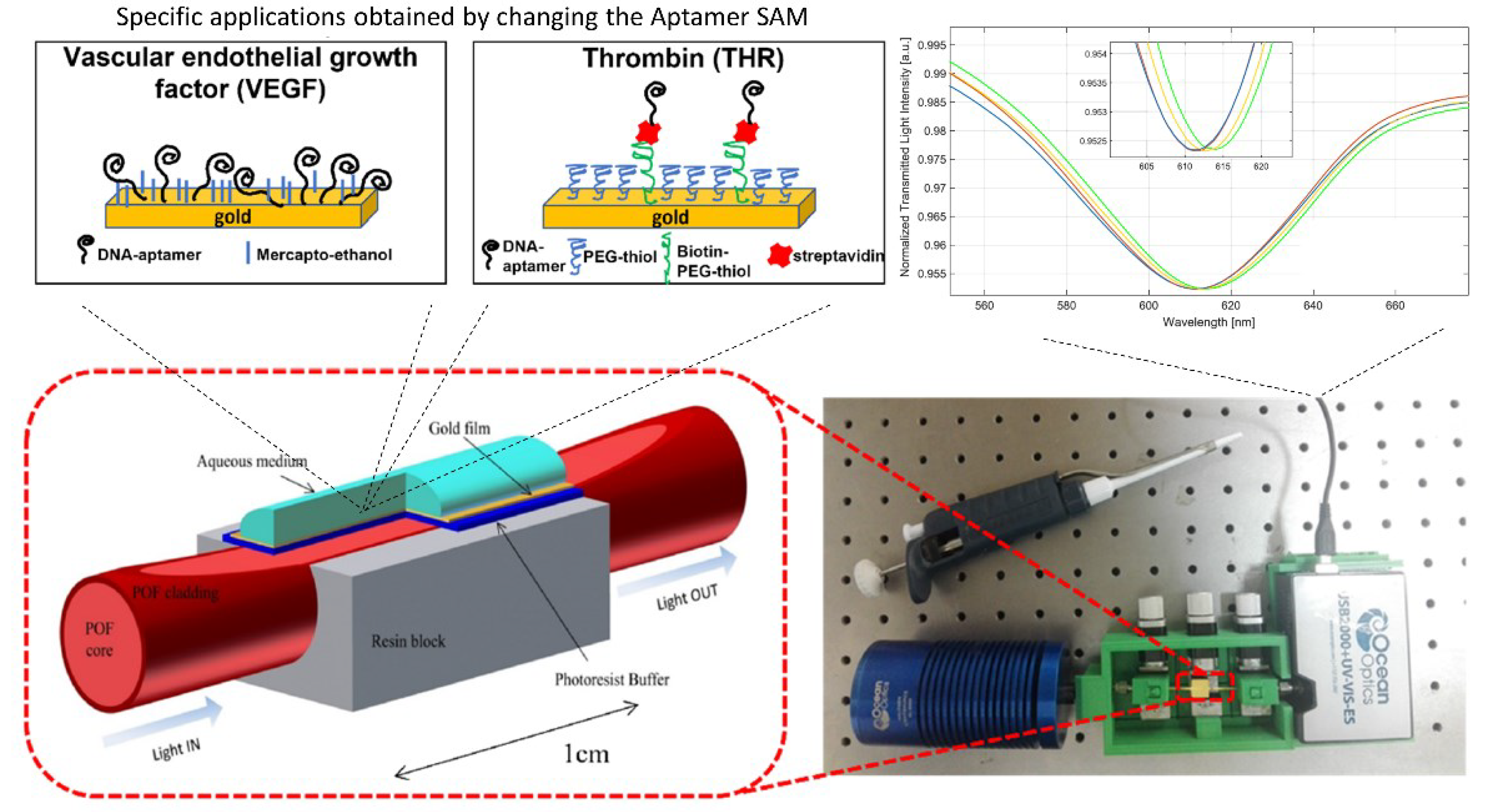
4.2. Disease Biomarkers Detection

Clinical biomarkers are fundamental not only for early disease discovery but also for monitoring the disease treatments. Aptamers are increasingly employed as MRE in this field [80,81,82]. In 2015, our group developed an aptamer-based POF-SPR sensor for the detection of Vascular endothelial growth factor (VEGF), selected as a circulating protein potentially associated with cancer [48]. A thiolated aptamer was directly immobilized on the gold film deposited on plastic optical fiber, followed by a passivation step with mercapto-ethanol. From the obtained results, it was figured out that passivation was an important element of the interface to guarantee a proper conformation of the aptamers, and VEGF detection was achieved up to 0.8 nM. Figure 4 reports the optical sensor system combined with two different aptamer layers, specific for VEGF and thrombin detection in the nanomolar range.
Even if our results agreed with other detection systems, we found that the dissociation constant of the aptamer immobilized on the surface was two orders of magnitude lower with respect to that measured in solutions. This means that we lost affinity on our platform, probably due to the direct immobilization of the gold layer.
For this reason, in the subsequent works, we changed the approach and developed an interface based on a short Poly Ethylene Glycol (PEG) layer. A PEG layer can reduce the non-specific adsorption of nucleic acid on the gold due to its high hydrophilic character and the steric repulsion resulting from the compression of PEG chains. In this way, the better orientation of the aptamer can be achieved.
The detection of thrombin (THR), a clinical marker of the blood coagulation cascade and homeostasis, was performed by modifying the gold layer on the POF with a PEG Self Assembled Monolayer (SAM) to provide the highest accessibility for the analyte to the aptamer immobilized on the plasmonic surface, decreasing at the same time the non-specific adsorption [49]. A mixed interface was built: a short-PEG and a biotinylated-PEG were mixed in an 8:2 molar ratio at 0.2 mM final concentration, and streptavidin was then immobilized on it. THR binding aptamer (TBA) was coupled to these PEG-SAMs through the avidin–biotin chemistry (see Figure 4). The performances of the obtained interface were confirmed by the SPR-POF measurement, resulting in detection in the nanomolar range (see Figure 4) and increasing the dissociation constant of one order of magnitude.
The THR-aptasensor was prepared according to [49]. Showing the details of this protocol could be of interest, considering it a paradigm that could also be used for other aptamers. Therefore, briefly, the gold surface was cleaned using an argon plasma (6.8 W of power to the RF coil for one minute), and then a water solution of 0.2 mM of PEGthiol: BiotinPEGlipo in an 8:2 molar ratio was incubated overnight. M-dPEG®₈-Thiol and Biotin-dPEG®₃-Lipoamide for the self-assembled monolayer (SAM) were purchased from Stratech (United Kingdom). After overnight incubation and washing in MilliQ water, 5μg/mL streptavidin (from Streptomyces avidinii, Sigma-Aldrich) solution in phosphate buffer (10 mM phosphate buffer, 138 mM NaCl, 2,7 mM KCl, pH 7.4) was applied for one hour. Finally, 10 μM of aptamer (5′-BiotinTEG-AG TCC GTG GTA GGG CAG GTT GGG GTG ACT-3′ for thrombin, IDT Integrated DNA technologies, Belgium) was incubated for three hours in the same phosphate buffer, after thermal treatment (95 °C for 1 min). After being washed in the same buffer, the aptasensor was ready for the optical measurements.
In order to increase the interface stability (which turned out to be 12 days in phosphate buffer saline (PBS) at 4 °C [71]), further experiments were conducted, freezing the prepared interface in PBS solution and testing it at selected times with the immunochemiluminescence protocol. At the selected time, 50 nM of thrombin (Sigma) was incubated on surfaces (kept in PBS at 4° or −20 °C) in buffer (Tris 50 mM, EDTA 1 mM, MgCl2 1 mM, KCl 150 mM pH 7.4) for one hour on a slow orbital shaker and followed by a washing step in the same buffer. After a passivation step with 3% w/v BSA for 30 min, a primary mouse anti-thrombin antibody at 2.5 μg/mL concentration was applied for 30 min. The excess antibodies were removed with three extensive washing steps, and the samples were then incubated for 30 min with an antimouse HRP-conjugated secondary antibody at 2 μg/mL concentration. After washing three times in buffer, the chemiluminescence signal was developed (SuperSignal West Femto Chemiluminescent Substrate kit, Thermo Fisher) and acquired for 0.5 s using a standard imaging system ChemDoc-It (Bio-Rad). The signal measured with the standard imaging system was quantified using the ImageJ software [83].
Figure 5 reports the obtained results, highlighting that the conservation of the interface at −20 °C seems to preserve the aptamer’s ability to recognize its target in an unaltered way up to 34 days.
A different system was developed by Sun and coworkers, who used a gold film-supported graphene sheet on a POF device to detect dopamine, a biomarker for neurological disorders [53]. The specific detection of dopamine was achieved, at sub-nanomolar concentration, taking advantage of the π-stacking interaction between the graphene sheet and aptamers, highlighting the high sensitivity of the system.
Another application was recently proposed by Sanjay et al. [55], based on a U-bend POF configuration coupled to an aptamer layer and connected to a smartphone camera for the detection of Plasmodium falciparum glutamate dehydrogenase (PfGDH), a potential malaria biomarker. A co-deposition of thiolated aptamer and mercapto-hexanol was used to derivatize the gold layer sputtered on the U-bend POF. They used the smartphone flash as the light source, and the camera was used as the detector.
The high performance of the system allowed the PfGDH detection in the subnanomolar range, well low its physiologically relevant range, not only in buffer solution but also in 20 times diluted human serum. Moreover, a shelf life study reported, on the 20th day, an efficiency of the probes equal to 91.38% of the initial output, suggesting the high platform stability.

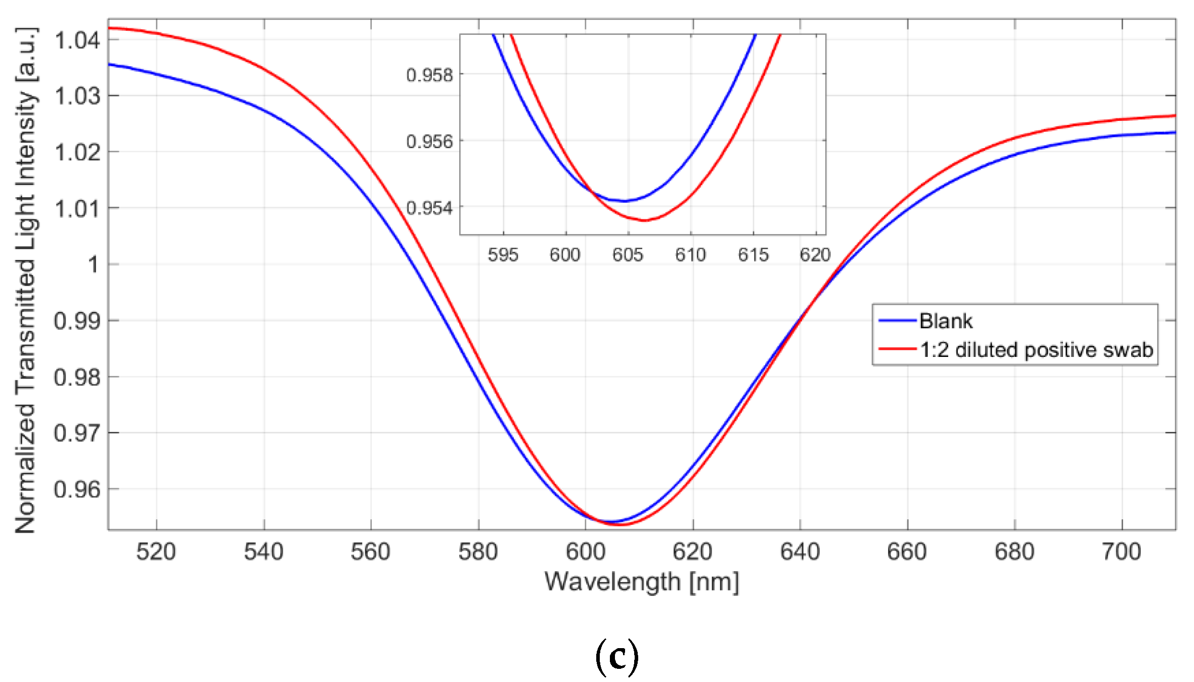
4.3. Virus Detection

The growing and spreading of the global population on the world caused in the last century an increase in the incidence of pathogens. There is, therefore, the requirement for rapid and efficient biosensors in order to detect viruses early and allow immediate counteractions. In this context, aptamers found several applications [84,85,86]. Recently, we tested the PEG-SAM-based aptasensor for the detection of the receptor-binding domain (RBD) of the SARS-CoV-2 spike glycoprotein [71]. A longer aptameric sequence was used (51 nucleotides with respect to 29 for THR and 26 for VEGF), and a thorough characterization was performed in order to ensure the right recognition.
The interface reported in Figure 6a was tested not only on the specific target (SARS-CoV-2 Spike protein) but also on aspecific targets such as other proteins (bovine serum albumin (BSA), AH1N1 hemagglutinin protein, and MERS spike protein) and in diluted human serum (50%) [4,71]. A limit of detection in the nanomolar range was achieved (see Figure 6), confirming the good performances of our aptamer-based optical platform. Figure 6b shows the SPR spectra obtained at different concentrations of the SARS-CoV-2 Spike protein [4].
A preliminary test was performed using real samples (positive Universal Transport Medium (UTM) swab sample), as described in [4] and shown in Figure 6c. A clear wavelength red shift was observed only on positive samples, confirming the ability of the aptamer-based platform to recognize the target.

5. Pros and Cons of Apta-POF SPR Sensors

Apta-POF SPR sensors are characterized by several advantages due to both components: Aptamers and POFs. For instance, the simple manufacturing process and the high flexibility can be obtained by exploiting the plastic fibers that allow easy modification and integration in small devices. Examples of these POF-based approaches were reported by Irawan et al., who embedded a POF in a disposable microfluidic chip in an integrated fluorescence detection system for biomedical devices [87]; by Kelb et al., who proposed a respiratory monitoring device suited for application in environments with strong magnetic fields [88]; or by Aitkulov and Tosi, who integrated a smartphone with a POF for the diagnosis of respiratory diseases [89]. Instead, the aptamer element guarantees the high specificity of this kind of POF SPR sensor. Moreover, the different configurations that can be simply implemented on the polymeric fiber, such as the D-shaped or U-bend or grating, allowed great flexibility in the sensor design depending on the system requirements [2,41]. On the other hand, with respect to silica fibers, POFs can efficiently work only in a limited range (visible), are characterized by a wide width at half height, and can suffer from environmental or external treatments; high-temperature treatments are, for instance, poor compatible with POF, even if thermal resistance can be improved [90]. Pro and cons of Apta-POF SPR sensors are summarized in Table 3.
The coupling of POFs and aptamers generates new challenges in the bio-detection field and will require major technological breakthroughs in the future.

6. Conclusions

Aptamers represent an emerging class of biorecognition elements, increasingly used in biosensors development. Their robustness, specificity, and dimensions make them ideal elements to be immobilized on plasmonic optical fiber devices. In particular, POF-based platforms can be combined with aptamers to develop sensitive biosensor chips easily integrable in a portable, small-size, and simple device due to the great flexibility and easy manipulation of POFs. In this review, we collected the literature on Apta-POF sensors suggesting up to now applications mainly in disease biomarker detection (VEGF, THR, PfGDH, and dopamine) and virus (SARS-CoV-2), unlike silica fiber-based sensors present applications also in agri-food. The possibility to easily connect POFs to smart devices, considering the great numerical aperture and the high specificity guaranteed by the aptamer layer, will pave the way for a new class of devices with a broad application area, from agriculture to the environment up to human and animal health. We believe that in the near future, more examples will appear thanks to the discovery of new aptamer sequences, also facilitated by the in-silico modeling.

Author Contributions

Conceptualization, L.P. and N.C. Writing—original draft L.P., Writing—review and editing, L.P., N.C., F.A. and L.Z. All authors have read and agreed to the published version of the manuscript.

Funding

This research received no external funding.

Institutional Review Board Statement

This study does not require ethical approval.

Informed Consent Statement

This study does not require informed consent.

Data Availability Statement

The data are available on reasonable request from the corresponding author.

Acknowledgments

This work was supported by the VALERE program of the University of Campania “Luigi Vanvitelli” (Italy).

Conflicts of Interest

The authors declare no conflict of interest.

References

  1. Jin, Y.; Granville, A.M. Polymer Fiber Optic Sensors—A Mini Review of their Synthesis and Applications. J. Biosens. Bioelectron. 2016, 7, 194. [Google Scholar] [CrossRef]
  2. Cennamo, N.; Pesavento, M.; Zeni, L. A review on simple and highly sensitive plastic optical fiber probes for bio-chemical sensing. Sens. Actuators B Chem. 2021, 331, 129393. [Google Scholar] [CrossRef]
  3. Kadhim, R.A.; Abdul, A.K.K.; Yuan, L. Advances in Surface Plasmon Resonance-Based Plastic Optical Fiber Sensors. IETE Tech. Rev. 2020. In preprint. [Google Scholar] [CrossRef]
  4. Cennamo, N.; D’Agostino, G.; Pasquardini, L.; Arcadio, F.; Perri, C.; Coppola, N.; Angelillo, I.F.; Altucci, L.; Di Marzo, F.; Parisio, E.M.; et al. (INVITED)Quantitative detection of SARS-CoV-2 virions in aqueous mediums by IoT optical fiber sensors. Results Opt. 2021, 5, 100177. [Google Scholar] [CrossRef]
  5. Ellington, A.D.; Szostak, J.W. In vitro selection of RNA molecules that bind specific ligands. Nature 1990, 346, 818–822. [Google Scholar] [CrossRef]
  6. Tuerk, C.; Gold, L. Systematic Evolution of Ligands by Exponential Enrichment: RNA Ligands to Bacteriophage T4 DNA Polymerase. Science 1990, 249, 505–510. [Google Scholar] [CrossRef]
  7. Ștefan, G.; Hosu, O.; De Wael, K.; Lobo-Castañón, M.J.; Cristea, C. Aptamers in biomedicine: Selection strategies and recent advances. Electrochim. Acta 2021, 376, 137994. [Google Scholar] [CrossRef]
  8. Wang, T.; Chen, C.; Larcher, L.M.; Barrero, R.A.; Veedu, R.N. Three decades of nucleic acid aptamer technologies: Lessons learned, progress and opportunities on aptamer development. Biotechnol. Adv. 2019, 37, 28–50. [Google Scholar] [CrossRef]
  9. Navien, T.N.; Thevendran, R.; Hamdani, H.Y.; Tang, T.-H.; Citartan, M. In silico molecular docking in DNA aptamer development. Biochimie 2021, 180, 54–67. [Google Scholar] [CrossRef]
  10. Tombelli, S.; Minunni, M.; Mascini, M. Analytical applications of aptamers. Biosens. Bioelectron. 2005, 20, 2424–2434. [Google Scholar] [CrossRef]
  11. Odeh, F.; Nsairat, H.; Alshaer, W.; Ismail, M.A.; Esawi, E.; Qaqish, B.; Al Bawab, A.; Ismail, S.I. Aptamers Chemistry: Chemical Modifications and Conjugation Strategies. Molecules 2019, 25, 3. [Google Scholar] [CrossRef] [PubMed] [Green Version]
  12. Arshavsky-Graham, S.; Urmann, K.; Salama, R.; Massad-Ivanir, N.; Walter, J.-G.; Scheper, T.; Segal, E. Aptamers vs. antibodies as capture probes in optical porous silicon biosensors. Analyst 2020, 145, 4991–5003. [Google Scholar] [CrossRef] [PubMed]
  13. Hianik, T. Affinity Biosensors for Detection Immunoglobulin E and Cellular Prions. Antibodies vs. DNA Aptamers. Electroanalysis 2016, 28, 1764–1776. [Google Scholar] [CrossRef]
  14. Liu, L.S.; Wang, F.; Ge, Y.; Lo, P.K. Recent Developments in Aptasensors for Diagnostic Applications. ACS Appl. Mater. Interfaces 2021, 13, 9329–9358. [Google Scholar] [CrossRef] [PubMed]
  15. Haupt, K.; Rangel, P.X.M.; Bui, B.T.S. Molecularly Imprinted Polymers: Antibody Mimics for Bioimaging and Therapy. Chem. Rev. 2020, 120, 9554–9582. [Google Scholar] [CrossRef] [PubMed]
  16. Bhalla, N.; Jolly, P.; Formisano, N.; Estrela, P. Introduction to biosensors. Essays Biochem. 2016, 60, 1–8. [Google Scholar] [CrossRef] [Green Version]
  17. Morales, M.; Halpern, J.M. Guide to Selecting a Biorecognition Element for Biosensors. Bioconjug. Chem. 2018, 29, 3231–3239. [Google Scholar] [CrossRef] [PubMed]
  18. Chiavaioli, F.; Gouveia, C.A.J.; Jorge, P.A.S.; Baldini, F. Towards a Uniform Metrological Assessment of Grating-Based Optical Fiber Sensors: From Refractometers to Biosensors. Biosensors 2017, 7, 23. [Google Scholar] [CrossRef] [Green Version]
  19. Yan, S.-R.; Foroughi, M.M.; Safaei, M.; Jahani, S.; Ebrahimpour, N.; Borhani, F.; Baravati, N.R.Z.; Aramesh-Boroujeni, Z.; Foong, L.K. A review: Recent advances in ultrasensitive and highly specific recognition aptasensors with various detection strategies. Int. J. Biol. Macromol. 2020, 155, 184–207. [Google Scholar] [CrossRef]
  20. Feng, C.; Dai, S.; Wang, L. Optical aptasensors for quantitative detection of small biomolecules: A review. Biosens. Bioelectron. 2014, 59, 64–74. [Google Scholar] [CrossRef]
  21. Gaudin, V. The Growing Interest in Development of Innovative Optical Aptasensors for the Detection of Antimicrobial Residues in Food Products. Biosensors 2020, 10, 21. [Google Scholar] [CrossRef] [PubMed] [Green Version]
  22. Majdinasab, M.; Daneshi, M.; Marty, J.L. Recent developments in non-enzymatic (bio)sensors for detection of pesticide residues: Focusing on antibody, aptamer and molecularly imprinted polymer. Talanta 2021, 232, 122397. [Google Scholar] [CrossRef] [PubMed]
  23. Khedri, M.; Ramezani, M.; Rafatpanah, H.; Abnous, K. Detection of food-born allergens with aptamer-based biosensors. TrAC Trends Anal. Chem. 2018, 103, 126–136. [Google Scholar] [CrossRef]
  24. Cunha, I.; Biltes, R.; Sales, M.; Vasconcelos, V. Aptamer-Based Biosensors to Detect Aquatic Phycotoxins and Cyanotoxins. Sensors 2018, 18, 2367. [Google Scholar] [CrossRef] [PubMed] [Green Version]
  25. Zhou, Q.; Tang, D. Recent advances in photoelectrochemical biosensors for analysis of mycotoxins in food. TrAC Trends Anal. Chem. 2020, 124, 115814. [Google Scholar] [CrossRef]
  26. Hassan, E.M.; DeRosa, M.C. Recent advances in cancer early detection and diagnosis: Role of nucleic acid based aptasensors. TrAC Trends Anal. Chem. 2020, 124, 115806. [Google Scholar] [CrossRef]
  27. Citartan, M.; Tang, T.-H. Recent developments of aptasensors expedient for point-of-care (POC) diagnostics. Talanta 2019, 199, 556–566. [Google Scholar] [CrossRef]
  28. Zahra, Q.U.A.; Khan, Q.A.; Luo, Z. Advances in Optical Aptasensors for Early Detection and Diagnosis of Various Cancer Types. Front. Oncol. 2021, 11, 632165. [Google Scholar] [CrossRef]
  29. Majdinasab, M.; Hayat, A.; Marty, J.L. Aptamer-based assays and aptasensors for detection of pathogenic bacteria in food samples. TrAC Trends Anal. Chem. 2018, 107, 60–77. [Google Scholar] [CrossRef]
  30. Sharifi, S.; Vahed, S.Z.; Ahmadian, E.; Dizaj, S.M.; Eftekhari, A.; Khalilov, R.; Ahmadi, M.; Hamidi-Asl, E.; Labib, M. Detection of pathogenic bacteria via nanomaterials-modified aptasensors. Biosens. Bioelectron. 2020, 150, 111933. [Google Scholar] [CrossRef]
  31. Safarpour, H.; Dehghani, S.; Nosrati, R.; Zebardast, N.; Alibolandi, M.; Mokhtarzadeh, A.; Ramezani, M. Optical and electrochemical-based nano-aptasensing approaches for the detection of circulating tumor cells (CTCs). Biosens. Bioelectron. 2020, 148, 111833. [Google Scholar] [CrossRef] [PubMed]
  32. Drolet, D.W.; Moon-McDermott, L.; Romig, T.S. An enzyme-linked oligonucleotide assay. Nat. Biotechnol. 1996, 14, 1021–1025. [Google Scholar] [CrossRef] [PubMed]
  33. Davis, K.A.; Abrams, B.; Lin, Y.; Jayasena, S.D. Use of a High Affinity DNA Ligand in Flow Cytometry. Nucleic Acids Res. 1996, 24, 702–706. [Google Scholar] [CrossRef] [PubMed] [Green Version]
  34. Aptamer Science Inc. Available online: https://www.aptsci.com/en/technology/aptamer.php (accessed on 15 April 2022).
  35. Neoventures Biotechnologies. Available online: https://neoaptamers.com/ (accessed on 15 April 2022).
  36. Somalogic. Available online: https://somalogic.com/technology/ (accessed on 15 April 2022).
  37. Bashir, A.; Yang, Q.; Wang, J.; Hoyer, S.; Chou, W.; McLean, C.; Davis, G.; Gong, Q.; Armstrong, Z.; Jang, J.; et al. Machine learning guided aptamer refinement and discovery. Nat. Commun. 2021, 12, 2366. [Google Scholar] [CrossRef] [PubMed]
  38. Cennamo, N.; Maniglio, D.; Tatti, R.; Zeni, L.; Bossi, A.M. Deformable molecularly imprinted nanogels permit sensitivity-gain in plasmonic sensing. Biosens. Bioelectron. 2020, 156, 112126. [Google Scholar] [CrossRef]
  39. Bilro, L.; Alberto, N.; Pinto, J.L.; Nogueira, R. Optical Sensors Based on Plastic Fibers. Sensors 2012, 12, 12184–12207. [Google Scholar] [CrossRef] [Green Version]
  40. Peters, K. Polymer optical fiber sensors—A review. Smart Mater. Struct. 2010, 20, 13002. [Google Scholar] [CrossRef]
  41. Loyez, M.; DeRosa, M.C.; Caucheteur, C.; Wattiez, R. Overview and emerging trends in optical fiber aptasensing. Biosens. Bioelectron. 2022, 196, 113694. [Google Scholar] [CrossRef]
  42. Cennamo, N.; D’Agostino, G.; Donà, A.; Dacarro, G.; Pallavicini, P.; Pesavento, M.; Zeni, L. Localized Surface Plasmon Resonance with Five-Branched Gold Nanostars in a Plastic Optical Fiber for Bio-Chemical Sensor Implementation. Sensors 2013, 13, 14676–14686. [Google Scholar] [CrossRef]
  43. Gowri, A.; Sai, V.V.R. Development of LSPR based U-bent plastic optical fiber sensors. Sens. Actuators B Chem. 2016, 230, 536–543. [Google Scholar] [CrossRef]
  44. Pollet, J.; Delport, F.; Janssen, K.P.; Jans, K.; Maes, G.; Pfeiffer, H.; Wevers, M.; Lammertyn, J. Fiber optic SPR biosensing of DNA hybridization and DNA–protein interactions. Biosens. Bioelectron. 2009, 25, 864–869. [Google Scholar] [CrossRef] [PubMed]
  45. Bekmurzayeva, A.; Dukenbayev, K.; Shaimerdenova, M.; Bekniyazov, I.; Ayupova, T.; Sypabekova, M.; Molardi, C.; Tosi, D. Etched Fiber Bragg Grating Biosensor Functionalized with Aptamers for Detection of Thrombin. Sensors 2018, 18, 4298. [Google Scholar] [CrossRef] [PubMed] [Green Version]
  46. Albert, J.; Lepinay, S.; Caucheteur, C.; DeRosa, M.C. High resolution grating-assisted surface plasmon resonance fiber optic aptasensor. Methods 2013, 63, 239–254. [Google Scholar] [CrossRef] [PubMed] [Green Version]
  47. Hu, W.; Huang, Y.; Chen, C.; Liu, Y.; Guo, T.; Guan, B.-O. Highly sensitive detection of dopamine using a graphene functionalized plasmonic fiber-optic sensor with aptamer conformational amplification. Sens. Actuators B Chem. 2018, 264, 440–447. [Google Scholar] [CrossRef]
  48. Cennamo, N.; Pesavento, M.; Lunelli, L.; Vanzetti, L.; Pederzolli, C.; Zeni, L.; Pasquardini, L. An easy way to realize SPR aptasensor: A multimode plastic optical fiber platform for cancer biomarkers detection. Talanta 2015, 140, 88–95. [Google Scholar] [CrossRef]
  49. Cennamo, N.; Pasquardini, L.; Arcadio, F.; Vanzetti, L.E.; Bossi, A.M.; Zeni, L. D-shaped plastic optical fibre aptasensor for fast thrombin detection in nanomolar range. Sci. Rep. 2019, 9, 18740. [Google Scholar] [CrossRef]
  50. Sun, J.; Jiang, S.; Xu, J.; Li, Z.; Li, C.; Jing, Y.; Zhao, X.; Pan, J.; Zhang, C.; Man, B.Y. Sensitive and selective surface plasmon resonance sensor employing a gold-supported graphene composite film/D-shaped fiber for dopamine detection. J. Phys. D Appl. Phys. 2019, 52, 195402. [Google Scholar] [CrossRef]
  51. Lao, J.; Han, L.; Wu, Z.; Zhang, X.; Huang, Y.; Tang, Y.; Guo, T. Gold Nanoparticle-Functionalized Surface Plasmon Resonance Optical Fiber Biosensor: In Situ Detection of Thrombin With 1 n·M Detection Limit. J. Light. Technol. 2019, 37, 2748–2755. [Google Scholar] [CrossRef]
  52. Sypabekova, M.; Korganbayev, S.; González-Vila, Á.; Caucheteur, C.; Shaimerdenova, M.; Ayupova, T.; Bekmurzayeva, A.; Vangelista, L.; Tosi, D. Functionalized etched tilted fiber Bragg grating aptasensor for label-free protein detection. Biosens. Bioelectron. 2019, 146, 111765. [Google Scholar] [CrossRef]
  53. Sun, D.; Sun, L.-P.; Guo, T.; Guan, B.-O. Label-Free Thrombin Detection Using a Tapered Fiber-Optic Interferometric Aptasensor. J. Light. Technol. 2018, 37, 2756–2761. [Google Scholar] [CrossRef]
  54. Dillen, A.; Mohrbacher, A.; Lammertyn, J. A Versatile One-Step Competitive Fiber Optic Surface Plasmon Resonance Bioassay Enabled by DNA Nanotechnology. ACS Sens. 2021, 6, 3677–3684. [Google Scholar] [CrossRef] [PubMed]
  55. Sanjay, M.; Singh, N.K.; Ngashangva, L.; Goswami, P. A smartphone-based fiber-optic aptasensor for label-free detection of Plasmodium falciparum glutamate dehydrogenase. Anal. Methods 2020, 12, 1333–1341. [Google Scholar] [CrossRef]
  56. Shevchenko, Y.; Francis, T.J.; Blair, D.A.D.; Walsh, R.; DeRosa, M.C.; Albert, J. In Situ Biosensing with a Surface Plasmon Resonance Fiber Grating Aptasensor. Anal. Chem. 2011, 83, 7027–7034. [Google Scholar] [CrossRef] [PubMed]
  57. Loyez, M.; Hassan, E.M.; Lobry, M.; Liu, F.; Caucheteur, C.; Wattiez, R.; DeRosa, M.C.; Willmore, W.G.; Albert, J. Rapid Detection of Circulating Breast Cancer Cells Using a Multiresonant Optical Fiber Aptasensor with Plasmonic Amplification. ACS Sens. 2020, 5, 454–463. [Google Scholar] [CrossRef]
  58. Daems, D.; Pfeifer, W.; Rutten, I.; Saccà, B.; Spasic, D.; Lammertyn, J. Three-Dimensional DNA Origami as Programmable Anchoring Points for Bioreceptors in Fiber Optic Surface Plasmon Resonance Biosensing. ACS Appl. Mater. Interfaces 2018, 10, 23539–23547. [Google Scholar] [CrossRef]
  59. Allsop, T.; Mou, C.; Neal, R.; Mariani, S.; Nagel, D.; Tombelli, S.; Poole, A.; Kalli, K.; Hine, A.; Webb, D.; et al. Real-time kinetic binding studies at attomolar concentrations in solution phase using a single-stage opto-biosensing platform based upon infrared surface plasmons. Opt. Express 2017, 25, 39. [Google Scholar] [CrossRef] [Green Version]
  60. Qian, H.; Huang, Y.; Duan, X.; Wei, X.; Fan, Y.; Gan, D.; Yue, S.; Cheng, W.; Chen, T. Fiber optic surface plasmon resonance biosensor for detection of PDGF-BB in serum based on self-assembled aptamer and antifouling peptide monolayer. Biosens. Bioelectron. 2019, 140, 111350. [Google Scholar] [CrossRef]
  61. Coelho, L.; De Almeida, J.M.M.M.; Santos, J.L.; da Silva Jorge, P.A.; Martins, M.C.L.; Viegas, D.; Queirós, R.B. Aptamer-based fiber sensor for thrombin detection. J. Biomed. Opt. 2016, 21, 087005. [Google Scholar] [CrossRef]
  62. Arghir, I.; Spasic, D.; Verlinden, B.E.; Delport, F.; Lammertyn, J. Improved surface plasmon resonance biosensing using silanized optical fibers. Sens. Actuators B Chem. 2015, 216, 518–526. [Google Scholar] [CrossRef] [Green Version]
  63. Zubiate, P.; Zamarreño, C.; Sánchez, P.; Matias, I.; Arregui, F. High sensitive and selective C-reactive protein detection by means of lossy mode resonance based optical fiber devices. Biosens. Bioelectron. 2017, 93, 176–181. [Google Scholar] [CrossRef]
  64. Lee, B.; Park, J.-H.; Byun, J.-Y.; Kim, J.H.; Kim, M.-G. An optical fiber-based LSPR aptasensor for simple and rapid in-situ detection of ochratoxin A. Biosens. Bioelectron. 2018, 102, 504–509. [Google Scholar] [CrossRef] [PubMed]
  65. Allsop, T.D.; Neal, R.; Wang, C.; Nagel, D.A.; Hine, A.V.; Culverhouse, P.; Castañón, J.D.A.; Webb, D.J.; Scarano, S.; Minunni, M. An ultra-sensitive aptasensor on optical fibre for the direct detection of bisphenol A. Biosens. Bioelectron. 2019, 135, 102–110. [Google Scholar] [CrossRef] [PubMed]
  66. Xu, Y.; Xiong, M.; Yan, H. A portable optical fiber biosensor for the detection of zearalenone based on the localized surface plasmon resonance. Sens. Actuators B Chem. 2021, 336, 129752. [Google Scholar] [CrossRef]
  67. Luo, Z.; Zhang, J.; Wang, Y.; Chen, J.; Li, Y.; Duan, Y. An aptamer based method for small molecules detection through monitoring salt-induced AuNPs aggregation and surface plasmon resonance (SPR) detection. Sens. Actuators B Chem. 2016, 236, 474–479. [Google Scholar] [CrossRef]
  68. Tran, D.T.; Knez, K.; Janssen, K.P.F.; Pollet, J.; Spasic, D.; Lammertyn, J. Selection of aptamers against Ara h 1 protein for FO-SPR biosensing of peanut allergens in food matrices. Biosens. Bioelectron. 2013, 43, 245–251. [Google Scholar] [CrossRef]
  69. Antohe, I.; Schouteden, K.; Goos, P.; Delport, F.; Spasic, D.; Lammertyn, J. Thermal annealing of gold coated fiber optic surfaces for improved plasmonic biosensing. Sens. Actuators B Chem. 2016, 229, 678–685. [Google Scholar] [CrossRef]
  70. Galatus, R.M.; Cristea, C.; Feier, B.; Cennamo, N.; Zeni, L. SPR based hybrid electro-optic biosensor for β-lactam antibiotics determination in water. Proc. SPIE 2017, 10405, 104050C. [Google Scholar] [CrossRef]
  71. Cennamo, N.; Pasquardini, L.; Arcadio, F.; Lunelli, L.; Vanzetti, L.; Carafa, V.; Altucci, L.; Zeni, L. SARS-CoV-2 spike protein detection through a plasmonic D-shaped plastic optical fiber aptasensor. Talanta 2021, 233, 122532. [Google Scholar] [CrossRef]
  72. Lobry, M.; Loyez, M.; Hassan, E.M.; Chah, K.; DeRosa, M.C.; Goormaghtigh, E.; Wattiez, R.; Caucheteur, C. Multimodal plasmonic optical fiber grating aptasensor. Opt. Express 2020, 28, 7539–7551. [Google Scholar] [CrossRef]
  73. Loyez, M.; Lobry, M.; Hassan, E.M.; DeRosa, M.C.; Caucheteur, C.; Wattiez, R. HER2 breast cancer biomarker detection using a sandwich optical fiber assay. Talanta 2021, 221, 121452. [Google Scholar] [CrossRef]
  74. Tripathi, S.M.; Dandapat, K.; Bock, W.J.; Mikulic, P.; Perreault, J.; Sellamuthu, B. Gold coated dual-resonance long-period fiber gratings (DR-LPFG) based aptasensor for cyanobacterial toxin detection. Sens. Bio-Sens. Res. 2019, 25, 100289. [Google Scholar] [CrossRef]
  75. Aptamer Group. Available online: https://aptamergroup.com/ (accessed on 15 April 2022).
  76. Zahra, Q.; Luo, Z.; Ali, R.; Khan, M.; Li, F.; Qiu, B. Advances in Gold Nanoparticles-Based Colorimetric Aptasensors for the Detection of Antibiotics: An Overview of the Past Decade. Nanomaterials 2021, 11, 840. [Google Scholar] [CrossRef] [PubMed]
  77. Mehlhorn, A.; Rahimi, P.; Joseph, Y. Aptamer-Based Biosensors for Antibiotic Detection: A Review. Biosensors 2018, 8, 54. [Google Scholar] [CrossRef] [PubMed] [Green Version]
  78. Yang, Y.; Yin, S.; Li, Y.; Lu, D.; Zhang, J.; Sun, C. Application of aptamers in detection and chromatographic purification of antibiotics in different matrices. TrAC Trends Anal. Chem. 2017, 95, 1–22. [Google Scholar] [CrossRef]
  79. Zahra, Q.A.; Fang, X.; Luo, Z.; Ullah, S.; Fatima, S.; Batool, S.; Qiu, B.; Shahzad, F. Graphene Based Nanohybrid Aptasensors in Environmental Monitoring: Concepts, Design and Future Outlook. Crit. Rev. Anal. Chem. 2022, 95, 1–22. [Google Scholar] [CrossRef]
  80. Varty, K.; O’Brien, C.; Ignaszak, A. Breast Cancer Aptamers: Current Sensing Targets, Available Aptamers, and Their Evaluation for Clinical Use in Diagnostics. Cancers 2021, 13, 3984. [Google Scholar] [CrossRef]
  81. Negahdary, M. Aptamers in nanostructure-based electrochemical biosensors for cardiac biomarkers and cancer biomarkers: A review. Biosens. Bioelectron. 2020, 152, 112018. [Google Scholar] [CrossRef]
  82. Huang, J.; Chen, X.; Fu, X.; Li, Z.; Huang, Y.; Liang, C. Advances in Aptamer-Based Biomarker Discovery. Front. Cell Dev. Biol. 2021, 9, 571. [Google Scholar] [CrossRef]
  83. Schneider, C.A.; Rasband, W.S.; Eliceiri, K.W. NIH Image to ImageJ: 25 Years of image analysis. Nat. Methods 2012, 9, 671–675. [Google Scholar] [CrossRef]
  84. Krüger, A.; de Jesus Santos, A.; de Sá, V.; Ulrich, H.; Wrenger, C. Aptamer Applications in Emerging Viral Diseases. Pharmaceuticals 2021, 14, 622. [Google Scholar] [CrossRef]
  85. Sánchez-Báscones, E.; Parra, F.; Lobo-Castañón, M.J. Aptamers against viruses: Selection strategies and bioanalytical applications. TrAC Trends Anal. Chem. 2021, 143, 116349. [Google Scholar] [CrossRef]
  86. Zou, X.; Wu, J.; Gu, J.; Shen, L.; Mao, L. Application of Aptamers in Virus Detection and Antiviral Therapy. Front. Microbiol. 2019, 10, 1462. [Google Scholar] [CrossRef] [PubMed] [Green Version]
  87. Irawan, R.; Tjin, S.C.; Fang, X.; Fu, C.Y. Integration of optical fiber light guide, fluorescence detection system, and multichannel disposable microfluidic chip. Biomed. Microdevices 2007, 9, 413–419. [Google Scholar] [CrossRef] [PubMed]
  88. Kelb, C.; Körner, M.; Prucker, O.; Rühe, J.; Reithmeier, E.; Roth, B. PDMAA Hydrogel Coated U-Bend Humidity Sensor Suited for Mass-Production. Sensors 2017, 17, 517. [Google Scholar] [CrossRef] [PubMed] [Green Version]
  89. Aitkulov, A.; Tosi, D. Optical Fiber Sensor Based on Plastic Optical Fiber and Smartphone for Measurement of the Breathing Rate. IEEE Sens. J. 2019, 19, 3282–3287. [Google Scholar] [CrossRef]
  90. Uyor, U.O.; Popoola, A.P.I.; Popoola, O.M.; Aigbodion, V.S. Polymeric cladding materials under high temperature from optical fibre perspective: A review. Polym. Bull. 2020, 77, 2155–2177. [Google Scholar] [CrossRef]
Figure 1. Comparison of different MREs based on the most significant parameters for biosensing. Strengths are relative to the most performing MRE for each parameter, and the higher value is at the periphery of the plot.
Applsci 12 04584 g001
Figure 2. Comparison of antibody-, MIP-, or aptamer-based POF sensors during years (The number of items were obtained from Web of Knowledge website for searching specific keywords; aptamer or MIP or antibodies and plastic optical fiber).
Applsci 12 04584 g002
Figure 3. Scheme of the hybrid sensor platform developed [3].
Applsci 12 04584 g003
Figure 4. SPR D-shaped POF aptasensor. Zoom of the resonance sensing region with two examples of self-assembled monolayer of aptamers.
Applsci 12 04584 g004
Figure 5. Apt-THR layer aged in PBS solution at 4 °C (circles) [4] or −20 °C (stars). Data are reported as mean value of two samples. Error bars represent the standard deviation. Exposition time: 0.5 s.
Applsci 12 04584 g005
Figure 6. (a) Scheme of the POF’s gold surface derivatization based on aptamer SAM. (b) Transmission spectra wide range and zoom (inset) for different SARS-CoV-2 spike protein concentrations (25 ÷ 1000 nM); (c) SPR spectra of the aptasensor at a SARS-CoV-2 positive swab in UTM 1:2 diluted with a physiological solution (0.9% NaCl) [4].
Applsci 12 04584 g006aApplsci 12 04584 g006b
Table 1. Aptamers versus antibodies.
AptamersAntibodies
Molecular basisNucleic acids Proteins
Molecular modificationsMany examples (significant benefits for clinical applications)Rare case (reduced affinity to antigens)
SizeSmall—10/15 KDa Big—150 KDa
StabilityStable even in high temperature and non-physiological pH environment
Susceptible to nucleases and rapid clearance rate in vivo
Susceptible to denaturation in high temperature and non-physiological pH environment
Less susceptible to nucleases and low clearance rate in vivo
TargetsWide range, from ions to whole living cellsOnly immunogenic molecules
ImmobilizationBy physical adsorption or chemical bondsBy random or oriented or chemical bonds
ImmunotoxicityNon-toxicMay cause a serious response in some patients
SpecificityHighHigh
Biochemical reactionsNot limited to physiological conditionsLimited to physiological conditions
CostLess expensive (solid-phase synthesis)More expensive (animals extraction)
ProductionChemical modifications or SELEXAnimals
ReproducibilityHigh batch to batch reproducibilityLow batch to batch reproducibility
Ethical concernsNoneProduction and development depend on living animals
Table 2. Aptamer-based silica or plastic optical fiber plasmonic sensor systems.
Fiber TypeAnalyteOptical
Detection
Sensitivity [nm/RIU]LoD [nM]Reference
Silica fiber
Bragg grating imprinted on silica fiber coated with gold (50 nm)ThrombinSPR-22Albert et al. 2013 [46]
Silica fiber coated with Au (50 nm)IgESPR~15002 ± 1Pollet et al. 2009 [44]
Silica fiber coated with Au (50 nm)Ara h1SPR-75Tran et al. 2013 [68]
Bragg grating imprinted on silica fiber coated with gold (50 nm)ThrombinSPR400 ÷ 100022.6Shevchenko et al. 2011 [56]
Tilted silica fiber Bragg gratings coated with gold (35 nm)HER2SPR102.03~8.6 × 10⁻⁶Lobry et al. 2020 [72]
Tilted silica fiber Bragg gratings coated with gold (50 nm)ThrombinSPR and LSPR-1Lao et al. 2019 [51]
Bragg grating imprinted on silica fiber coated with gold (50 nm)CTC (circulating tumor cells)SPR-49 cells/mL or 10 cells/mL (with amplification)Loyez et al. 2020 [57]
Tilted silica fiber Bragg gratingsThrombinSPR23.380.075–0.11Sypabekova et al. 2019 [52]
Silica fiber with annealed gold layer (50 nm)Ara h1SPR>1000 1.6Antohe et al. 2016 [69]
Silica fiber with gold coating (45 nm)HER2SPR1573.90.077Loyez et al. 2021 [73]
Gold-coated silica fiberThrombinSPR-6.1Daems et al. 2018 [58]
Silica fiber with gold coating (50 nm)ATP/Thombin/DNASPR-72,000/36/30Dillen et al. 2021 [54]
Silica fiber in gold nanoantennas configuration (Ge/SiO2/Au 36/24/22 nm)ThrombinLSPR3.4 × 1045 × 10−7Allsop et al. 2017 [59]
Gold nanorods on silica fiberOchratoxin ALSPR601.050.012Lee et al. 2018 [64]
Silica fiber in gold nanoantennas configuration (Ge/SiO2/Au 36/24/22 nm)Bisphenol ALSPR~7 × 1033.3 ± 0.7 × 10⁻⁷Allsop et al. 2019 [65]
Gold nanoparticles on silica fiberZearalenoneLSPR-0.3Xu et al. 2021 [66]
Silica U-bend fiber coated with silver (68 nm)Bisphenol ALSPR17170.02Luo et al. 2016 [67]
Gold coated silica fiberPDGF-BB (Platelet-derived growth factor)SPR2294.503.5 × 10−4Qian et al. 2019 [60]
Silica fiber coating (Cr/Au-/TiO2 2/16/100 nm)ThrombinSPR314310Coehlo et al. 2016 [61]
Silanized silica fiber coated with gold (50 nm)ThrombinSPR1931>34Arghir et al. 2015 [62]
Long-period fiber grating coated with gold layer (10 nm)Microcystin-LRSPR3891.5~5Tripathi et al. 2019 [74]
Bragg grating imprinted on silica fiber coated with gold (32 nm)DopamineSPR-1.7 × 10⁻⁴Hu et al. 2018 [47]
Plastic fiber
Plastic optical fiber coated with gold (60 nm)VEGFSPR-0.8Cennamo et al. 2015 [48]
Plastic optical fiber coated with gold (50 nm)AmpicillinSPR1627.9-Galatus et al. 2017 [70]
Plastic optical fiber coated with gold (60 nm)ThrombinSPR-1.6Cennamo et al. 2019 [49]
Plastic optical fiber coated with gold (50 nm)DopamineSPR1539>0.1Sun et al. 2019 [50]
Plastic optical fiber coated with gold (60 nm)SARS-CoV-2 spike proteinSPR-36.7Cennamo et al. 2021 [71]
Plastic U-bend optical fiber coated with goldPfGDH (Plasmodium falciparum glutamate dehydrogenase) SPR-0.264Sanjay et al. 2020 [55]
Table 3. Advantages and disadvantages of Apta-POF-SPR sensors.
AdvantagesDisadvantages
Highly flexible (easily integrable in small device)Moderate cost due to the aptamers
Highly specific due to the aptamersPossible susceptible to environmental modification (humidity, temperature)
Different configurations allowed (D-shaped, U-bend, etc.)Wide value of the SPR spectra width at half maximum (FWHM)
Easy to produce and manipulateWorking efficiency in a limited range (visible)
Publisher’s Note: MDPI stays neutral with regard to jurisdictional claims in published maps and institutional affiliations.

