Abstract
Eagle Syndrome is an underestimated syndrome with broad and often unspecific signs and symptoms. Both the neuropathic and vascular patterns need a thorough investigation in terms of all their clinical and radiological aspects. A positional/dynamic study is mandatory in the case of suspicion of Eagle Syndrome due to the strong influence of head and neck positions. This work aims to propose a new virtual technique able to predict conflicts between the styloid process and neck vascular structures.
1. Technical Note: Eagle Syndrome (ES) Is an Underestimated Syndrome with Broad and Often Unspecific Signs and Symptoms
Eagle Syndrome (ES) is a rare condition characterized by an elongated and/or misshapen styloid process, causing a broad spectrum of signs and symptoms usually related to neuropathic and vascular patterns []. It has also been shown how the swallowing process and changing of the head position can trigger compression of neurovascular structures and that these probably play a major role in the pathophysiology of the syndrome.
The strong influence of head position, from head-turning to tilting and flexo-extension, suggests the importance of performing positional imaging of the head and neck to assess the relationship between the styloid process (SP) and vascular structures of the neck. For this purpose, different tools such as Transoral Carotid Ultrasonography (TOCU), Magnetic Resonance Angiography (MRA), and Computer Tomography Angiography (CTA) have been proposed in recent literature [,]. Some authors also recently proposed the use of Cone Beam CT (CBCT) in the differentiation of true from mimicking ES [].
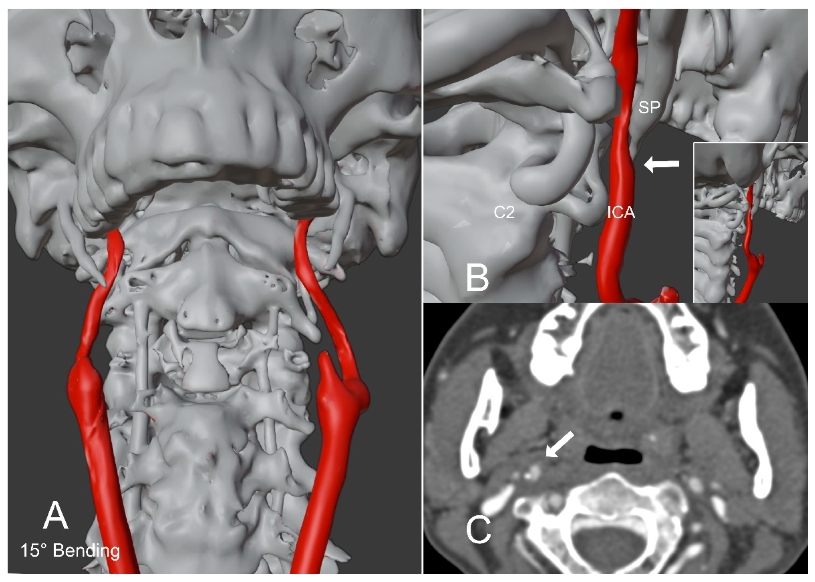
We propose a novel CT-based 3D virtual technique able to investigate in a dynamic and positional manner the osteo-vascular interactions of the neck region. A patient affected by right carotid ES with right periorbital pain and ipsilateral tinnitus was studied with head-and-neck CTA acquisition in both rest position and 15° head flexion, where the head flexion was capable of eliciting the SP impingement on ICA []. The CTA confirmed the hypothesis of right SP impingement on the ipsilateral internal carotid artery (ICA) in the 15° head flexion. After the right carotid ES diagnosis, a virtual dynamic study was performed using a 3D modeling and a rendering software (3D-Slicer [] and Blender [] to evaluate the feasibility of a virtual prediction model of ES. Nonetheless, it is worth noticing that motion simulation may not always be sufficient in predicting ES since even a simple swallowing act can also lead to ES symptoms. Furthermore, a direct SP impingement on the ICA may not always lead to arterial dissection and related ES symptoms.
Only free software was used for this study. CTA DICOM (Digital Imaging and COmmunications in Medicine) images were imported in a 3D-slicer (https://www.slicer.org, accessed on 29 April 2022) [], and internal carotid arteries, skull, and cervical spine were semiautomatically segmented with the same software and then exported as a solid 3D Volume (file extension *.stl). This volume was further imported into Blender (http://www.blender.org, accessed on 29 April 2022) [], and the virtual rotation pivot was set at the level of C1, to simulate reliable movements of the head on the neck in different planes, especially flexo-extension and rotation.
From the head and neck rest position CTA, Blender was able to reproduce the right stylo-carotid impingement, which had previously been shown with the flexed CTA, by performing a virtual 15° head flexion (Figure 1 and Figure 2, and Video S1).

Figure 1.
Patient with Eagle Syndrome in a rest position. (A,B) show a 3D reconstruction model of a patient with. The model was created from the rest position CTA of the head and neck. The right styloid process abuts the ipsilateral internal carotid artery (arrow in (B)). The enclosed image in (B) represents a postero-lateral perspective with a zoomed-out view. (C) shows the CTA image of the styloid process abutting the ipsilateral internal carotid artery (arrow). SP = Styloid process; ICA = Internal carotid artery; C2 = C2 Vertebral body.

Figure 2.
Patient with Eagle Syndrome in a flexed position of the head. (A,B) show a 3D reconstruction model of a patient with Eagle Syndrome in a flexed position of the head (15°). The model was created from the rest position CTA of the head and neck, applying a virtual bending of 15°. The right styloid process impinges the ipsilateral internal carotid artery tightening its caliber (arrow in (B). The enclosed image in (B) represents a postero-lateral perspective with a zoomed-out view. (C) shows the CTA image of the subject in a flexed position of the head (15°). The right styloid process impinges the ipsilateral internal carotid artery (arrow). SP = Styloid process; ICA = Internal carotid artery; C2 = C2 Vertebral body.
Video S1 A dynamic 3D virtual reconstruction model of a CTA of the head and neck in a patient with Eagle syndrome is shown. The simulation starts from a rest position to a 15° bending position, going back to a rest position and reaching a 20° right rotation of the head. In the second part of the video, the same movements are shown from a different angle. In both flexed and rotated positions, the impingement of the right styloid process on the ipsilateral internal carotid artery is shown. Common and internal carotid arteries are shown in red (external carotid arteries are not shown in this simulation).
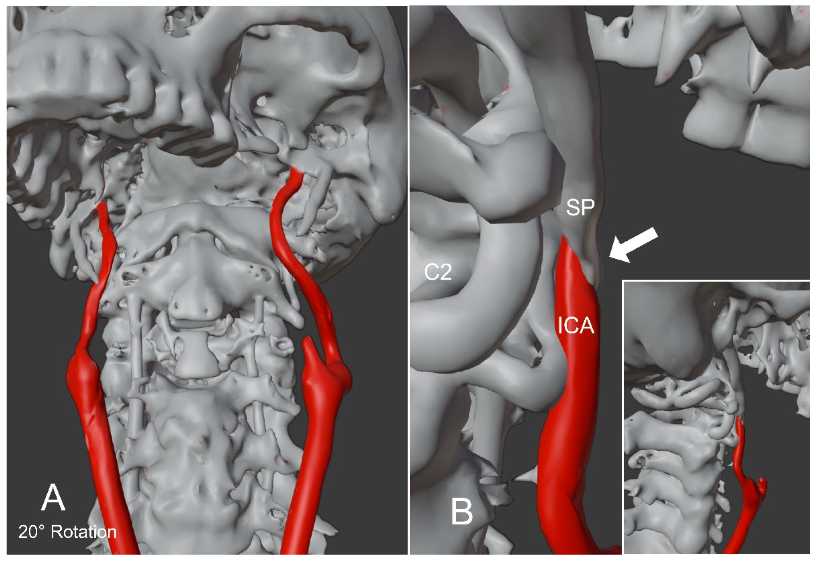
Encouraged by this outcome, a further head virtual 20° rotation movement has been obtained using the same method. The simulation showed how even in the right rotation of the head, a direct SP impingement on the ICA is capable of tightening its caliber (Figure 3 and Video S1). These latter data have not been studied in the previous CTA, which had been performed only in flexion and rest position to avoid further increment of the radiation burden.

Figure 3.
Patient with Eagle Syndrome in a right rotation of the head. (A,B) show a 3D reconstruction model in a 20° right rotation of the head. The model was created from the rest position CTA of the head and neck by applying a virtual 20° right rotation of the head. The right styloid process impinges the ipsilateral internal carotid artery tightening its caliber (arrow in (B)). The 3D reconstruction model has been created from the rest position CTA of the head and neck. The enclosed image in (B) represents a postero-lateral perspective with a zoomed-out view. SP = Styloid process; ICA = Internal carotid artery; C2 = C2 Vertebral body.
This model suggests that some limits related to dose radiation and contrast medium required in different positional CTA acquisitions can be overcome by the use of this virtual prediction model, above all in the study of dynamic structures like the neck. This study highlights the importance of virtual models in the prediction of some pathological conditions such as ES, which might be otherwise overlooked with standard radiological tools. Further integrations of our virtual prediction model with artificial intelligence and machine learning could help its implementation and the reduction of potential operator-dependent bias when used in routine clinical settings.
Supplementary Materials
The following supporting information can be downloaded at: https://www.mdpi.com/article/10.3390/app12094564/s1, Video S1: Dynamic 3D virtual reconstruction model of a CTA of the head and neck in a patient with Eagle Syndrome.
Author Contributions
Conceptualization, E.N.S., E.M. and S.C.; methodology, E.N.S., E.M. and S.C.; software, S.C.; validation, E.N.S., F.S.D.P., F.G. and S.L.V.; formal analysis, K.G. and L.C.; investigation, K.G. and L.C.; writing—original draft preparation, E.N.S. and E.M.; writing—review and editing, E.N.S. and E.M. All authors have read and agreed to the published version of the manuscript.
Funding
This research received no external funding.
Institutional Review Board Statement
The study was conducted in accordance with the Declaration of Helsinki, and approved by the Ethics Committee of the University of Messina (CEME7721—13.7.2021).
Informed Consent Statement
The study was conducted in accordance with the Declaration of Helsinki, and approved by the Ethics Committee of the University of Messina (CEME7721—13.7.2021). Informed consent was obtained from all subjects involved in the study.
Data Availability Statement
Not applicable.
Conflicts of Interest
The authors declare no conflict of interest.
References
- Galletta, K.; Siniscalchi, E.N.; Marco, C.; Mariano, V.; Francesca, G. Eagle syndrome: A wide spectrum of clinical and neuroradiological findings from cervico-facial pain to cerebral ischemia. J. Craniofacial Surg. 2019, 30, E424–E428. [Google Scholar] [CrossRef] [PubMed]
- Eagle, W.W. The symptoms, diagnosis and treatment of the elongated styloid process. Am Surg. 1962, 28, 1–5. [Google Scholar] [PubMed]
- Tijanić, M.; Burić, N.; Burić, K. The Use of Cone Beam CT(CBCT) in Differentiation of True from Mimicking Eagle’s Syndrome. Int. J. Environ. Res. Public Health 2020, 17, 5654. [Google Scholar] [CrossRef] [PubMed]
- Brassart, N.; Deforche, M.; Goutte, A.; Wery, D. A rare vascular complication of Eagle syndrome highlight by CTA with neck flexion. Radiol. Case Rep. 2020, 15, 1408–1412. [Google Scholar] [CrossRef] [PubMed]
- Pieper, S.; Lorensen, B.; Schroeder, W.; Kikinis, R. The NA-MIC Kit: ITK, VTK, Pipelines, Grids and 3D Slicer as an Open Platform for the Medical Image Computing Community. In Proceedings of the 3rd IEEE International Symposium on Biomedical Imaging: From Nano to Macro, Arlington, VA, USA, 6–9 April 2006; Volume 1, pp. 698–701. [Google Scholar]
- Community, B.O. Blender—A 3D Modelling and Rendering Package. Stichting Blender Foundation, Amsterdam. 2018. Available online: http://www.blender.org (accessed on 29 April 2022).
Publisher’s Note: MDPI stays neutral with regard to jurisdictional claims in published maps and institutional affiliations. |
© 2022 by the authors. Licensee MDPI, Basel, Switzerland. This article is an open access article distributed under the terms and conditions of the Creative Commons Attribution (CC BY) license (https://creativecommons.org/licenses/by/4.0/).