Article

Clinical-Pathological Study on Expressions β-APP, GFAP, NFL, Spectrin II, CD68 to Verify Diffuse Axonal Injury Diagnosis, Grade and Survival Interval

1
Morphofunctional Sciences Department 2, Faculty of Medicine, “Grigore T. Popa” University of Medicine and Pharmacy of Iasi, 700115 Iasi, Romania
2
Institute of Forensic Medicine, 700455 Iasi, Romania
3
Morphofunctional Sciences Department 1, Faculty of Medicine, “Grigore T. Popa” University of Medicine and Pharmacy of Iasi, 700115 Iasi, Romania
4
Forensic Science Department, Faculty of Medicine, “Grigore T. Popa” University of Medicine and Pharmacy of Iasi, 700115 Iasi, Romania
5
Providenta Hospital, 707317 Iasi, Romania
*
Authors to whom correspondence should be addressed.
Appl. Sci. 2022, 12(7), 3638; https://doi.org/10.3390/app12073638
Submission received: 23 February 2022 / Revised: 30 March 2022 / Accepted: 2 April 2022 / Published: 4 April 2022
(This article belongs to the Special Issue Updates in Forensic Medicine: New Tools Applied in Forensic Science)

Abstract

:
Traumatic brain injury (TBI) is one of the leading causes of death worldwide, particularly in young people. Diffuse axonal injuries (DAI) are the result of strong rotational and translational forces on the brain parenchyma, leading to cerebral oedema and neuronal death. DAI is typically characterized by coma without focal lesions at presentation and is defined by localized axonal damage in multiple regions of the brain parenchyma, often causing impairment of cognitive and neuro-vegetative function. Following TBI, axonal degeneration has been identified as a progressive process that begins with the disruption of axonal transport, leading subsequently to axonal swelling, axonal ballooning, axonal retraction bulges, secondary disconnection and Wallerian degeneration. The objective of this paper is to report on a series of patients who have suffered fatal traumatic brain injury, in order to verify neurological outcomes in dynamics, relative to the time of injury, using antibodies for neurofilament (NFL), spectrin II, beta-amyloid (β-APP), glial fibrillary acidic protein (GFAP) and cluster of differentiation 68 (CD68). From the studied cases, a total of 50 cases were chosen, which formed two study groups. The first study group comprises 30 cases divided according to survival interval. The control group comprises 20 cases with no history of traumatic brain injury. Cardiovascular disease and history of stroke, cases suffering from loss of vital functions, a post-traumatic survival time of less than 15 min, autolysis and putrefaction were established as criteria for exclusion. Based on their expression, we tested for diagnosis and degree of DAI as a strong predictor of mortality. Immunoreactivity was significantly increased in the DAI group compared to the control group. The earliest changes were recorded for GFAP and CD68 immunolabeling, followed by β-APP, spectrin II and NFM. The most intense changes in immunostaining were recorded for spectrin II. Comparative analysis of brain apoptosis, reactive astrocytosis and inflammatory reaction using specific immunohistochemical markers can provide important information on diagnosis of DAI and prognosis, and may elucidate the timing of the traumatic event in traumatic brain injury.

1. Introduction

With an incidence of 235-556/100,000 inhabitants, closed or open traumatic brain injury is the most studied area in forensic medicine. In the case of traumatic brain injury accompanied by exitus, traumatic brain injury is the leading cause of violent death, in its various forms, and may or may not be accompanied by legal implications [1]. Sabina J. Strich first reported DAI in 1956 [2]. In her paper, she presented a series of autopsied cases at an interval of 5–15 months after trauma, highlighting a diffuse degeneration of cerebral white matter, which led to lifelong psycho-behavioral changes associated with neurodegenerative pathologies. Adams et al. further explained the biomechanics of DAI, identifying that angular acceleration together with the acceleration–deceleration mechanism cause true shear forces at the boundary between white matter and gray matter. It was concluded, using primate models, that extensive axonal injury is the primary cause of post-traumatic unconsciousness without focal lesions [2,3]. The most common changes have been shown in the subcortical white matter of the frontal and temporal lobes, the corpus and splenium of the corpus callosum, and in the most severe cases, the rostral superior region of the brainstem [4,5,6]. Today, DAI is considered a common finding in patients suffering from TBI, existing in about half of the cases of closed TBI in combination with focal lesions [1,2,3]. Diffuse axonal lesions in isolation, however, are less common [2]. DAI has a negative impact on vital prognosis and quality of life in case of survival, as they are left with severe motor, cognitive and behavioral impairments. Traumatic brain injury is the most common type of trauma encountered in current forensic practice. Investigations into the mechanisms of injury, the timing of traumatic brain injury, and the causes of death remain topical. Determining the time of trauma involves both distinguishing between antemortem and postmortem injuries and considering the post-traumatic survival interval [3,4,5,6,7]. The elucidation of the time of traumatic brain injury from a forensic point of view has not yet been satisfactorily achieved. Finding objective criteria for assessing the time of trauma is essential. Therefore, the present study aims to investigate immunohistochemically apoptotic brain cellular changes, brain reactive astrocytosis processes and tissue inflammatory reactions in relation to the survival interval [8,9]. The classical hypothesis on the development of traumatic brain injury shows that it is the result of primary traumatic injury, due to cell necrosis combined with the brain inflammatory response leading to secondary brain injury [10]. In recent decades, multiple studies have been conducted on the role of the inflammatory response, in the perpetuation of traumatic brain injury, with the observation that microglial and macrophage cells recruited to the brain exacerbate primary neuronal injury. Much experimental research related to traumatic brain injury has focused on the occurrence and progression of tissue necrotic lesions, largely due to direct post-traumatic mechanical effect at the brain level. A clear disjunction has been made between necrosis and apoptosis. Necrosis is cell death that occurs accidentally due to external factors and is unprogrammed, whereas apoptosis represents programmed and directed cell death, with a role in maintaining tissue homeostasis. These changes also apply to diffuse axonal injury, where the necrosis/apoptosis ratio dictates the subsequent course [11,12]. This ratio is dependent on the cellular influx of calcium ions, which will lead, depending on the intracellular concentration of these ions, to the activation of one or another of the caspase or calpain pathways. Post-traumatic neuronal loss has been described as being strictly due to necrosis, while inflammation and cell apoptosis are physiological processes that have no role in this process [13]. Due to recent experimental data, brain cell apoptosis is being re-evaluated. The pathophysiology of traumatic brain injury is far from being fully understood, and the idea that apoptosis may play an even more important role than initially thought is beginning to emerge. More specifically, injured brain cells release neuromodulatory substances that can lead to late neuronal destruction that occurs long after brain necrotic and inflammatory phenomena have ceased to act. These neuronal cell injuries are responsible for the development of various neurological deficits and post-traumatic sequelae [5,7,14].

1.1. Aims and Objectives

The present study aims to be a clinico-pathological study on β-APP, GFAP, NFL, spectrin II and CD68 expressions to verify the diagnosis, severity and survival interval for subjects with diffuse axonal lesions.
The present study aims to highlight whether brain apoptotic processes play a role in the pathophysiological evolution of traumatic brain injury, contributing to the development of secondary traumatic brain injury. For a long time, astrocytic cells were thought to play more of a structural role in nervous tissue [12,15]. With advances in immunohistochemistry and electron microscopy techniques, it has been observed that they also play multiple functional roles in the brain essential for cerebral homeostasis [2,16]. Astrocytic cell activation occurs as a result of various structural lesions in the central nervous system (CNS). As there is currently no general scientific consensus on the definition of reactive astrocytosis or criteria for assessing the severity of astrocytic lesions, the present study aims to highlight the specific changes in characteristics of reactive astrocytosis following traumatic brain injury [17,18]. GFAP is not detected by immunohistochemical techniques in non-reactive astrocytes, and the investigation of this specific protein in astrocytic cells may provide information on the phenomena of reactive cerebral astrocytosis occurring post-traumatically [6,9,19].

1.2. Biomechanics and Pathophysiology of Axonal Injury

Under normal conditions, brain tissue is elastic and resilient to mechanical stress stretching, having the ability to double its length, due to a capacity called viscoelasticity. Despite this property, in the case of DAI, the main force applied is rotational acceleration-deceleration, leading to dynamic shear, tensile deformation and compression of the brain tissue, dependent on the magnitude and abruptness of the force acting on the cephalic extremity. The higher these forces, the stiffer and more fragile the brain tissue becomes [2], thus damaging the axons and damaging the axonal cytoskeleton. Moreover, the difference in density between white and gray matter causes mass effects, leading to tension in the interface. Although deformations rarely lead to axonal disconnections on impact, these events trigger secondary axotomy. This is a consequence of axonal membrane permeability, which leads to a marked influx of Ca ions, with injury to the cytoskeleton, a phenomenon that starts at the level of the nodule Ranvier, rich in Na channels, Ca channels, Na/Ca exchangers, ATP-dependent Ca pumps, the presence of mitochondria, microtubules and neurofilaments, and ankyrin-3 and alpha-II spectrin-rich cytoskeleton, susceptible to injury by Ca-dependent calpain. Spectrin alpha chain, non-erythroid 1 (UniProt: Q13813; also known as alpha-II spectrin, fodrin alpha chain, spectrin, non-erythroid alpha subunit) is encoded by the SPTAN1 (also known as NEAS, SPTA2) gene (gene ID: 6709) in humans. Non-erythroid spectrin is essential for maintaining membrane stability and cell shape by connecting the cytoskeleton to plasma membranes or intracellular vesicles. Spectrin II is an abundant structural protein in central nervous system neurons and is cleaved into signature fragments by proteases involved in necrotic and apoptotic cell death. It is one of the primary targets cleaved by activated caspases during apoptosis. Spectrin II is involved in secretion, interacts with calmodulin in a calcium-dependent manner and is thus a candidate for calcium-dependent movement of the cytoskeleton at the membrane. Important forces act mainly at the level of Na-voltage and mechano-dependent channels, which have the ability to transform mechanical stimulus into electrical stimulus, thanks to mechanotransduction. This massive activation of Na channels will lead to a massive influx of Ca via Ca/Na exchanger and activation of voltage-dependent L and N calcium channels with depolarization, which should normally maintain a longer action potential at the axonal level. Calcium will activate numerous proteolytic enzymes, including Ca-dependent calpain, which will lead to proteolysis of the H-gate, inactivating Na-voltage-dependent channels. We aimed to identify cases with traumatic brain injury showing diffuse axonal lesions by immunohistochemical study with antibodies to GFAP, β-APP, spectrin II, CD68 and NFL in the analyzed samples. We also wanted to analyze the correlation between these lesions and the degree of survival, as well as their dynamics as a function of survival interval.

2. Materials and Methods

This study was conducted in accordance with the standards developed by the Declaration of Helsinki, with the approval of the management of the Institute of Forensic Medicine Iasi, Romania (no. 12 C/2020).

2.1. Inclusion and Exclusion Criteria

The study is based on the analysis of cases with a history of cranio-cerebral trauma autopsied at the Institute of Legal Medicine Iasi Romania, between 2019 and 2021, having a retrospective component, but also a prospective one. From the studied cases, we selected a total of 50 cases, which formed two study groups. The first study group comprises 30 cases, the analysis being performed both on paraffin-embedded tissue fragments taken from 18 cases, and on fresh tissue taken from 12 cases. In turn, the 30 cases were divided according to survival interval into 4 categories: death in the supra-acute period (within 1 h of the traumatic event), death in the acute period (within 24 h of the event), death in the subacute period (between 2 days and 2 weeks after the trauma), and death in the chronic period (2 weeks after the event). The second group is the control group, made up of 20 cases with no history of traumatic brain injury. As criteria for exclusion for the second group, we established cardiovascular disease and history of stroke, cases suffering from loss of vital functions, having a post-traumatic survival time of less than 15 min, autolysis and putrefaction.

2.2. Working Technique Used

Five brain tissue fragments were collected at necropsy from each of the cases belonging to group I with craniocerebral trauma (thalamus, hypothalamus, striatum, temporal and frontal subcortical regions). One brain tissue fragment was collected from each of the control cases belonging to lot II. The brain fragments harvested at autopsy were subsequently placed in 10% buffered formalin fixative solution and sealed in containers with a volume of formalin 5 times the mass of the specimen for a period of 2 weeks. Tissue fragments were subsequently embedded in paraffin blocks. The usual technique of staining sections with H-E, the IHC technique of staining sections as well as the microscopic examination of the harvested brain tissue fragments were performed in the Pathological Anatomy Department of the Emergency Clinical Hospital Prof. Dr. N. Oblu Iasi Romania, and at the IHC Laboratory of the Institute of Forensic Medicine Iasi. Using a SAKURA Accu-Cut SRM microtome from each paraffin block, 3 sections of 1.5–2 μm thickness were cut in the coronal plane. The sections were thoroughly embedded in a thermostated water bath at 400 °C and subsequently etched on special slides, pre-treated with polysine (Polysine Slides, Polysciences, Hirschberg, Germany ). The slides obtained were kept for 4–6 h at 600 °C. The sections were subjected to 3 successive 8–10 min baths in SLIDE BRITE solution (Biocare Medical, Pacheco, United States of America). Subsequently, baths were performed with absolute ethanol in successive dilutions, 95–80%, for 8–10 min. Sections were rehydrated in deionized water for 10–15 min. Sections were initially stained using the usual hematoxylin–eosin section staining technique.

2.3. Immunohistochemical Staining Technique of Sections

The immunohistochemical technique was performed manually, using labeled antibodies to determine anti-β-APP+, anti-GFAP+, anti-NFL+, anti-spectrin II+ and CD68+ cells. We performed endogenous peroxidase blocking treatment of sections for 5 min at room temperature with Peroxidazed 1 (Merck, Berlin, Germany), and then slides were washed with pH 7.6 TBS plus buffer (Merck). Antigen demosaicing was performed by boiling for 45 min at 95 °C (HIER—heat-induced epitope retrieval) using a decloaking chamber and a citrate-based buffer solution with high pH 6 and stability at high temperature. Sections were treated with Carezyme I—Trypsin (Merck). Slides were then immersed in pH 7.6 TBS plus buffer (Merck). For nonspecific blocking, sections were treated with Background Sniper solution (Biocare Medical) for 10 min at room temperature. At the end, slides were washed with pH 7.6 TBS plus buffer solution (Biocare Medical). In the application of primary antibodies, the following primary Ac was applied to the sections for 60 min at room temperature: antibodies (Merck). At the end of each step, the slides were washed with buffer solution pH 7.6 TBS 21 plus (Merck). Each primary Ac was used in dilutions according to the manufacturer’s recommendations. For the probe/polymer detection system, a universal detection system was used—MACH 4 Universal HRP-Polymer (Biocare Medical), which can detect both anti-mouse and anti-rabbit antibodies. The probe was first applied to the sections for 10 min and then the universal polymer for 20 min. At the end of the step, the slides were washed with pH 7.6 TBS plus buffer (Biocare Medical). Next, 3,3’-diaminobenzidine (DAB) Biocare Medical Chromogen was applied to the sections at the dilution recommended by the manufacturer. At the end of the step, the slides were washed for 1–2 min in deionized water. For overstaining with hematoxylin CAT, slides were immersed in hematoxylin CAT (Biocare Medical) for 1 min. For the dehydration, clarification and mounting procedures, sections were immersed in Ottix Shaper bath (Diapath, Bergamo, Italy) for 10 min and then in Ottix Plus bath (Diapath) for 10 min and then mounted on slides using Micromount mounting medium (Diapath).

2.4. Microscopic Examination

Brain fragments were examined by light microscopy using different magnification objectives with the Leica ICC 50W transmitted light microscope with the included photographic system.
For standard hematoxylin–eosin staining, 6 slides were made in cases belonging to lot I (LOT TCC). In the cases belonging to the control batch, 3 slides were taken. Five microscopic fields were analyzed at 20× magnification from the three/six slides of each case studied. Semi-quantitative examination of histological changes in the brain aimed at (a) determination of the type of brain lesions (focal (1); diffuse (2)) and (b) determination of the degree of vascular congestion, post-traumatic brain oedema, cerebral inflammatory reaction, cerebral hemorrhage and severity of neuronal and glial lesions. Quantification of histological parameters was as follows: 0 = no changes; 1 = slight changes; 2 = moderate changes; 3 = severe changes.

2.5. Immunohistochemical Staining

A quantitative analysis of the number of anti-β-APP, anti-GFAP, anti-NFL, anti-spectrin II and CD68 cells was performed for each of the cases analyzed. On each of the three/six slides of a case, as many 5 microscopic fields were analyzed, with 20× magnification. With the help of a standardized ocular grid, a high random power count of the number of anti-β-APP+, anti-GFAP+, anti-NFL+, anti-spectrin II+ and CD68+ cells per field was performed. The final mean number of positive cells for each case was determined by counting the cell types listed on each slide, averaging the determinations from every 3/6 slides of the case. The density of positive cells for the above-mentioned antibodies was determined by reporting the mean final number of cells/mm2 brain tissue. For the traumatic brain injury group, a regional quantitative analysis of the number of anti-β-APP+, anti-GFAP+, anti-NFL+, anti-spectrin II+ and CD68+ cells was performed, determining the mean final cell count in the lesional areas. The density of immuno-positive brain cells in the lesional areas was determined by reporting the mean final cell count/mm2 brain tissue.

2.6. Statistical Analysis
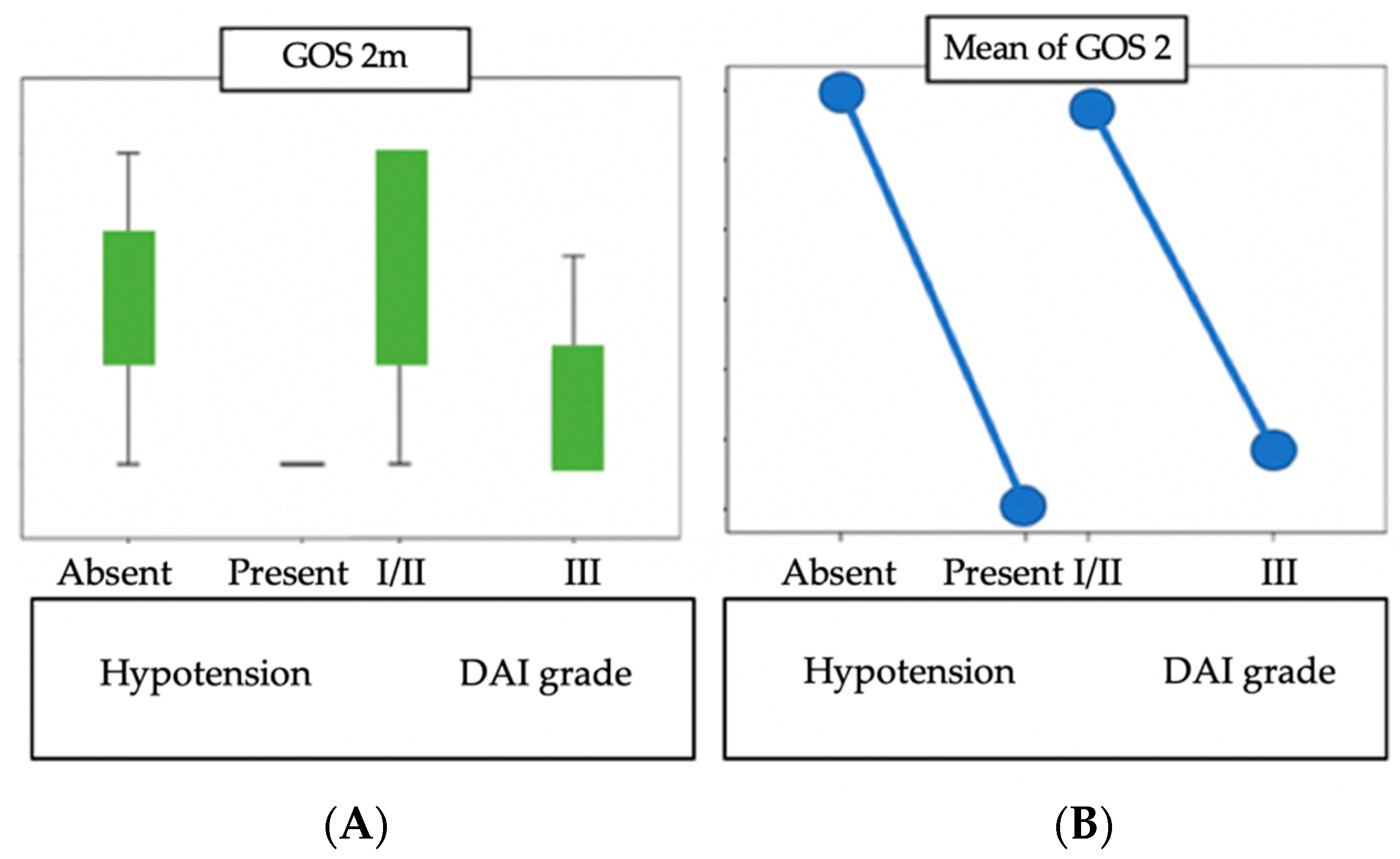
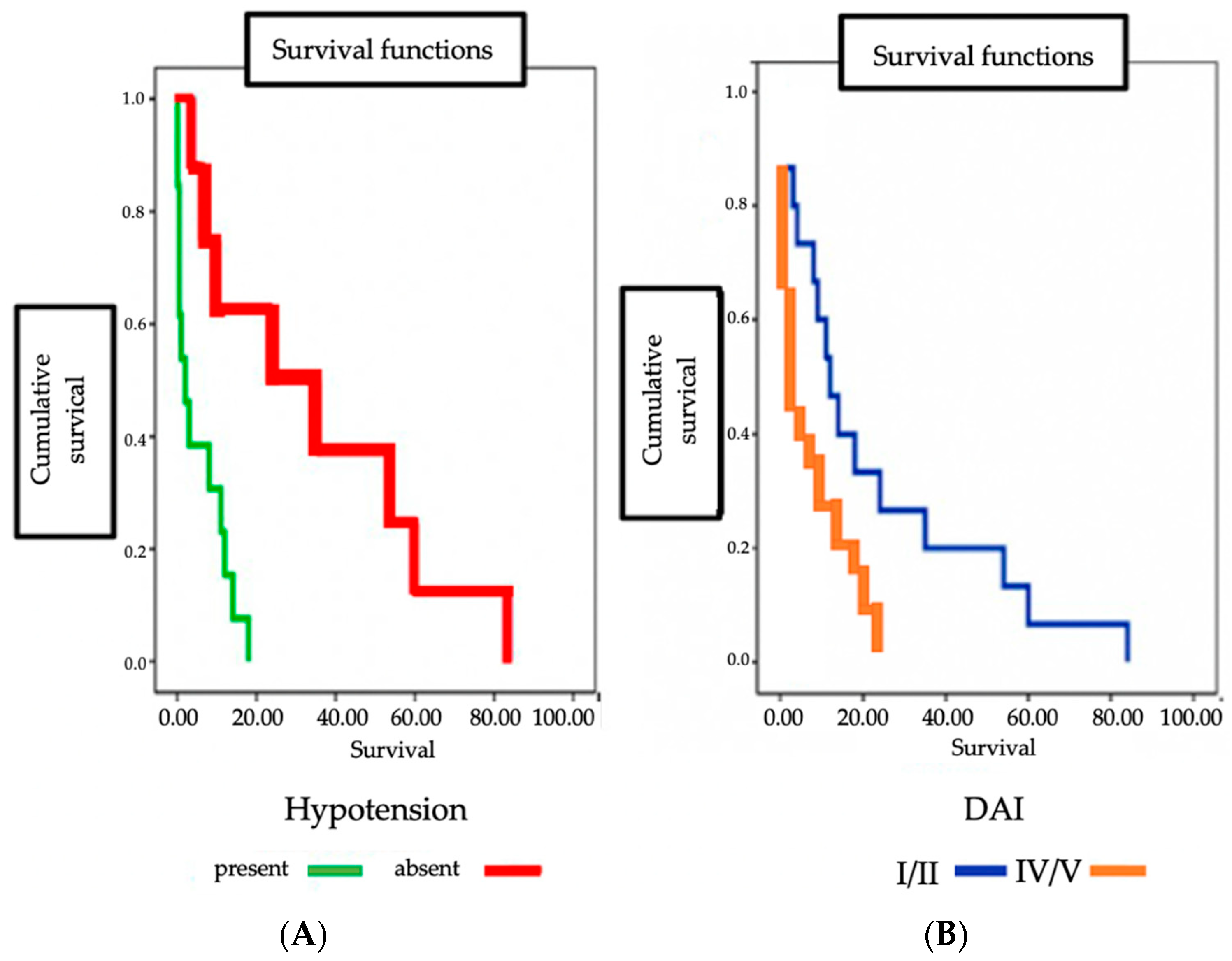
Data were statistically processed using SPSS software (SPSS for Windows, version 28.01, G00L3EN, New York, United States of America). In the analytical stage, the comparison of groups and validation of the study hypothesis was performed by applying the parametric Student t-test, the variant independent samples t-test and the nonparametric Mann–Whitney U-test. The analysis of variance (ANOVA) test as well as the nonparametric Kruskal–Wallis test were used to determine the differences between the parameters of the groups analyzed. Differences between groups were considered “statistically significant” at an accepted p threshold less than or equal to 0.05. We applied the chi-square test or Fisher’s exact test to compare qualitative variables. The relationship between brain cell densities and different variables of interest was tested using correlation analysis. In addition, we performed a Kaplan–Meier analysis to find differences in survival between different subgroups.

3. Results

In terms of neurological outcome at two months (GOS) or survival, these aspects were not influenced by the gender of the patients. Severity of diffuse axonal lesions predicted the survival and GOS at two months. The p-value for the correlation between axonal injury severity and survival interval is statistically significant (p 0.003); the correlation between axonal injury severity and GOS is statistically significant (p 0.070). Uncontrolled hypotension revealed a similar statistical association (p 0.010, respectively p 0.060). As shown in Figure 1, uncontrolled hypotension is associated with a small survival interval (p 0.010) and is also predictive of severe GOS (p 0.060) (Figure 1).
Regarding the mechanism of trauma and the association of traumatic brain injury with polytrauma, we found that survival time was not influenced by these aspects. A Kaplan–Meier analysis showed a lower survival rate in the group with a higher degree of DAI and in the group with uncontrolled hypotension on admission (Figure 1 and Figure 2).
Table 1 illustrates the differences identified in terms of IHC between the two batches analyzed.

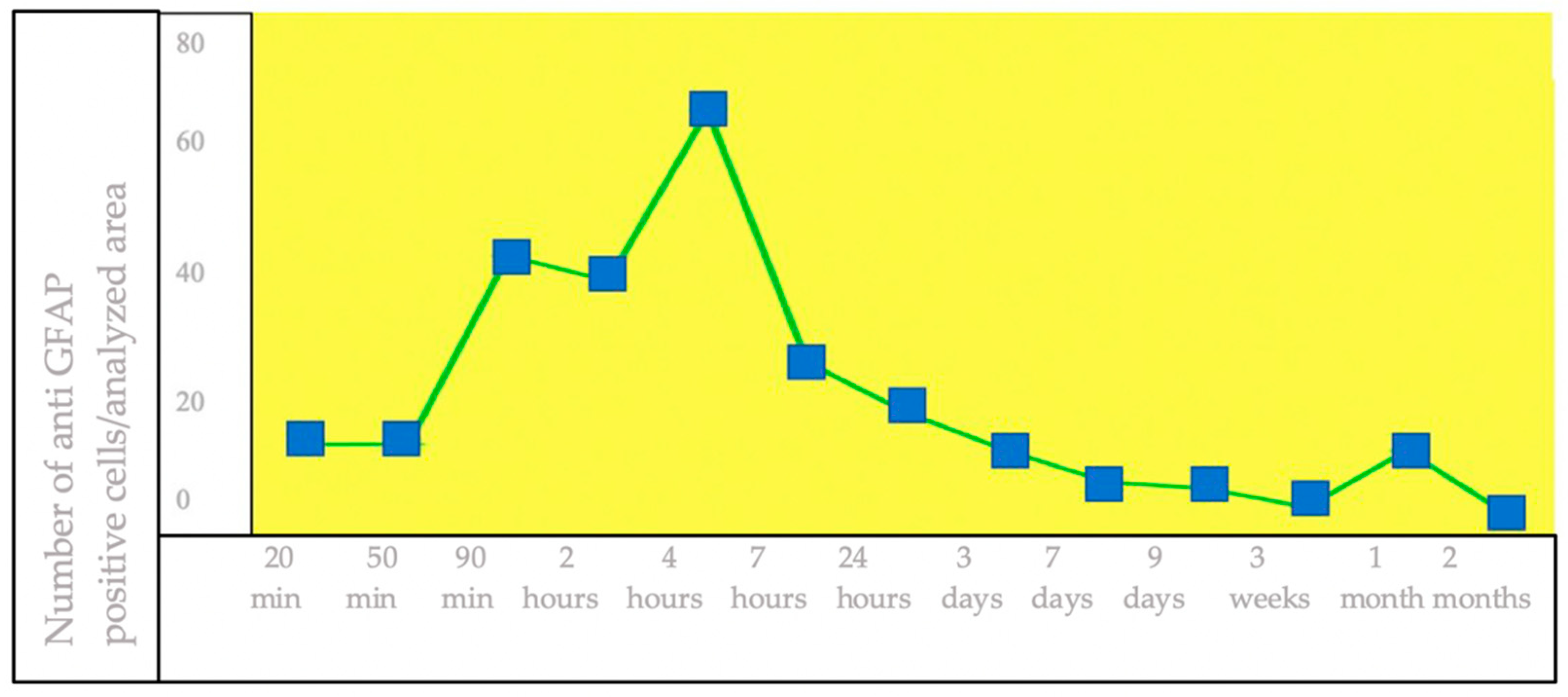
3.1. IHC Staining for the Detection of GFAP+ Brain Cells

Analysis of the study group with traumatic brain injury with variable post-traumatic survival intervals revealed the following: Statistical analysis of GFAP+ brain cell density showed statistically significant differences in cell density between the two groups analyzed (Figure 3).
In relation to the duration of post-traumatic survival, GFAP+ brain cell density shows a variable, upward trend with a maximum at 4 h, followed by an approximately continuous decrease until 2 months post-trauma (Figure 4).

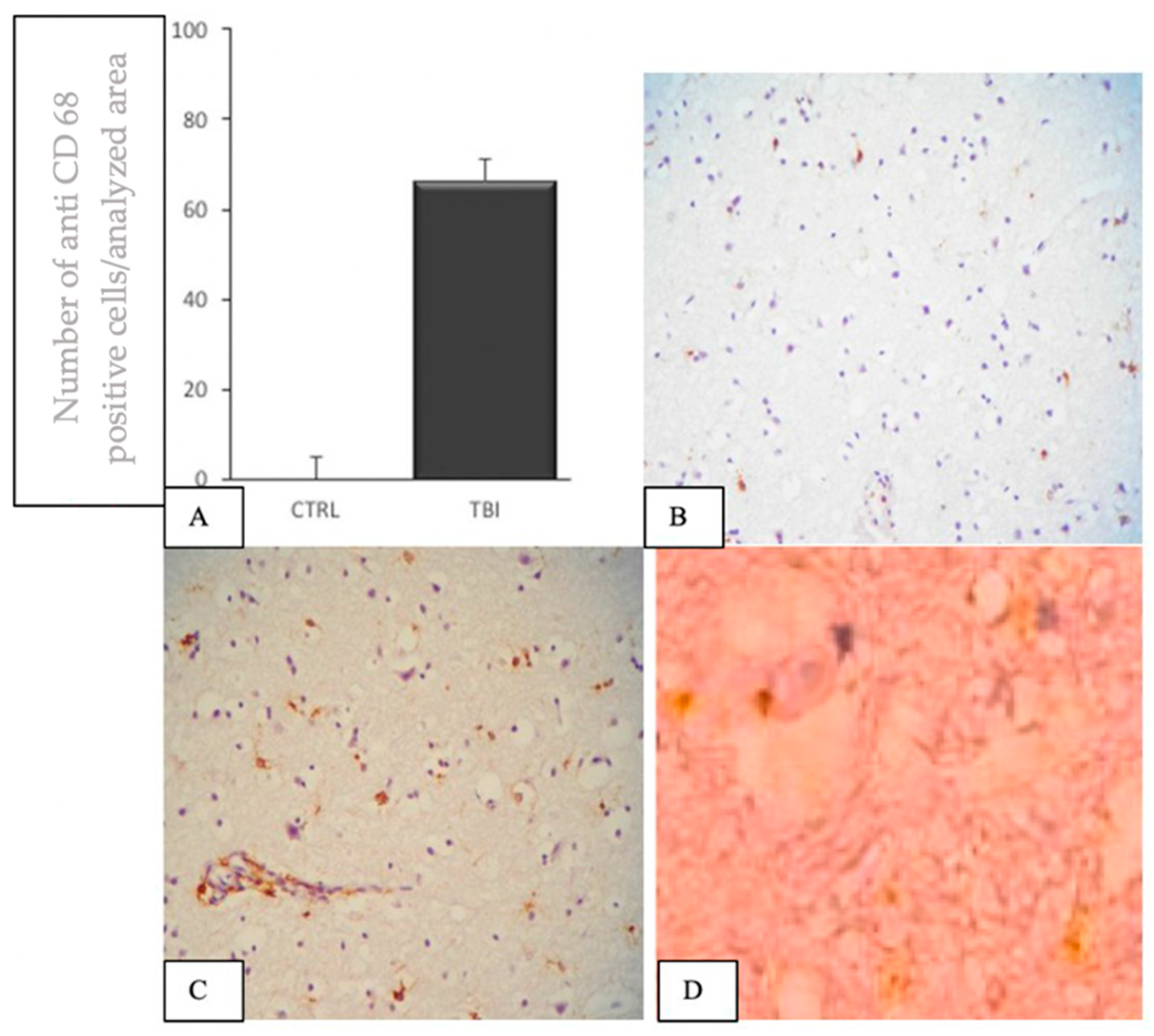
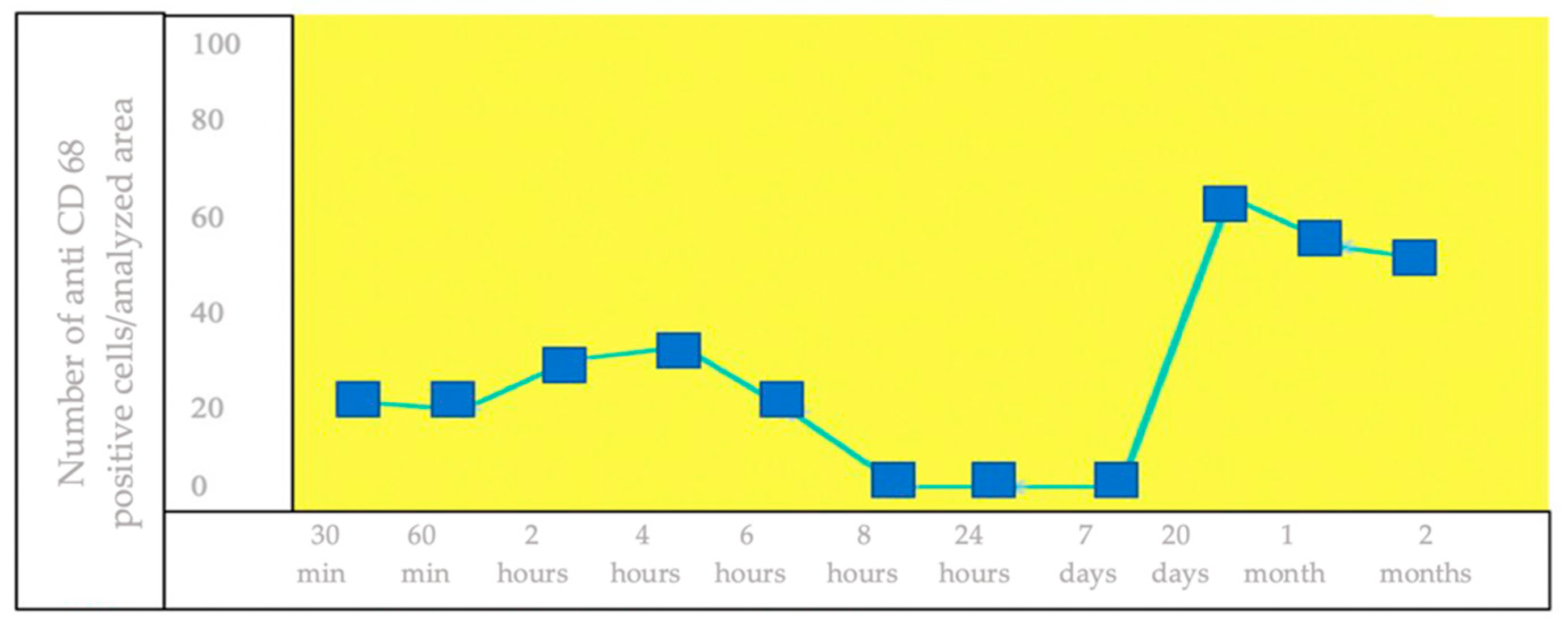
3.2. IHC Staining for the Detection of CD68+ Brain Cells

Analysis of the study group with traumatic brain injury with variable post-traumatic survival intervals revealed the following: Statistical analysis of CD68+ brain cell density showed statistically significant differences in cell density between the two analyzed groups (Figure 5).
From death after 30 min post-trauma to 2 months of survival post-trauma, a variable evolution of brain density of CD68+ brain cells was observed. Between 8 h and 7 days post-trauma, the lowest density of CD68+ cells was observed. The highest brain density of CD68+ cells was recorded at 20 days post-traumatic brain injury survival (Figure 6).

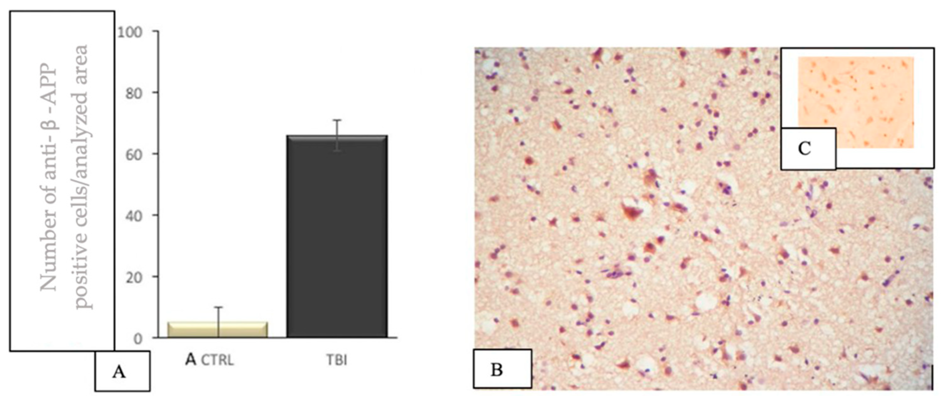
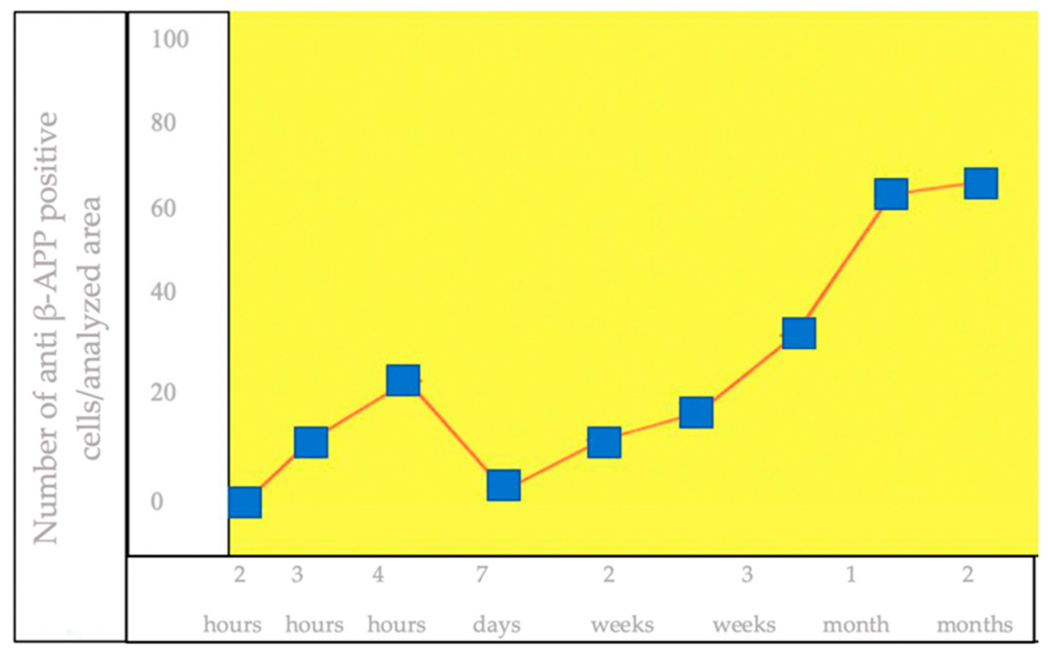
3.3. IHC Staining for the Detection of β-APP+ Brain Cells

Statistical analysis of β-APP+ brain cell density showed statistically significant differences in cell density between the two analyzed groups (Figure 7).
From death after 2 h post-trauma to 2 months post-traumatic survival, a variable evolution of brain β-APP+ brain cell density was observed. Cells started to become positive at 2 h post-trauma, with an upward evolution in the next hour, after which a decrease in immunopositivity was observed in the days 1–7 post-trauma range. After this period, there was a progressive upward evolution of positivity, with a peak at 2 months (Figure 8).

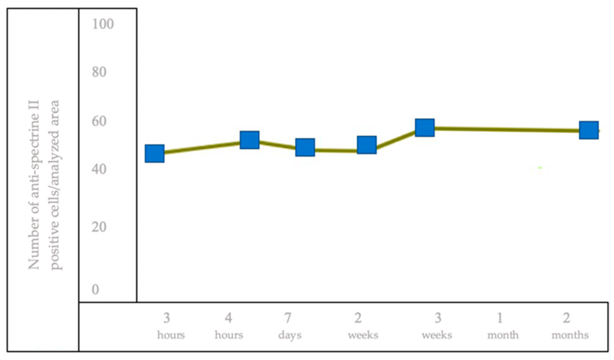
3.4. IHC Staining for the Detection of Brain Cells of Anti-Spectrin II+

Statistical analysis of spectrin II+ brain cell density showed statistically significant differences in cell density between the two analyzed groups (Figure 9).
From death after almost 3 h post-trauma to 2 months of post-traumatic survival, an approximately linear evolution of spectrin II+ brain cell density was observed. Cells started to become positive at 3 h post-trauma, with a discrete upward evolution over the next 4 h, after which there was a discrete decrease in immunopositivity up to 1 week, after which a plateau phase was observed between 7 and 14 days. In the interval 14–21 days, there was a slight increase in immunopositivity, with a peak in the third week after the trauma. In the interval up to 2 months, a plateau phase was recorded. We appreciate that the dynamics of immunoreactivity for spectrin II, unlike the rest of the antibodies studied, showed discrete variations over the studied interval (Figure 10).

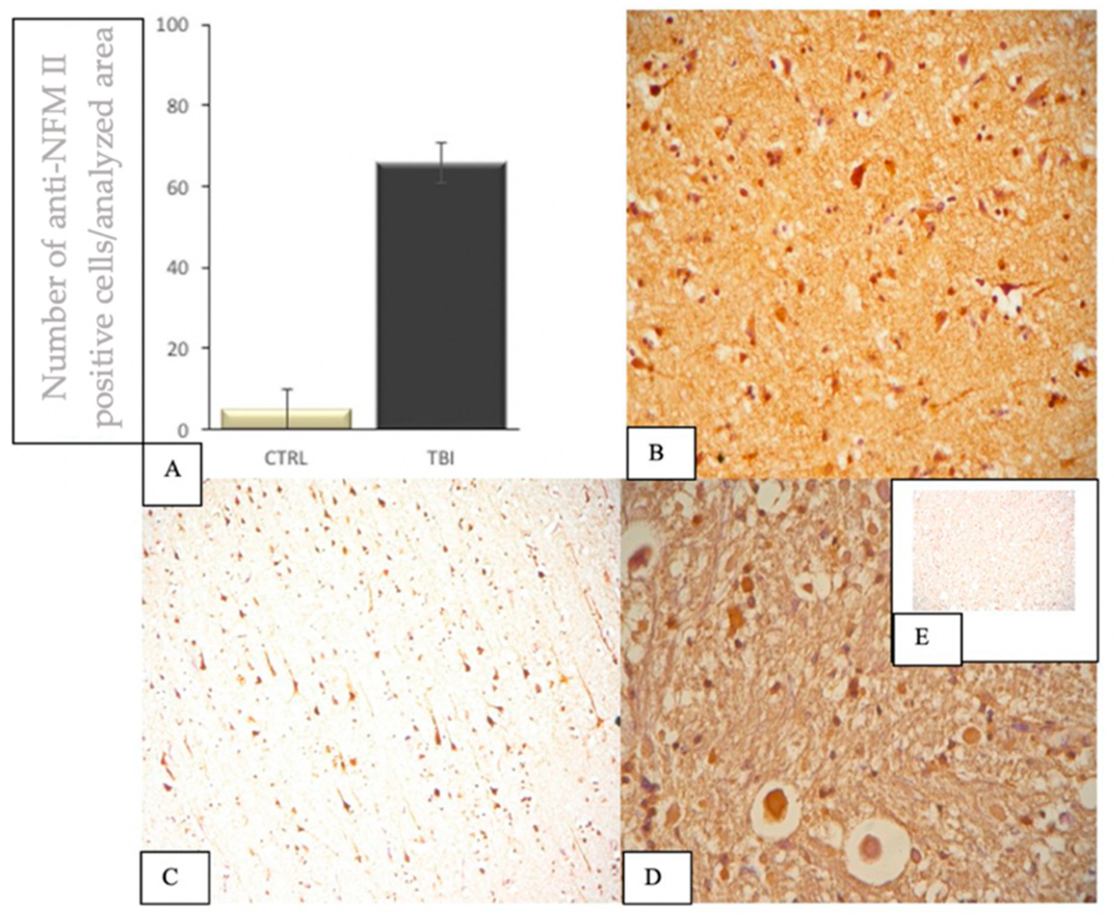
3.5. IHC Staining to Highlight Brain Cells of NFM II+

Statistical analysis of spectrin II+ brain cell density showed statistically significant differences in cell density between the two analyzed groups (Figure 11).
From death after almost 3 h post-trauma to 7 days post-trauma survival, a hyperbolic evolution of NFM II+ brain cell density was observed, with an ascending evolution between 3 h and 24 h post-trauma. Between 1 and 3 days, there is a slowly ascending evolution, with a maximum at 3 days, after which there is a marked and sudden decline in immunoreactivity until the 7th day post-trauma (Figure 12).

4. Discussion

A promising area of research in brain injury assessment, in addition to identifying possible blood and brain markers useful for clinical assessment and staging of DAI, is the search for new immunohistochemical identification methods that could be useful for determining the timing of traumatic brain injury (TBI). Biomarker dynamics can provide important information regarding the timing of the trauma. An open area of research remains the identification of markers to distinguish between traumatic and ischemic neuronal injury. Current markers do not differentiate between acute and sub-acute TBI (<48 h after the traumatic event). The main immunohistochemical markers used to assess traumatic brain injury include β-APP (β-amyloid precursor) and GFAP [20,21]. β-APP is the gold standard of diffuse axonal injury. In fact, β-APP-positive immunohistochemistry is not detectable in uninjured brain tissue samples, whereas an increase in immunoreactivity has been observed starting 2 h after traumatic brain injury [16,22]. More recent studies have also detected how it is possible to reveal different immunohistochemical patterns of β-APP immunoreactivity, which may help discriminate traumatic from ischemic brain injury. Thus, we observed that in ischemic-type lesions, even if β-APP immunoreactivity is present, that of neurofilaments and spectrin II is absent [22]. We also showed chronodependent changes, by increasing neurofilament reactivity after 3 h, with a peak at 2–3 days, especially for corpus callosum neurons. Neurofilaments are the main cytoskeletal components in neuronal cells, important for maintaining axon caliber. After axonal cell injury caused by direct trauma, increased levels of neurofilaments have been observed [15,21]. We also observed that neurofilaments and β-APP immunoreactivity are proportional during the first 7 days, after which NF immunoreactivity disappears, while that of β-APP decreases over the next few weeks and increases again after 1 month, as a sign of Wallerian degeneration, a phenomenon similar to the mechanism of Alzheimer’s disease. Immunoreactivity for neurofilaments and CD68 are also proportional in the first week, after which neurofilaments disappear, while CD68 reappears after about 3 weeks, especially at a distance from the lesion. Immunohistochemical analysis of activated CD68+ macrophages and microglial cells revealed a strong and persistent post-traumatic brain inflammatory response, increased densities being observed even after 2 weeks of post-traumatic brain injury survival. Peak CD68+ is observed in late death. The minimum CD68+ cell density was noted in the acute range. Most CD68+ cells were observed at the perilesional level. The reaction of macrophages and microglial CD68+ cells was rapid, these cells appearing early post-traumatically, with different lesional and perilesional densities. SNTF proportional to GFAP (gliosis) in the first hours, after 5–6 days, is IHC negative. Immunoreactivity for GFAP and CD68 are proportional to cerebral edema and hemorrhage. However, GFAP appears to be a better biomarker of mild TBI, as extracerebral expression of GFAP has not been detected. Li suggests that GFAP immunopositivity showed subacute or late-onset death, with increases after about 1 month. Our study found concordant information, but the increase was not significant at one month after trauma [7,18,22]. The study of reactive astrocytosis processes found in traumatic brain injury cases, as revealed by GFAP immunohistochemical brain staining, identified a broad spectrum of astrocytic cell transformations depending on the post-traumatic interval. The highest brain density of GFAP+ reactive astrocytes was observed in the acute period of death and the lowest in the chronic period (˃2 weeks). In supra-acute, acute and subacute post-traumatic death periods, most reactive astrocytes GFAP+ evidenced were localized at the perilesional level. The gradual and differentiated appearance of reactive astrocytes according to the interval post-traumatic brain interval with specific lesional, perilesional and remote distribution shows that the study of reactive astrocytosis can be an objective index in the assessment of the post-traumatic interval.
The earliest changes were recorded for GFAP and CD68 immunolabeling, followed by β-APP, spectrin II and NFM. The most intense changes in immunostaining were recorded for spectrin II, an aspect explained by the fact that spectrin II is one of the primary targets cleaved by activated caspases during apoptosis. Alpha-II spectrin is involved in secretion, interacts with calmodulin in a calcium-dependent manner and is thus a candidate for calcium-dependent movement of the cytoskeleton at the membrane.
Comparative analysis of brain apoptosis, reactive astrocytosis and inflammatory reaction using specific immunohistochemical markers can provide important information on the diagnosis of DAI and prognosis, and may elucidate the timing of the traumatic event in a traumatic brain injury.
The limitations of the study are the small number of cases studied. Moreover, in current practice, immunohistochemical staining is not used. Other disadvantages are the relatively high cost of immunohistochemical examination and the large number of samples to be taken from each case studied. Additionally, changes that occur over time in β-APP+ cells may be pre-existing and need to be interpreted in combination with other biomarkers.

5. Conclusions

Lesion biomechanics and pathophysiological mechanisms of chronologically revealed axonal injury are essential for further progress in the diagnosis, dating and treatment of DAI. Immunoreactivity was significantly increased in the DAI group compared to the control group. The earliest changes were recorded for GFAP and CD68 immunolabeling, followed by β-APP, spectrin II and NFM. The most intense changes in immunostaining were recorded for spectrin II. Comparative analysis of brain apoptosis, reactive astrocytosis and inflammatory reactions using specific immunohistochemical markers can provide important information on the diagnosis of DAI and prognosis, and may elucidate the timing of the traumatic event in a traumatic brain injury. The present study emphasizes that the earliest possible detection of DAI is essential for the diagnosis of deaths that apparently may have an unknown cause, but also for the assessment of survival interval. It should be noted that immunohistochemistry, although more sensitive than the usual stains, is not specific for the detection of traumatic neuronal lesions, as there are positive results also in hypoxic ischemic brain injury.
As directions for future studies, we propose to extend the study to a larger number of cases and to correlate the immunohistochemical examination with the dynamics of these markers in the cerebrospinal fluid, which may extend the applicability of the findings to patients with traumatic brain injury.

Author Contributions

Conceptualization, I.H. and L.R.; methodology, M.D. and N.G.; software, C.F.; formal analysis, S.D.; investigation, I.H., N.G. and C.L.; data curation, I.H. and I.C.D.; writing—original draft preparation, I.H.; writing—review and editing, A.K.; supervision, D.B.I. and M.C. All authors have read and agreed to the published version of the manuscript.

Funding

This research received no external funding.

Institutional Review Board Statement

This study was conducted according to the guidelines of the Declaration of Helsinki. The study was approved by the Research Ethics Commission of Institute of Forensic Medicine, Iasi, Romania.

Informed Consent Statement

Informed consent for the autopsy is not required; the autopsy is mandatory under Romanian Law (Romanian Criminal Procedure Code Law no. 135/2010, chapter VII, article 185).

Data Availability Statement

The study did not report any data, apart from those reported already in the article.

Conflicts of Interest

The authors declare no conflict of interest.

References

  1. Mata-Mbemba, D.; Mugikura, S.; Nakagawa, A.; Murata, T.; Kato, Y.; Tatewaki, Y.; Li, L.; Takase, K.; Ishii, K.; Kushimoto, S.; et al. Intraventricular hemorrhage on initial computed tomography as marker of diffuse axonal injury after traumatic brain injury. J. Neurotrauma 2015, 32, 359–365. [Google Scholar] [CrossRef] [PubMed]
  2. Hunea, I.; Damian, S.I.; David, S.; Diac, M.M.; Iliescu, D.B.; Ciocoiu, M. Brief literature Review of the Mechanism involved in producing diffuse axonal injury. Brain 2017, 8, 91–97. [Google Scholar]
  3. Currie, S.; Saleem, N.; Straiton, J.A.; Macmullen-Price, J.; Warren, D.J.; Craven, I.J. Imaging assessment of traumatic brain injury. Postgrad. Med. J. 2016, 92, 41–50. [Google Scholar] [CrossRef] [PubMed]
  4. Thelin, E.P.; Nelson, D.W.; Vehviläinen, J.; Nyström, H.; Kivisaari, R.; Siironen, J.; Svensson, M.; Skrifvars, M.B.; Bellander, B.M.; Raj, R. Evaluation of novel computerized tomography scoring systems in human traumaticbrain injury: An observational, multicenter study. PLoS Med. 2017, 14, e1002368. [Google Scholar] [CrossRef]
  5. Ripoll, M.A.; Siösteen, B.; Hartman, M.; Raininko, R. MR detectability and appearance of small experimental intracranial hematomas at 1.5 T and 0.5 T. A 6–7-month follow-up study. Acta Radiol. 2003, 44, 199–205. [Google Scholar] [CrossRef]
  6. Schiavone, S.; Neri, M.; Trabace, L.; Turillazzi, E. NADPH oxidase NOX2 mediates loss of parvalbumin interneurons in traumatic brain injury: Human autoptic immunohistochemical evidence. Sci. Rep. 2017, 7, 8752. [Google Scholar] [CrossRef] [Green Version]
  7. Yokobori, S.; Hosein, K.; Burks, S.; Sharma, I.; Gajavelli, S.; Bullock, R. Biomarkers for the Clinical Diferential Diagnosis in Traumatic Brain Injury—A Systematic Review. CNS Neurosci. Ther. 2013, 19, 556–565. [Google Scholar] [CrossRef]
  8. Graham, E.M.; Burd, I.; Everett, A.D.; Northington, F.J. Blood Biomarkers for Evaluation of Perinatal Encephalopathy. Front. Pharmacol. 2016, 7, 196. [Google Scholar] [CrossRef] [Green Version]
  9. Redell, J.B.; Zhao, J.; Dash, P.K. Altered Expression of miRNA-21 and Its Targets in the Hippocampus Afer Traumatic Brain Injury. J. Neurosci. Res. 2011, 89, 212–222. [Google Scholar] [CrossRef]
  10. Valiyaveettil, M.; Alamneh, Y.A.; Miller, S.A.; Hammamieh, R.; Arun, P.; Wang, Y.; Wei, Y.; Oguntayo, S.; Long, J.B.; Nambiar, N.P. Modulation of cholinergic pathways and infammatory mediators in blast-induced traumatic brain injury. Chem. Biol. Interact. 2013, 203, 371–375. [Google Scholar] [CrossRef]
  11. Sun, T.Y.; Chen, X.R.; Liu, Z.L.; Zhao, L.L.; Jiang, Y.X.; QU, G.Q.; Wang, R.S.; Huang, S.Z.; Liu, L. Expression Profling of MicroRNAs in Hippocampus of Rats Following Traumatic Brain Injury. J. Huazhong Univ. Sci. Technol. Med. Sci. 2014, 34, 548–553. [Google Scholar] [CrossRef] [PubMed]
  12. Farkas, O.; Lifshitz, J.; Povlishock, J.T. Mechanoporation induced by diffuse traumatic brain injury: An irreversible or reversible response to injury? J. Neurosci. 2006, 26, 3130–3140. [Google Scholar] [CrossRef] [PubMed] [Green Version]
  13. Celeghin, A.; Galetto, V.; Tamietto, M.; Zettin, M. Emotion Recognition in Low-Spatial Frequencies Is Partly Preserved following Traumatic Brain Injury. Biomed Res. Int. 2019, 2019, 1–10. [Google Scholar] [CrossRef] [PubMed] [Green Version]
  14. Iacono, D.; Lee, P.; Hallett, M.; Perl, D. Possible Post-Traumatic Focal Dystonia Associated with Tau Pathology Localized to Putamen-Globus Pallidus. Mov. Disord. Clin. Pract. 2018, 5, 492–498. [Google Scholar] [CrossRef]
  15. Hendricks, H.T.; Heeren, A.H.; Vos, P.E. Dysautonomia after severe traumatic brain injury. Eur. J. Neurol. 2010, 17, 1172–1177. [Google Scholar] [CrossRef] [PubMed]
  16. Van Eijck, M.M.; Schoonman, G.G.; Van der Naalt, J.; De Vries, J.; Roks, G. Diffuse axonal injury after traumatic brain injury is a prognostic factor for functional outcome: A systematic review and meta-analysis. Brain Inj. 2018, 32, 395–402. [Google Scholar] [CrossRef] [PubMed]
  17. Weber, M.T.; Arena, J.D.; Xiao, R.; Wolf, J.A.; Johnson, V.E. Clarity reveals a more protracted temporal course of axon swelling and disconnection than previously described following traumatic brain injury. Brain Pathol. 2019, 29, 437–450. [Google Scholar] [CrossRef]
  18. Williams, A.F.; McCartt, A.T.; Sims, L.B. History and current status of state graduated driver licensing (GDL) laws in the United States. J. Saf. Res. 2016, 56, 9–15. [Google Scholar] [CrossRef]
  19. Curtin, S.C.; Warner, M.; Hedegaard, H. Increase in suicide in the United States, 1999–2014. NCHS Data Briefs 2016, 241, 1–8. [Google Scholar]
  20. Davceva, N.; Sivevski, A.; Basheska, N. Traumatic axonal injury, a clinical-pathological correlation. J. Forensic Leg. Med. 2017, 48, 35–40. [Google Scholar] [CrossRef]
  21. Humble, S.S.; Wilson, L.D.; Wang, L.; Long, D.A.; Smith, M.A.; Siktberg, J.C.; Mirhoseini, M.F.; Bhatia, A.; Pruthi, S.; Day, M.A.; et al. Prognosis of diffuse axonal injury with traumatic brain injury. J. Trauma Acute Care Surg. 2018, 85, 155–159. [Google Scholar] [CrossRef] [PubMed]
  22. Ma, J.; Zhang, K.; Wang, Z.; Chen, G. Progress of Research on Diffuse Axonal Injury after Traumatic Brain Injury. Neural Plast. 2016, 2016, 1–7. [Google Scholar] [CrossRef] [PubMed]
Figure 1. (A) GOS results at 2 months by DAI grade; (B) mean of GOS at 2 months by DAI grade.
Applsci 12 03638 g001
Figure 2. Survival differences for groups according to the presence of (A) hypotension; (B) degree of DAI.
Applsci 12 03638 g002
Figure 3. Statistical analysis of the immunohistochemical findings and gradation of the immunohistochemical reactions; anti-GFAP expressions in brain specimens. Statistical analysis of the immunostaining results and the intensity of changes in the thalamus (B), hypothalamus (C) and striatum (D) was intensely positive mainly in TCC neurons compared to the control group (E), which is illustrated in the first graph (A).
Applsci 12 03638 g003
Figure 4. Variations in GFAP+ brain cell density in relation to the duration of post-traumatic brain injury survival encountered in the study group.
Applsci 12 03638 g004
Figure 5. Statistical analysis of the immunohistochemical findings and gradation of the IHC reactions; (A) CD68 expressions in brain specimens (striatum) after 4 h (B) and after 3 weeks (C) with perivascular and perineuronal microglia, compared to the control group (D).
Applsci 12 03638 g005
Figure 6. Variations in CD68+ brain cell density in relation to the duration of post-traumatic survival in traumatic brain injury cases.
Applsci 12 03638 g006
Figure 7. (A) Statistical analysis of the IHC reactions; (B) anti-β-APP expressions in brain specimens (thalamus) after one month; (C) control group.
Applsci 12 03638 g007
Figure 8. Variations in brain β-APP+ cell density in relation to post-traumatic survival time in traumatic brain injury cases.
Applsci 12 03638 g008
Figure 9. IHC reactions for anti-spectrin II with statistical analysis of immunohistochemical results and reaction grading. (B) The anti-spectrin II reaction was intensely positive in patients with TBI as opposed to those in the control group (C), as shown in graph (A).
Applsci 12 03638 g009
Figure 10. Variations in spectrin II+ brain cell density in relation to post-traumatic survival time in traumatic brain injury cases.
Applsci 12 03638 g010
Figure 11. Statistical analysis of IHC results and gradation of anti-NFM II antibody reactions in brain tissue fragments. Immunolabeling for anti-NFM II with statistical analysis of immunohistochemical findings and gradation of reactions; anti-NFM II in thalamus (B), hypothalamus (C) and striatum (D) was mainly positive in TBI neurons compared to control group (E), as shown in graph (A).
Applsci 12 03638 g011
Figure 12. Variations in NFM II+ brain cell density in relation to post-traumatic survival time in traumatic brain injury cases.
Applsci 12 03638 g012
Table 1. Statistical analysis of the immunohistochemical findings and gradation of the immunohistochemical reactions. Responses about antibody anti-β-APP, anti-GFAP, anti-NFL, anti-spectrin II and CD68 expressions in brain specimens.
AntibodyControl
Group
TBI GroupThalamusHypothalamusStriatumStatistical Value TBI vs. Control
anti-β-APP−/+++++++++++++p < 0.04
anti-GFAP+++++++++++++p < 0.03
anti-NFL−/+++++++++++++p < 0.03
anti-spectrin II−/+++++++++++++p < 0.02
anti-CD68−/+++++++++++++p < 0.04
Publisher’s Note: MDPI stays neutral with regard to jurisdictional claims in published maps and institutional affiliations.

