Article

Dementia Prediction Support Model Using Regression Analysis and Image Style Transfer

1
Department of Computer Science, Kyonggi University, Suwon-si 16227, Korea
2
Division of AI Computer Science and Engineering, Kyonggi University, Suwon-si 16227, Korea
*
Author to whom correspondence should be addressed.
Appl. Sci. 2022, 12(7), 3536; https://doi.org/10.3390/app12073536
Submission received: 16 February 2022 / Revised: 26 February 2022 / Accepted: 28 March 2022 / Published: 30 March 2022

Abstract

:
It is difficult to provide information to patients because the cause of Alzheimer’s disease is not accurately identified. Therefore, there are difficulties in management and prevention. However, if one can manage the basic influencing factors, one can maintain a healthy brain. Therefore, this study proposes a prediction support model for dementia based on regression analysis using an image style transfer. The proposed method collects images of factors extracted from text information about Alzheimer’s disease, images of a normal brain, and images of a brain with Alzheimer’s disease to provide precautions for the factors affecting Alzheimer’s disease. Accordingly, it transforms the brain’s style by transferring image features of the factors affecting it onto the normal brain image. The transformed results allow for discovery of the factors that affect Alzheimer’s disease, compared to the brain with Alzheimer’s disease, and allow the medical team or the patients themselves to prevent and manage it. In addition, performance evaluation compares the similarities in style transmission results for factors affecting it according to each stage of the dementia condition. A comparison of similarities shows that a brain with cerebral hemorrhage and the brain of an alcoholic have the highest similarities to all stages of dementia.

1. Introduction

In modern society, interest in health management increases with the increase in life expectancy. However, it is difficult for patients or a medical team to predict sudden onset diseases. For example, dementia is an acquired symptom in which intelligence, learning, language, cognitive, and mental functions deteriorate as the normal brain is damaged due to trauma, disease, or heredity. The cognitive function is a mental function that allows decision-making through the processes of storing, perceiving, and judging a variety of information generated in daily life in the brain. Cognitive disorder means the disorder in all processes that can be utilized intellectually, such as memory or learning [1,2]. Although there are various causes for dementia, Alzheimer’s disease is the disease that occurs most frequently. Alzheimer’s disease is a disease in which one cannot control one’s body due to impaired memory and language functions; loss of judgment, personality, and sense of direction; and change in personality with a gradual degenerative change in cerebral cortical cells [3,4]. However, the causes of Alzheimer’s disease have not been identified accurately, and, for diagnosis, it is classified into a possible state, a predicted state, and a clear state through DSM-IV and NINCDS–ADRDA. In addition, it is diagnosed through features such as general brain atrophy, ventricular enlargement, multiple lesions in nerve fibers, and presenile spots in MRI image data. An MRI is useful for judging structural changes in the brain with a higher resolution than that of a CT and can observe cerebral blood vessels with poor visibility. However, since an MRI is costly and time consuming to shoot, it takes a long time for diagnosis. In addition, it has problems; e.g., noises occur from the operation of the machine. MRI scanning is impossible, and the image quality deteriorates when there are medical apparatuses that react to magnets or metal foreign substances, due to the characteristic of its use of the magnetic field [5]. Thus, since continuous monitoring requires a considerable cost, it is necessary to find a way to solve the problem of cost for continuous monitoring. In addition, it is necessary to find a way to raise attention to the need for health management [6,7].
With the development of artificial intelligence technology and the development of hardware and software, studies of deep learning are actively conducted. Of them, there is image style transfer; this keeps the main form of the image, using two images, and transforms the image style by additionally applying the desired style. There are two methods of image style transfer. First, the method of learning to transfer from one domain to another domain if images are provided from two different domains. Feedforward is used when it is applied to a new image after learning the network once. However, because it learns a new network, multiple image data are needed for each domain, and it takes much time to learn that. Second, the method of using the network that is learned in advance. Each feature map is stored, which is output when the content image and style image pass through the network, and the image data are optimized, so that the feature map of the newly synthesized image data can have similar features to the feature map extracted from the content image and style image. Style transfer is possible with two images, but it takes a long operation time to optimize the images [8,9]. Consequently, it is necessary to find ways to avoid distorting the data and to reduce operation time.
Thus, this study proposes a prediction support model for dementia based on regression analysis using an image style transfer. The contributions of the proposed method are as follows:
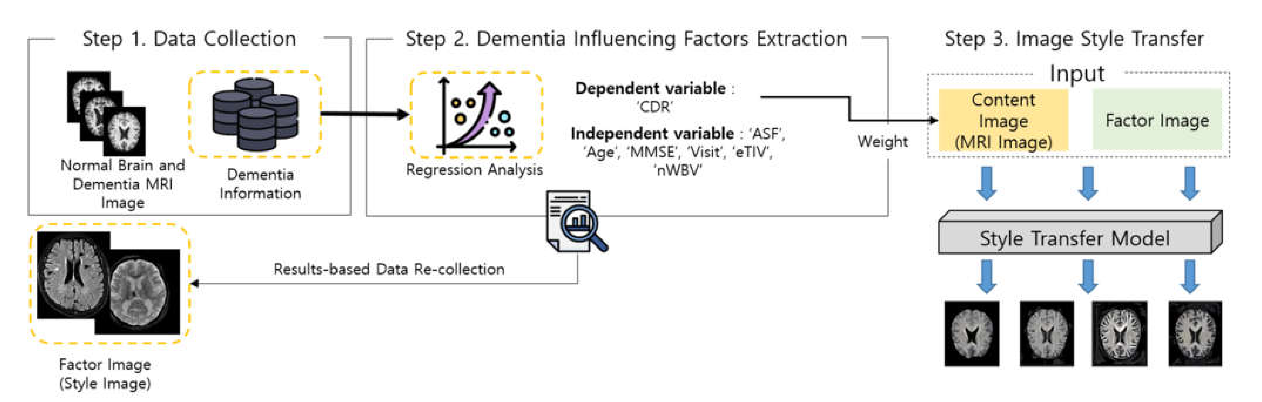
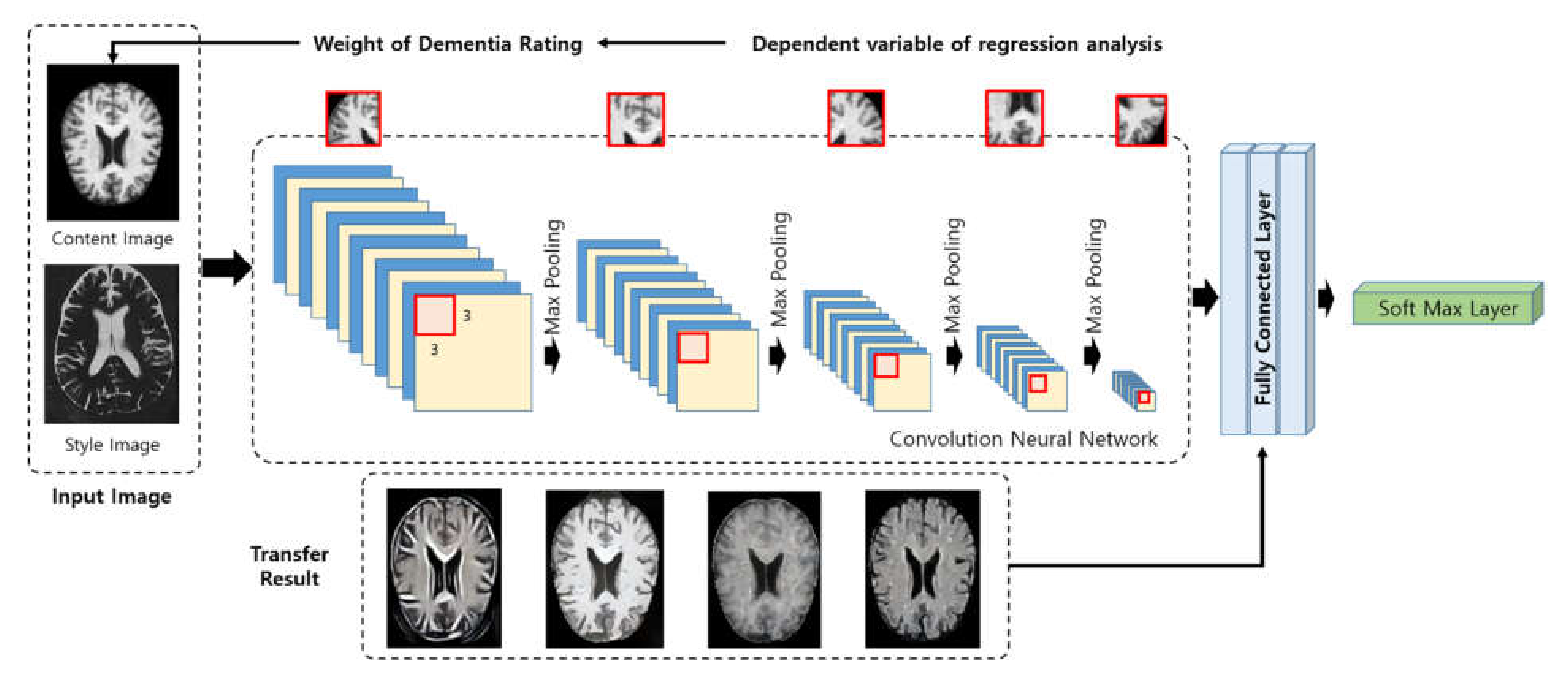
  • This is a method for collecting features and evidence needed for management, prevention, and diagnosis through an analysis of various factors that affect Alzheimer’s disease and monitoring the dementia condition according to changes in the brain. The factors affecting dementia are extracted through regression analysis, and the regression coefficient is used as the degree of impact. In addition, images of the factors affecting it are extracted, and results from the regression analysis are collected.
  • The dimension is reduced by removing unnecessary data through regression analysis. The loss of accuracy occurs according to the degree of dimension reduction, and it is possible to provide a model that can reduce the return time, with higher accuracy than other estimation models.
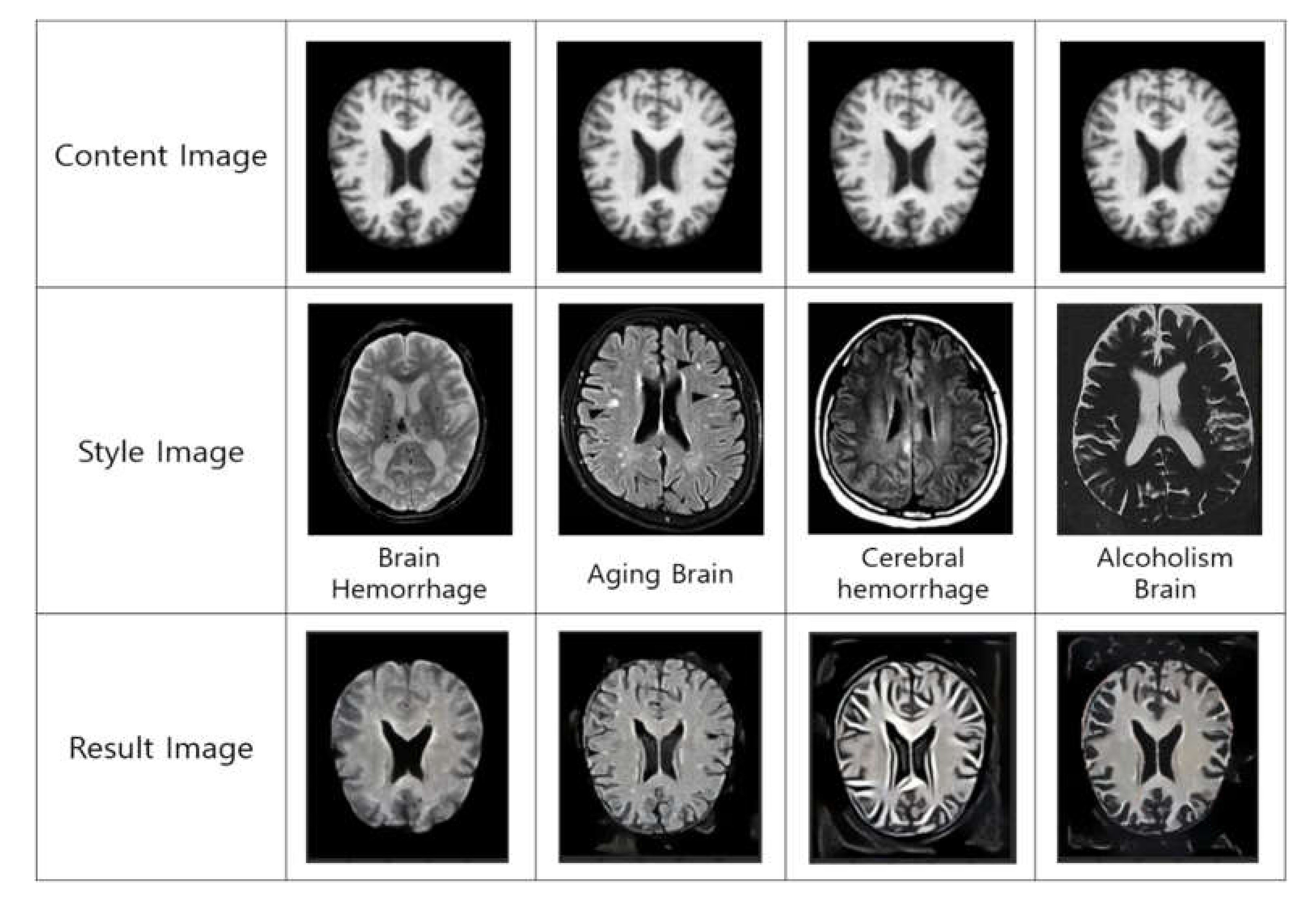
  • Image style transfer is a method for transforming image style by keeping the main form of the image using two-image data in the computer vision and, additionally, applying the desired style. Through this, changes in the brain are predicted by transferring the factors affecting dementia.
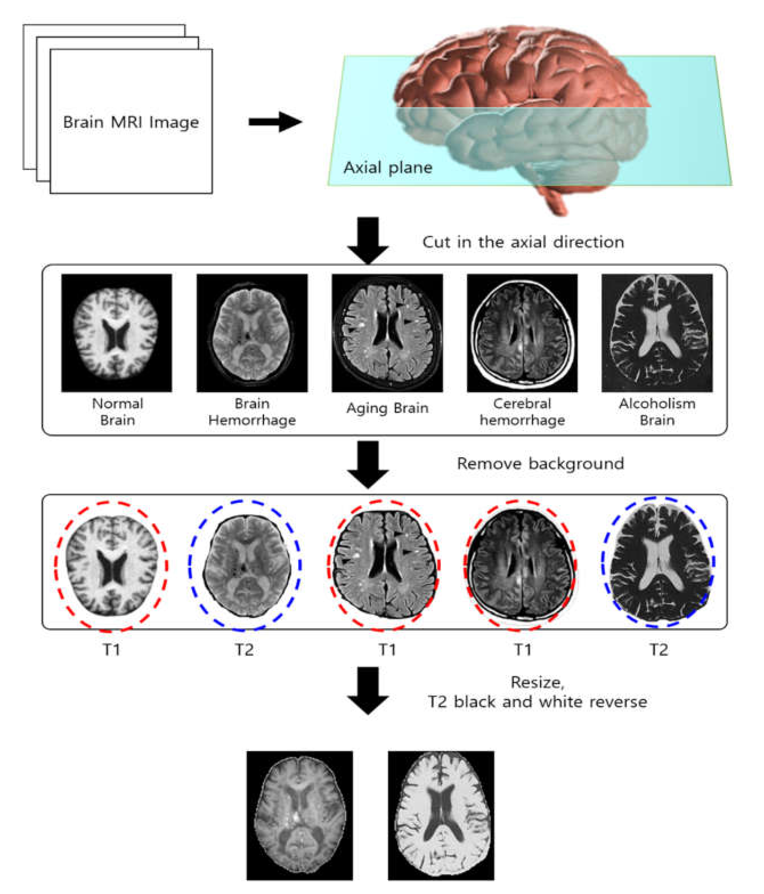
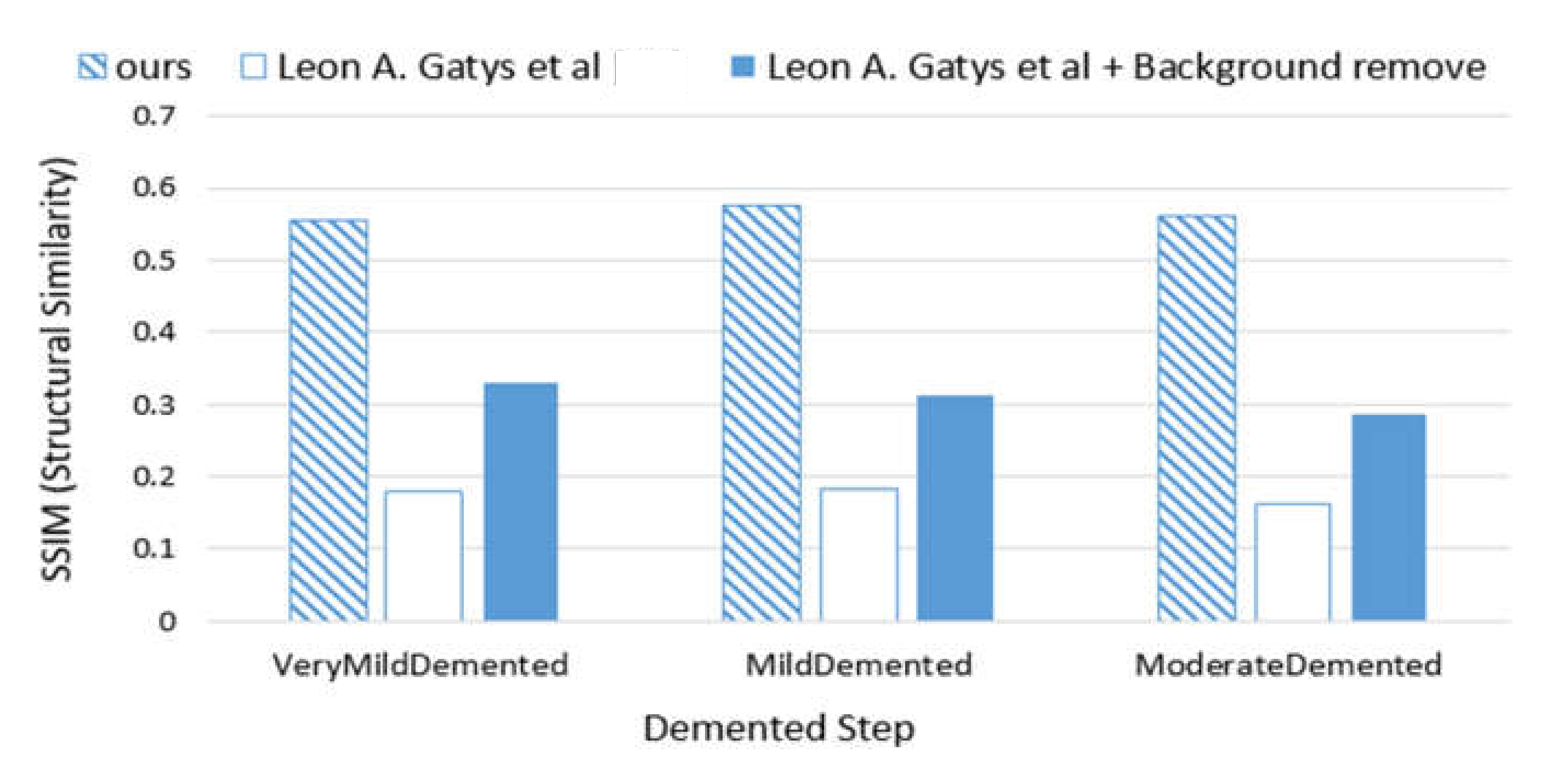
  • Since the background of the image was not removed in the existing method, unnecessary attributes were applied to the image style transmission. In order to solve this problem, the ‘existing method+background removal’ removes unnecessary attributes, but the performance is evaluated poorly because differentiated weights are not applied in each step. However, the proposed method has a different weight and reduces the amount of computation by removing the background in order to prevent unnecessary attribute style transmission, so the performance is evaluated well.
  • The need for health care is alleviated since it awakens patients as well as diagnostic supports of physicians. Thus, it is possible to improve the quality of life.
This study consists of the following: Section 2 describes image style transfer technology and health image predictive analytics technology. Section 3 describes the proposed dementia prediction support model based on regression analysis using image style transfer. Section 4 describes the result of Alzheimer’s disease and performance evaluation using image style transfer, and Section 5 draws conclusions.

2. Related Work

2.1. Image Style Transfer Technology

With the development of artificial intelligence technology, deep learning studies are actively conducted and utilized in various industrial sectors. Of the deep learning algorithms, the CNN-based algorithm is applied to computer vision, used in close relation to real life. In addition, the algorithm changing the image style utilizing CNN is in the limelight. This can be applied to video as well as an image. The style transfer algorithm for the transformation of image style is the one that keeps the information on the object, like the outline of the given image and applies the style of another image [10,11]. In other words, it separates and recombines style and content in a natural image to form a new image. Style transfer is made in two methods. First, the method of using the network that is learned in advance. This is a method of storing the feature map of content image and style image from a network and optimizing the feature map of the images to be newly synthesized so that it has similar features to those from the content image and style image. This has the advantage that style transfer is possible with two images, but it has a disadvantage that it takes much time since the image is optimized each time. The second method is to have the style transfer network learned. This is a method for learning the change of the style from one domain to another when there are images in two different domains. It has the advantage that it only needs feedforward when a network is applied to a new image after having been learned once. In contrast, because it is necessary to learn a new network, multiple images are needed for each domain, and it takes much time to learn it (which is a disadvantage).
H. H. Zhao et al. [12] proposed automatic semantic style transfer using deep convolutional neural networks and soft masks. This is a method for automatically separating objects to keep the content and structure of the image, while delivering the style and extracting soft semantic masks from the style and content images. The proposed method shows the specific part of the style image that falls under the soft mask of the content image with the same meaning in each soft mark of the style image. Both the soft mask and the source image are used as multichannel inputs in the deep convolutional neural network framework augmented for style transfer that integrates the generative Markov random field model. Accordingly, information on various images enhances the quality of style transfer.

2.2. Health Image Prediction Analysis Technology Using Style Transfer

In modern society, concern for health increases with the growing aging population. Accordingly, smart health care is studied continuously throughout the world. Health care is a comprehensive personalized medical service in which health-related services and medical IT are combined into individual health and information related to medical care, devices, systems, and platforms [13,14]. It is possible to analyze lifestyle, bio information, and medical information, etc. through deep learning, machine learning, and statistical analysis, etc. Health data exist in various forms, such as 2D and 3D images, sensors, and texts, etc. Of these, the information on the diagnosis and treatment of lung disease and heart disease, etc., through 3D medical image analysis, is relevant. C. Ma et al. [15] proposed a method for improving the 3D cardiovascular MR image separation of inconsistent data through nerve style transmission. The proposed method applies a style transfer algorithm to unlabeled data to minimize the difference in the attributes of the images of the unlabeled data. In addition, stochastic adjustment is applied to network output, and multiple predictions are integrated through ensemble learning. It is possible to resolve the problem of data inconsistency for the generalization of a deep learning model for image data analysis. Figure 1 shows the process of health image prediction analysis technology using style transfer.
J. Islam et al. [16] proposed an MRI analysis method for diagnosing Alzheimer’s disease using an ensemble system of deep convolutional neural networks. This is a fast approach for training deep learning models using unbalanced datasets. The proposed method directly identifies various stages through a deep convolutional neural network model that learns features of MRI data, and multiple classifications are possible for early-stage diagnosis.
N. M. Khan et al. [17] proposed transfer learning through intelligent training data selection for Alzheimer’s disease prediction. It is fine-tuned according to the layers being trained on MRI images using predefined layer groups. Select the most efficient slice for analysis by using image entropy to reduce the size of the training data. The proposed method helps medical staff make decisions by focusing on differential image areas through transfer learning that is initialized with pre-trained weights through the VGG structure. However, users who receive information through this are limited to medical staff. Since health management requires patients to be able to prevent it in advance, it is necessary to provide a method that enables not only medical staff, but also patients, to receive information for disease decision-making.

3. Dementia Prediction Support Model Using Regression Analysis and Image Style Transfer

Recently, studies have actively been conducted on a model that can play a role in assisting with disease diagnosis for doctors in the development of deep learning and health care. Accordingly, since it is possible to make an accurate diagnosis for patients, the deep learning-based model is in the limelight in the medical industry. However, the causes of Alzheimer’s disease have not been accurately identified. Thus, patients are diagnosed with a disease by the doctor’s judgment through MRI results, and there may be differences in opinions according to the doctors. Since dementia can be prevented through personal health care, it is necessary to make people aware of dementia and feel the importance of health care. This study proposes a prediction support model for dementia based on regression analysis using image style transfer. The proposed method is a method for transferring data on the factors affecting Alzheimer’s disease; well-known causes for it, such as brain damage; and aging of blood vessels to the brain image data as well as for providing results from predicting or preventing dementia through changes in the brain. This is conducted in three steps. First, MRI data and medical information data from patients with dementia are collected and pre-processed. The second step extracts the features of the factors affecting dementia through medical information data. This uncovers the factors affecting dementia and the degree of impact through regression analysis. In the final step, diseases are predicted and judged by monitoring changes and by transferring the images related to factors affecting it to the MRI brain image. Figure 2 shows the process of a prediction support model for dementia based on regression analysis using image style transfer.

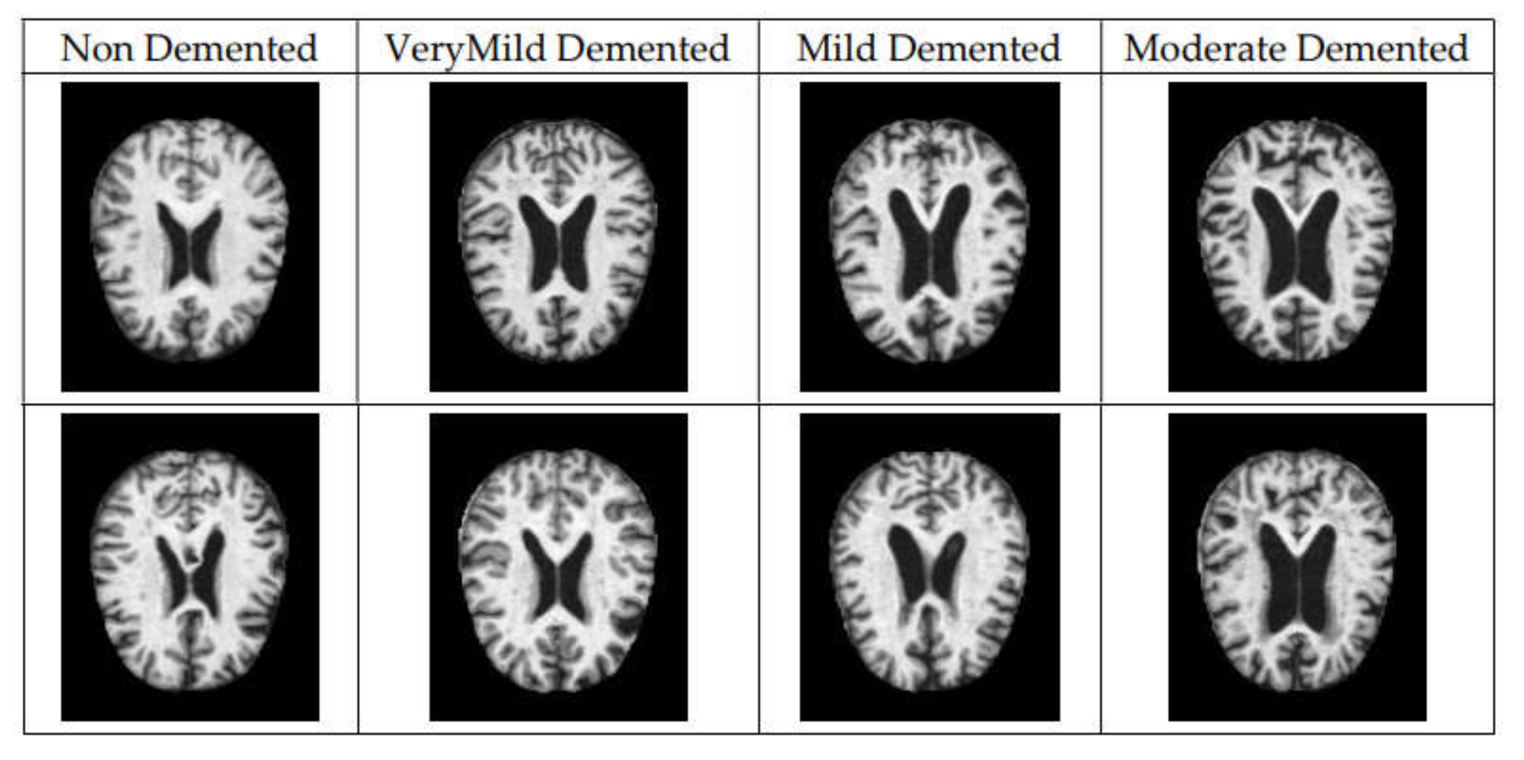
3.1. Data Collection

Dementia is not a specific disease; it is a category that accounts for the symptoms associated with severe loss of memory or thinking ability that reduces the ability to perform daily activities. Specifically, Alzheimer’s disease takes up 60–80% of dementia cases. In addition, vascular dementia that occurs after stroke is the second most common type of dementia and may cause dementia symptoms, including other factors such as thyroid problems and vitamin deficiencies. With the data used in this study, cases of Alzheimer’s disease are investigated. Alzheimer’s disease causes problems with memory, thinking, and behavior and is a disease that may disturb daily life as its symptoms are slowly developed and aggravated with the flow of time. Proven to date, the factors that affect cognitive function include age, sex, education, income, depression, obesity, and poverty. However, since there is no specific cause for Alzheimer’s disease, it is difficult to discover the factors affecting it [18]. Thus, from the Alzheimer’s disease-related data, an epidemiological analysis is conducted to discover the factors affecting it as they relate to the variables affecting it, based on the variability of the variable changing with the symptoms. The data used in this study are the Alzheimer patients’ brain MRI image data and patient information data provided by the Open Access Series of Imaging Studies (OASIS) [19]. OASIS provides data from OASIS-1, OASIS-2, and OASIS-3, of which OASIS-3 is used. This is a dataset of vertical neuroimages as well as clinical andcognitive biomarkers for normal aging and Alzheimer’s disease. In addition, data from 609 adults with normal cognitive abilities and 489 people in various cognitive decline stages are included, and identifiers are assigned to protect personal information. Table 1 shows the dataset of the information on Alzheimer patients. A total of 15 variables are included, such as Subject ID, MRI ID, Group, Visit, MR Delay, M/F (Gender (M if Male, F if Female)), Hand (Handedness), Age (Age in years), EDUC (Years of education), SES (Socioeconomic Status), MMSE (Mini Mental State Examination), CDR (Clinical Dementia Rating), eTIV (Estimated Total Intracranial Volume), nWBV (normalized Whole Brain Volume), and ASF (Atlas Scaling Factor).
Figure 3 shows MRI data according to the dementia condition. The data are divided into Non Demented, Very Mild Demented, Mild Demented, Moderate Demented, according to the dementia condition.
Figure 3 notes that the volume of the vacuum increases in the brain with an increase in the stage.

3.2. Dementia Influencing Factors Extraction Using Regression Analysis

Dimension reduction and feature selection may enhance the prediction performance. To do so, conditions affecting dementia are extracted through regression analysis. Regression analysis extracts the independent variables that can best explain the dependent variables by modeling the relationships among the variables. There are various types of regression analyses, including basic linear regression analysis, multiple linear regression analysis, logistic regression analysis, LASSO regression analysis, etc. Regression analysis has the limitation that predictability decreases while variability increases, if there are too many variables or the correlation between variables is high. To resolve this problem, the number of variables is reduced, or the predicting model is normalized [20]. If logistic regression analysis is used, there is a disadvantage that reduces the accuracy of the classification of unnecessary features. In addition, if the dimension of test data is larger than the number of samples of training data, the problem of overfitting may occur. Data on Alzheimer’s disease have a small number of samples and various features. To enhance the accuracy of the prediction of Alzheimer’s disease, it is necessary to choose features with the best explanation power.
The LASSO (Least Absolute Shrinkage Selector Operator) regression analysis is a linear regression method with an L1-norm penalty. This applies the same level of normalization regardless of the size of the parameters. Normalization can perform characteristic selection and model learning simultaneously. Accordingly, it has advantages that it is possible to create a model simply by creating parameters with a small value being created as 0 or a value approximate to 0 by removing the relevant variables from the model, and it is useful for interpretation. The L1-norm refers to the sum of the absolute values of vector elements. It is possible to understand changes in factor value. In addition, it is possible to resolve the complexity of the model [21]. However, logistic and LASSO can be used if binary classification is possible. Therefore, the data used in this study consist of more than two independent variables. Therefore, it uses multiple regression analysis as a regression analysis method. This removes the variables, such as ‘Subject ID’, ‘MRI ID’, ‘MR Delay’, ‘SES’, ‘Group’, and ‘EDUC’ which are unnecessary for analysis, and uses ‘CDR’, which indicates the clinical dementia rating as a dependent variable, and ‘ASF’, ‘Age’, ‘MMSE’, ‘Visit’, ‘eTIV’, and ‘nWBV’ as independent variables. Table 2 shows the result of the multiple regression analysis to extract the factors that affect dementia. “Coef.” refers to regression coefficient; “Std. error” refers to standard error; and “t” refers to t-test, and they are shown through the regression coefficient/standard error. This means that the smaller the standard error, the bigger the t value becomes, which means that there are high correlations between independent variables and dependent variables. Therefore, the bigger the t value, the more significant the results become. The equation p > |t| shows the p-value.
Table 2 shows that MMSE has the highest correlation. MMSE is the method that is the most widely used dementia screening test, which simply evaluates cognitive function and dementia in a short time. Thus, it is inferred that the factors affecting cognitive function may change the dementia condition. This conducts a test based on 30 points, and the questions consist of time and place orientation, registration, attention and calculation, memory recall, language, and visuospatial function. MMSE judges higher than 24 points as the normal range; 20–23 points as suspected dementia and cognitive dysfunction; and less than 19 points as a high likelihood of having dementia and cognitive dysfunction. Accordingly, this study recollected data concerning the factors affecting cognitive function. This is based on the information in the National Center for Mental Health [22] and Dementia Center [23]. The recollected data are from an alcoholism brain, an aging brain, a brain with cerebral hemorrhage, and hemorrhage brain. Thus, the brain image data are collected from patients who have symptoms related to these. Figure 4 shows the recollected image data.
In Figure 4, in the traumatic brain injury, the blood spreads and pools, and the aging brain is contracted (wrinkled) in the upper parts on the left and right. In addition, if there is cerebral hemorrhage, the brain expands, and the head is full. In the alcoholic brain, the brain is shrunken.

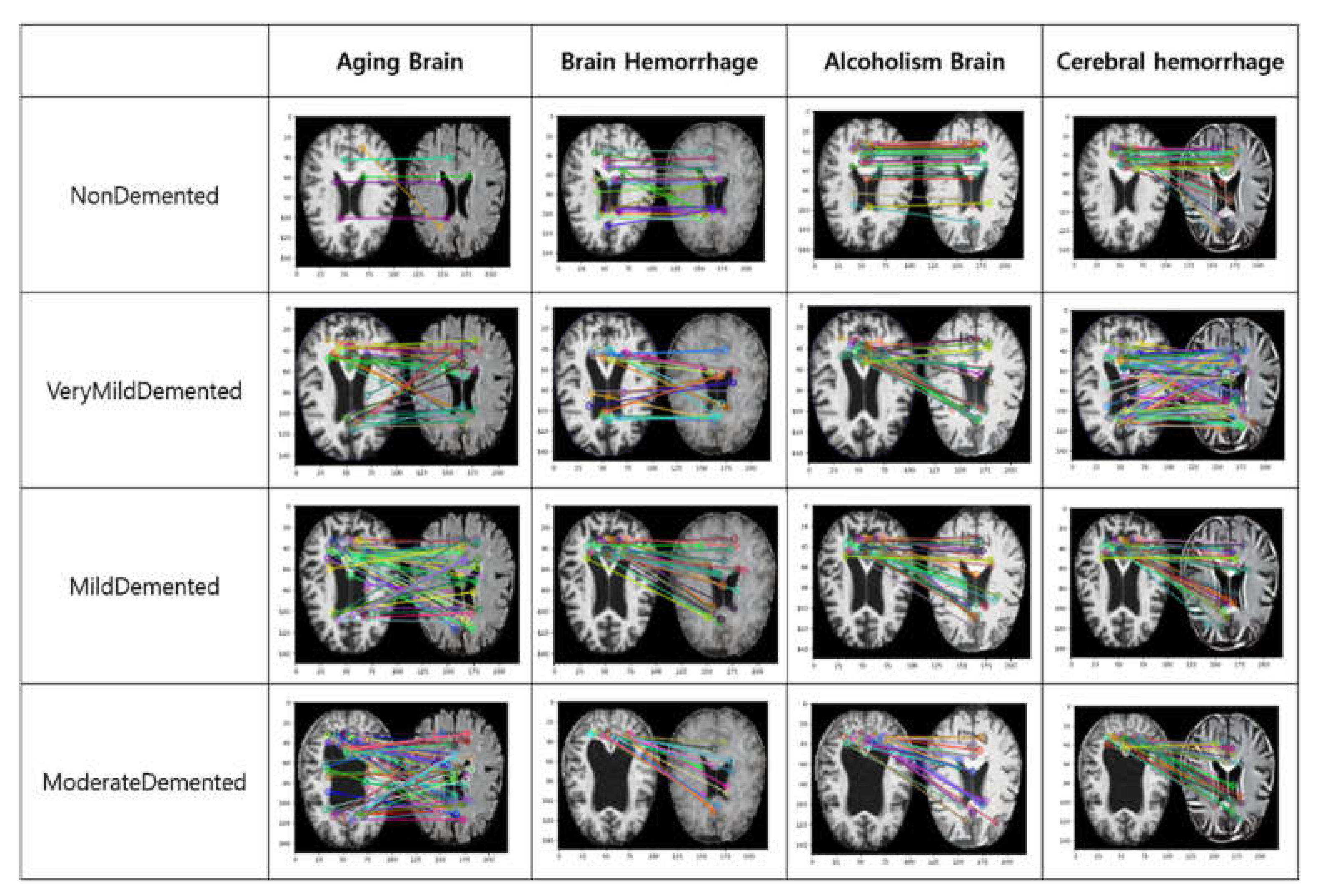
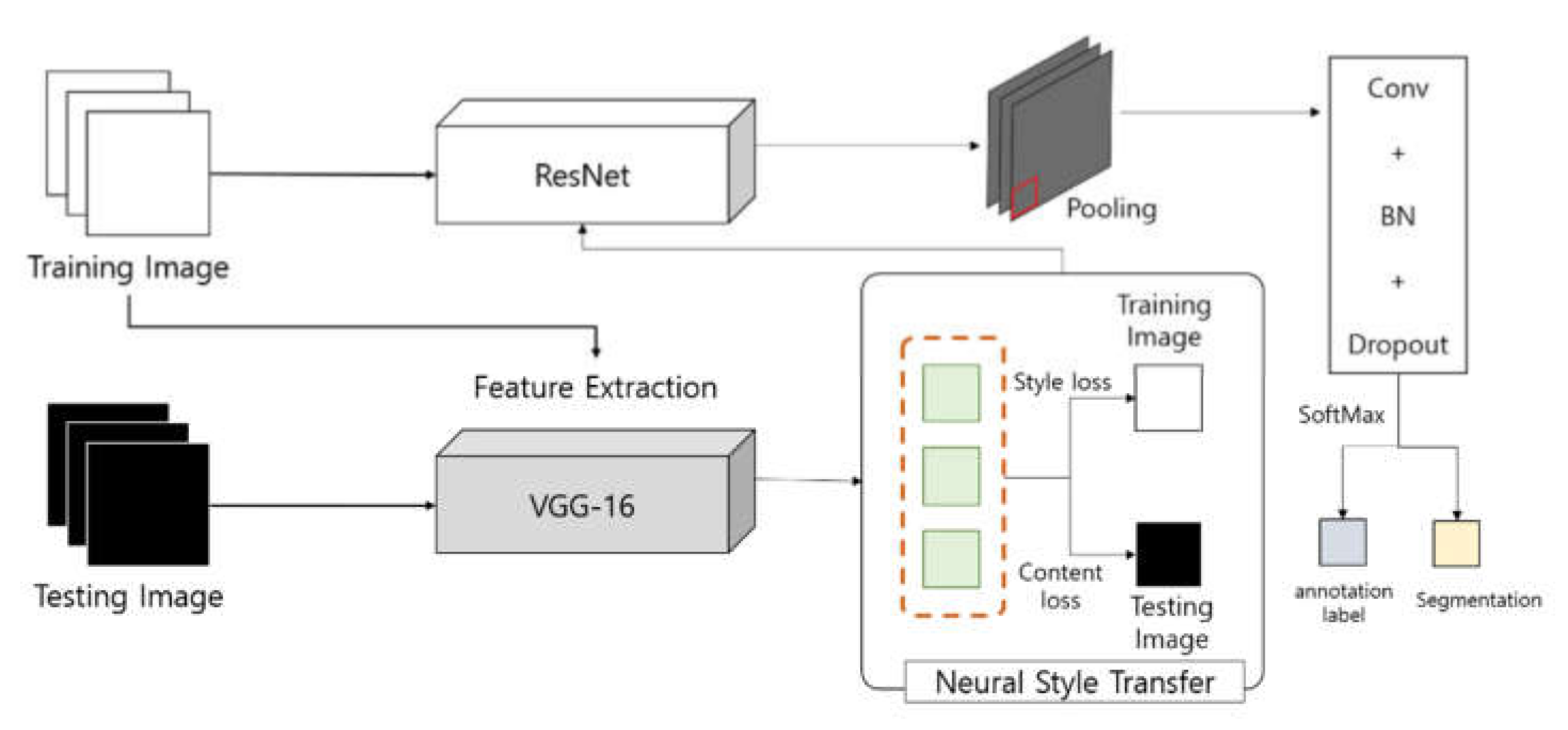
3.3. Dementia Prediction Support Model Using Image Style Transfer

NST (Neural Style Transfer) is the process for rendering images of various style contents, using CNN. It includes Image-Optimization-Based Online Neural Methods (IOB-NST) that transfer styles by repeatedly optimizing the image and Model-Optimization-Based Offline Neural Methods (MOB-NST), which optimizes the generated model offline and generates the image styled with a single forward pass. However, firstly, it costs a lot to calculate the procedure of repeatedly optimizing the image, and there is an efficiency issue. In addition, generally, NST captures only the characteristic correlation per pixel and does not limit the space layout. Thus, there is a disadvantage in that the result is visually unstable (less plausible) for a realistic style. To resolve this, C. Li et al. [24] modeled the style with a non-parametric method and applied a new style loss function, which included patch-based MRF. In contrast, secondly, the method of optimizing the model offline can resolve problems with speed and calculation cost by reconstructing the styled result. The dementia diagnosis support model proposed in this study is in an optimization-based style transfer method. However, to resolve the problem of the unstable results from the optimization-based style transfer method, the clinical dementia rating is taken as the weighted value of the content image.
The model transforms the normal brain into a brain with a disease by transferring information about the attributes of the image. For example, the attributes of an alcohol-addicted brain, a brain with cerebral hemorrhage, and a traumatic brain injury, recollected in the images of the normal brain, are transferred in order to transform the brain into the brain of Alzheimer’s disease. Accordingly, it is possible to monitor the factors affecting dementia and the transformation process, as well as to predict the disease.
Different positions of the object and different numbers of layers in the convolution operation represent very different visual results. Thus, the selection of contents and style layers are important factors in the style transfer process. Image attributes include image color and texture, etc. The proposed dementia prediction support model is designed based on the VGG network. In VGG, the size of an input image is fixed to 110 × 150 and pre-processing removes the average value of RGB. The convolution layer uses a 3 × 3 filter to consider the pixel values at the top, bottom, left, and right. The filter’s stride is one, and padding is applied for operation. In the pooling layer, maximum pooling is applied next to the convolution layer, which consists of a total of five. Figure 5 shows the diagram of the proposed dementia prediction support model.
Style transfer uses the features of the VGG19 network consisting of convolution, pooling layers, and an FC layer. The model in Figure 5 is divided into the extractor and the classifier of features. The input image is divided into a content image and a style image. From each convolution layer and each pooling layer, the features of the content image and style image are extracted, respectively. In addition, the FC layer and SoftMax layer features are calculated, classified, and predicted to transfer the style. Figure 6 shows the result of the image style transfer. The content image is the normal human brain image. The style image is the recollected brain image. The resulting image is the result extracted by transferring the attributes of the style image to the content image.
As a result of the transfer of image style in Figure 6, the style of dementia factors in normal brain conditions is applied; therefore, there are changes in brain volume. Thus, dementia factors have serious impacts on dementia as well as cognitive disorder; it is necessary to be careful when managing and preventing it.

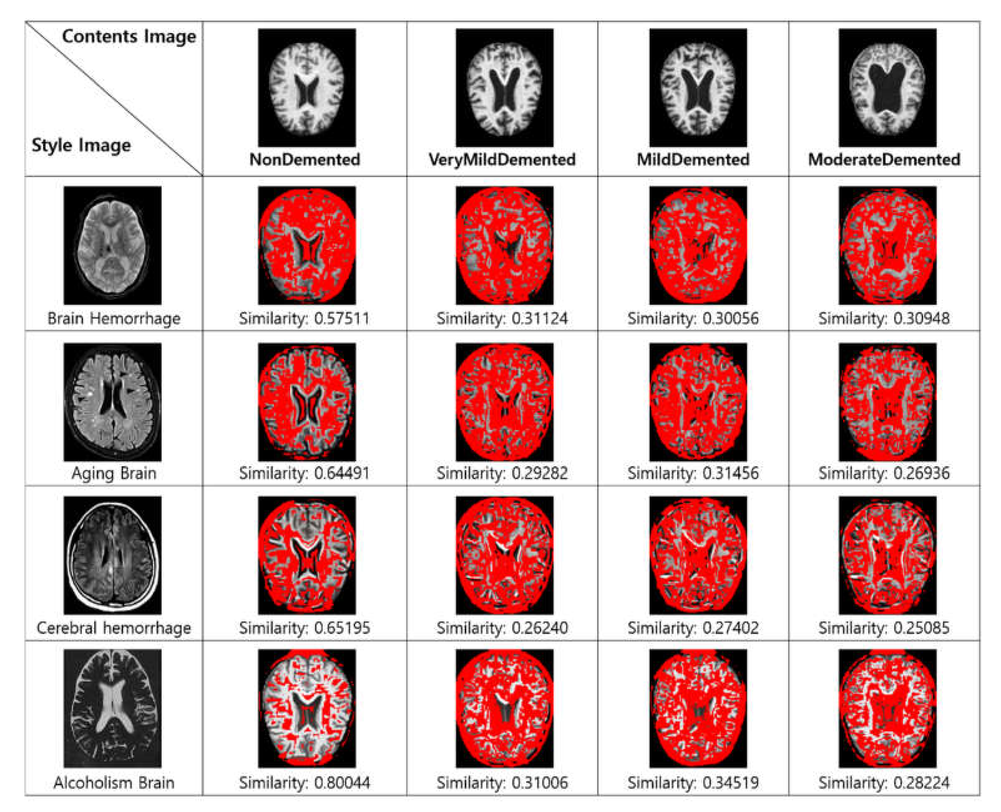
4. Results and Performance Evaluation

The data used in this study are the brain images and patient information data according to the dementia stages provided by OASIS. In the patient information data, preprocessing is made; e.g., the removal of unnecessary variables, etc., and, with the clinical dementia rating as the dependent variable and other variables as independent variables, regression analysis is conducted. The brain condition related to MMSE is recollected and extracted accordingly, and, unless the size of the collected image and that of the recollected image are the same, there may be an error in the CNN operation. Thus, all images are adjusted to the same size to transfer the style. In addition, the weighted value of the content image in the model is that of the clinical dementia stage, and 0 means non-demented; 0.5, very mild; 1, mild; 2, moderate. To prevent the transfer of information about unnecessary attributes, the background of all images is removed before proceeding. The development environment of the research is hardware AMD Ryzen 9 5900X 12-Core Processor 3.70 GHz, 64GB RAM, GeForce RTX 3090(NVIDIA). In addition, as a software environment, Python 3.7 and Anaconda 4.5.11 are employed. The performance evaluation makes a comparison with similarity to the image transferred in style according to the dementia rating. Figure 7 shows the data processing process for performance evaluation.
The data processing process for performance evaluation in Figure 7 generates MRI data that cut the brain on the axial plane. Accordingly, unnecessary backgrounds are removed, leaving a part of the brain where each character can be identified. Finally, the remaining image data unify T1 and T2. In the case of T1, the water component appears in black, and the fat component appears in white. Alternatively, in the case of T2, contrary to T1, the water component appears white, and the fat component appears black. Therefore, in order to unify this, in the case of T2, black and white are reversed so that the same characteristics as in T1 are displayed. Further, the size of the image is created to be the same as 110 × 150. Accordingly, images are processed under the same conditions, the results are extracted through the proposed model, and a performance evaluation is performed.
The evaluation method for style transfer includes qualitative evaluation and quantitative evaluation. The qualitative evaluation depends on the subjective aesthetic judgment of the observer. Conversely, a quantitative evaluation can conduct an objective evaluation according to the evaluation index, including time complexity or data loss change, etc. It is complex to define the concept of style, and, since the definition of the standard is subjective, it is difficult to evaluate the result [25]. Thus, in this study, the evaluation of the proposed model is conducted using a quantitative evaluation method. First, the similarity is compared between the styled image and the actual image of the brain of the patients with dementia. As an evaluation index for a comparison of image similarity, Structural Similarity (SSIM) is used. SSIM is a method for evaluating image quality, which evaluates the original image. This has a value between zero and one, and the closer to one, the higher the similarity to the natural image. It is possible to evaluate image similarities by considering Luminance, Contrast, and Structure [26,27]. Equation (1) shows the SSIM. X and Y respectively mean the image; L is Luminance, C is Contrast, and S is Structure.
S S I M X , Y = I X , Y × C X , Y × S X , Y
Through Equation (1), the results of the transfer of dementia stages and the styles of the factors affecting dementia are compared and extracted. In addition, the parts different from the dementia brain are marked to compare the SSIM value.
The first performance evaluation makes a comparison of similarity between the image in which a style transfer was made with the proposed method and the brain MRI image in which there is a dementia symptom according to the dementia stage. Figure 8 shows the comparison result of the similarity between the style transmitted image and the dementia stage. This compares the similarity with the contents image, and the difference is expressed in red.
As a result of Figure 8, the result most similar to the brain of a normal person is extracted. This is because we applied the style transfer to a normal brain. In addition, the comparison between very weak dementia, mild dementia, and intermediate stage dementia identifies the influence factors of dementia through the result of the normal brain receiving the disease style and the result similar to dementia. Thus, the results of the transmission of alcohol brain properties were found to be the most similar to those with dementia on average. Therefore, it means that drinking can have the greatest effect on dementia. However, in the case of similarity comparison, there is a problem in that performance is evaluated poorly not only because brain characteristics are considered, but also because unnecessary parts are also compared.
A second assessment compares the style-transferred image with similar features to the dementia stage. It uses a feature-matching algorithm. The feature-matching algorithm extracts various features from the image and has the advantage of being able to extract features even if the image is rotated, enlarged, or reduced. It is judged to be a similar image as more extracted features appear [28]. It is possible to determine which part of the style transmitted image is similar to the dementia image. Figure 9 shows the result of comparing the characteristics of the style transmitted image with the dementia stage.
As a result of comparing the features in Figure 9, the most common features of the aged brain were extracted in all dementia stages. In addition, in the case of cerebral hemorrhage, very weak dementia and mild dementia showed similar characteristics, and, in the case of alcohol brain and traumatic brain, many similar characteristics were extracted, not only in all dementia stages, but also in the brain without dementia.
The third performance evaluation compares the performance of the existing neural style transfer model of Leon A. Gatys et al. [29] and the proposed method. This compares the similarity of the result of style transfer of the brain image in the dementia stage and compares the average similarity of each dementia stage. The proposed method took the weighted value of the content image in the model of Leon A. Gatys et al. as that in the stage of clinical diagnosis of dementia, and the image background is removed to prevent the transfer of unnecessary attributes. Figure 10 shows a graph of the average similarity of the existing method and the proposed method, according to the stage of dementia. The horizontal axis represents the dementia stage, while the vertical axis represents the SSIM value.
In the results from Figure 10, the performance of the proposed method was most excellent. Since the existing method did not remove the background, unnecessary attributes were also transferred [30]. In addition, ‘the existing method+background removal’ removed unnecessary attributes, but no differentiated weighted value was given according to each step like in ‘the existing method.’ Meanwhile, the proposed method gave different weighted values according to each clinical step, but the backgrounds of the images were removed to reduce computation by preventing the transfer of unnecessary attributes; its performance was more excellent than that of the compared model. Thus, it is possible to judge changes in the dementia stages according to the factors affecting dementia through the proposed method.

5. Conclusions

This study proposed a prediction support model for dementia based on regression analysis using image style transfer. The proposed method was composed of three steps. First, MRI data and medical information data from patients with dementia were collected and pre-processed. In the second step, through the medical information data, the features of the factors affecting dementia were extracted. Through a regression analysis, the factors affecting dementia and their impacts on it were observed. The factors affecting dementia extracted as a result of the regression analysis are alcohol addicted brain, aging brain, cerebral hemorrhage, and traumatic brain injury. Accordingly, the images of the factors affecting it were recollected. In the final step, the backgrounds of all images used in the analysis were removed to resolve the problem of transferring unnecessary features. Accordingly, the images related to the factors affecting it were transferred to the images of the MRI normal brain. Thus, the normal brain was transformed into a patient’s brain through the style of the factors affecting it. For performance evaluation, the similarities were compared between the brain images of very mild demented, mild demented, and middle dementia stages and the style transferred brain image. In order to evaluate this performance, a comparison of similarities and features between the style transmitted image and the dementia stage, and a performance comparison with the existing method were conducted. As a result of the similarity comparison, when the properties of the alcohol brain were transmitted, on average, the results were most similar to those of dementia. In addition, in the case of aging brains and alcohol brains, features similar to those of dementia were extracted most. In addition, the proposed method was excellent in its similarity assessment rate to the existing methods. Thus, the proposed method provides the results necessary to predict or prevent dementia through changes in the brain. In addition, changes can be observed by transferring the styles of each influencing factor to images, such as joints and cancers, in the form of image data.
However, the proposed method proceeded with a limited assumption, due to a lack of public data. Since the brain has been transformed through the most fundamental factors, there are limitations on the information that has been studied in a small number of cases and provided to patients or medical staff. Further, since the proposed model is in a black box format, there would be a limitation that Alzheimer’s disease risk information may be predicted through the result of style transfer. In addition, it takes much time and computational cost to empirically choose parameters through repeated experiments. In the future, in order to improve the amount of computation, accuracy, and performance used in MRI image analysis, the MRI data will be restructured and pre-processed through Realign, Segment, Normalize, and Smooth. In addition, in order to provide reliable information on Alzheimer’s disease, the relationship between the influencing factors and the basis for selecting the parameters of the model will be additionally studied to explore a white box-based auxiliary model.

Author Contributions

Conceptualization, J.-W.B.; methodology, J.-W.B. and K.C.; validation, J.-W.B. and K.C.; writing—original draft preparation, J.-W.B. and K.C.; writing—review and editing, J.-W.B. and K.C.; visualization, J.-W.B. and K.C.; supervision, K.C.; project administration, K.C.; funding acquisition, K.C. All authors have read and agreed to the published version of the manuscript.

Funding

This work was supported by Kyonggi University Research Grant 2021.

Institutional Review Board Statement

Not applicable.

Informed Consent Statement

Not applicable.

Data Availability Statement

Data sharing not applicable.

Conflicts of Interest

The authors declare no conflict of interest.

References

  1. Kivipelto, M.; Mangialasche, F.; Ngandu, T. Lifestyle interventions to prevent cognitive impairment, dementia and Alzheimer disease. Nat. Rev. Neurol. 2018, 14, 653–666. [Google Scholar] [CrossRef] [PubMed]
  2. Günak, M.M.; Barnes, D.E.; Yaffe, K.; Li, Y.; Byers, A.L. Risk of suicide attempt in patients with recent diagnosis of mild cognitive impairment or dementia. JAMA Psychiatry 2021, 78, 659–666. [Google Scholar] [CrossRef] [PubMed]
  3. Guo, T.; Zhang, D.; Zeng, Y.; Huang, T.Y.; Xu, H.; Zhao, Y. Molecular and cellular mechanisms underlying the pathogenesis of Alzheimer’s disease. Mol. Neurodegener. 2020, 15, 1–37. [Google Scholar] [CrossRef] [PubMed]
  4. Sims, R.; Hill, M.; Williams, J. The multiplex model of the genetics of Alzheimer’s disease. Nat. Neurosci. 2020, 23, 311–322. [Google Scholar] [CrossRef] [PubMed]
  5. Liu, Q.; Dou, Q.; Yu, L.; Heng, P.A. MS-Net: Multi-site network for improving prostate segmentation with heterogeneous MRI data. IEEE Trans. Med. Imaging 2020, 39, 2713–2724. [Google Scholar] [CrossRef] [PubMed] [Green Version]
  6. Vostrý, M.; Zilcher, L. Combination therapy for patients after ischemic stroke from the point of view of complex rehabilitation. J. Educ. Cult. Soc. 2020, 11, 119–125. [Google Scholar] [CrossRef]
  7. Vostrý, M.; Fischer, S.; Lanková, B. The Effect of Combined Therapy on the Support and Development of Social Skills of people with Multiple Sclerosis in Senior Age. Neuroendocrinol. Lett. 2020, 41, 101–105. [Google Scholar]
  8. Yoo, J.; Uh, Y.; Chun, S.; Kang, B.; Ha, J.W. Photorealistic style transfer via wavelet transforms. In Proceedings of the IEEE/CVF International Conference on Computer Vision, Seoul, Korea, 27 October–2 November 2019; pp. 9036–9045. [Google Scholar]
  9. Shen, F.; Yan, S.; Zeng, G. Neural style transfer via meta networks. In Proceedings of the IEEE Conference on Computer Vision and Pattern Recognition, Salt Lake City, UT, USA, 18–23 June 2018; pp. 8061–8069. [Google Scholar]
  10. Luan, F.; Paris, S.; Shechtman, E.; Bala, K. Deep Photo Style Transfer. In Proceedings of the IEEE Conference on Computer Vision and Pattern Recognition, Honolulu, HI, USA, 21–26 July 2017. [Google Scholar]
  11. Jing, Y.; Yang, Y.; Feng, Z.; Ye, J.; Yu, Y.; Song, M. Neural style transfer: A review. IEEE Trans. Vis. Comput. Graph. 2019, 26, 3365–3385. [Google Scholar] [CrossRef] [Green Version]
  12. Zhao, H.H.; Rosin, P.L.; Lai, Y.K.; Wang, Y.N. Automatic semantic style transfer using deep convolutional neural net-works and soft masks. Vis. Comput. 2020, 36, 1307–1324. [Google Scholar] [CrossRef] [Green Version]
  13. Baek, J.W.; Chung, K. Multi-level health knowledge mining process in P2P edge network. IEEE Access 2021, 9, 61623–61634. [Google Scholar] [CrossRef]
  14. Yoo, H.; Park, R.C.; Chung, K. IoT-Based Health Big-Data Process Technologies: A Survey. KSII Trans. Internet Inf. Syst. 2021, 15, 974–992. [Google Scholar]
  15. Ma, C.; Ji, Z.; Gao, M. Neural Style Transfer Improves 3D Cardiovascular MR Image Segmentation on Inconsistent Data. In Proceedings of the International Conference on Medical Image Computing and Computer-Assisted Intervention, Shenzhen, China, 13–17 October 2019. [Google Scholar]
  16. Islam, J.; Zhang, Y. Brain MRI analysis for Alzheimer’s disease diagnosis using an ensemble system of deep convolutional neural networks. Brain Inform. 2018, 5, 1–14. [Google Scholar] [CrossRef] [PubMed]
  17. Khan, N.M.; Abraham, N.; Hon, M. Transfer learning with intelligent training data selection for prediction of Alzheimer’s disease. IEEE Access 2019, 7, 72726–72735. [Google Scholar] [CrossRef]
  18. Alzheimer’s Association. Alzheimer’s disease facts and figures. Alzheimer’s Dement. 2018, 14, 367–429. [Google Scholar]
  19. OASIS (Open Access Series of Imaging Studies). Available online: https://www.oasis-brains.org/ (accessed on 12 December 2021).
  20. Baek, J.W.; Chung, K. Context deep neural network model for predicting depression risk using multiple regression. IEEE Access 2020, 8, 18171–18181. [Google Scholar] [CrossRef]
  21. Yang, X.; Wen, W. Ridge and lasso regression models for cross-version defect prediction. IEEE Trans. Reliab. 2018, 67, 885–896. [Google Scholar] [CrossRef]
  22. National Center for Mental Health. Available online: http://www.mentalhealth.go.kr/ (accessed on 10 October 2021).
  23. Dementia Center. Available online: https://www.nid.or.kr/ (accessed on 10 October 2021).
  24. Li, C.; Wand, M. Combining markov random fields and convolutional neural networks for image synthesis. In Proceedings of the IEEE Conference on Computer Vision and Pattern Recognition, Las Vegas, NV, USA, 27–30 June 2016; pp. 2479–2486. [Google Scholar]
  25. Wang, W.; Yang, S.; Xu, J.; Liu, J. Consistent video style transfer via relaxation and regularization. IEEE Trans. Image Processing 2020, 29, 9125–9139. [Google Scholar] [CrossRef]
  26. Sara, U.; Akter, M.; Uddin, M.S. Image quality assessment through FSIM, SSIM, MSE and PSNR—A comparative study. J. Comput. Commun. 2019, 7, 8–18. [Google Scholar] [CrossRef] [Green Version]
  27. Peng, J.; Shi, C.; Laugeman, E.; Hu, W.; Zhang, Z.; Mutic, S.; Cai, B. Implementation of the structural SIMilarity (SSIM) index as a quantitative evaluation tool for dose distribution error detection. Med. Phys. 2020, 47, 1907–1919. [Google Scholar] [CrossRef] [PubMed]
  28. Ma, J.; Jiang, X.; Fan, A.; Jiang, J.; Yan, J. Image matching from handcrafted to deep features: A survey. Int. J. Comput. Vis. 2021, 129, 23–79. [Google Scholar] [CrossRef]
  29. Gatys, L.A.; Ecker, A.S.; Bethge, M. Image Style Transfer Using Convolutional Neural Networks. In Proceedings of the IEEE Conference on Computer Vision and Pattern Recognition (CVPR), Las Vegas, NV, USA, 27–30 June 2016; pp. 2414–2423. [Google Scholar]
  30. Yoo, H.; Chung, K. Deep learning-based evolutionary recommendation model for heterogeneous big data integration. KSII Trans. Internet Inf. Syst. 2020, 14, 3730–3744. [Google Scholar]
Figure 1. Process of health image prediction analysis technology using style transfer.
Applsci 12 03536 g001
Figure 2. Process of dementia prediction support model using regression analysis and image style transfer.
Applsci 12 03536 g002
Figure 3. MRI data according to the dementia condition.
Applsci 12 03536 g003
Figure 4. Recollected image data.
Applsci 12 03536 g004
Figure 5. Diagram of the proposed dementia prediction support model.
Applsci 12 03536 g005
Figure 6. Result of image style transfer.
Applsci 12 03536 g006
Figure 7. Data processing process for performance evaluation.
Applsci 12 03536 g007
Figure 8. Comparison result of the similarity between the style transmitted image and the dementia stage.
Applsci 12 03536 g008
Figure 9. The result of comparing the characteristics of the style transmitted image with the dementia stage.
Applsci 12 03536 g009
Figure 10. Graph of the average similarity of the existing method and the proposed method according to the stage of dementia.
Applsci 12 03536 g010
Table 1. Dataset of the information on Alzheimer patients.
Subject IDMRI IDeTIVnWBVASF
OAS2_0001OAS2_0001_MR119870.6960.883
OAS2_0001OAS2_0001_MR220040.6810.876
OAS2_0002OAS2_0002_MR116780.7361.046
OAS2_0002OAS2_0002_MR217380.7131.01
OAS2_0002OAS2_0002_MR316980.7011.034
OAS2_0004OAS2_0004_MR112150.711.444
Table 2. Result of the regression analysis to extract the factors that affect dementia.
Coef.Std. Errortp > |t|
ASF−0.59760.688−0.8690.385
Age−0.0030.002−1.3530.177
MMSE−0.06440.004−15.4980
Visit−0.01840.015−1.2030.23
eTIV−0.00040.001−0.8050.421
nWBV−1.60920.492−3.2740.001
Publisher’s Note: MDPI stays neutral with regard to jurisdictional claims in published maps and institutional affiliations.

