Article

Maxillary Sinusitis of Odontogenic Origin: Prevalence among 3D Imaging—A Retrospective Study

1
School of Dentistry, University of Jordan, Amman 11942, Jordan
2
Division of Dentistry, School of Medical Sciences, University of Manchester, Manchester M13 9PL, UK
3
Independent Researcher, Amman 11942, Jordan
*
Author to whom correspondence should be addressed.
Appl. Sci. 2022, 12(6), 3057; https://doi.org/10.3390/app12063057
Submission received: 16 February 2022 / Revised: 11 March 2022 / Accepted: 14 March 2022 / Published: 17 March 2022
(This article belongs to the Section Applied Dentistry and Oral Sciences)

Abstract

:
The aim of this study was to investigate the prevalence of maxillary sinus mucosal thickening and compare it to the presence of odontogenic pathology of adjacent teeth. Three hundred and twenty-one full maxilla cone-beam computed tomography scans were examined. The parameters investigated included signs of maxillary sinus mucosal thickening and the diagnosis and proximity of the adjacent teeth. Statistical analysis was conducted using IBM SPSS version-16.0 (SPSS Inc., Chicago, IL, USA). Out of the 2001 teeth examined, 888 (44.4%) were associated with mucosal thickening with a significant difference between the different tooth types (p = 0.04). Mucosal thickening in a maxillary sinus was more likely if the neighboring teeth had apical disease. Interruption of the maxillary sinus floor was noticed more in specific teeth, particularly the first and second molars (p < 0.001). Sinusitis of odontogenic origin can be more prevalent than clinicians expect. The treatment of odontogenic sinusitis should be straightforward, whereby the diseased tooth is root treated or extracted. The sinusitis will then resolve if there are no other etiological factors.

1. Introduction

Sinusitis of odontogenic origin is a disease that is often overlooked. Patients suffering from sinusitis would normally present to their general medical practitioner and then be referred to an ENT surgeon long before there would be an assessment by a dentist [1]. The norm is referral to the dentist after all other sinusitis-causing factors have been medically excluded.
Sinusitis is defined as inflammation of the sinus mucosal lining epithelium. The etiology of sinusitis might include the common cold, influenza, measles, tumors, and anatomical problems and, in some cases, can be of odontogenic origin. Sinusitis of odontogenic origin was first reported and documented in 1943 by Bauer [2], with a reported prevalence of around 10% [3]. More recent studies—using three-dimensional imaging devices such as cone-beam computed tomography (CBCT)—identified sinusitis of odontogenic origin in up to 40% of cases. According to Ferguson [4], odontogenic sinusitis (OS) can be caused by apical periodontitis, periodontal disease, dental implants impinging on the maxillary sinus, or extrusion of dental materials. Patients were found to have a higher risk for postoperative OS when large amounts of bone grafts were used during sinus lifting procedures [5].
Unilateral sinus mucosal thickening (MT) is an important factor to consider in sinusitis of an odontogenic cause. Further dental examination and investigations of this precise area should be carried out to determine the etiology of the sinusitis [6,7,8]. Troeltzsch, Pache et al. [9] examined 173 cases of unilateral sinusitis, and upon diagnosing the etiology retrospectively, they found that 75% of the cases were linked to an odontogenic infection. In a study by Hoskison, Daniel et al. [10] conducted in the United Kingdom, where the authors reviewed the incidence of odontogenic sinusitis, they reported that 73% of the OS cases were caused by periapical (PA) lesions, 23% by oroantral fistula, and 4% by retained teeth; the study sample was small and consisted of twenty-six patients. Severe periodontal bone loss was also suggested as a major cause of OS. Phothikhun, Suphanantachat et al. [11] concluded that periodontal disease may be a major cause of OS, as it has been found that periodontal disease increases the incidence of apical pathology associated with increasing bone loss. Of all the maxillary teeth, it was found that the first molar, which lies in close proximity to the sinus membrane, is the most common tooth to cause OS. Of the three roots of the maxillary first molar, it has been stated that the palatal roots, followed by the mesiobuccal roots, were most commonly associated with the maxillary sinus [12,13].
Determining that sinusitis is of odontogenic origin can be challenging, as many factors should be considered, such as the degree of MT, and at what point the odontogenic factor should be considered as an etiology and when to exclude it, which proves that the diagnostic criteria are heterogenous [14]. Additionally, diagnosing OS using different imaging modalities by relying on unilateral MT without a proper clinical examination is not accurate and can end up in misdiagnosis. Other possible causes of unilateral MT can be caused by a cyst or tumor, and in both cases, further investigations would be necessary [15,16].
The American Association of Endodontists (AAE) issued a position paper [17] in 2018 on maxillary sinusitis of endodontic origin (MSEO). The presentation of MSEO was divided into three main categories: Periapical Osteoperiostitis (PAO), Periapical Mucositis (PAM), and Sinus Obstruction from MSEO. The position paper emphasized the importance of limited field of view (FOV) CBCT in the source detection of MSEO. It was concluded that MSEO can be easily overlooked, and emphasis was placed on the importance of endodontists in the process of the detection and treatment of MSEO.
The aim of this study was to detect the presence and prevalence of maxillary sinus mucosal thickening and assess its relationship to the adjacent teeth’s health and proximity.

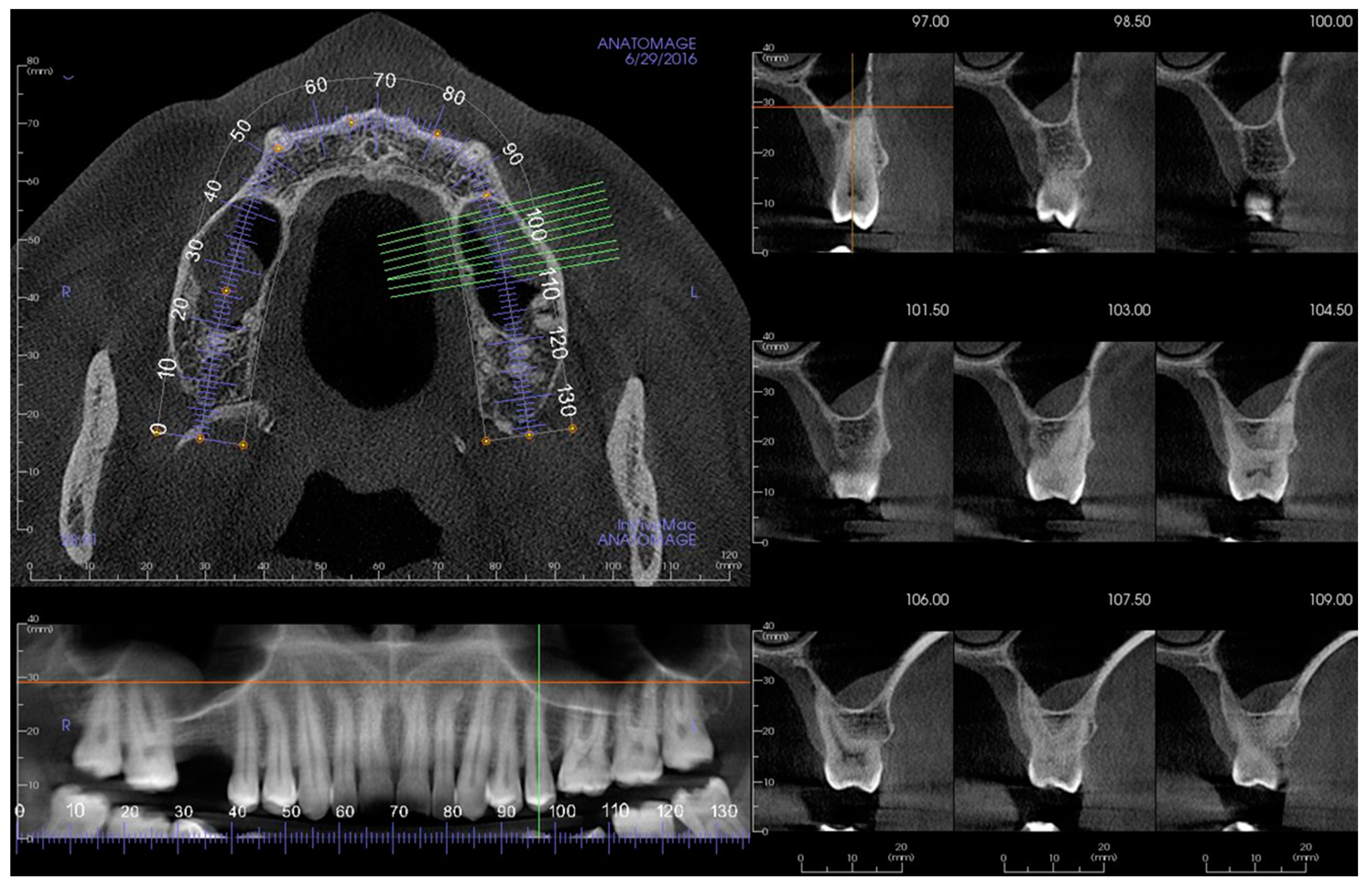
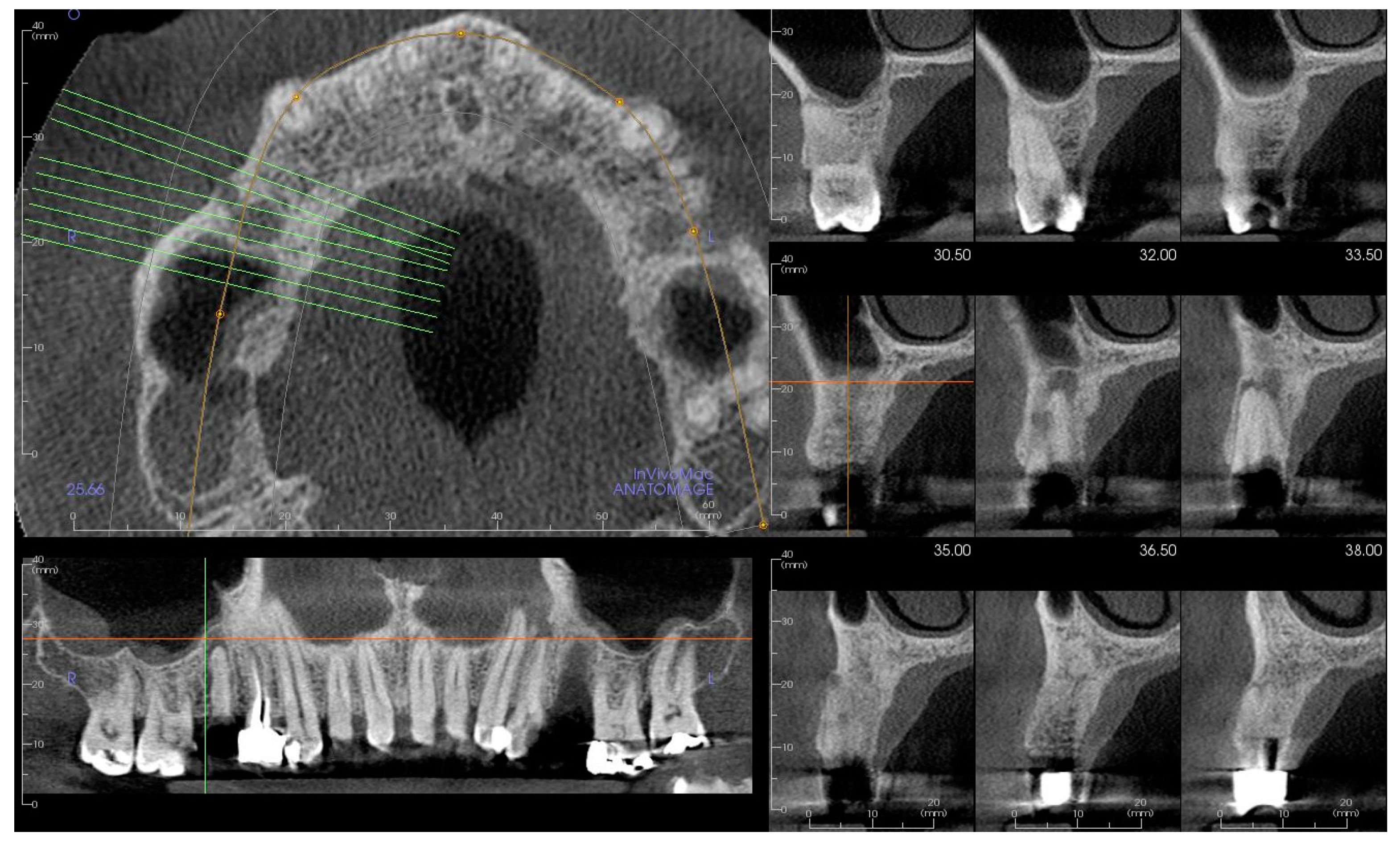
2. Materials and Methods
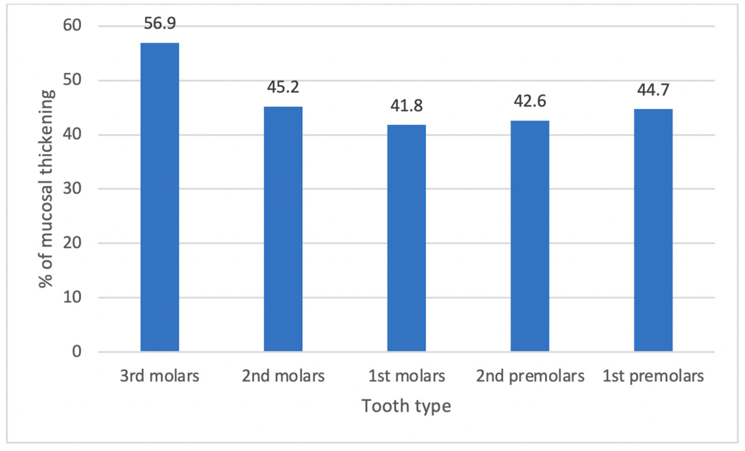
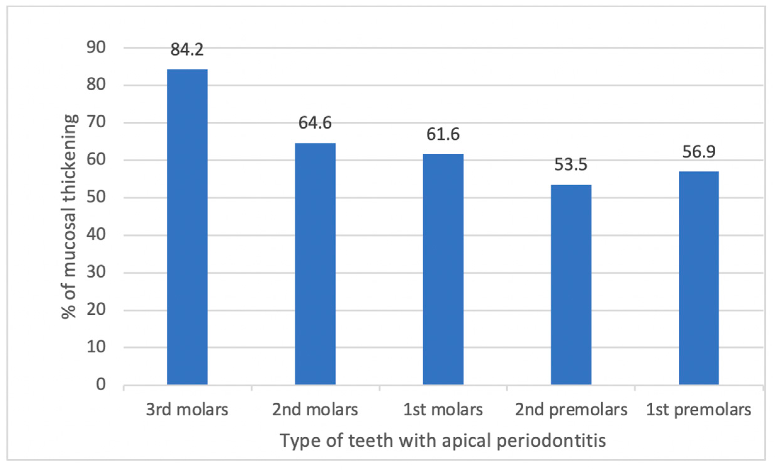
Ethical approval for this study was granted by the ethical committee of the School of Dentistry at the University of Jordan (reference IRB 234/2020). All methods were performed in accordance with the university’s guidelines and regulations that align with the Helsinki Declaration. Due to the anonymity of the patients’ personal data and details, informed consent was waived by the ethical committee of the School of Dentistry at the University of Jordan. Four hundred and twenty-one cone-beam computed tomography (CBCT) scans were used in this study, undertaken between 7 October 2019 and 22 February 2020. The scans were identified from the radiology department logbook for patients seen by different specialty clinics at the Jordan University Hospital, Amman, Jordan. The scans were justified and needed for either treatment or follow-up. All identified scans included in this retrospective study were taken at the Jordan University Hospital (JUH). The number of scans included in this study was based on a previous similar study by de Lima, Devito [18]. After looking through the identified scans, the final number of scans which met the inclusion criteria was three hundred and twenty-one CBCT maxillary scans. Included scans needed to have a full view of the maxilla with at least one permanent maxillary first molar. Scans with a poor imaging technique, artifacts, or pathological features impeding visualization were excluded.
Two qualified general dentists were trained by an oral and maxillofacial radiologist to read CBCT scans and to identify the parameters investigated in this study. General dentists were chosen as the vast majority of CBCT scans are reported by the prescribing dentists. A sample of twenty scans was not included in the study, as they were used for the training of the reviewers. The results were then discussed with the radiologist, another round of training was conducted 2 weeks later to ensure consistency, and intra-examiner feedback was given. The first fifty scans included in the study were analyzed first by the two general dentists, and then all of them were reassessed with the radiologist to make sure the examiners correctly identified all the parameters. All the scans were examined by both general dentists, and they had to agree on the result. In the case of disagreement, the final decision was that of the maxillofacial radiologist. Data extraction was conducted ensuring standardized viewing conditions of a low-lit room and using a software that allows brightness and contrast adjustments. All CBCT scans were analyzed on the same workstation to minimize any screen bias. The extracted data consisted of the patients’ gender and number, type, and location of teeth, in addition to the parameters. Data were entered into the statistical data analysis software (IBM SPSS Data Editor) (Table 1).
Statistical analysis was performed using SPSS for Windows release 16.0 (SPSS Inc., Chicago, IL, USA). Descriptive statistics were generated, and the chi-square test was used to examine differences between groups. Cramér’s V was used to express the effect size. The Cramér’s V value (abbreviated as V) was interpreted depending on the degree of freedom (df), as follows: df = 1 (0.10 = small effect) (0.30 = medium effect) (0.50 = large effect); df = 2 (0.07 = small effect) (0.21 = medium effect) (0.35 = large effect); df = 3 (0.06 = small effect) (0.17 = medium effect) (0.29 = large effect); df = 4 (0.05 = small effect) (0.15 = medium effect) (0.25 = large effect). The significance level was stated as p < 0.05.

3. Results

A total of 321 patients (176 females and 145 males) were examined, which resulted in 605 (300 right, 305 left) exposed maxillary sinuses that met our inclusion criteria. There were 2001 teeth examined, and of these, there were 468 first premolars, 455 second premolars, 488 first molars, 467 second molars, and 123 third molars.
There was mucosal thickening of the sinus lining in either or both of the maxillary sinuses in 178 (55.5%) of the patients (Figure 1).
Mucosal thickening was seen in 55.1% of the female patients compared to 55.9% of the males (p = 0.89). Among all the 605 exposed maxillary sinuses, 271 (44.8%) were associated with sinus mucosal thickening. Mucosal thickening was seen in 42.3% of the right maxillary sinuses compared with 47.2% of the left maxillary sinuses (p = 0.23). Out of the 2001 premolars and molars examined, 888 (44.4%) were associated with mucosal thickening, with a significant difference between the different tooth types (p = 0.04) (df = 4, V = 0.07) (Figure 2).
The periapical regions of the teeth were examined, and 688 teeth (34.4%) had periapical periodontitis (Figure 3). Among the teeth with periapical periodontitis, 38 were third molars, 147 were second molars, 185 were first molars, 144 were second premolars, and 174 were first premolars.
Mucosal thickening in a maxillary sinus was more likely if the neighboring tooth was associated with radiographic signs of apical disease (Figure 4).
Almost 54% of quadrants, of those that had periapical periodontitis, were associated with mucosal thickening of the adjacent maxillary sinus, compared to 31.6% when there was no periapical periodontitis (p < 0.001) (df = 1, V = 0.22). The odds ratio for having mucosal thickening when apical periodontitis was present was 2.52, while the relative risk for having mucosal thickening in the presence of apical periodontitis was 1.71. The prevalence of sinus mucosal thickening for each tooth type with periapical periodontitis is shown in Figure 5.
This section may be divided into subheadings. It should provide a concise and precise description of the experimental results, their interpretation, and the experimental conclusions that can be drawn.
When periapical periodontitis was present, third molars had the highest prevalence rate of sinus mucosal thickening (p = 0.007) (df = 4, V = 0.14).
Out of the 2001 teeth examined, 302 (15.1%) received root canal treatment. Only 28.2% of teeth with periapical lesions received root canal treatment. No association was found between sinus mucosal thickening and the presence of root canal treatment (p = 0.38) or the type of tooth with root canal treatment (p = 0.24).
Interruption of the maxillary sinus floor was noticed in relation to 79 teeth (3.9%), particularly the first and second molars (p < 0.001) (df = 4, V = 0.15). The vast majority (93.7%) of teeth associated with interruption of the sinus floor had periapical periodontitis (p < 0.001) (df = 1, V = 0.25), but no relation was found between sinus floor interruption and root canal treatment (p = 0.52).
The effect of the anatomical relationship between the root tip of the different posterior maxillary teeth and the sinus floor on the rate of sinus mucosal thickening is seen in Table 2. Generally, the rate of sinus mucosal thickening increased as the distance between the root tips and the sinus floor decreased, which was confirmed previously by Kuligowski, Jaroń et al. [19]. In addition, as shown in Table 3, the rate of sinus mucosal thickening increased when the roots were associated with periapical lesions.

4. Discussion

There are anatomical variations in human maxillary sinuses and paranasal sinuses. These structures begin to develop in the third week of gestation, and the development is not complete until early adulthood [20]. Studies conflict on the gender difference in the size of the sinuses—some studies found the size of the sinuses is larger in males [21], whilst others stated there is no difference between males and females [22,23]. Some believe that smaller sinuses have a reduced flow of air that can lead to low oxygen and reduced motility of the cilia. With both these situations, there may be mucus stagnation and bacterial growth [24].
This study found a difference in mucosal thickening between males (55.9%) and females (55.1%), but this was not significant p < 0.89. No significant difference was also found in another study [25]. However, others have found that there was a significant difference in males and females, with mucosal thickening being higher in males [26]. These differences could be accounted for by environmental factors such as smoking or occupation.
The prevalence of incidental paranasal inflammatory changes in the Jordanian population was 64.3%. This was found in patients who had MRI scans for neurological signs and symptoms [27]. The present study found a lower incidence (55.5%), but the previous study was conducted in a different area of Jordan, and environmental or demographic factors may be the reason for the difference.
There was a slightly higher percentage of thickening found in the right and left sinuses, with 47.2% and 42.3%, respectively. Even though the degree of nasal septum deviation was not among the parameters investigated, one explanation for this could be a deviated septum which can be influenced by the presence of a concha bullosa. The use of computerized tomography has been utilized to answer this question, and it was found that nasal septal deviation had a significant influence on developing maxillary sinusitis [23].
The relationship between different teeth and thickening of the mucosa of the maxillary sinus was explored, as can be seen in Figure 1. There was MT associated with 44.4% of molars and premolars, all of which did not undergo any type of apical surgery, e.g., root resection or apicectomy. There was a significant difference between the teeth, and the lowest percentage was found for the first molars. If looking at the relationship of teeth to the sinus, other studies assessed the relationship between the maxillary molar and premolar roots and the maxillary sinus [28,29,30,31]. In one of the studies, the mean distance between the maxillary posterior teeth and the maxillary sinus floor was found to be 1.97 mm [28]. In another study, the maxillary first and second molars were assessed, and it was found that they had a close relationship with the maxillary sinus floor in 40% of cases [30]. The palatal roots of molars have been shown to be closer to the antral floor and closer to the maxillary sinus than the palatal vault in 20% of cases [29]. This is probably due to the divergence of the roots and how the buccal and palatal roots saddle the sinus.
In a study by Kilic, Kamburoglu et al. [31], they looked at the distance and relationship between the tips of posterior teeth and the maxillary sinus. They classified the relationship into three groups:
  • Group 1: root tips in contact with the sinus floor;
  • Group 2: root tips penetrating into the sinus;
  • Group 3: root tips below the sinus floor.
They reported that for the right side, 60% of the root tips were in Group 3, 30% were in Group 1, and 10% were in Group 2; for the left side, 68% were in Group 3, 21% were in Group 1, and 11% were in Group 2. There was no significant difference in the right or left. They concluded in the results that the distance from the root tip to the sinus floor was the longest for the first premolar root tip and the shortest for the second molar distobuccal root tip [31]. This close relationship may explain the results of the presence of periapical pathology and mucosal thickening with an odds ratio of 2.52 CI of having mucosal thickening with periapical pathology.
In other areas of medicine, it was recognized that infections in sinuses such as the frontal, ethmoidal, or maxillary sinuses could be sources of orbital infections and that spread potentially occurs by direct extension through the sinus wall and/or by local thrombophlebitis in areas where the bone is thin and in close proximity [32]. The reverse may also happen with infection within bone from apical disease, which causes the spread of infection to the sinus.
In relation to root-treated teeth, in this study, it was found that there was no association between the presence of root canal treatment and mucosal thickening (p = 0.38). One study looked at the association of dental condition and maxillary thickening. This retrospective study on CBCT found that 42.1% had mucosal thickening of >2 mm where periapical pathology was absent, and 53.6% when pathology was present (p < 0.05) [33]. In the present study, it was also found that teeth with periapical pathology had an influence on mucosal thickening and that the more posterior molars had an increased percentage of thickening (Figure 2). This reflects that the closer the association with the sinus, the more posterior position the tooth occupies [31].
Thickening and periapical pathology were the highest in third molars, followed by second molars, first molars, and then first premolars. This reflects the prevalence of teeth with periapical pathology, and in other studies, it was found that the teeth most likely to have periapical pathology were molars, followed by first premolars [34]. In one study, the size of the lesion was compared using the periapical index, and it was concluded that the larger the periapical lesion, the greater the prevalence of mucosal thickening, and if more than one tooth had periapical pathology, the more severe the sinus disease recorded [35]. The fact that teeth have been root treated and the size of the lesion may be reduced or even healed would explain the results of no association with root canal treatment in the present study.
On exploring sinus floor perforation, 93.7% of teeth with periapical pathology had perforation of the sinus floor (p < 0.001). With the close relationship of the posterior teeth and the maxillary sinus [31], and the spread of infection in thin bone [32], this finding is what one would expect.

5. Conclusions

The reported incidence of the spread of infection from periapical tissues to the sinus is between 4.6 and 47% [36]. With the results of the present study, the close relationship of posterior teeth, the spread of infection, and the perforation of the sinus floor, the reported association with periapical disease and maxillary sinusitis can be understood.
The term endo-antral syndrome is used for the spread of periapical disease into the maxillary sinus [37,38,39]. All this shows there is a need to include dental investigation in cases of maxillary sinusitis, in particular where the etiology does not show ENT causes for the disease.
Computerized tomography is used for the evaluation of sinus disease within ENT. ENTs should routinely look for periapical pathology as a potential cause for sinusitis and regularly refer to dentists to assess these teeth before invasive sinus surgery.
With the major limitation of this study being that it was purely reliant on radiographic imaging to diagnose OS, it seems clear that future work should focus on conducting studies that possess both a clinical examination component and a radiographic one, to assess the presence or absence of signs and symptoms and monitor the change in symptoms after undergoing dental treatments.

Author Contributions

All authors contributed to the study conception and design. Conceptualization: S.A.M. and J.C.; methodology: S.A.M. and A.A.-H.; formal analysis and investigation: F.A.S., S.A.-N. and Y.B.; writing—original draft preparation: S.A.M. and J.C.; writing—review and editing: S.A.M., A.A.-H. and Y.H. All authors have read and agreed to the published version of the manuscript.

Funding

This research did not receive any specific grants from funding agencies in the public, commercial, or not-for-profit sectors. All authors certify that they have no affiliations with or involvement in any organization or entity with any financial or non-financial interest in the subject matter or materials discussed in this manuscript.

Institutional Review Board Statement

Ethical approval was granted from the ethical committee of the School of Dentistry at the University of Jordan (reference IRB 234/2020). All methods were performed in accordance with the university’s guidelines and regulations that align with the Helsinki Declaration. Due to the anonymity of the patients’ personal data and details, informed consent was waived by the ethical committee of the School of Dentistry at the University of Jordan.

Informed Consent Statement

Not applicable.

Data Availability Statement

The datasets generated and analyzed during the current study are not publicly available due to this manuscript discussing only a part of the data extracted (Other parameters were recorded and are to be used in a completely different manuscript that is looking into different aspects not related to this manuscript. At this point, it will be very difficult to separate the data keeping only relevant data.) but are available from the corresponding author on reasonable request. Once all data have been published, all data extracted can be shared publicly.

Conflicts of Interest

The authors declare no conflict of interest.

References

  1. Garry, S.; O’Riordan, I.; James, D.; Corbett, M.; Barry, T.; Thornton, M. Odontogenic Sinusitis—Case Series and Review of Literature. J. Laryngol. Otol. 2021, 136, 49–54. [Google Scholar] [CrossRef]
  2. Bauer, W.H. Maxillary sinusitis of dental origin. Am. J. Orthod. Oral Surg. 1943, 29, B133–B151. [Google Scholar] [CrossRef]
  3. Mehra, P.; Murad, H. Maxillary sinus disease of odontogenic origin. Otolaryngol. Clin. N. Am. 2004, 37, 347–364. [Google Scholar] [CrossRef]
  4. Ferguson, M. Rhinosinusitis in oral medicine and dentistry. Aust. Dent. J. 2014, 59, 289–295. [Google Scholar] [CrossRef] [PubMed]
  5. Kayabasoglu, G.; Nacar, A.; Altundag, A.; Cayonu, M.; Muhtarogullari, M.; Cingi, C. A retrospective analysis of the relationship between rhinosinusitis and sinus lift dental implantation. Head Face Med. 2014, 10, 53. [Google Scholar] [CrossRef] [PubMed] [Green Version]
  6. Guerra-Pereira, I.; Vaz, P.; Faria-Almeida, R.; Braga, A.-C.; Felino, A. CT maxillary sinus evaluation—A retrospective cohort study. Med. Oral Patol. Oral Cir. Bucal 2015, 20, e419. [Google Scholar] [CrossRef]
  7. Turfe, Z.; Ahmad, A.; Peterson, E.I.; Craig, J.R. Odontogenic sinusitis is a common cause of unilateral sinus disease with maxillary sinus opacification. Int. Forum Allergy Rhinol. 2019, 9, 1515–1520. [Google Scholar] [CrossRef]
  8. Whyte, A.; Boeddinghaus, R. Imaging of odontogenic sinusitis. Clin. Radiol. 2019, 74, 503–516. [Google Scholar] [CrossRef]
  9. Troeltzsch, M.; Pache, C.; Troeltzsch, M.; Kaeppler, G.; Ehrenfeld, M.; Otto, S.; Probst, F. Etiology and clinical characteristics of symptomatic unilateral maxillary sinusitis: A review of 174 cases. J. Cranio-Maxillofac. Surg. 2015, 43, 1522–1529. [Google Scholar] [CrossRef]
  10. Hoskison, E.; Daniel, M.; Rowson, J.; Jones, N. Evidence of an increase in the incidence of odontogenic sinusitis over the last decade in the UK. J. Laryngol. Otol. 2012, 126, 43. [Google Scholar] [CrossRef]
  11. Phothikhun, S.; Suphanantachat, S.; Chuenchompoonut, V.; Nisapakultorn, K. Cone-beam computed tomographic evidence of the association between periodontal bone loss and mucosal thickening of the maxillary sinus. J. Periodontol. 2012, 83, 557–564. [Google Scholar] [CrossRef] [PubMed]
  12. Ugincius, P.; Kubilius, R.; Gervickas, A.; Vaitkus, S. Chronic odontogenic maxillary sinusitis. Stomatologija 2006, 8, 44–48. [Google Scholar] [PubMed]
  13. Arias-Irimia, O.; Barona-Dorado, C.; Santos-Marino, J.A.; Martínez-Rodríguez, N.; Martínez-González, J.M. Meta-analysis of the etiology of odontogenic maxillary sinusitis. Med. Oral Patol. Oral Cir. Bucal 2010, 15, e70–e73. [Google Scholar] [CrossRef] [Green Version]
  14. Allevi, F.; Fadda, G.L.; Rosso, C.; Martino, F.; Pipolo, C.; Cavallo, G.; Felisati, G.; Saibene, A.M. Diagnostic criteria for odontogenic sinusitis: A systematic review. Am. J. Rhinol. Allergy 2021, 35, 713–721. [Google Scholar] [CrossRef]
  15. Park, M.S.; Eo, M.Y.; Myoung, H.; Kim, S.M.; Lee, J.H. Early diagnosis of jaw osteomyelitis by easy digitalized panoramic analysis. Maxillofac. Plast. Reconstr. Surg. 2019, 41, 6. [Google Scholar] [CrossRef]
  16. Workman, A.D.; Granquist, E.J.; Adappa, N.D. Odontogenic sinusitis: Developments in diagnosis, microbiology, and treatment. Curr. Opin. Otolaryngol. Head Neck Surg. 2018, 26, 27–33. [Google Scholar] [CrossRef]
  17. Tataryn, R.; Lewis, M.; Horalek, A.; Thompson, C.; Cha, B.; Pokorny, A. Maxillary Sinusitis of Endodontic Origin: AAE Position Statement. 2018. Available online: https://www.aae.org/specialty/wp-content/uploads/sites/2/2018/04/AAE_PositionStatement_MaxillarySinusitis.pdf (accessed on 15 February 2022).
  18. de Lima, C.O.; Devito, K.L.; Vasconcelos, L.R.B.; do Prado, M.; Campos, C.N. Correlation between endodontic infection and periodontal disease and their association with chronic sinusitis: A clinical-tomographic study. J. Endod. 2017, 43, 1978–1983. [Google Scholar] [CrossRef] [PubMed]
  19. Kuligowski, P.; Jaroń, A.; Preuss, O.; Gabrysz-Trybek, E.; Bladowska, J.; Trybek, G. Association between Odontogenic and Maxillary Sinus Conditions: A Retrospective Cone-Beam Computed Tomographic Study. J. Clin. Med. 2021, 10, 2849. [Google Scholar] [CrossRef]
  20. Wang, R.-G.; Jiang, S.; Gu, R. The cartilaginous nasal capsule and embryonic development of human paranasal sinuses. J. Otolaryngol. 1994, 23, 239–243. [Google Scholar]
  21. Emirzeoglu, M.; Sahin, B.; Bilgic, S.; Celebi, M.; Uzun, A. Volumetric evaluation of the paranasal sinuses in normal subjects using computer tomography images: A stereological study. Auris Nasus Larynx 2007, 34, 191–195. [Google Scholar] [CrossRef]
  22. Pirner, S.; Tingelhoff, K.; Wagner, I.; Westphal, R.; Rilk, M.; Wahl, F.; Bootz, F.; Eichhorn, K.W. CT-based manual segmentation and evaluation of paranasal sinuses. Eur. Arch. Oto-Rhino-Laryngol. 2009, 266, 507–518. [Google Scholar] [CrossRef] [PubMed]
  23. Kucybała, I.; Janik, K.A.; Ciuk, S.; Storman, D.; Urbanik, A. Nasal Septal Deviation and Concha Bullosa—Do They Have an Impact on Maxillary Sinus Volumes and Prevalence of Maxillary Sinusitis? Pol. J. Radiol. 2017, 82, 126–133. [Google Scholar] [CrossRef] [PubMed] [Green Version]
  24. Lana, J.P.; Carneiro, P.M.; Machado Vde, C.; de Souza, P.E.; Manzi, F.R.; Horta, M.C. Anatomic variations and lesions of the maxillary sinus detected in cone beam computed tomography for dental implants. Clin. Oral Implant. Res. 2012, 23, 1398–1403. [Google Scholar] [CrossRef] [PubMed]
  25. Raghav, M.; Karjodkar, F.R.; Sontakke, S.; Sansare, K. Prevalence of incidental maxillary sinus pathologies in dental patients on cone-beam computed tomographic images. Contemp. Clin. Dent. 2014, 5, 361–365. [Google Scholar] [CrossRef]
  26. Hsiao, Y.J.; Yang, J.; Resnik, R.R.; Suzuki, J.B. Prevalence of Maxillary Sinus Pathology Based on Cone-Beam Computed Tomography Evaluation of Multiethnicity Dental School Population. Implant. Dent. 2019, 28, 356–366. [Google Scholar] [CrossRef]
  27. Hiari, M.; Hiari, M.A. Incidental paranasal sinus inflammatory changes in a Jordanian population. East. Mediterr. Health J. 1998, 4, 308–311. [Google Scholar] [CrossRef]
  28. Eberhardt, J.A.; Torabinejad, M.; Christiansen, E.L. A computed tomographic study of the distances between the maxillary sinus floor and the apices of the maxillary posterior teeth. Oral Surg. Oral Med. Oral Pathol. 1992, 73, 345–347. [Google Scholar] [CrossRef]
  29. Waite, D.E. Maxillary sinus. Dent. Clin. N. Am. 1971, 15, 349–368. [Google Scholar]
  30. Wallace, J.A. Transantral endodontic surgery. Oral Surg. Oral Med. Oral Pathol. Oral Radiol. Endod. 1996, 82, 80–83. [Google Scholar]
  31. Kilic, C.; Kamburoglu, K.; Yuksel, S.P.; Ozen, T. An assessment of the relationship between the maxillary sinus floor and the maxillary posterior teeth root tips using dental cone-beam computerized tomography. Eur. J. Dent. 2010, 4, 462–467. [Google Scholar] [CrossRef]
  32. Mills, R.P.; Kartush, J.M. Orbital wall thickness and the spread of infection from the paranasal sinuses. Clin. Otolaryngol. Allied Sci. 1985, 10, 209–216. [Google Scholar] [CrossRef] [Green Version]
  33. Aksoy, U.; Orhan, K. Association between odontogenic conditions and maxillary sinus mucosal thickening: A retrospective CBCT study. Clin. Oral Investig. 2019, 23, 123–131. [Google Scholar] [CrossRef] [PubMed]
  34. van der Borden, W.G.; Wang, X.; Wu, M.-K.; Shemesh, H. Area and 3-dimensional volumetric changes of periapical lesions after root canal treatments. J. Endod. 2013, 39, 1245–1249. [Google Scholar] [CrossRef] [PubMed]
  35. Lu, Y.; Liu, Z.; Zhang, L.; Zhou, X.; Zheng, Q.; Duan, X.; Zheng, G.; Wang, H.; Huang, D. Associations between maxillary sinus mucosal thickening and apical periodontitis using cone-beam computed tomography scanning: A retrospective study. J. Endod. 2012, 38, 1069–1074. [Google Scholar] [CrossRef]
  36. Melén, I.; Lindahl, L.; Andréasson, L.; Rundcrantz, H. Chronic maxillary sinusitis: Definition, diagnosis and relation to dental infections and nasal polyposis. Acta Oto-Laryngol. 1986, 101, 320–327. [Google Scholar] [CrossRef] [PubMed]
  37. Selden, H.S. The endo-antral syndrome. J. Endod. 1977, 3, 462–464. [Google Scholar] [CrossRef]
  38. Selden, H.S. The endo-antral syndrome: An endodontic complication. J. Am. Dent. Assoc. 1989, 119, 397–398, 401–402. [Google Scholar] [CrossRef] [PubMed]
  39. Selden, H.S. Endo-Antral syndrome and various endodontic complications. J. Endod. 1999, 25, 389–393. [Google Scholar] [CrossRef]
Figure 1. Mucosal thickening: no apical disease.
Applsci 12 03057 g001
Figure 2. Different tooth types and their relation to sinus pathology.
Applsci 12 03057 g002
Figure 3. Apical disease present: no mucosal thickening.
Applsci 12 03057 g003
Figure 4. Mucosal thickening: apical disease present.
Applsci 12 03057 g004
Figure 5. Types of teeth with apical pathology and their relation to sinus pathology.
Applsci 12 03057 g005
Table 1. Parameters investigated.
(1)
Presence of apical disease around the apices of any of the posterior teeth;
(2)
Presence of endodontic therapy of any of the posterior teeth;
(3)
Thickening of the mucosal lining of the maxillary sinuses;
(4)
Interruption of the bony floor of the maxillary sinus;
(5)
Distance between the sinus floor and the root end.
Table 2. The effect of the distance between the root tips and the sinus floor on the rate of sinus mucosal thickening.
Root and Tooth NumberRate of Sinus Mucosal Thickening
(%)
p ValuedfV
Apex-to-Sinus Distance
0 mm≤5 mm>5 mm
SR 471.480.677.80.7820.06
BR 481.876.2640.0920.14
PR 476.980.562.30.04820.18
SR 585.364.867.30.0120.21
BR 594.157.155.60.0120.41
PR 590.566.755.60.0220.36
MBR 676.772.261.10.0620.12
DBR 676.172.256.50.0720.12
PR 677.269.158.80.0220.14
MBR 785.368.527.3<0.00120.32
DBR 785.968.346.7<0.00120.27
PR 787.167.652.6<0.00120.28
MBR 891.590.966.70.3720.17
DBR 893.690.5500.0320.33
PR 890.995.2750.4020.20
SR: single rooted, BR: buccal root, PR: palatal root, MBR: mesiobuccal root, DBR: distobuccal root, df: degree of freedom, V: Cramér’s V test.
Table 3. The effect of the presence or absence of periapical lesions around the different tooth roots on the rate of sinus mucosal thickening.
Root and Tooth NumberRate of Sinus Mucosal Thickening (%)p ValuedfV
Presence of Periapical Lesion
YesNo
SR 463.136.3<0.00110.25
BR 452.439.50.0410.13
PR 452.838.90.0310.14
SR 553.836.70.00210.16
BR 545.541.20.7310.04
PR 547.843.40.7210.04
MBR 663.130.5<0.00110.31
DBR 662.530.8<0.00110.31
PR 661.831.1<0.00110.30
MBR 763.837.7<0.00110.24
DBR 764.236.8<0.00110.25
PR 765.735.9<0.00110.28
MBR 882.444.6<0.00110.35
DBR 883.343.2<0.00110.37
PR 884.445.3<0.00110.35
SR: single rooted, BR: buccal root, PR: palatal root, MBR: mesiobuccal root, DBR: distobuccal root, df: degree of freedom, V: Cramér’s V test.
Publisher’s Note: MDPI stays neutral with regard to jurisdictional claims in published maps and institutional affiliations.

