Article

Beta vulgaris subsp. maritima: A Valuable Food with High Added Health Benefits

1
Team of Experimental Oncology and Natural Substances, Cellular and Molecular Immuno-Pharmacology, Faculty of Science and Technology, Sultan Moulay Slimane University, Beni-Mellal 23000, Morocco
2
Institute of Biological Sciences (ISSB-P), Mohammed VI Polytechnic University (UM6P), Ben-Guerir 43150, Morocco
3
Laboratory of Biochemistry and Molecular Genetics, Faculty of Sciences and Technologies of Tangier, B.P. 416, Tangier 90000, Morocco
4
Laboratory of Analytical Chemistry and Bromatology, Faculty of Medicine and Pharmacy, Mohammed V University in Rabat, Rabat 10100, Morocco
5
Laboratory of Natural Resources and Environment, Polydisciplinary Faculty of Taza, Sidi Mohamed Ben Abdellah University of Fez, B.P. 1223 Taza-Gare, Taza 35000, Morocco
6
Department of Biomedical, Dental, Morphological and Functional Imaging Sciences, University of Messina, 98125 Messina, Italy
*
Authors to whom correspondence should be addressed.
Appl. Sci. 2022, 12(4), 1866; https://doi.org/10.3390/app12041866
Submission received: 14 January 2022 / Revised: 3 February 2022 / Accepted: 7 February 2022 / Published: 11 February 2022
(This article belongs to the Special Issue Innovative Separation Methods in Food Chemistry and Analysis)

Abstract

:
The present study was conducted to evaluate a natural extract, obtained from the Beta vulgaris plant, for its phytochemical composition and its beneficial health effects. Therefore, total phenolic and flavonoid contents, as well as identification and quantification of phenolic compounds by HPLC, were assessed in leaves’ extract. Moreover, antioxidant activities were investigated using free radical scavenging tests, (ABTS+ and DPPH+) and reducing power assay (FRAP) as well as ferrous ions’ (Fe2+) chelating activity. The Antiglycation effect was also evaluated, using the BSA-fructose model, and the antidiabetic effect was determined by inhibition of α-amylase and α-glucosidase enzymes. Additionally, the in vitro antitumor effect was quantified using the MTT assay, and the antibacterial activity was evaluated using the agar disc diffusion and broth microdilution methods. Both aqueous and methanolic extracts exhibited potential antioxidant capacity with a higher effect for the methanolic extract. Furthermore, the in vitro antitumor activity of the methanolic extracts exhibited potent cytotoxic effects against two breast cancer cell lines, MDA-MB-468 and MCF-7. Moreover, Beta vulgaris extracts inhibit not only α-amylase and α-glucosidase, but also advanced glycation end-products’ (AGEs) formation, which would prevent diabetes’ complications. Beta vulgaris methanolic extract revealed also a high antibacterial effect against Proteus mirabilis and Bacillus subtilis. Taken together, these results revealed that Beta vulgaris leaves’ extracts constitute a valuable food and natural source of bioactive molecules that could be used for the development of new, natural drugs against cancer and diabetes.

1. Introduction

Breast cancer (BC) is known to be a highly invasive cancer that affects women of different ages in the different parts of the world [1]. In fact, it is considered as the most heterogeneous cancer that contains more than 20 subtypes and, despite this classification, BC is almost considered as a dilemma in treatment, especially the triple negative breast cancer (TNBC), which makes breast cancer extremely intractable.
Currently, BC treatments face several problems, especially the development of therapy resistance and malignant progression. Therefore, new chemical drugs are permanently sought, causing a huge economic burden worldwide [2,3].
Diabetes, as with several other chronic diseases, has harmful impacts on people’s health and can lead to a variety of complications. The different complications associated to diabetes could be linked to a common feature, which is protein glycation. This process could cover a wide range of protein and may, therefore, cause several other pathologies, such as nephropathy, retinopathy, neuropathy, etc. [4]. (Diabetes’ complications are mediated by several factors, among them, the non-enzymatic glycation and oxidative stress are the most known; thus, antiglycation and antioxidant activities are the most known determinants in plants’ extracts to be assessed to explore new natural compounds to treat diabetes and its complications [4,5].)
For centuries, natural products (NP) represented an invaluable source of inspiration in the search for new drug candidates. Therefore, NP have been suggested as alternative therapy due to rich sources, multiple targets, and low toxicity with fewer side effects, as well as being considered as a new safe source of effective antioxidant, anti-diabetic, and anticancer bioactive compounds [6,7,8].
Beta vulgaris subsp. maritima (B. vulgaris) is a plant species that belongs to the Chenopodiaceae family and that is widely distributed throughout the world. Additionally, Beta vulgaris L. genus has been proven to possess a wide array of pharmacological effects such as anti-inflammatory, antioxidant, neuroprotective, hyperglycemic, and anticancer [9,10,11,12]. Moreover, a few previous studies proved that the Beta vulgaris L. possesses antitumor activities against tumors cells, in particular, breast cancer. B. vulgaris subsp. maritima is used as an old medicinal plant and traditional food. It is employed in folk medicine to treat different diseases, in particular, different cancers such as leukemia, esophagus, glands, prostate, and breast tumors [13]. However, presently, no previous investigation pertaining to the Cytotoxicity of Beta vulgaris subsp. maritima towards human breast has been reported, especially, ER-positive and TNBC cancer cell lines. In view of this, the goal of this study was to seek, on the one hand, the phytochemical composition and the antioxidants capabilities of both the aqueous and methanolic extracts of B. vulgaris subsp. maritima leaves. On the other hand, the anti-glycation, α-amylase, and α-glucosidase inhibitory potential of the methanolic extract were investigated in vitro. Antibacterial and cytotoxic effects against two breast cancer cell lines, the luminal subtype line (MCF-7) and the triple-negative breast cancer (TNBC) subtype line (MDA-MB-468), were also assessed. Furthermore, the antibacterial activity of this methanolic extract was investigated.

2. Materials and Methods

2.1. Plant Material

The B. vulgaris subsp. maritima leaves were collected in January 2018, in the suburbs of Taza, Morocco, and identified and authenticated by Pr. Younes ABBAS (Ph.D., botanist, Sultan Moulay Slimane University). Voucher specimens are kept at the Herbarium of the Faculty of Sciences and Techniques. The leaves were washed with distilled water, shade dried, and powdered with a mechanical grinder; the powder was stored in an airtight container until further use.

2.2. Plant Extraction

The extraction of plant material was conducted at room temperature with continuous shaking and in dark conditions for 24 h using either dH2O or methanol 80%. Centrifugation at 5.0× g for 10 min and filtration with the Whatman filter were conducted. The water and methanol solvents were then dried in an incubator at 40 °C. The final yields were 21.71% for aqueous extracts and 18.82% for methanolic extracts.

2.3. HPLC-DAD Analysis

Liquid chromatography analyses of polyphenolic compounds from the methanolic extracts of B. vulgaris subsp. maritima were conducted on a Shimadzu (Kyoto, Japan) liquid chromatography system. The same protocol of separation and quantification of the compounds was already published in the work of Asraoui et al. [14].

2.4. Determination of Total Phenolic Content

To measure the total phenolic contents, we referred to the method of Folin–Ciocalteu (FC) after some slight modifications, as reported by Kabach et al. [15]. Briefly, 0.1 mL from the B. vulgaris subsp. maritima extracts was added to 0.4 mL of the FC reagent; then, to this mixture, 1 mL of Na2CO3 (prepared at a concentration of 7%) and 0.1 mL of dH2O were added. A final step of incubation in the dark for 30 min was followed by the measurement of absorbance at 725 nm. To quantify the total phenolic contents, we used a gallic acid calibration curve.

2.5. Determination of Total Flavonoid Content

The protocol for the determination of flavonoid contents was the same as reported by Kabach et al. [15]. Briefly, to 40 µL of each sample of the B. vulgaris subsp. maritima extracts, 10 µL of 1 M of acetate potassium and 10% aluminum chloride were added. After mixing, a volume of 100 µL of methanol at 50% was added and the final volume was then made up to 400 µL using dH2O. Absorbances were read at 415 nm and quercetin was used as standard.

2.6. Antioxidant Activities

2.6.1. ABTS+ Radical Scavenging Assay

The radical scavenging effect of the methanolic and aqueous extracts obtained from B. vulgaris subsp. maritima leaves was assessed against the radical ABTS+ as reported by Ben Mrid et al. [8]. From a diluted solution of ABTS+, 185 µL was taken up and mixed with 15 µL of each sample. The mixtures were kept for 10 min before measuring the absorbance at 734. The following equation was used to determine the radical scavenging activity:
% Scavenging activity = [(AABTS+ − AS)/AABTS+] × 100
AS and AABTS+ refer to the absorbance of the sample and ABTS+ solution, respectively.

2.6.2. DPPH Radical Scavenging Assay

The radical scavenging ability of aqueous and methanolic extracts of B. vulgaris subsp. maritima leaves was determined using DPPH (2,2-diphenyl-1-picrylhydrazyl), as reported in the study of Ben Mrid et al. [8] with some modifications. Indeed, 150 µL of a freshly prepared solution of DPPH was mixed with 50 µL from each extract. After mixing, the solutions were kept in the dark for 30 min at room temperature (RT). The absorbance was measured in a spectrophotometer at 517 nm. The equation below was used to determine the scavenging activity of the extracts on DPPH:
% Scavenging effect = [(ADPPH − AS)/ADPPH] × 100
AS refers to the sample absorbance; ADPPH is the absorbance of the DPPH solution.

2.6.3. Ferric Reducing Antioxidant Power Assay (FRAP)

The FRAP assay was carried out to determine the reducing power of the B. vulgaris subsp. maritima extracts, using the protocol reported by Oyazu [16]. Then, 200 µL of the sample extract was added to 500 µL of phosphate buffer (0.2 M, pH 6.6) and 500 µL of potassium ferricyanide (1%). After 20 min at 50 °C, 500 µL of trichloroacetic acid (10%) was added to each tube and the mixtures were centrifuged at 1500× g for 10 min. Then, 500 µL from the supernatants were added to 500 µL of dH2O and 0.1 mL of FeCl3 (0.1%). Ascorbic acid was used with various concentrations (7.8–62.5 µg/mL) as a standard curve. The absorbance of each sample was read at 700 nm.

2.6.4. Metal Chelating Activity

This effect was demonstrated using the protocol described by Dinis et al. [17]. In glass tubes, increasing concentrations of each sample were added to 10 µL of 0.6 mM FeCl2. After vigorous mixing, the tubes were maintained at RT for 10 min. Thereafter, 50 µL of ferrozine (5 mM) was added to begin the reaction. The absorbance was determined after 10 min at 562 nm and the IC50 was calculated.

2.7. Antiglycation Activities

A glycated BSA formation was achieved following a previous method with some modifications [18]. Fructose was prepared using phosphate buffer solution (PBS, 100 mM, pH 7.4) to form a 500 mM solution. BSA (10 mg/mL) was added to a fructose solution and incubated for 4 weeks at 37 °C. Then, 0.05% sodium azide was used to inhibit the microorganism’s development. The advanced glycation end products (AGEs) were quantified by a spectrofluorometer (excitation wavelength: 355; emission wavelength: 460 nm). In this study, a positive control consisting of aminoguanidine (AG) was used. The inhibition rates of the AGEs’ formation were measured following the equation below:
Inhibition   ( % ) = [ 1 ( FLs FLsb ) ( FLc FLcb ) ] × 100
where FLs represents the fluorescence intensity of the mixture, FLsb corresponds to the fluorescence intensity of the sample blank (without fructose), FLc is the fluorescence intensity of the control mixture, and FLcb refers to the fluorescence intensity of the control blank mixture.

2.7.1. Determination of Fructosamine

The fructosamine assay was tested after 28 days of incubation. Fructosamine reduces NBT and produces coloration, which had an absorption at 530 nm [19]. In brief, 40 µL of the glycated BSA was incubated with 160 µL of 0.3 mM NBT in sodium carbonate buffer (100 mM, pH 10.35) at 37 °C for 30 min. The absorbance of the mixture was determined at the wavelength of 530 nm. The concentration of fructosamine was calculated from the standard curve using 1-deoxy-1-morpholino-fructose (1-DMF).

2.7.2. Determination of Protein Thiol Group

To determine the level of free thiol in glycated materials, Ellman’s reagent was used after four weeks of incubation. Briefly, 250 µL of glycated samples were incubated with 750 µL of DTNB solutions (0.5 mM; PBS 0.1 M, pH 7.4) for 15 min at 37 °C, and the absorbance was determined at 410 nm. Various concentrations of L-cysteine (1.95–500 mM) were used as a standards’ curve.

2.8. Antidiabetic Enzymatic Activities

2.8.1. α-Amylase Inhibition Effect

The α-amylase inhibition potential of B. vulgaris maritima extract was evaluated following the modified protocol described by Dong et al. [20]. Then, 100 µL of various concentrations of sample was premixed with 100 µL of 0.1 U/mL of α-amylase solution solubilized in phosphate buffer, pH = 6.9, kept at 37 °C for 30 min. Then, 100 µL of 0.25% starch solution prepared in phosphate buffer (pH 6.9) was added. The combinations were maintained at 37 °C for 30 min before adding 200 µL of the DNS reagent that was composed of a mixture of sodium potassium tartrate (12% in 0.4 M NaOH) and 3,5-dinitrosalicylic acid (1%). The next step consisted of boiling the samples for 5 min and, after cooling the temperature down (at RT), a spectrophotometer was used to determine the absorbance at 540 nm.
The rate of inhibition was measured following the equation:
Inhibition   ( % ) = [ ( A c A c b ) ( A s A s b ) ( A c A c b ) ] × 100
Ac is the absorbance of the control that was composed of the enzyme; Acb refers to the blank control that was composed of the buffer only; As and Asb correspond to the absorbance of the sample and the sample blank, respectively.

2.8.2. α-Glucosidase Inhibitory Assay

The determination of inhibitory effect against α-glucosidase was assessed following the protocol reported by Asraoui et al. [14].

2.9. In Vitro Cytotoxic Activity

2.9.1. Cell Culture

Two different subtypes of cancer cell lines, the MCF-7, representing the luminal subtype of breast cancer, and MDA-MB-468, representing the triple-negative subtype, were grown in a culture medium composed of RPMI-1640, to which fetal bovine serum, at a concentration of 5%, L-glutamine at 0.2%, and penicillin G-streptomycin at 1% were added. The incubator was settled at 5% CO2 and 37 °C.

2.9.2. MTT Assay

Approximately 105 and 7 × 10⁴ cells per well for MCF-7 and MDA-MB-468, respectively, were seeded in 96-well microplates that contained 0.1 mL of complete medium to undergo an in vitro cytotoxic screening, as previously described [2,21].

2.10. Antibacterial Activity

2.10.1. Microorganisms and Growth Conditions

The pathogens’ bacterial strains that were tested for this study were Gram-negative (Proteus mirabilis (The strain was collected clinically and was provided as a kind of gift from the National Institute of Hygiene, Rabat, Morocco: NIH), Escherichia coli K12, and Pseudomonas aeruginosa CECT 118) and Gram-positive (Staphylococcus aureus CECT 976 and Bacillus subtilis DSM 6633). These strains were cultivated in Mueller–Hinton agar (MHA) at 37 °C and used for the antibacterial tests at a concentration of 106 CFU/mL.

2.10.2. Agar Disc Diffusion Method and Broth Microdilution Method

The antibacterial effect was performed by the agar disc diffusion method, as previously described [22]. Briefly, sterile disks (6-mm diameter) contained 20 µL of extract of plants at a concentration of 50 mg/mL. DMSO and Gentamicin were used as negative and positive controls, respectively. The plates were incubated at 37 °C for 24 h. After the incubation, the antimicrobial effect was assessed by calculating the diameter of the inhibition zones. For the determination of minimal inhibitory (MIC) and minimal bactericidal concentration (MBC), the broth microdilution method was performed, following the Gulluce et al. [23] protocol. After the incubation of microplates, a volume of 20 μL of an indicator of microorganism’s growth was added in each well, and the 2,3,5-triphenyl-tetrazoliumchloride (TTC) was prepared at a concentration of 5 mg/mL in sterile distilled water. The microplate was re-incubated for 2 to 4 h at a temperature of 30 °C. Where microbial growth was inhibited, the solution kept the initial color of TTC. Then, the MIC corresponded to the lowest concentration of extract capable of retaining the initial color of TTC, i.e., to inhibit the bacterial growth. To determine the minimum bactericidal concentration (MBC) value, 20 μL of broth from the uncolored wells was inoculated in MHA and incubated for 24 h at 37 °C. CMB corresponded to a clear agar without bacterial growth.
Samples were taken from the uncolored wells and then deposited “in streak” on MHA. Seeded boxes were incubated for 24 h at 37 °C. The first box devoid of bacteria corresponded to the CMB (the lowest concentration of extract capable of killing more than 99% of the initial bacterial inoculum).

2.11. Statistical Analysis

The results represented means’ values ± standard deviation (SD). One-way analysis of variance (ANOVA) was used to determine the statistically significant results (p < 0.05), using version 18 of SPSS statistics.

3. Results and Discussion

3.1. Total Polyphenols’ and Flavonoid Contents

Polyphenols are among the most diverse phytochemicals found in natural products, making them an important part of the human diet [24]. Table 1 shows the phytochemical screening results of Beta vulgaris subsp. maritima extracts. The presence of the highest content of phenolic and flavonoid compounds was found in the methanolic extract, which contained 37.117 mg GAE/g dw and 38.785 mg QE/g dw, respectively, while, compared to the methanolic extract, the polyphenols’ and flavonoids’ contents were lower in the aqueous extract (12.377 mg GAE/g dw and 26.023 mg QE/g dw), respectively. Several studies have shown that using an organic solvent to extract phytochemicals such as phenolic from natural products is effective. The content of polyphenols was found to be similar in our study to those obtained on Beta vulgaris subsp. vulgaris reported by Biondo et al. [25], which had an average of 15.27 mg GAE/g dw. In another study, polyphenols’ and flavonoids’ contents present in beet (Beta vulgaris) leaves extract were 51 GAE mg/g dw and 17.3 RE mg/g dw, respectively [26]. The differences between these results could be attributed to the extraction method, the species, or the plant’s developmental stage.

3.2. Identification and Quantification of Phenolic Compounds by HPLC

The phenolic composition of Beta vulgaris subsp. maritima extracts was assessed by HPLC analysis. Most of the external standards used in this study were phenolic acids, flavonoids, and terpenes that commonly occur in vegetables. The phenolic compositions of the aqueous and the methanolic extracts were found to be different. The salicylic acid was the most abundant molecule in the aqueous extract and not detected in the methanolic extract. Nine compounds were identified in the aqueous extract and four compounds in methanolic extract (Table 2). The most abundant compound in the aqueous extract was salicylic acid (758.52 mg/100 g), followed by hesperidin (189.07 mg/100 g). In the methanolic extract, the most abundant compound was hesperidin (68.82 mg/100 g). However, some compounds that were present in the aqueous extract were not detected in the methanolic extract such as caffeic acid, salicylic acid, syringic acid, p-hydroxybenzoic acid, and rutin. Furthermore, the gallic acid and limonene were not detected in both extracts from this plant.
There are only a few studies that have been conducted to analyze the phytochemical composition of the Beta vulgaris genus and even just one exploration has focused on Beta vulgaris subsp. maritima. To the best of our knowledge, this is the second study that reported the chemical composition of B. maritima. Therefore, HPLC analysis of some Beta vulgaris species demonstrated the presence of phenolic acids such as vanillic acid [27], catechin-hydrate, epicatechin, and rutin [28] and 4-hydroxybenzoic acid, caffeic acid, and chlorogenic acid in concentrations ranging from 0.12 to 0.047 mg/g of dry extract [29]. Considering the fact that there is one datum concerning the phytochemical composition of B. maritima essential oil, the present investigation could be of great interest and the newly identified compounds can be added to the known compounds identified in this species.

3.3. Antioxidant Capacity

The antioxidant properties of phenolic and flavonoid compounds in food are well-known. Plants with high phenolic components can be a great source of antioxidants [30]. Numerous studies have linked low antioxidant capacity, free radicals, and other reactive oxygen species to oxidative stress and the development of chronic diseases like cardiovascular disease, aging, and cancer [31]. As a result, antioxidants can reduce oxidative stress and subsequently certain chronic diseases such as cancer and diabetes. Despite the effectiveness of synthetic molecules, their use has presented undesirable side effects on health [32]. As a result, secondary metabolites derived from medicinal plants could provide a safe antioxidant potential. For these reasons, the antioxidant properties of aqueous and methanolic extracts prepared from B.v. sub maritima leaves were evaluated using four complementary, in vitro assays based on different mechanisms, including free radical scavenging assays (DPPH and ABTS+•), Ferric Reducing Antioxidant Power Assay (FRAP), and ferrous ions’ (Fe2+) chelating activity.
The scavenging effects of both the radical scavenging assays DPPH• and ABTS+• were expressed by IC50, as shown in Table 2. On both radical scavenging assays, both methanol and aqueous extracts showed significant, very outstanding antioxidant activity, at p < 0.05. The methanol extract had the highest activity in the DPPH radical scavenging assay, with an IC50 of 0.919 mg/mL, compared to the IC50 of 1.789 mg/mL in the aqueous extract. The leaves’ methanol extract had the highest capacity to quench ABTS+ with an IC50 of 1.218 mg/mL, whereas the aqueous extract had an IC50 of 1.551 mg/mL in the ABTS assay. These findings are consistent with previous research that found Beta vulgaris genus subsp. maritima, var. cicla to have high antioxidant activity [33,34]. The antioxidant properties of the methanolic extract can be attributed to its high phenolic content. As a result of this research, the radical scavenging capacity of each extract may be related to its polyphenol concentration. Furthermore, the transition metal ion Fe2+ is the most potent lipid oxidation pro-oxidant, and higher levels of ferrous ions contribute to oxidative damage, which can lead to a variety of diseases in humans. As a result, determining the antioxidant activity of plant extracts was considered necessary. Table 3 shows that the aqueous extract had higher metal chelating activity than the methanolic extract, with an IC50 of 0.484 mg/mL compared to 0.739 mg/mL for the methanolic extract.
In the FRAP assay, antioxidants in extracts influence the reduction of Fe3+/Ferricyanide complex to ferrous form [35]. Table 3 shows the results of the ferric reducing antioxidant power assay of Beta vulgaris subsp. maritima extracts as ascorbic acid equivalents. Compared to the aqueous extracts (98.498 mg EA/g dw), the results obtained revealed the highest reducing power in methanolic extract (300.057 mg EA/g dw). Our result is not in line with the results obtained on the essential oil from the aerial parts of B. vulgaris subsp. maritima (L.) to scavenge DPPH and ABTS cation, which was higher with an IC50 equal to 0.055 mg/mL and 0.079 mg/mL, respectively [33]. Furthermore, according to Valifard et al. [36], phenolic compounds can play a key role in absorbing and neutralizing free radicals.
The high content of phenolic and flavonoid compounds in B. vulgaris subsp. maritima can explain their high antioxidant activity. The metal chelating activity, on the other hand, yielded unexpected results. In fact, the IC50 values in the aqueous extract were significantly higher than those in the methanolic extract. This result was not in correlation with the phenolic and flavonoid contents showing that phenols are not the unique element responsible for the antioxidant activity. Furthermore, the results of DPPH, ABTS, and FRAP assays were in correlation with the phenolic and flavonoid contents (Table 3). We concluded that B. vulgaris subsp. maritima can be considered as good food ingredients with high free radical-scavenger compounds and, thus, high antioxidant activity.

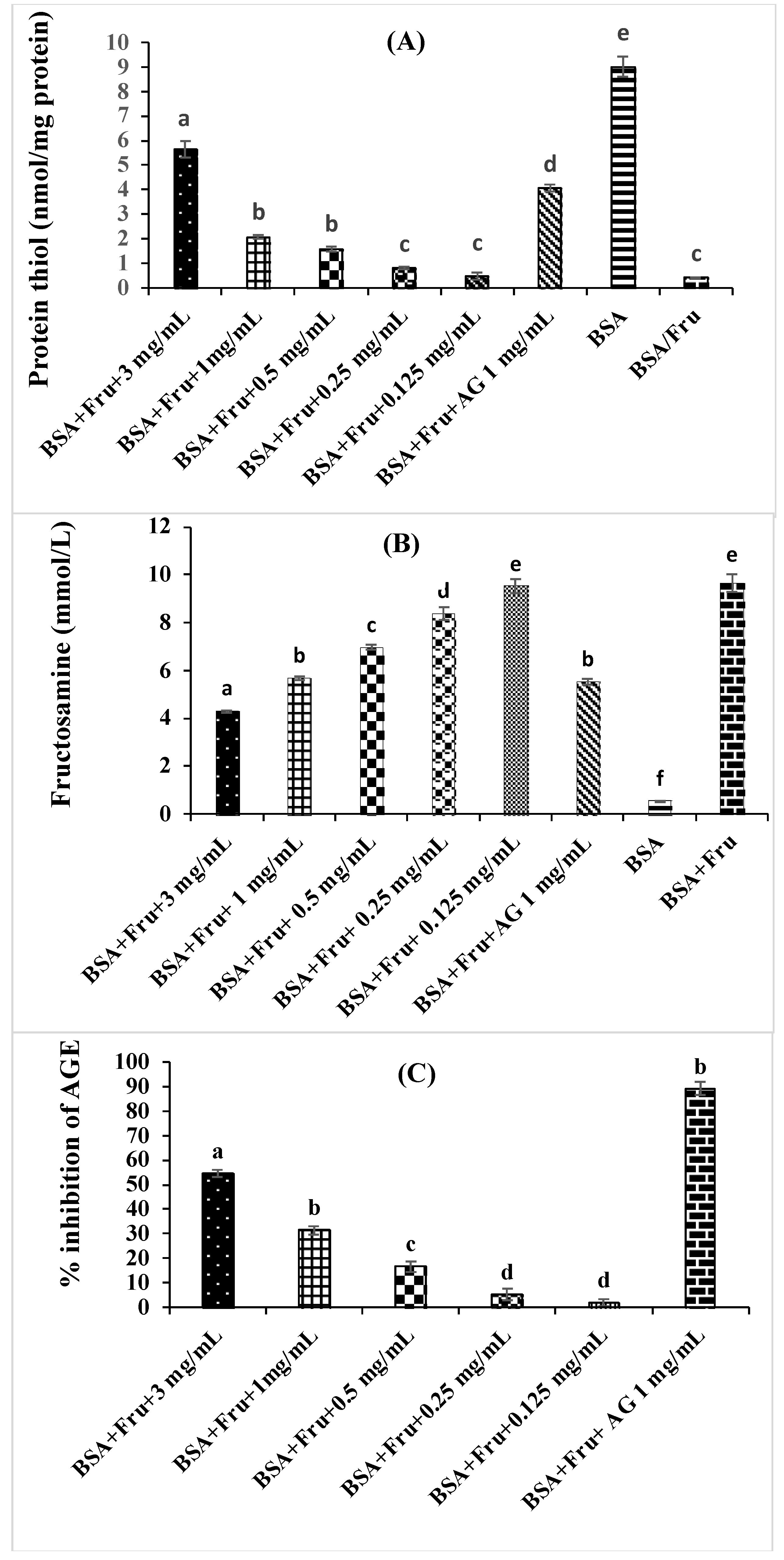
3.4. Inhibition of Beta vulgaris subsp. maritima Leaves on Non-Enzymatic Glycation Process

Glycation of proteins is a non-enzymatic chemical modification of proteins or amino acids by reducing sugars such as fructose [18]. Advanced glycation end products (AGEs) are irreversible heterogeneous byproducts of this reaction that are implicated in the development of aging as well as the pathogenesis of age-related disorders such as diabetes’ complications [37,38]. The extract of B. vulgaris subsp. maritima leaves was evaluated with BSA assay to determine its ability to reduce AGEs’ formation. The extract (0.125–3 mg/mL) showed a concentration-dependent anti-glycation effect (Figure 1C). After 4 weeks of incubation, the highest concentration reduced BSA glycation by 54.63%, while 1 mg/mL of aminoguanidine, the positive control, inhibited glycation by 89.20%.
In the early stages of glycation, unstable Schiff’s bases form, which are then converted into Amadori products such as fructosamine. As a result, reducing fructosamine is a therapeutic strategy for the onset of diabetes’ complications [18]. The extract of B. vulgaris subsp. maritima leaves significantly reduced fructosamine and AGE production. Figure 1B shows the amount of fructosamine produced by Amadori. After 4 weeks, monosaccharide-induced glycated BSA was found to have significantly higher fructosamine levels than non-glycated BSA. The addition of B. vulgaris subsp. maritima extract and AG significantly suppressed the generation of fructosamine. At the end of the study period, the utilization of B. vulgaris subsp. maritima extract led to a reduction in the fructosamine levels in a dose-dependent manner. Compared to the positive control, the content of fructosamine in the fructose-glycated BSA was the same in the extract at the concentration of 1 mg/mL. The effects of B. vulgaris subsp. maritima extract on the oxidation of protein thiols are shown in Figure 1A. BSA incubated with fructose had significantly decreased thiol groups’ content when compared to BSA alone. At week 4, fructose caused a decrease in thiol group content in BSA (0.5 nmol/mg protein). There was a significant improvement in the level of thiol after the addition of B. vulgaris subsp. maritima extract at various concentrations (3–0.125 mg/mL) as well as AG. At week 4 of incubation, B. vulgaris subsp. maritima extract and AG mediated protect the thiol groups at the different concentrations with the highest content observed when using 3 mg/mL of the extract.
AGE production begins under hyperglycemic or oxidative stress conditions and is characterized by the conversion of reversible Schiff-base adducts to covalently bound Amadori products, which undergo further rearrangements that terminate in the formation of irreversibly bound molecules known as AGEs [39].
To the best of our knowledge, the effects of B. vulgaris subsp. maritima leaves have not yet been examined for bioactivities relevant to diabetes; therefore, our studies on the effects of B. vulgaris subsp. maritima extract on glycation inhibition add significant further information to current research.
In this work, we showed that B. vulgaris subsp. maritima extract had significant effects on the different steps of glycation. These effects could be the result of the chemical composition of the leaves of B. vulgaris subsp. maritima. Moreover, the antioxidant activity of polyphenols may highlight major mechanisms for the prevention of AGE formation by reducing free radical generation [40]. Recent studies showed that polyphenolic compounds from edible plants may play a protective role against sugars induced protein glycation. Grzegorczyk et al. [41] reported a strong correlation between the polyphenolic content in the plant extracts and the ability to inhibit protein glycation. According to the findings, it may be postulated that B. vulgaris subsp. maritima extract can inhibit glycation through different pathways such as blocking the early glycation product (fructosamine), reducing the generation of a reactive group either from fructosamine or glucose, and inhibiting the AGEs’ formation. The results of the study put forward the protective effect of polyphenolic compounds of B. vulgaris subsp. maritima toward glycation-induced cellular damage and inhibition of AGEs.

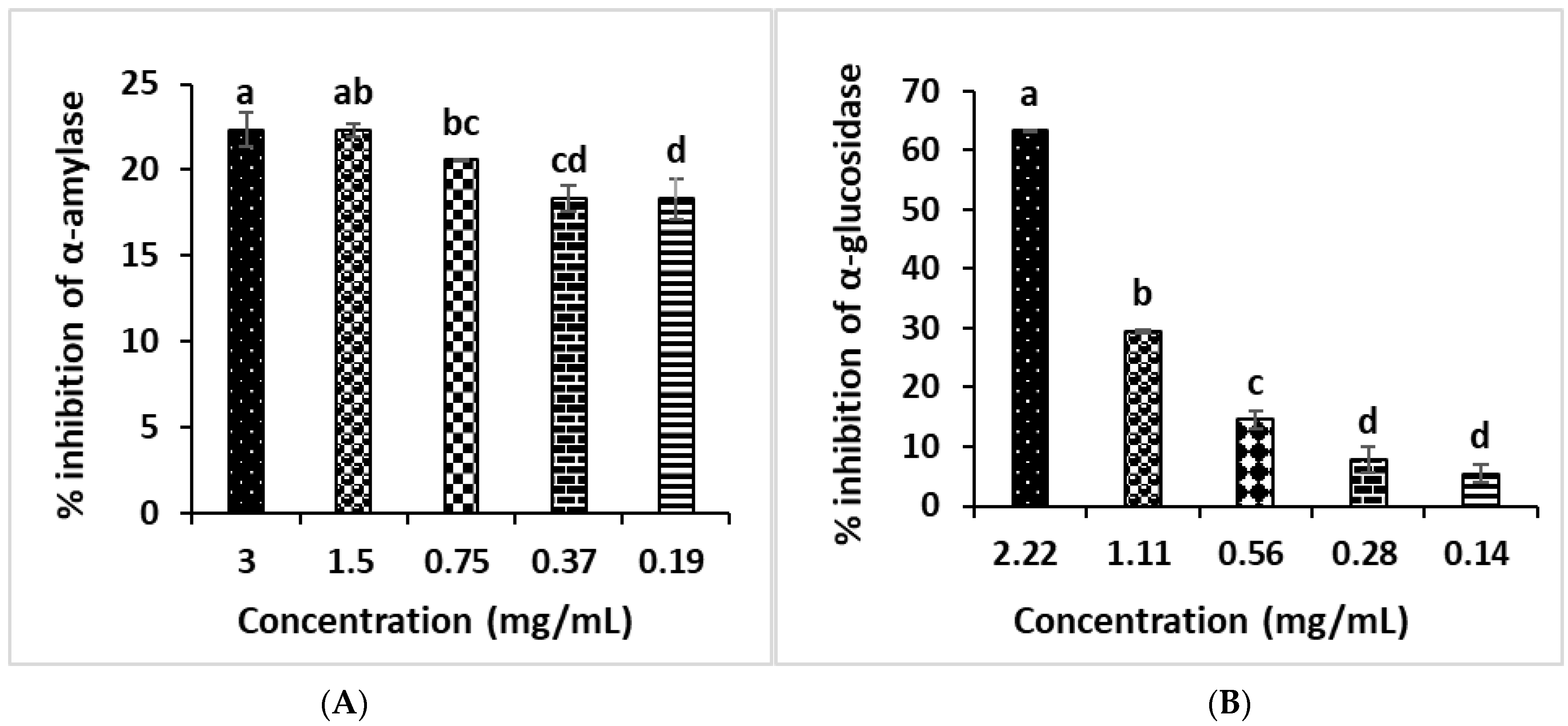
3.5. In Vitro Study of Anti-Diabetic Activities of B. vulgaris subsp. maritima Leaves’ Extract

The ability of α-amylase and α-glucosidase to inhibit complex carbohydrates’ hydrolysis during digestion, such as starch, was investigated as a strategy for controlling post-prandial plasma glucose levels by retarding or avoiding complex carbohydrates’ hydrolysis during digestion, resulting in lower glucose absorption [42]. Acarbose is an antidiabetic drug that works by inhibiting the enzymes α-amylase and α-glucosidase. Although this medication is effective at lowering blood glucose levels, long-term use can cause liver toxicity and uncomfortable gastrointestinal symptoms [43]. As a result, there is a demand for new α-amylase and α-glucosidase inhibitors derived from natural sources, especially plants, that do not have any negative or unwanted side effects in diabetic patients. Beta vulgaris is one of the most medicinal plants that could be employed by diabetics [44]. The inhibitory effects of the methanolic extract of B. vulgaris subsp. maritima leaves against the enzymes α-glucosidase and α-amylase were also investigated in our investigation. The results are shown in Table 4 and Figure 2. Both α-glucosidase and α-amylase inhibition had a moderate effect on B. vulgaris subsp. maritima leaves. The inhibitory effect of α-amylase was 25.93% at 3 mg/mL, while the inhibitory concentration of acarbose was 92.43% at 1 mg/mL and the percentage of α-glucosidase inhibition was 63.28% at 2.22 mg/mL compared to the acarbose, which showed 58.71 inhibition percent at 0.33 mg/mL (Figure 2A,B). The presence of some molecules such as saponin may explain the potential of B. vulgaris subsp. maritima to inhibit enzymes related to diabetes disease [34]. Indeed, the flavonoid content of B. vulgaris subsp. maritima extract may be responsible for its enzyme inhibitory capacity. Flavonoids inhibit glucose transporters. For example, quercetin has been shown to have anti-diabetic properties by inhibiting the intestinal glucose transporter GLUT2 [45]. Another complementary mechanism could be flavonoid-induced inhibition of α-amylase and α-glucosidase activity. For example, two flavonol glycosides isolated from Salsola kali that have B. vulgaris leaves and seeds, such as vitexin, isovitexin, orientin, and isoorientin, have been found to inhibit α-glucosidase and may be the most likely cause of the enzyme inhibitory activity shown in diabetes patients [46,47].

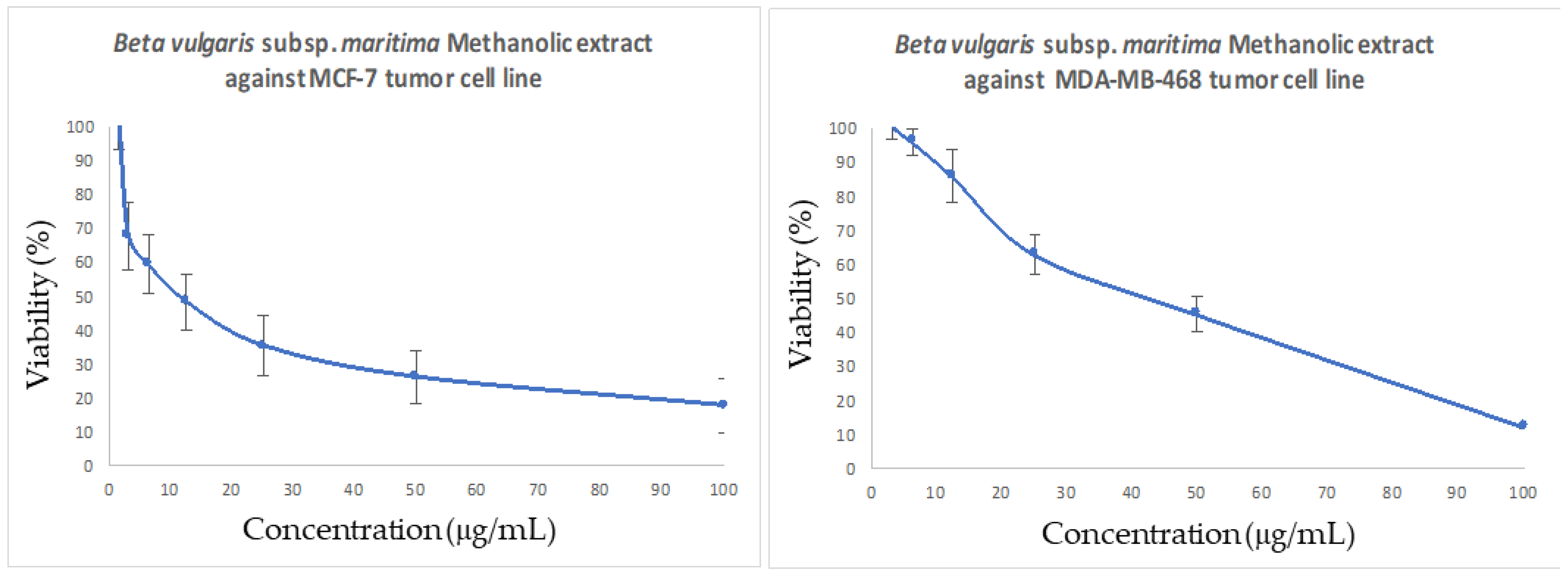
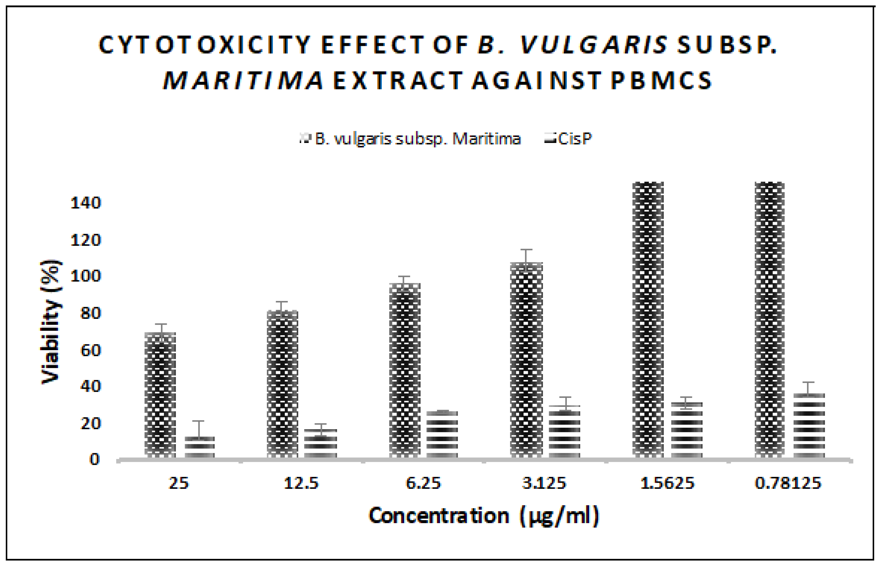
3.6. In Vitro Antitumor Activity of B. vulgaris subsp. maritima Leaves against Breast Cancer

Methanolic extract of B. vulgaris subsp. maritima leaves was assessed in our study for its effects against two human breast cancers: the triple negative breast cancer (MDA-MB-468) and the ER-positive (MCF-7) (Figure 3).
In our work, we treated both tumor cells lines with various concentrations of the methanolic extract of B. vulgaris (0.78, 1.56, 3.12, 6.25, 12.5, 25, 50, and 100 µg/mL) for 48 h. The results showed a dose-dependent, significant cytotoxic effect against both breast cancer cell lines. Furthermore, the B. vulgaris methanolic extract had moderate potency against both MCF-7 (82.28 percent for 100 g/mL) and MDA-MB-468 (87.86 percent for 100 g/mL) mammalian cell lines. Indeed, the IC50 values for MCF-7 and MDA-MB-468 were found to be 11.57 5.09 and 42.50 7.27 g/mL, respectively (Table 5).
Our findings are in the same line with those obtained by Gennari et al. [48], who found that the other Beta vulgaris genus such as Beta vulgaris var. cicla had multiple compounds with chemopreventive and moderate cytotoxic effects against human colon cancer (RKO) cell line.
To our knowledge, there are no studies yet that have demonstrated the qualification of the species B. vulgaris subsp. maritima extracts as an anticancer agent towards human breast cancer, in particular on TNBC and ER-positive. The results obtained in this work were found to be consistent with those obtained by some compounds extracted from essential oil of B. maritima against the HeLa and A549 cells lines. Accordingly, the in vitro antitumor activity of both extracts may be attributed to their specific components, such as hesperidin and thymoquinone, which were reported to have an antitumor effect towards MCF-7 [49] and MDA-MB-231 [50].
In addition, B. vulgaris subsp. maritima ethanolic extract has proven markedly effective as an anti-genotoxic [51], which make it a source of natural bioactive molecules that can be used as a safe food source and as a chemopreventive.
Similarly, the cytotoxic effect on human normal cells was considered in this study. The cytotoxicity of B. vulgaris subsp. maritima leaf extracts on human peripheral blood mononuclear cells (PBMCs) was also assessed (Figure 4).
The methanolic extract of B. vulgaris subsp. maritima leaves was found to have no cytotoxic effect against normal cells (PBMCs) (IC50 > 50 µg/mL). Therefore, our extract is considered to have a highly selective killing ability against TNBC and ER-positive breast cancer cell lines (MDA-MB-468 and MCF7). As a result, the percentage of lysis obtained for the MDA-MB-468 and MCF-7 cancer cell lines demonstrated promising chemopreventive properties for possible synergic drug development.

3.7. Antibacterial Activity of B. vulgaris subsp. maritima Leaves’ Extract

The antimicrobial effect of B. vulgaris subsp. maritima leaves’ extracts against the different microorganisms belonging to Gram (−) and Gram (+) was quantitatively and qualitatively demonstrated by the diameter of the zone of inhibition, in addition to the MIC and MBC results (Table 6 and Table 7). According to the in vitro test, our results showed that the extract of B. vulgaris subsp. maritima leaves exhibited antimicrobial activity against two bacteria tested with a variable degree. Indeed, the most sensitive bacteria was Bacillus subtilis with a diameter inhibitory zone value of 10.7 ± 0.6 mm with MIC equal to 25 mg/mL, followed by Proteus mirabilis (9.0 ± 0.0 mm). Staphylococcus aureus, Pseudomonas aeruginosa, and Escherichia coli were not sensitive to the extract.
Antibacterial properties of various phytochemicals have been studied to prove the capacity to treat infectious diseases. In their natural state, plants produce chemicals that are toxic to microorganisms [52]. The presence of secondary metabolites such as terpenoids, phenolics, and flavonoids in B. vulgaris subsp. maritima leaves’ extract was discovered in this study (Table 2). According to available evidence, secondary metabolites such as alkaloids, flavonoids, tannins, and other phenolic compounds are thought to be responsible for antimicrobial effects in plants [53,54]. These compounds act on the lipid membrane and its permeability through destabilization of the cytoplasmic membrane or by disrupting the ions’ transports [55].

4. Conclusions

In conclusion, high levels of phenolic and flavonoid content were found in both aqueous and methanolic extracts. Likewise, these bioactive components were suggested to be responsible for the potent antioxidant activity. The analysis of the in vitro antitumor activity of methanolic extract towards both human breast cancer cell lines (MCF-7 and MDA-MB-468) showed potent cytotoxicity effects in a dose-dependent manner. Moreover, the methanolic extract appears to be not cytotoxic against normal cells (PBMCs). These findings suggest a highly selective killing ability towards TNBC and ER-positive breast cancer cell lines (MDA-MB-468 and MCF7). Our study found that extracts from B. vulgaris subsp. maritima leaves inhibit not only α-amylase and α-glucosidase but also advanced glycation end-product formation, which could help to prevent diabetes’ complications. However, the methanolic extract of B. vulgaris subsp. maritima leaves have a strong antibacterial effect against Proteus mirabilis and Bacillus subtilis. For all these reasons, these promising chemopreventive properties might be of use in possible synergic drug development. Taken together, B. vulgaris subsp. maritima leaves’ extracts constitute a rich, medicinal, natural source of antioxidants that can be used as a promising food.

Author Contributions

Conceptualization, N.B. and R.B.M.; methodology, N.B., R.B.M., I.K. and Z.Z.; software, H.C. and K.K.; validation, N.B. and R.B.M.; formal analysis, N.B., R.B.M., I.K. and Z.Z.; investigation, N.B. and R.B.M.; resources, F.C.; data curation, N.B., R.B.M., I.K., Z.Z., H.C. and K.K.; writing—original draft preparation, N.B., R.B.M., I.K., Z.Z., H.C., K.K. and F.C.; writing—review and editing, N.B., R.B.M., A.Z., M.N. and F.C.; visualization, F.C.; supervision, A.Z. and M.N.; project administration, N.B. and R.B.M.; funding acquisition, A.Z. and M.N. All authors have read and agreed to the published version of the manuscript.

Funding

This research received no external funding.

Institutional Review Board Statement

Not applicable.

Informed Consent Statement

Not applicable.

Data Availability Statement

Not applicable.

Conflicts of Interest

The authors declare no conflict of interest.

References

  1. Saeedian, M.-E.; Amini, M. 2-[2-Methyl-5-phenyl-1-(3, 4, 5-trimethoxyphenyl)-1H-pyrrol-3-yl]-2-oxo-N-(pyridin-4-yl) acetamide. Molbank 2018, 2018, 1002. [Google Scholar] [CrossRef] [Green Version]
  2. Bouchmaa, N.; Ben Mrid, R.; Boukharsa, Y.; Nhiri, M.; Ait Mouse, H.; Taoufik, J.; Zyad, A. Cytotoxicity of new pyridazin-3 (2H)-one derivatives orchestrating oxidative stress in human triple-negative breast cancer (MDA-MB-468). Arch. Pharm. 2018, 351, 1800128. [Google Scholar] [CrossRef] [PubMed]
  3. Bouchmaa, N.; Tilaoui, M.; Boukharsa, Y.; Jaâfari, A.; Mouse, H.A.; Oukerrou, M.A.; Taoufik, J.; Ansar, M.; Zyad, A. In vitro antitumor activity of newly synthesized pyridazin-3 (2H)-one derivatives via apoptosis induction. Pharm. Chem. J. 2018, 51, 893–901. [Google Scholar] [CrossRef]
  4. Zhu, A.-G.; Jiang, Z.-S.; Gong, P.-Y.; Zhang, D.-M.; Zou, Z.-W.; Zhang, Q.; Ma, H.-M.; Guo, Z.-g.; Zhao, J.-y.; Dong, J.-j.; et al. Efficacy of low-protein diet for diabetic nephropathy: A systematic review of randomized controlled trials. Lipids Health Dis. 2018, 17, 141. [Google Scholar] [CrossRef] [PubMed] [Green Version]
  5. Ben Mrid, R.; Bouargalne, Y.; El Omari, R.; Nhiri, M. New insights into the therapeutic effects of phenolic acids from sorghum seeds. J. Rep. Pharm. Sci. 2019, 8, 91. [Google Scholar]
  6. Li, Y.; Li, S.; Meng, X.; Gan, R.Y.; Zhang, J.J.; Li, H.B. Dietary natural products for prevention and treatment of breast cancer. Nutrients 2017, 9, 728. [Google Scholar] [CrossRef] [PubMed] [Green Version]
  7. Oukerrou, M.A.; Tilaoui, M.; Mouse, H.A.; Bouchmaa, N.; Zyad, A. Differential cytotoxic activity of essential oil of Lippia citriodora from different regions in Morocco. Chem. Biodivers. 2017, 14, e1600497. [Google Scholar] [CrossRef]
  8. Ben Mrid, R.; Bouchmaa, N.; Bouargalne, Y.; Ramdan, B.; Karrouchi, K.; Kabach, I.; Nhiri, M. Phytochemical characterization, antioxidant and in vitro cytotoxic activity evaluation of Juniperus oxycedrus Subsp. oxycedrus needles and berries. Molecules 2019, 24, 502. [Google Scholar] [CrossRef] [Green Version]
  9. Kazimierczak, R.; Hallmann, E.; Lipowski, J.; Drela, N.; Kowalik, A.; Püssa, T.; Matt, D.; Luik, A.; Gozdowski, D.; Rembiałkowska, E. Beetroot (Beta vulgaris L.) and naturally fermented beetroot juices from organic and conventional production: Metabolomics, antioxidant levels and anticancer activity. J. Sci. Food Agric. 2014, 94, 2618–2629. [Google Scholar] [CrossRef]
  10. Nade, V.S.; Kawale, L.A.; Zambre, S.S.; Kapure, A.B. Neuroprotective potential of Beta vulgaris L. in Parkinson’s disease. Ind. J. Pharmacol. 2015, 47, 403. [Google Scholar] [CrossRef] [Green Version]
  11. Oztay, F.; Sacan, O.; Kayalar, O.; Bolkent, S.; Ipci, Y.; Kabasakal, L.; Sener, G.; Yanardag, R. Chard (Beta vulgaris var. cicla) extract improved hyperglycemia-induced oxidative stress and surfactant-associated protein alterations in rat lungs. Pharm. Biol. 2015, 53, 1639–1646. [Google Scholar] [CrossRef] [PubMed]
  12. Citores, L.; Iglesias, R.; Gay, C.; Ferreras, J.M. Antifungal activity of the ribosome-inactivating protein BE 27 from sugar beet (Beta vulgaris L.) against the green mould Penicillium digitatum. Mol. Plant Pathol. 2016, 17, 261–271. [Google Scholar] [CrossRef] [PubMed]
  13. Duke, J.A. Handbook of Energy Crops; Purdue University, Center for New Crops & Plants Products: West Lafayette, IN, USA, 1983. [Google Scholar]
  14. Asraoui, F.; Kounnoun, A.; El Cadi, H.; Cacciola, F.; Oulad El Majdoub, Y.; Alibrando, F.; Mandolfino, F.; Dugo, P.; Mondello, L.; Louajri, A. Phytochemical Investigation and Antioxidany Activity of Globularia alypum L. Molecules 2021, 26, 759. [Google Scholar] [CrossRef] [PubMed]
  15. Kabach, I.; Mrid, R.; Bouchmaa, N.; Bouargalne, Y.; Nhiri, A. Phytochemical screening, antioxidant and cytotoxic activities of M. vulgare. Int. J. Pharm. Res. 2019, 11, 338–345. [Google Scholar]
  16. Oyaizu, M. Studies on products of browning reaction antioxidative activities of products of browning reaction prepared from glucosamine. Jpn. Nutr. Diet. 1986, 44, 307–315. [Google Scholar] [CrossRef] [Green Version]
  17. Dinis, T.C.; Madeira, V.M.; Almeida, L.M. Action of phenolic derivatives (acetaminophen, salicylate, and 5-aminosalicylate) as inhibitors of membrane lipid peroxidation and as peroxyl radical scavengers. Arch. Biochem. Biophys. 1994, 315, 161–169. [Google Scholar] [CrossRef]
  18. Adisakwattana, S.; Sompong, W.; Meeprom, A.; Ngamukote, S.; Yibchok-Anun, S. Cinnamic acid and its derivatives inhibit fructose-mediated protein glycation. Int. J. Mol. Sci. 2012, 13, 1778–1789. [Google Scholar] [CrossRef]
  19. Zhang, L.; Meng, Z.; Jiang, Z.; Liu, Z.; Hou, L.; Cai, G.; Ou, H.; Huang, S.; Song, Q.; Fang, J.; et al. Indicators of glucose dysregulation and the relationship with iron overload in Chinese children with beta thalassemia major. Pediatric Diabetes 2021. [Google Scholar] [CrossRef]
  20. Dai, J.; Dong, A.; Xiong, G.; Liu, Y.; Hossain, M.S.; Liu, S.; Gao, N.; Li, S.; Wang, J.; Qiu, D. Production of highly active extracellular amylase and cellulase from Bacillus subtulis ZIM3 and a recombinant strain with a potential application in tobacco fermentation. Front. Microbiol. 2020, 11, 1539. [Google Scholar] [CrossRef]
  21. Bouchmaa, N.; Ben Mrid, R.B.; Boukharsa, Y.; Bouargalne, Y.; Nhiri, M.; Idir, A.; Taoufik, J.; Ansar, M.; Zyad, A. Reactive Oxygen Species-Mediated Apoptosis and Cytotoxicity of Newly Synthesized Pyridazin-3-Ones In P815 (Murin Mastocytoma) Cell Line. Drug Res. 2019, 69, 528–536. [Google Scholar] [CrossRef]
  22. Benali, T.; Chtibi, H.; Bouyahya, A.; Khabbach, A.; Hammani, K. Detection of antioxidant and antimicrobial activities in phenol components and essential oils of cistus ladaniferus and mentha suaveolens extracts. Biomed. Pharmacol. J. 2020, 13, 603–612. [Google Scholar] [CrossRef]
  23. Gulluce, M.; Sahin, M.; Sokmen, M.; Ozer, H.; Daferera, D.; Sokmen, A.; Polissiou, M.; Adiguzel, A.; Ozkan, H. Antimicrobial and antioxidant properties of the essential oils and methanol extract of Mentha longifolia L. ssp longifolia. Food Chem. 2007, 103, 1449–1456. [Google Scholar] [CrossRef]
  24. Shahidi, F.; Chandrasekara, A. Millet grain phenolics and their role in disease risk reduction and health promotion: A review. J. Funct. Food. 2013, 5, 570–581. [Google Scholar] [CrossRef]
  25. Biondo, P.B.F.; Boeing, J.S.; Barizão, É.O.; Souza, N.E.D.; Matsushita, M.; Oliveira, C.C.D.; Boroski, M.; Visentainer, J.V. Evaluation of beetroot (Beta vulgaris L.) leaves during its developmental stages: A chemical composition study. Food Sci. Technol. 2014, 34, 94–101. [Google Scholar] [CrossRef] [Green Version]
  26. Hajihosseini, S.; Setorki, M. The antioxidant activity of Beta vulgaris leaf extract in improving scopolamine-induced spatial memory disorders in rats. Avic. J. Phytomed. 2017, 7, 417. [Google Scholar]
  27. Gennari, L.; Felletti, M.; Blasa, M.; Angelino, D.; Celeghini, C.; Corallini, A.; Ninfali, P. Total extract of Beta vulgaris var. cicla seeds versus its purified phenolic components: Antioxidant activities and antiproliferative effects against colon cancer cells. Phytochem. Anal. 2011, 22, 272–279. [Google Scholar] [CrossRef]
  28. Kujala, T.S.; Vienola, M.S.; Klika, K.D.; Loponen, J.M.; Pihlaja, K. Betalain and phenolic compositions of four beetroot (Beta vulgaris) cultivars. Eur. Food Res. Technol. 2002, 214, 505–510. [Google Scholar] [CrossRef]
  29. Georgiev, V.G.; Weber, J.; Kneschke, E.M.; Denev, P.N.; Bley, T.; Pavlov, A.I. Antioxidant activity and phenolic content of betalain extracts from intact plants and hairy root cultures of the red beetroot Beta vulgaris cv. Detroit dark red. Plant Food. Hum. Nutr. 2010, 65, 105–111. [Google Scholar] [CrossRef]
  30. Barros, L.; Morales, P.; Carvalho, A.M.; Ferreira, I.C. Antioxidant potential of wild plant foods. In Mediterranean Wild Edible Plants; Springer: New York, NY, USA, 2016; pp. 209–232. [Google Scholar]
  31. Cheng, D.; Li, W.; Wang, L.; Li, T.; Poiani, G.; Wassef, A.; Hudilikar, R.; Ondar, P.; Brunetti, L.; Kong, A.-N. Pharmacokinetics, Pharmacodynamics, and PKPF Modeling of Curcumin in Regulating Antioxidant and Epigenetic Gene Expression I Healthy Human Volunteers. Mol. Pharm. 2019, 16, 1881–1889. [Google Scholar] [CrossRef]
  32. Xu, X.; Guo, S.; Hao, X.; Ma, H.; Bai, Y.; Huang, Y. Improving antioxidant and antiproliferative activities of colla corii asini hydrolysates using ginkgo biloba extracts. Food Sci. Nutr. 2018, 6, 765–772. [Google Scholar] [CrossRef]
  33. Zardi-Bergaoui, A.; Ben Nejma, A.; Harzallah-Skhiri, F.; Flamini, G.; Ascrizzi, R.; Ben Jannet, H. Chemical composition and biological studies of the essential oil from aerial parts of Beta vulgaris subsp. maritima (L.) arcang. growing in Tunisia. Chem. Biodivers. 2017, 14, e1700234. [Google Scholar] [CrossRef] [PubMed]
  34. Mzoughi, Z.; Chahdoura, H.; Chakroun, Y.; Cámara, M.; Fernández-Ruiz, V.; Morales, P.; Mosbah, H.; Flamini, G.; Snoussi, M.; Majdoub, H. Wild edible Swiss chard leaves (Beta vulgaris L. var. cicla): Nutritional, phytochemical composition and biological activities. Food Res. Int. 2019, 119, 612–621. [Google Scholar] [CrossRef]
  35. Aktumsek, A.; Zengin, G.; Guler, G.O.; Cakmak, Y.S.; Duran, A. Antioxidant potentials and anticholinesterase activities of methanolic and aqueous extracts of three endemic Centaurea L. species. Food Chem. Toxicol. 2013, 55, 290–296. [Google Scholar] [CrossRef] [PubMed]
  36. Valifard, M.; Mohsenzadeh, S.; Kholdebarin, B.; Rowshan, V. Effects of salt stress on volatile compounds, total phenolic content and antioxidant activities of Salvia mirzayanii. S. Afr. J. Bot. 2014, 93, 92–97. [Google Scholar] [CrossRef] [Green Version]
  37. Rowan, S.; Bejarano, E.; Taylor, A. Mechanistic targeting of advanced glycation end-products in age-related diseases. Biochim. Biophys. Acta 2018, 1864, 3631–3643. [Google Scholar] [CrossRef]
  38. Gill, V.; Kumar, V.; Singh, K.; Kumar, A.; Kim, J.J. Advanced glycation end products (AGEs) may be a striking link between modern diet and health. Biomolecules 2019, 9, 888. [Google Scholar] [CrossRef] [Green Version]
  39. Sarmah, S.; Roy, A.S. A review on prevention of glycation of proteins: Potential therapeutic substances to mitigate the severity of diabetes complications. Int. J. Biol. Macromol. 2021, 195, 565–588. [Google Scholar] [CrossRef]
  40. Wu, C.H.; Huang, S.M.; Lin, J.A.; Yen, G.C. Inhibition of advanced glycation endproduct formation by foodstuffs. Food Funct. 2011, 2, 224–234. [Google Scholar] [CrossRef]
  41. Grzegorczyk-Karolak, I.; Gołąb, K.; Gburek, J.; Wysokińska, H.; Matkowski, A. Inhibition of advanced glycation end-product formation and antioxidant activity by extracts and polyphenols from Scutellaria alpina L. and S. altissima L. Molecules 2016, 21, 739. [Google Scholar] [CrossRef] [Green Version]
  42. Fernández-Fernández, A.M.; Iriondo-DeHond, A.; Dellacassa, E.; Medrano-Fernandez, A.; del Castillo, M.D. Assessment of antioxidant, antidiabetic, antiobesity, and anti-inflammatory properties of a Tannat winemaking by-product. Eur. Food Res. Technol. 2019, 245, 1539–1551. [Google Scholar] [CrossRef]
  43. Lordan, S.; Smyth, T.J.; Soler-Vila, A.; Stanton, C.; Ross, R.P. The a-Amylase and a-Glucosidase Inhibitory Effects of Irish Seaweed Extracts. Food Chem. 2013, 141, 2170–2176. [Google Scholar] [CrossRef]
  44. Ozsoy-Sacan, O.; Karabulut-Bulan, O.; Bolkent, S.; Yanardag, R.; Ozgey, Y. Effects of chard (Beta vulgaris L. var cicla) on the liver of the diabetic rats: A morphological and biochemical study. Biosci. Biotechnol. Biochem. 2004, 68, 1640–1648. [Google Scholar] [CrossRef] [PubMed] [Green Version]
  45. Song, J.; Kwon, O.; Chen, S.; Daruwala, R.; Eck, P.; Park, J.B.; Levine, M. Flavonoid inhibition of sodium-dependent vitamin C transporter 1 (SVCT1) and glucose transporter isoform 2 (GLUT2), intestinal transporters for vitamin C and glucose. J. Biol. Chem. 2002, 277, 15252–15260. [Google Scholar] [CrossRef] [PubMed] [Green Version]
  46. Tundis, R.; Loizzo, M.R.; Statti, G.A.; Menichini, F. Inhibitory effects on the digestive enzyme α-amylase of three Salsola species (Chenopodiaceae) in vitro. Int. J. Pharm. Sci. 2007, 62, 473–475. [Google Scholar]
  47. Li, H.; Song, F.; Xing, J.; Tsao, R.; Liu, Z.; Liu, S. Screening and structural characterization of α-glucosidase inhibitors from hawthorn leaf flavonoids extract by ultrafiltration LC-DAD-MS n and SORI-CID FTICR MS. J. Am. Soc. Mass Spectrom. 2009, 20, 1496–1503. [Google Scholar] [CrossRef] [PubMed] [Green Version]
  48. Gennari, A.; Stockler, M.; Puntoni, M.; Sormani, M.; Nanni, O.; Amadori, D.; Wilcken, N.; D’Amico, M.; DeCensi, A.; Bruzzi, P. Duration of chemotherapy for metastatic breast cancer: A systematic review and meta-analysis of randomized clinical trials. J. Clin. Oncol. 2011, 29, 2144–2149. [Google Scholar] [CrossRef] [Green Version]
  49. Kongtawelert, P.; Wudtiwai, B.; Shwe, T.H.; Pothacharoen, P.; Phitak, T. Inhibitory effect of Hesperidin on the expression of programmed death ligand (PD-L1) in breast cancer. Molecules 2020, 25, 252. [Google Scholar] [CrossRef] [Green Version]
  50. Al-Mutairi, A.; Rahman, A.; Rao, M.S. Low doses of thymoquinone and ferulic acid in combination effectively inhibit proliferation of cultured MDA-MB 231 breast adenocarcinoma cells. Nutr. Canc. 2021, 73, 282–289. [Google Scholar] [CrossRef]
  51. Mekki, L.; Mansour, H.; Eldean, E.G.; Nasser, A.A. Evaluation of anti-genotoxicity of the ethanolic plant extract of Beta vulgaris Maritima using Allium cepa root assay. Egypt J. Exp. Biol. 2015, 11, 147–153. [Google Scholar]
  52. Anand, U.; Jacobo-Herrera, N.; Altemimi, A.; Lakhssassi, N. A comprehensive review on medicinal plants as antimicrobial therapeutics: Potential avenues of biocompatible drug discovery. Metabolites 2019, 9, 258. [Google Scholar] [CrossRef] [Green Version]
  53. Othman, L.; Sleiman, A.; Abdel-Massih, R.M. Antimicrobial activity of polyphenols and alkaloids in middle eastern plants. Front. Microbiol. 2019, 10, 911. [Google Scholar] [CrossRef] [PubMed]
  54. Jubair, N.; Rajagopal, M.; Chinnappan, S.; Abdullah, N.B.; Fatima, A. Review on the antibacterial mechanism of plant-derived compounds against multidrug-resistant bacteria (MDR). Evid.-Based Complement. Alternat. Med. 2021, 2021, 3663315. [Google Scholar] [CrossRef] [PubMed]
  55. Mishra, A.; Sharma, A.K.; Kumar, S.; Saxena, A.K.; Pandey, A.K. Bauhinia variegata leaf extracts exhibit considerable antibacterial, antioxidant, and anticancer activities. Biomed. Res. Int. 2013, 2013, 915436. [Google Scholar] [CrossRef] [PubMed] [Green Version]
Figure 1. The effects of B. vulgaris subsp. maritima leaves extract (0.125–3 mg/mL) and aminoguanidine (1 mg/mL) on (A) the level of protein thiol group, (B) the level of fructosamine, and (C) the percentage of AGE inhibition in BSA/fructose system. Each value represents the mean of three replicates. Bars represent the standard error. Different letters indicate significant differences among treatments at p < 0.05.
Applsci 12 01866 g001
Figure 2. Percentage of α-amylase inhibition (A) and α-glucosidase inhibition (B) versus different concentrations of B. vulgaris subsp. maritima leaves’ extract. Each value represents the mean of three replicates. Bars represent the standard error. Different letters indicate significant differences among treatments at p < 0.05.
Applsci 12 01866 g002
Figure 3. The cytotoxicity effect against the human breast adenocarcinoma MDA-MB-468 and MCF-7 cell lines after treatment at different concentrations with methanolic extracts of B. vulgaris subsp. maritima leaves for 48 h. Each value represents the mean ± standard deviation of three independent replicates. Different letters indicate significant differences (p < 0.05) within the same concentration.
Applsci 12 01866 g003
Figure 4. The viability of PBMCs treated at various concentrations of methanolic extracts of B. vulgaris subsp. maritima leaves’ extracts for 48 h. Each value represents the mean ± standard deviation of three independent replicates. Different letters indicate significant differences (p < 0.05) within the same concentration.
Applsci 12 01866 g004
Table 1. Bioactive compounds (mean ± SD) obtained for the studied Beta vulgaris subsp. maritima.
Extract Yield (%)Polyphenols (mg GAE/g dw)Flavonoids (mg QE/g dw)
Aqueous extract21.7112.377 ± 3.324 a26.023 ± 3.565 a
Methanolic extract18.8237.117 ± 6.336 b38.785 ± 1.148 b
The values are mean ± standard deviation. GAE: Gallic Acid Equivalent; dw: dry weight; QE: Quercetin Equivalent. Different letters indicate significant differences between conditions (p < 0.05).
Table 2. Major phenolic compounds identified in Beta vulgaris subsp. maritima extract by HPLC.
Phenolic Compounds
mg/100 g dw
Aqueous ExtractMethanolic Extract
Hydroxycinnamic acids
Caffeic acid0.61ND
P-coum acid 0.490.49
Hydroxybenzoic acids
Salicylic acid758.52ND
Syringic acid2.16ND
Gallic acidNDND
p-hydroxybenzoic acid0.58ND
Flavonoids
Hesperidin189.0768.82
Naringinin5.2811.12
Rutin47.86ND
Terpernes
Thymoquinone0.5716.48
LimoneneNDND
dw: dry weight. ND: Not Detected.
Table 3. IC50 values (mean ± SD) obtained in the antioxidant activity assays of Beta vulgaris subsp. maritima.
Antioxidants’ Properties (IC50 Values; mg/mL)Reducing Power
(mg AAE/g dw)
DPPH Scavenging ActivityABTSMetal Chelating Activity
Aqueous extract1.789 ± 0.062 a1.551 ± 0.094 a0.484 ± 0.007 a98.498 ± 4.198 a
Methanolic extract0.919 ± 0.044 b1.218 ± 0.044 a0.739 ± 0.057 b300.057 ±10.824 b
All the values are mean ± standard deviation. IC50: the extract concentration providing 50% inhibition; DPPH: 2,2-diphenyl-1-picrylhydrazyl; ABTS: 2,2′-azino-bis (3ethylbenzothiazoline-6-sulphonic acid); AAE: Ascorbic acid Equivalent; dw: dry weight. Different letters (a & b) within each column indicate statistically significant differences at p < 0.05.
Table 4. Inhibition results of Beta vulgaris subsp. maritima extract on both α-amylase and α-glucosidase enzymes.
IC50 (mg/mL)
α-Amylaseα-Glucosidase
Beta vulgaris subsp. maritima leaves-1.792 ± 0.005 a
Acarbose0.046 ± 0.001 b0.329 ± 0.041 b
Values are the mean of three replicates ± standard deviation. Different letters (a & b) within each column indicate statistically significant differences at p < 0.05.
Table 5. IC50 values of cytotoxic activity towards MDA-MB-468, MCF-7, and PBMCs and percentage of viability on PBMCs at various concentrations of the extract and CisP. Cells were treated with methanolic extracts from B. vulgaris subsp. Maritima leaves.
Sample TestedIC50 of Cytotoxic Activity against Tumor Cells% of Viability in PBMCs
MCF-7MDA-MB-468PBMCsConcentration (µg/mL)
12.53.1250.78125
B. v. subsp. Maritima11.57 ± 5.09 a42.50 ± 7.27 a>50 a81.15 ±5.91 a108.02 ±1.49 a224.9 ±6.46 a
CisP0.20 ± 0.0 c2.20 ± 0.40 c0.27 b16.08 ± 3.39 b30.08 ± 3.58 b37.96 ± 3.44 b
Values are the mean of three replicates ± standard deviation. Different letters (a & b& c) within each column indicate statistically significant differences at p < 0.05.
Table 6. Antimicrobial activity of B. vulgaris subsp. maritima leaves’ extract using the disc diffusion method against Gram-positive and -negative strains.
Bacterial StrainsInhibition Zone Diameter (mm)
B. vulgaris subsp. maritimaGentamicine
Proteus mirabilis9.0 ± 0.018.7± 0.6
Bacillus subtilis10.7 ± 0.619.7 ± 0.6
Staphylococcus aureusn.e15 ± 1
Pseudomonas aeruginosan.en.e
Escherichia colin.e16 ± 1
The diameter of the inhibition zones (mm), including diameter of disc (6 mm), is given as mean ± SD of triplicate experiments; n.e: no effect.
Table 7. Minimal inhibitory (MIC) and minimal bactericidal (MBC) concentrations (mg/mL) of B. vulgaris subsp. maritima leaves’ extract.
SpeciesProteus mirabilisBacillus subtilis
MIC (mg/mL)MBC (mg/mL)MIC(mg/mL)MBC(mg/mL)
B. vulgaris subsp. maritimaNDND25 ± 0>25
ND: not determined.
Publisher’s Note: MDPI stays neutral with regard to jurisdictional claims in published maps and institutional affiliations.

