
A Drug Stability Study Using Surface-Enhanced Raman Scattering on Silver Nanoparticles


Abstract
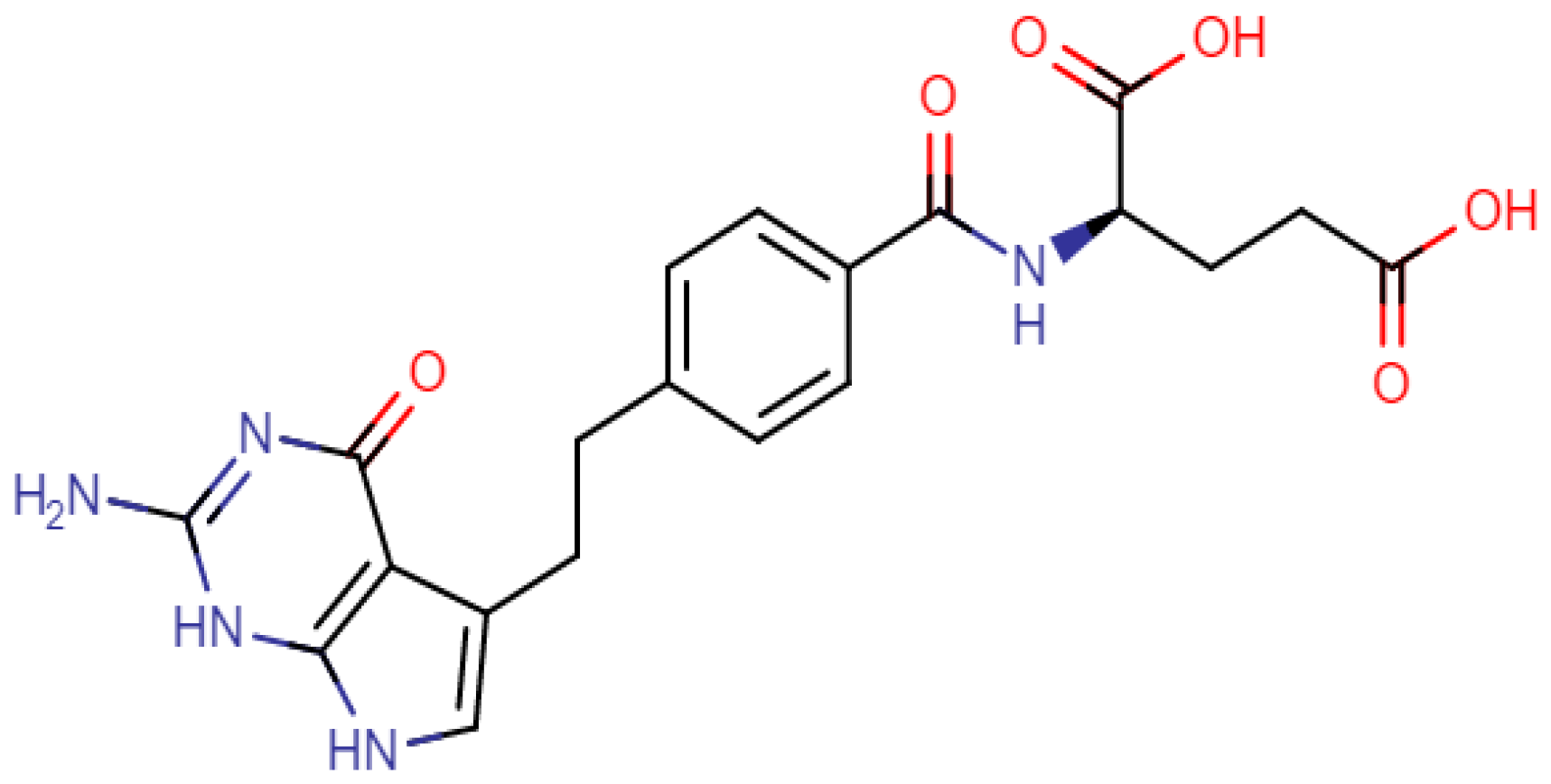
1. Introduction
2. Materials and Methods
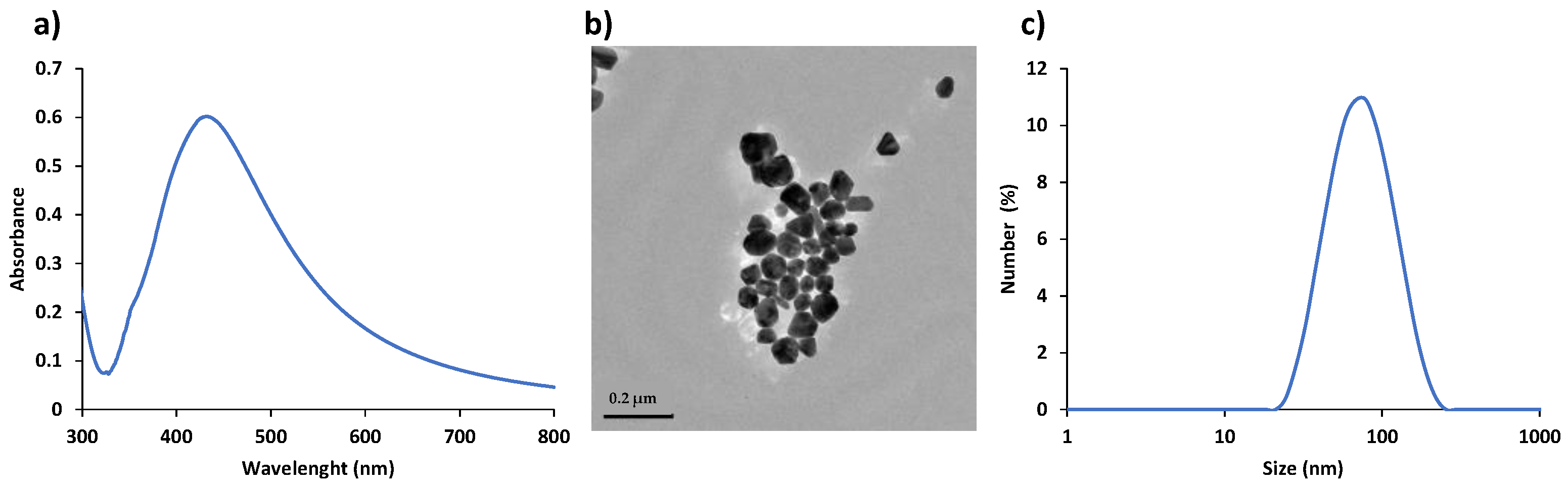
2.1. Silver Nanoparticles (AgNPs) Synthesis and Characterization
2.2. Forced Degradation Conditions
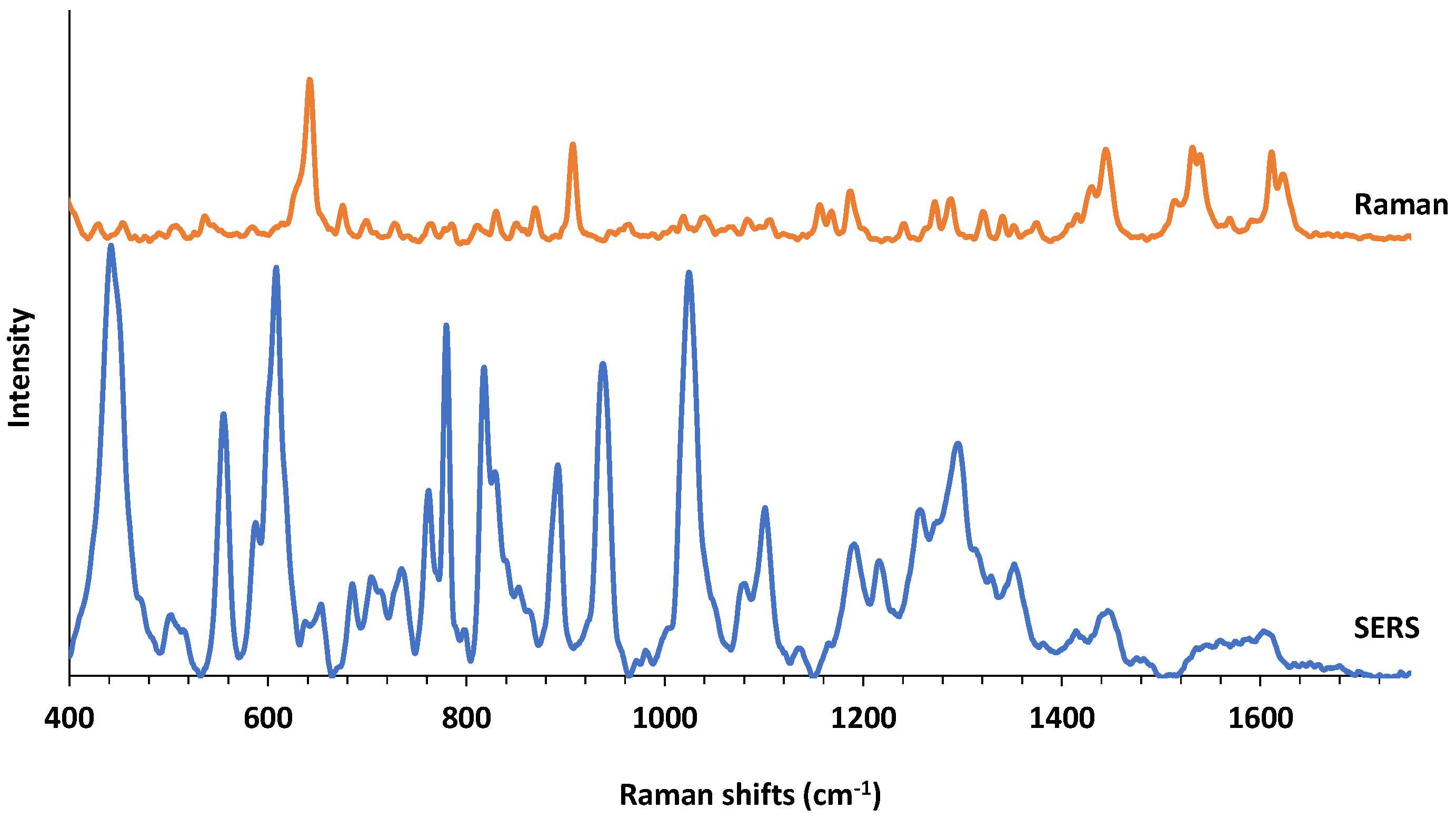
2.3. SERS Studies
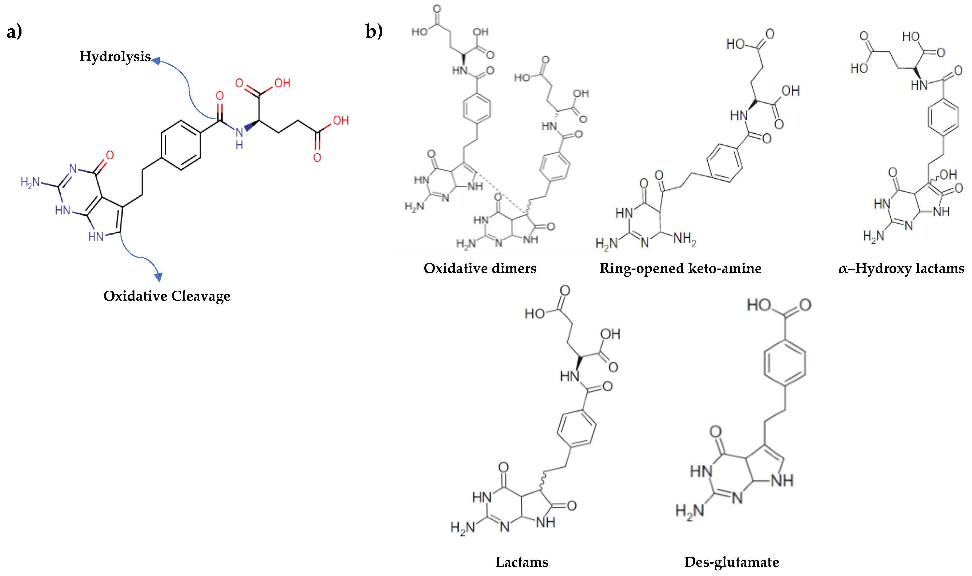
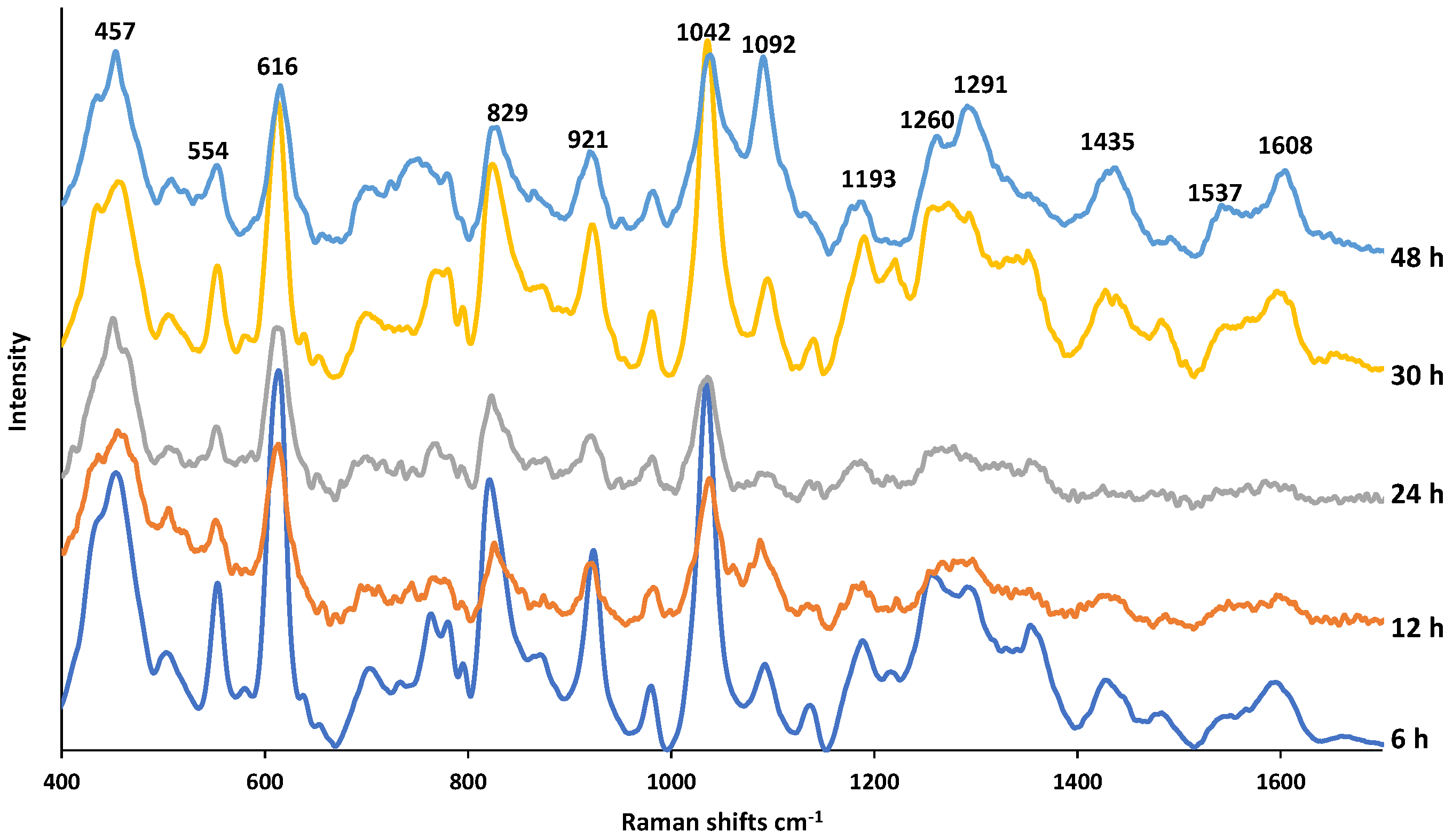
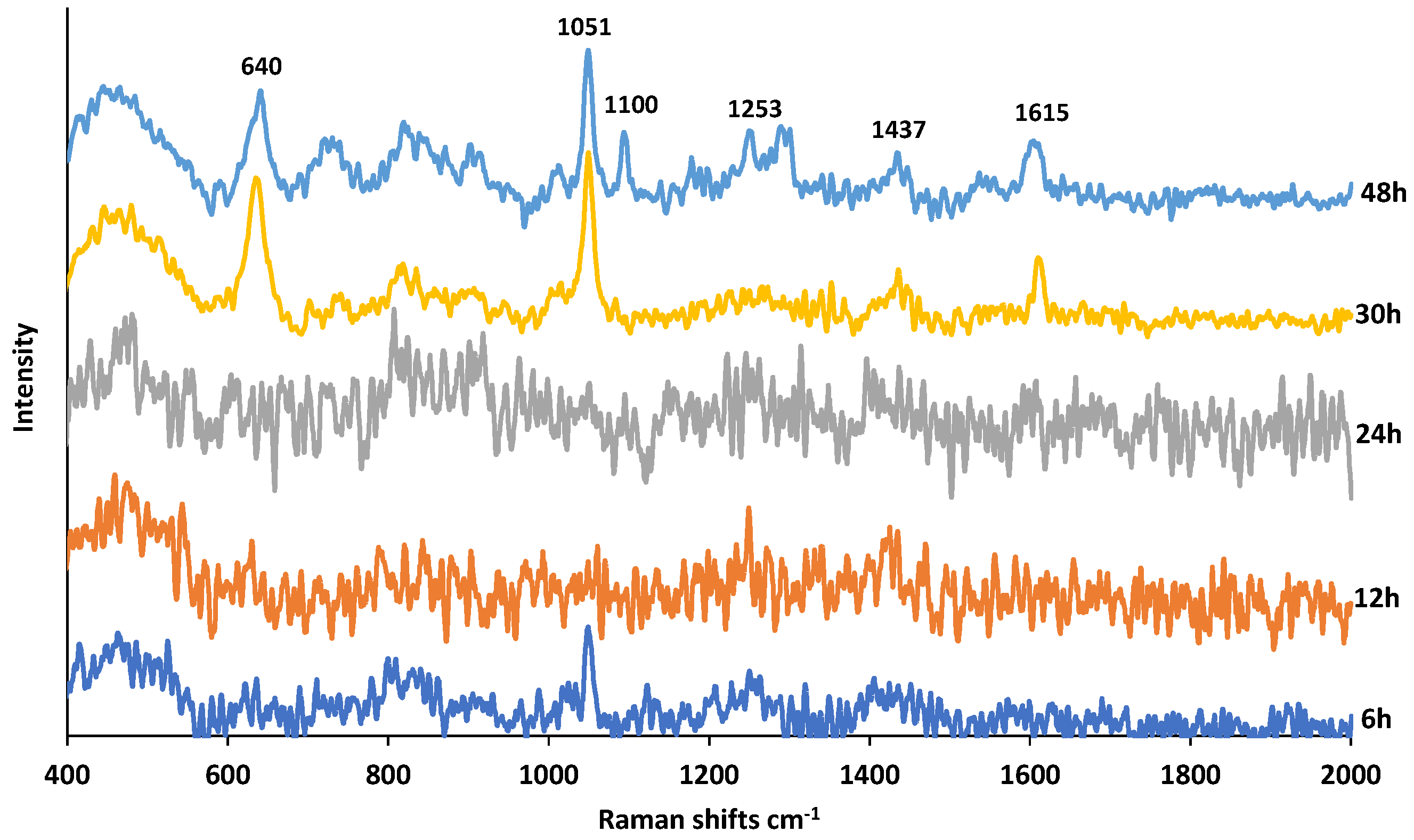
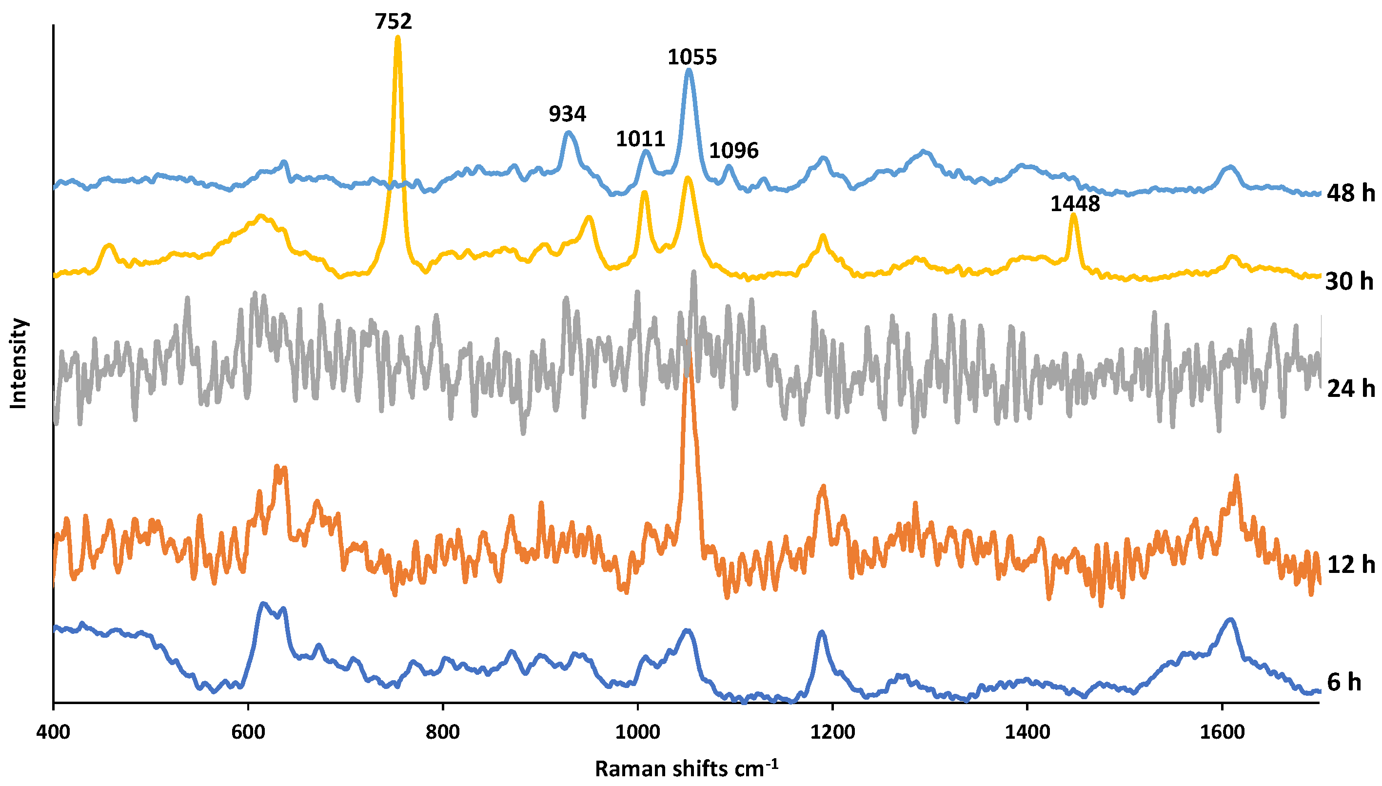
3. Results and Discussion
4. Conclusions
Supplementary Materials
Author Contributions
Funding
Institutional Review Board Statement
Informed Consent Statement
Data Availability Statement
Conflicts of Interest
References
- Lockwood, S.; Saïdi, N.; Morgan, V.A. Options for a Strategic Approach to Pharmaceuticals in the Environment Task 1 Report—Revised Version; INERIS (French National Institute for Industrial Environment and Risks); LSE (The London School of Economics and Political Science); Milieu Ltd.; European Commission-DG ENV: Tokyo, Japan, 2016. [Google Scholar]
- Bryn, S.R.; Xu, W.; Newman, A. Chemical reactivity in solid-state pharmaceuticals: Formulation implications. Adv. Drug Del. Rev. 2001, 48, 115–136. [Google Scholar] [CrossRef]
- Kawaguchi, W.H.; Fachi, M.M.; Cerqueira, L.B.; Campos, M.L.; Gonçalves, A.G.; Trindade, A.C.L.B.; Reason, I.J.M.; Pontarolo, R. Stability Indicating Method for Determination of Benznidazole and Its Degradation Products in Active Pharmaceutical Ingredient. J. Braz. Chem. Soc. 2020, 31, 1194–1202. [Google Scholar] [CrossRef]
- Serajuddin, A.T.M.; Thakur, A.B.; Ghoshal, R.N.; Fakes, M.G.; Ranadive, S.A.; Morris, K.R.; Varia, S.A. Selection of solid dosage form composition through drug-excipient compatibility testing. J. Pharm. Sci. 1999, 88, 696–704. [Google Scholar] [CrossRef] [PubMed]
- Alsante, K.M.; Huynh-Ba, K.C.; Baertschi, S.; Reed, R.A.; Landis, M.S.; Furness, S.; Olsen, B.; Mowery, M.; Russo, K.; Iser, R.; et al. Recent Trends in Product Development and Regulatory Issues on Impurities in Active Pharmaceutical Ingredient (API) and Drug Products. Part 2: Safety Considerations of Impurities in Pharmaceutical Products and Surveying the Impurity Landscape. AAPS Pharm. Sci. Tech. 2013, 15, 1. [Google Scholar] [CrossRef] [PubMed]
- Melo, S.R.D.O.; Homem-De-Mello, M.; Silveira, D.; Simeoni, L.A. Advice on Degradation Products in Pharmaceuticals: A Toxicological Evaluation. PDA J. Pharm. Sci. Technol. 2014, 68, 221–238. [Google Scholar] [CrossRef] [PubMed]
- Mudgal, S.; Toni, A.; Lockwood, S.; Salès, K.; Backhaus, T. Study on the Environmental Risks of Medicinal Products; Final Report; Executive Agency for Health and Consumers: Copenhagen, Denmark, 2013; pp. 1–310. [Google Scholar]
- Moreno, A.H.; Salgado, H.R.N. Stability Study and Degradation Kinetics of Ceftazidime in Pharmaceutical Preparations Advances in Analytical Chemistry. Adv. Anal. Chem. 2012, 2, 1–5. [Google Scholar]
- Larat, V.; Feltham, C. Raman Spectroscopy of Pharmaceutical Ingredients Under Humidity Controlled Atmosphere. HORIBA scientific. Application Note, Pharmaceutical RA61. Available online: https://static.horiba.com/ (accessed on 20 December 2021).
- Yilmaz, H. Forced Degradation Studies to Assess the Stability of a Janus Kinase Inhibitor Using RPLC Method. Suleyman Demirel Univ. J. Nat. Appl. Sci. 2021, 25, 134–141. [Google Scholar]
- ICH. Impurities in New Drug Products; IFPMA: Geneva, Switzerland, 1996. [Google Scholar]
- ICH. Final Guidance on Stability Testing of Biotechnological/ Biological Products Availability; International Conference on Harmonization: Geneva, Switzerland, 1996. [Google Scholar]
- Hazarika, M.; White, R.M.; Booth, B.P.; Wang, Y.C.; Ham, D.Y.L.; Liang, C.Y.; Rahman, A.; Gobburu, J.V.S.; Li, N.; Sridhara, R.; et al. Pemetrexed in Malignant Pleural Mesothelioma. Clin. Cancer Res. 2005, 11, 982–992. [Google Scholar]
- Joerger, M.; Omlin, A.; Cerny, T.; Fruh, M. The Role of Pemetrexed in Advanced Non-Small-Cell Lung Cancer: Special Focus on Pharmacology and Mechanism of Action. Curr. Drug Targets 2010, 11, 37–47. [Google Scholar] [CrossRef]
- Warner, A.; Piraner, I.; Weimer, H.; White, K. Development of a purity control strategy for pemetrexed disodium and validation of associated analytical methodology. J. Pharm. Biomed. Anal. 2015, 105, 46–54. [Google Scholar] [CrossRef]
- Saravanan, G.; Suryanarayana, M.V.; Jadhav, M.J.; Ravikumar, M.; Someswararao, N.; Acharyulu, P.V.R. A Stability-Indicating LC Assay Method for Pemetrexed Disodium. Chromatographia 2007, 66, 431–434. [Google Scholar] [CrossRef]
- Zhang, Y.; Trissel, L.A. Physical Instability of Frozen Pemetrexed Solutions in PVC Bags. Ann. Pharmacother. 2006, 40, 1289. [Google Scholar] [CrossRef] [PubMed]
- Jansen, P.J.; Smith, W.K.; Baertschi, S.W.; Dorman, D.E.; Kemp, C.A.; McCune, K.A. Determination of the degradation chemistry of the antitumor agent pemetrexed disodium. J. Pharm. Sci. 2016, 105, 3256–3268. [Google Scholar] [CrossRef] [PubMed]
- Respaud, R.; Tournamille, J.F.; Croix, C.; Laborie, H.; Elfakir, C.; Viaud-Massuard, M.C. Development of an ion-pairing reversed-phase liquid chromatography method using a double detection analysis (UV and evaporative light scattering detection to monitor the stability of Alimta®-pemetrexed preparations: Identification and quantification of l-glutamic acid as a potential degradation product. J. Pharm. Biomed. Anal. 2011, 54, 411–416. [Google Scholar]
- Galla, V.K.; Archana, V.; Jinka, R. A new rapid Stability indicating RP-PDA-UPLC method for the estimation of Assay of Pemetrexed disodium-An anti-Lung cancer drug from lyophilized parenteral formulation. J. Appl. Pharm. Sci. 2017, 7, 131–137. [Google Scholar]
- Hemchand, S.; Babu, R.R.C.; Annapurna, M.M. A new validated stability-indicating gradient RP-HPLC method for the determination of pemetrexed disodium and its process related substances. J. Drug Deliv. Ther. 2019, 9, 588–610. [Google Scholar]
- Adyanth, M.N.M.K.; Ravi, T.C.K.W.P. Determination of related substances in pemetrexed disodium (Form-IV) in bulk drug samples by HPLC. Pharm. Technol. 2014, 38. Available online: https://www.pharmtech.com/view/determination-related-substances-pemetrexed-disodium-form-iv-bulk-drug-samples-hplc (accessed on 20 December 2021).
- Narenderan, S.T.; Ramesh, J.; Babu, B.; Meyyanathan, S.N.A. stability-indicating LC–MS/MS method optimization for Pemetrexed through design of experiments: Identification and characterization of major oxidative degradation product. J. Pharm. Biomed. Anal. 2020, 183, 113150. [Google Scholar]
- Meesters, R.J.W.; Cornelissen, R.; van Klaveren, R.J.; de Jonge, R.; Boer, E.D.; Lindemans, J.; Luider, T.M. A new ultrafast and high-throughput mass spectrometric approach for the therapeutic drug monitoring of the multitargeted anti-folate pemetrexed in plasma from lung cancer patients. Anal. Bioanal. Chem. 2010, 398, 2943–2948. [Google Scholar] [CrossRef][Green Version]
- Rivory, L.P.; Clarke, S.J.; Boyer, M.; Bishop, J.F. Highly sensitive analysis of the antifolate pemetrexed sodium: A new cancer agent in human plasma and urine by High Performance Liquid Chromatography. J. Chromatogr. B Bio. Med. Sci. Appl. 2001, 765, 135–140. [Google Scholar] [CrossRef]
- Bobin-Dubigeon, C.; Amiand, M.B.; Herrenknecht, C.; Bard, J.M. Development and validation of an improved liquid chromatography–mass spectrometry method for the determination of pemetrexed in human plasma. J. Chromatogr. B 2009, 877, 2451–2456. [Google Scholar] [CrossRef] [PubMed]
- Kolmer, E.W.J.; Teulen, M.J.A.; Boosman, R.J.; Rouw, N.; Burgers, J.A.; Heine, R. Highly sensitive quantification of pemetrexed in human plasma using UPLC-MS/MS to support micro dosing studies. Biomed. Chromatogr. 2022, 36, e5277. [Google Scholar]
- Yilmaz, H.; Cobandede, Z.; Yilmaz, D.; Cinkilic, A.; Culha, M.; Demiralay, E.C. Monitoring forced degradation of drugs using silica coated AgNPs with surface-enhanced Raman scattering. Talanta 2020, 214, 120828. [Google Scholar] [CrossRef] [PubMed]
- El-Zahry, M.R.; Lendl, B. Structure elucidation and degradation kinetic study of Ofloxacin using surface enhanced Raman spectroscopy, Spectrochim. Acta Part A Mol. Biomol. Spectrosc. 2018, 193, 63–70. [Google Scholar] [CrossRef] [PubMed]
- El-Zahry, M.R.; Refaat, I.H.; Mohamed, H.A.; Rosenberg, E.; Lendl, B. Utility of surface enhanced Raman spectroscopy (SERS) for elucidation and simultaneous determination of some penicillins and penicilloic acid using hydroxylamine silver nanoparticles. Talanta 2015, 144, 710–716. [Google Scholar] [CrossRef]
- Wang, W.; Yin, Y.; Tana, Z.; Liu, J. Coffee-ring effect-based simultaneous SERS substrate fabrication and analyte enrichment for trace analysis. Nanoscale 2014, 6, 9588. [Google Scholar] [CrossRef]
- Avci, E.; Culha, M. Influence of droplet drying configuration on surface-enhanced Raman scattering performance. RSC Adv. 2013, 3, 17829–17836. [Google Scholar] [CrossRef]
- Li, L.; Zhu, Y.-J. High chemical reactivity of silver nanoparticles toward hydrochloric acid. J. Colloid Interface Sci. 2006, 303, 415–418. [Google Scholar] [CrossRef]
- Guideline, I.H.T. Stability testing of new drug substances and products. Q1A R2 Curr. Step 2003, 4, 1–24. [Google Scholar]
- Stolarczyk, E.U.; Stolarczyk, K.; Łaszcz, M.; Kubiszewski, M.; Leś, A.; Michalak, O. Pemetrexed conjugated with gold nanoparticles—Synthesis, characterization and a study of noncovalent interactions. Eur. J. Pharm. Sci. 2017, 109, 13–20. [Google Scholar] [CrossRef]
- Alula, M.T.; Mengesha, Z.T.; Mwenesongole, E. Advances in surface-enhanced Raman spectroscopy for analysis of pharmaceuticals: A review. Vib. Spectrosc. 2018, 98, 50–63. [Google Scholar] [CrossRef]
- Oztas, D.Y.; Altunbek, M.; Uzunoğlu, D.; Yılmaz, H.; Çetin, D.; Suludere, Z.; Çulha, M. Tracing size and surface chemistry dependent endosomal uptake of gold nanoparticles using surface-enhanced Raman scattering. Langmuir 2019, 35, 4020–4028. [Google Scholar] [CrossRef] [PubMed]


Publisher’s Note: MDPI stays neutral with regard to jurisdictional claims in published maps and institutional affiliations. |
© 2022 by the authors. Licensee MDPI, Basel, Switzerland. This article is an open access article distributed under the terms and conditions of the Creative Commons Attribution (CC BY) license (https://creativecommons.org/licenses/by/4.0/).
Share and Cite
Yilmaz, H.; Culha, M. A Drug Stability Study Using Surface-Enhanced Raman Scattering on Silver Nanoparticles. Appl. Sci. 2022, 12, 1807. https://doi.org/10.3390/app12041807
Yilmaz H, Culha M. A Drug Stability Study Using Surface-Enhanced Raman Scattering on Silver Nanoparticles. Applied Sciences. 2022; 12(4):1807. https://doi.org/10.3390/app12041807
Chicago/Turabian StyleYilmaz, Hulya, and Mustafa Culha. 2022. "A Drug Stability Study Using Surface-Enhanced Raman Scattering on Silver Nanoparticles" Applied Sciences 12, no. 4: 1807. https://doi.org/10.3390/app12041807
APA StyleYilmaz, H., & Culha, M. (2022). A Drug Stability Study Using Surface-Enhanced Raman Scattering on Silver Nanoparticles. Applied Sciences, 12(4), 1807. https://doi.org/10.3390/app12041807