Article

Bioelectroanalytical Detection of Lactic Acid Bacteria

1
Singapore Centre for Environmental Life Sciences Engineering (SCELSE), Nanyang Technological University, 60 Nanyang Drive, Singapore 637551, Singapore
2
Singapore Eye Research Institute (SERI), 20 College Rd., Level 6 Discovery Tower, Singapore 169856, Singapore
3
Centre for Biosensors, Bioelectronics and Biodevices (C3Bio), Department of Chemical Engineering, University of Bath, Bath BA2 7AY, UK
*
Author to whom correspondence should be addressed.
Appl. Sci. 2022, 12(3), 1257; https://doi.org/10.3390/app12031257
Submission received: 29 October 2021 / Revised: 4 January 2022 / Accepted: 5 January 2022 / Published: 25 January 2022
(This article belongs to the Special Issue Electrochemistry Approaches in Food Industry)

Abstract

:
Lactic acid bacteria (LAB) are an industrial important group of organisms that are notable for their inability to respire without growth supplements. Recently described bioelectroanalytical detectors that can specifically detect and enumerate microorganisms depend on a phenomenon known as extracellular electron transport (EET) for effective detection. EET is often described as a type of microbial respiration, which logically excludes LAB from such a detection platform. However, members of the LAB have recently been described as electroactive with the ability to carry out EET, providing a timely impetus to revisit the utility of bioelectroanalytical detectors in LAB detection. Here, we show that an LAB, Enterococcus faecalis, is easily detected bioelectroanalytically using the defined substrate resorufin-β-d-galactopyranoside. Detection is rapid, ranging from 34 to 235 min for inoculum sizes between 107 and 104 CFU mL−1, respectively. We show that, although the signal achieved by Enterococcus faecalis is comparable to systems that rely on the respiratory EET strategies of target bacteria, E. faecalis is not dependent on the electrode for energy, and it is only necessary to capture small amounts of an organism’s metabolic energy to, in this case 1.6%, to achieve good detection. The results pave the way for new means of detecting an industrially important group of organisms, particularly in the food industry.

1. Introduction

Lactic acid bacteria (LAB) are an economically important group of microorganisms that have utility in the food industry, in clinical settings, and the environment [1]. The ability to detect and enumerate a range of lactic acid bacteria is relevant to, for example, the identification of beer-spoilage organisms in the brewing industry [2], in characterizing persistent endodontic infections in dentistry [3], and in monitoring recreational water for fecal contamination in municipal settings [4]. Genera belonging to the LAB include Lactobacillus, Streptococcus, Vagococcus, and Enterococcus. Simple and rapid techniques for enumerating LAB that could be operated by non-specialists would, therefore, find application in numerous settings. Lactic acid bacteria belong to an exclusively Gram-positive phylum, the firmicutes, and are fermentative organisms distinguished by their inability to produce heme [5]. Even though LAB possess many of the components of respiratory chains, they do not respire on account of their inability to synthesize functional heme-containing cytochromes that typically act as the terminal reductases in oxidative respiratory chains [6].
Recently, methods for bioelectroanalytical detection of low numbers of organisms in environmental samples have been described [7]. The technique involves tagging enzyme-specific substrates to electrochemical reporters to achieve specific detection of a target organism by exploiting a phenomenon known as extracellular electron transfer (EET)—the metabolic process that transports electrons from the cytosol to the exterior of a cell [8,9]. When the electrochemical reporter, or redox mediator, is released into the medium, it is indicative of the presence of the target organism; the redox mediator is reduced metabolically and subsequently reduces the electrode. EET has been extensively studied, particularly in the model Gram-negative electrogens Geobacter spp. and Shewanella spp., and is usually described as a type of anaerobic respiration achieved by heme-containing electron transfer protein complexes [9,10]. The environmental significance of EET is in biogeochemical cycling of metal oxides in the subsurface and is achieved biologically, as microbes transport electrons across their membranes and reduce solid terminal electron acceptors, such as Fe (III) or Mn (V), in a process that yields metabolic energy in the form of ATP [11,12]. The practical role of respiration in achieving a good detection signal in bioelectroanalytical systems was recently deduced from the protracted detection times for Escherichia coli (E. coli) strains with deficient respiratory chains that are only able to grow by fermentation [13]. Therefore, the utility of bioelectroanalytical systems to rapidly detect lactic acid bacteria is unknown, as LAB are unable to respire and instead gain their metabolic energy exclusively by fermentation.
EET is well described in Gram-negative organisms but has typically remained more obscure in Gram-positive organisms, except for a few notable exceptions, and despite Firmicutes regularly turning up in the phylogeny of bioelectrochemical systems [14,15,16]. Recent research suggests that EET in Gram-positive bacteria is evolutionarily more ancient than in Gram-negative organisms [17]. Lately, it has become commonplace to describe EET mechanisms in Gram-positive organisms that include LAB. Light et al. (2019) recently described a flavin-based EET mechanism in Listeria monocytogenes and electrode-dependent growth that is distinct from more well-described mechanisms of EET [9]. Similarly, EET has been described in another clinically important LAB, Enterococcus faecalis (E. faecalis), and although the exact mechanism is yet to be elucidated, its ability to reduce an electrode and Fe (III) has been demonstrated [8,18,19]. In addition, for both Listeria monocytogenes and E. faecalis, EET has been implicated in virulence either through increased competitive capabilities, enhanced biofilm potential, or through EET mediated synergistic interactions with commensals. In these systems, however, basic quantitative assessments of the EET process is not always reported.
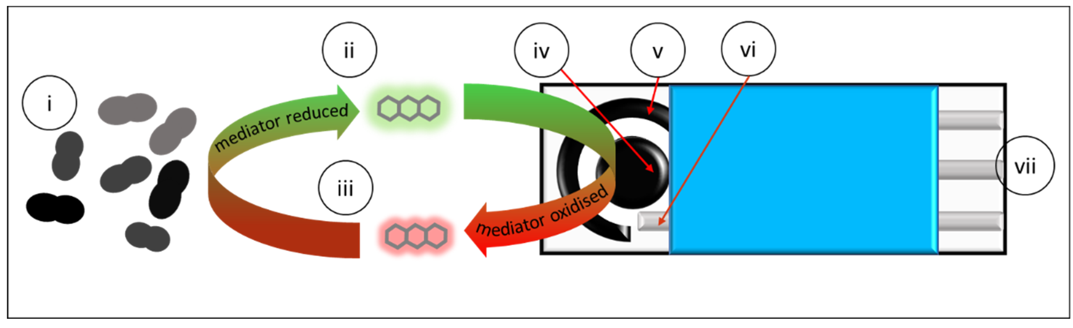
In light of the recent insights into the EET in LAB, we decided to revisit the idea of their compatibility in bioelectroanalytical detection. The aim of this contribution is to determine whether a clinically relevant member of the LAB, E. faecalis, is amenable to rapid detection and enumeration in a bioelectroanalytical system (Figure 1). A secondary aim of this contribution is to comment on the quantitative importance of EET mechanisms in E. faecalis by looking at EET efficiency. To our knowledge, this is the first report describing the potential for specific and swift bioelectroanalytical LAB detection and to quantitatively assess the efficiency of the EET process and how this relates to EET mechanisms of E. faecalis in the context of biosensing.

2. Materials and Methods

2.1. Growth Conditions for E. faecalis OG1RF and Other Lactoacid Bacilli (LABs)

Overnight growth of E. faecalis OG1RF was attained by inoculating a single E. faecalis colony grown on BHI agar into BHI broth (Acumedia, San Bernardino, CA, USA) and grown for 18 h at 37 °C at 200 rpm in a shaking incubator. Overnight bacterial cultures were centrifuged; the supernatant was discarded, and the pellet washed three times in phosphate-buffered saline (PBS) before adjusting to the desired inoculum density in fresh media.

2.2. Respiratory Stimulation (and Inhibition) of LAB

Stock concentrations of menaquinone (Mk-4 Cayman Chemical, Ann Arbor, MI, USA) (10 mg mL−1) and Heme (Sigma, Singapore) (0.5 mg mL−1) were made by dissolving in absolute ethanol and deionized H2O and were diluted to a final concentration of 0.02 mg mL−1 and 0.002 mg mL−1, respectively. Stock dissolved in water was filtered and stored in 4 °C for up to a week. The end point optical density of the microbial growth in each well with initial inoculum of 5 × 10⁵ CFU mL−1 was recorded at 48 h. For aerobic growth, flasks were incubated at 30 °C shaking at 200 rpm, while the bacteria incubated for anaerobic growth were kept in an anaerobic chamber (Bactron, Sheldon Manufacturing, Cornelius, OR, USA) with an N2, CO2, and H2 atmosphere, also at 30 °C, and only removed for periodic measurements. The absorbance at 600 nm wavelength was recorded with the Tecan Spark™ 10M microplate reader. End-point pH was recorded with a pH meter from pooled samples to ensure sufficient volume for accurate pH measurements from the small volumes incubated

2.3. Preparation of Electrochemical Reactors and Mini Electrochemical Cells

Conical electrochemical cells with stirrers were prepared as previously reported [7]. For screen-printed electrodes (SPE) reactors, the caps of 1.5 mL Eppendorf tubes were trimmed off and a hole was punctured at the conical portion of the Eppendorf tube using a 30G needle. After autoclaving (120 °C, 15 psi 15 min), the modified tubes were attached to the SPEs with epoxy resin. The SPEs consist of a circular carbon working electrode with a diameter of 4 mm, a carbon counter electrode, and a silver reference electrode on a ceramic support (Metrohm DropSens, DRP-C110, Herisau, Switzerland). SPEs were sterilized by soaking for one minute in 70% ethanol, followed by ultraviolet sterilization for 20 min. Reactors were assembled in a biological safety hood and the epoxy was allowed to cure for a minimum of 12 h before use. SPE technology is convenient for non-specialist users and can be commercially implemented at very low cost.
For electrochemical experiments, dissolved oxygen was displaced from the reactors by sparging N2 gas through the medium prior to dispensing the media into the reactor in an anaerobic chamber. The only inlet was sealed with tape before its removal from the anaerobic chamber and subsequent bench top operation in a bead bath at 37 °C.

2.4. Potentiometric Enumeration

To enhance E. faecalis communication with the electrode when required, resorufin or the electrochemically active selective agent for E. faecalis, resorufin-β-D-galactopyranoside, was added at a final concentration of 50 µM, as previously reported [7]. Potentiometric measurements were performed with a VSP-150 multichannel potentiostat (Bio-Logic SAS, Claix, France) or DRP-STAT8000 (Metrohm DropSens, Oviedo, Spain). All electrochemical potential values are reported with respect to an Ag reference electrode, and average current was recorded every 60 s during chronoamperometric measurements.

2.5. Estimation of Columbic Efficiency

To estimate the quantitative contribution of electrochemical output relative to overall metabolism, the consumption of carbon in a system inoculated with 102 CFU ml was determined by estimating the change in chemical oxygen demand (COD) over 20 h using a high-range COD kit (Hach, Loveland, CO, USA). COD measurements were conducted in triplicate and the mean value was used in the to calculate CE. The relationship between the number of electrons consumed and the number of electrons captured at the electrode (charge) was calculated as reported by Logan (2008):
C E = 8 0 t b I   d t F   v A n       Δ C O D
where CE is Coulombic efficiency, I is current, F is Faraday’s constant, VAn is the volume of the anode chamber, and ΔCOD is the change in COD over a 20 h period.

3. Results and Discussion

E. faecalis can easily be detected and enumerated using the proposed detection framework described previously [7,13]. Chronoamperometric analysis of E. facecalis reveals that all test inocula, with the exception of 107 CFU mL
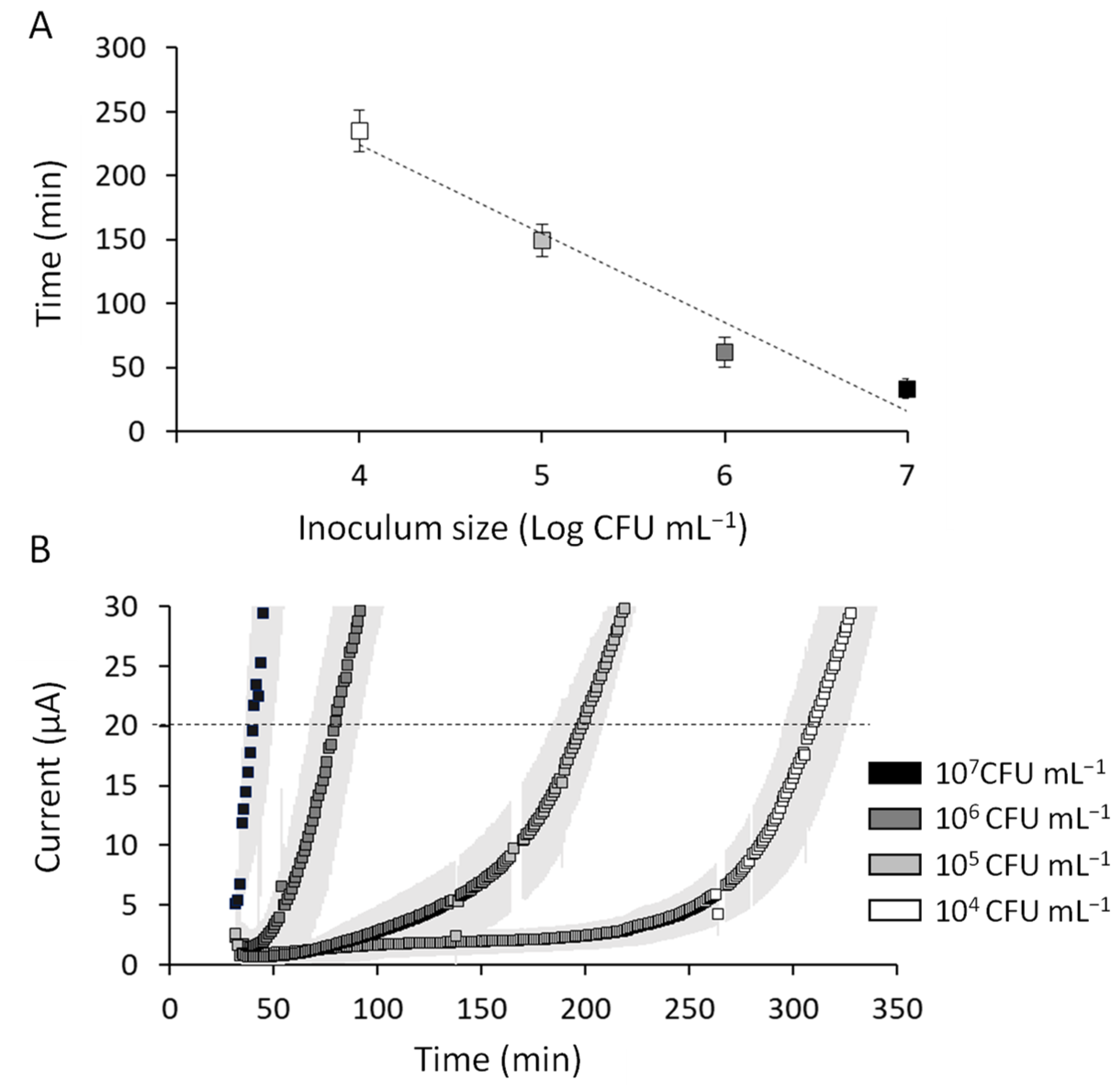
−1 where the current onset is almost instant, produce a distinctive curve showing an initial flat period of baseline current representing lag phase growth followed by a sharp increase in current generation resulting from increased metabolic activity (and thus EET) as the culture enters the exponential growth phase (Figure 2A). A previously reported method defines a detection event when the slope of the chronoamperometric readout exceeds five standard deviations of the baseline current for more than five consecutive time points [13]. We apply a more conservative definition here and define detection as the time at which the current passes a threshold value (20 µA). This is an objective and robust way to define a detection time and yields a linear calibration curve that inversely correlates with inoculum size. For every log fold increase in inoculum size ranging from 10,000 to 10,000,000 CFU mL−1, the detection time increased linearly, yielding mean detection times of 235 (±16), 148 (±12), 62 (±12), and 32 (±8) min for 104, 105, 106, and 107 CFU mL−1, respectively, where the values in parentheses represents the standard deviation of three replicates. This observation is easily interpreted from the relationship between inoculum density and the duration of the lag phase in classical growth curve theory. This property can be used to construct a linear standard curve with a regression coefficient (r2) of 0.96 (Figure 2B). Additionally, the standard deviation of the mean detection time for three replicates is between 7 and 23%, which is comparable to previous reports and also to commercially available systems for bacterial detection [4,7,20]. In short, the bioelectroanalytical system is an effective means of enumerating E. faecalis and should be further developed to effect real world detection of lactobacilli in the multitude of settings for which they are relevant. This will require development of selective medium to suppress non-target organisms that are specific to the individual settings.
The detection compounds used here are electroactive glycosides comprising glucose conjugated to resorufin. When the glycosidic bond is cleaved by native glucosidases expressed by E. faecalis, the electroactive resorufin is liberated from the pyranose ring and subsequently reduced by microbial electron carriers to dihydroresorufin [21]. Following its reduction by microbes, the mobile dihydroresorufin in turn reduces the electrode generating a current proportional to the metabolic activity of the culture [22]. Microbial metabolic coupling to an electrode via a mobile redox mediator is well documented and usually described as a component of an energy-yielding type of respiration called mediated EET [23]. The quantitative importance of respiration compared to fermentation in a similar bioelectroanalytical detector was recently demonstrated by contrasting mediated detection times achieved with wildtype E. coli vs. a mutant, E. coli SHSP 18—an E. coli K12 derivative that is auxotrophic for δ-aminolevulinic acid, a growth factor critical in heme synthesis and therefore respiration [13,24]. The wildtype E. coli detection time, for an inoculum size of 5000 CF, was about 6 h compared with the detection time of around 14 h for the SHSP 18 mutant that was unable to respire. Thus, from this previously reported study, it appears that fermentative metabolism is not ideally compatible with bioelectroanalytical detection even in the presence of mediators.
The detection time for E. faecalis observed here (Figure 2A) cannot be directly compared to the previous studies, but it is likely to be similar. For a higher inoculum size of 10,000 CFU, double that reported for the E. coli SHSP mutant, the detection time is around three hours vs. a six-hour detection time for 5000 CFU of wildtype E. coli and 14 h for the fermentative strain [13]. The detection time achieved here for 10,000 CFU E. faecalis resembles respiratory behavior.
The genome of E. faecalis is reportedly deficient in heme, but supplementation of this growth factor and or menaquinone induces respiratory behavior in E. faecalis by activating the redox center of cytrochrome bd and providing a quinone pool to transfer electrons from NADH+ to terminal reductases. Respiratory behavior is defined in this sense by observations of increased vitality or biomass production accompanied by less acidification of the medium from lactate accumulation [25].
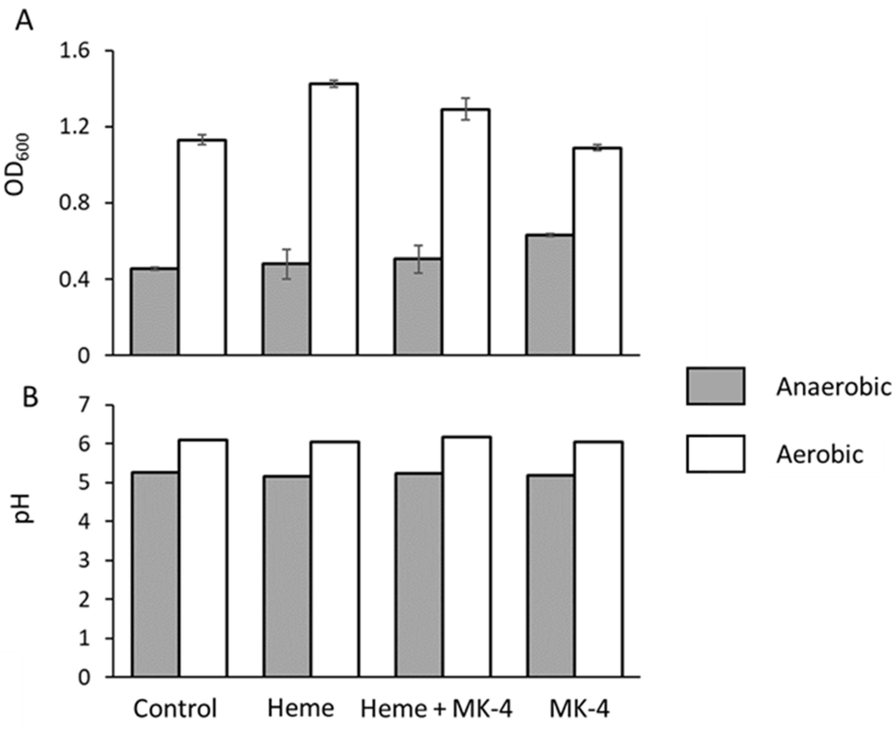
Upon analyzing the growth yields of E. faecalis in bench top experiments, we observed that the optical density (OD600) was always higher in aerobic conditions than it was in anaerobic ones (Figure 3A). This phenomenon was reported previously [26] and it is suggestive of respiratory behavior and could arise from residual heme or quinoids in the undefined medium that we used as was reported for the Todd Hewitt broth r by Del Papa and Perego (2008). Further analysis shows that the addition of heme, menaquinone, or both does not alter this trend, although menaquinone does give a slight boost in growth, observed by an increase in OD in anaerobic conditions, suggesting a role in managing reactive oxygen species (Figure 2A). A comparative analysis of Lactobacilli electron transport chain stimulation by heme and menaquinone by Brooijman et al. (2009) [25] showed similar behavior in E. faecalis, i.e., that E. faecalis growth is stimulated by menaquinone and, to greater extent, by the addition of both heme and menaquinone. However, the difference in pH between ‘fermentative’ and ‘respiratory’ growth ranged only between 0.2 pH units (5.62–5.82) [25], although growth increased by 38%. Under equivalent conditions, we observed a quantitatively lower increase in growth (OD600) of around 14% and the pH to be similarly stable (Figure 3B). We observed a pH with a delta (ΔpH) of only 0.13 pH units but an overall moderately lower pH of 5.14 and 5.27 between the unsupplemented control and supplemented conditions, respectively. The ΔpH between fermentative LAB and the supplemented treatment thathad been induced to respire was previously reported to be greater what we have observed here, and the observed ΔpH is usually close to a single pH unit; for example, the ΔpH between a heme and menaquinone-stimulated Streptococcus entericus culture compared to an unsupplemented control was 1.07, with the final pH being 4.42 and 5.49 for fermentative and respiratory conditions, respectively [25]. Taken together, although E. faecalis exhibits less respiratory stimulation upon the addition of heme in BHI medium relative to other LAB, it is clear that it does derive some growth benefits from medium supplementation with respiratory components. Despite the small difference in pH between respiratory and fermentative growth in E. faecalis, Broojiman (2009) still concluded that E. faecalis is both heme and menaquinone stimulated [25].
Concluding that E. faecalis OG1RF will grow vigorously and that its response to additive respiratory components is minimal, we proceeded to examine the quantitative extent of 
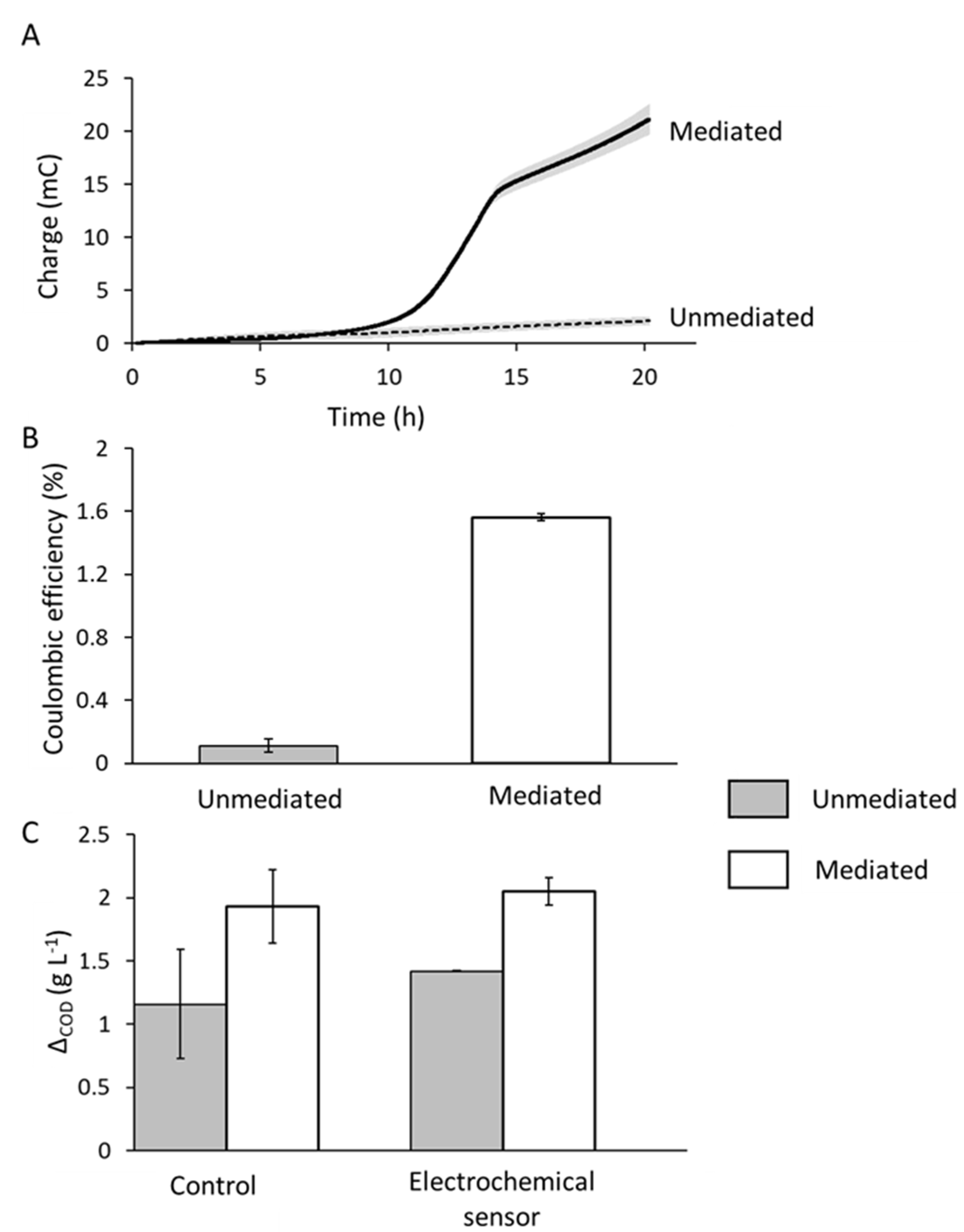
E. faecalis EET in our microscale detectors using BHI as a medium and the redox active aglycone used in the proposed detection approach, resorufin, to effect electron transport. The charge generated by E. faecalis was substantially greater (20 mC) when mediated with resorufin than the control current which barely increased above a baseline current of around 2 mC (Figure 4A). Over a 20 h period, around 600 mg L−1 COD more was consumed in the mediated system (ΔCOD = 2020 mg L−1) compared to the unmediated system (ΔCOD = 1420 mg L−1), which is suggestive of an increase in EET by E. faecalis upon the addition of the mediator (Figure 3B). However, the response of E. faecalis to mediator addition in terms of COD consumption was similar in both benchtop controls and in bioelectrochemical systems. The ΔCOD in equivalent reactors but without the electrode and incubated under identical conditions was 1160 and 1920 mg L−1 for the unmediated and mediated systems, respectively, equating to 760 mg L−1 more COD consumed in the benchtop system that was supplemented with resorufin compared with the unsupplemented control. Thus, while the electrochemical behavior of E. faecalis is stimulated with the addition of redox mediators, so too is a planktonic culture, and this is reflected in the observation in Figure 4A as well as with the growth studies in Figure 3A, where the addition of menaquinone only resulted in some growth stimulation under anaerobic conditions. This is also in keeping with observations by other researchers [25]. Coulombic efficiency (CE) of the electrochemical response of E. faecalis is low: 1.6% in the mediated system and 0.1% in the unmediated system (Figure 4B).
EET is increasingly being described in Firmicutes isolated from the gut [8,9,27,28] and, more recently, in situ directly by inserting electrodes into a mouse gut and comparing the output with germ-free organisms [29]. These studies, in the main, have lacked a quantitative assessment of the electron flux as a function of the carbon utilization in the system. Where columbic efficiency (CE) is reported, it is low (typically <1%) and of a comparative magnitude to that reported here. For example, similarly diminutive CE observations were made by Naradasu et al. (2019), where a CE of only 0.02% was reported for a gut-isolated Entrococcacea sp. with a 99% 16 S RNA sequence similarity to E. avium [27]. The question therefore remains whether EET in many LAB-based systems, either to an electrode or in the reduction in metal oxides (e.g., the commonly reported Fe (III) reduction), is a respiratory phenomenon or if it is a strategy for Firmicutes to exert redox control over their environment, e.g., to enable nutrient uptake or even mitigate metal toxicity, or even if it is just redox leakage. Since the first option is involved in energy conservation, and the other comes at a metabolic cost, the distinction is an important one. However, the low CE of EET in LAB systems reported to date suggest that such a strategy, if it used to conserve energy, is a minor one.
Mechanisms for EET in Firmicutes have been reported to be flavin dependent [9,28], although this does not necessarily mean that the flavins are mobile redox mediators as proposed in Shewanella spp. Light et al. (2018) and Hederstedt (2020) identified key genes for EET in Firmicutes. In L. monocytogenes, the important EET gene cluster appears to contain a flavoprotein (PplA) a type-two dehydrogenase (NdH2), and two small proteins, EetA and EetB, as well as genes for quinone synthesis. Orthologous genes in E. faecalis, ppl3 and ndhA, appear to have some role in EET, but alternative dehydrogenases Ndh2 and Ndh3, as well as EetA and EetB, also have a role depending on the type of EET mechanism at play. It appears that Ndh3 and EetA are essential to EET in wildtype E. faecalis, i.e., where there is no supplementary heme and, hence, they are not respiring (Hederstedt, 2020). Under such conditions, in our systems at least, there appears to be both a growth benefit and an increase in carbon utilization upon introducing a redox mediator. While a useful detection signal (current) from E. faecalis can only be recorded at an electrode in the presence of a mediator, the growth benefit to E. faecalis of such an addition appears to be independent of the electrode. Additionally, Hederstedt (2020) and Pankratova (2018) suggest that EET is always promoted by the presence of a mediator and that EET is curtailed by the activation of cytochromes [8]. Taken together, the relatively low bioelectricity yield of E. faecalis, the absence of advanced COD consumption at the electrode, and the fact that assembly of a fully functioning electron transport chain reportedly attenuates EET in E. faecalis, suggests that the observed current production in our systems is incidental. This may arise from the fact that the quinone pool (which has been shown essential to all EET described in Firmicutes to date) may become charged with electrons and can provide reducing power for other biological processes that are not essential to conserving energy. Alternatively, the current signal in these systems may arise from leakage, a phenomenon well known in mammalian electron transport chains and that produces free radicals [30]. Nonetheless, the reducing power captured here is sufficient to achieve detection that is likelybe more rapid than traditional techniques.
Accordingly, the question arises of whether it is helpful to describe the bioelectricity production observed here in E. faecalis detection systems as EET. If the phenomenon is not special, i.e., that it applies to most organisms and it is not quantitatively substantial, then the focus should be on the analytical procedure describing EET rather than the biological significance, i.e., the ability to sense the metabolism of a particular organism rather than the ‘special qualities’ of that organism’s metabolism. In this investigation, we observed CE comparable to that attributed to an electrochemically isolated organism in a member of the same genus. Recognizing the need to objectively screen for electrogenicity, Zhou et al. (2015) proposed a rapid colorimetric screening procedure to assign EET capabilities to different microbial genera. They only screened one member of the Firmicutes, and they did not assign to it the ability to carry out EET when it is objectively compared to known EET-capable genera.

4. Conclusions

The ability to detect microbial metabolism at an electrode is not dependent upon an organism’s respiratory behavior, nor is it constrained to genera with definitive and substantial EET capabilities afforded by specific electron-transporting outer membrane components. Such a finding renders an industrial important group of organisms, the LAB, amenable to rapid bioelectroanalytical detection. As a phylogenetically distinct group, LAB do not respire but conserve energy fermentatively. We showed that, to specifically detect LAB through bioelectroanalysis with confidence, redox mediator-based detection compounds are required. Using a simple but sometimes overlooked means to determine CE, we showed that it is only necessary to capture one or two percent of a microbe’s metabolic activity to set up an effective detection signal. While we have demonstrated this concept with E. faecalis, the technique is likely to be compatible with many other industrially or clinically relevant microorganisms. Here, we show the utility of a detection compound that has some specificity for the test organism; however, with medium development, it is possible that tests could be designed that specifically detect LAB under the range of conditions that reflects the diversity of applications where they are important.

Author Contributions

Study design: E.J.Y.H. and J.H., conducted the study, E.J.Y.H., supervised the study: J.H., secured funding: J.H. and S.W., data analysis: E.J.Y.H. and L.G.O., wrote the manuscript: E.J.Y.H., J.H., edited manuscript: S.W., L.G.O., E.J.Y.H. and J.H. All authors have read and agreed to the published version of the manuscript.

Funding

NRF Gap Fund Ignition Grant (NGF-2016-01-013).

Institutional Review Board Statement

Not applicable.

Informed Consent Statement

Not applicable.

Data Availability Statement

On request to CA.

Acknowledgments

This work was supported by seed funding from NTUs Sustainable Earth Office (SEO), NRF gap fund ignition grant (NGF-2016-01-013) administered by NTUitive Ltd., and SCELSE core funds. SCELSE is funded by the National Research Foundation, Ministry of Education, Nanyang Technological University (NTU) and National University of Singapore (NUS) and hosted by NTU in partnership with NUS. Thanks to Archie and Milo for their special help during the Singapore circuit breaker period.

Conflicts of Interest

The authors declare no competing conflict of interest.

References

  1. Makarova, K.; Slesarev, A.; Wolf, Y.; Sorokin, A.; Mirkin, B.; Koonin, E.; Pavlov, A.; Pavlova, N.; Karamychev, V.; Polouchine, N.; et al. Comparative genomics of the lactic acid bacteria. Proc. Natl. Acad. Sci. USA 2006, 103, 15611–15616. [Google Scholar] [CrossRef] [Green Version]
  2. Lewis, M.J. Rapid, species-specific detection of Lactic Acid Bacteria from beer using the polymerase chain reaction. J. Am. Soc. Brew. Chem. 1993, 51, 63–66. [Google Scholar] [CrossRef]
  3. Stuart, C.H.; Schwartz, S.A.; Beeson, T.J.; Owatz, C.B. Enterococcus faecalis: Its role in root canal treatment failure and current concepts in retreatment. J. Endod. 2006, 32, 93–98. [Google Scholar] [CrossRef]
  4. Erdem, Ö.; Saylan, Y.; Cihangir, N.; Denizli, A. Molecularly imprinted nanoparticles based plasmonic sensors for real-time Enterococcus faecalis detection. Biosens. Bioelectron. 2019, 126, 608–614. [Google Scholar] [CrossRef]
  5. Vinderola, G.; Ouwehand, A.; Salminen, S.; von Wright, A. Lactic Acid Bacteria: Microbiological and Functional Aspects; CRC Press: Boca Raton, FL, USA, 2019. [Google Scholar]
  6. Kanehisa, M.; Goto, S. KEGG: Kyoto Encyclopedia of Genes and Genomes. Nucleic Acids Res. 2000, 28, 27–30. [Google Scholar] [CrossRef]
  7. Hinks, J.; Han, E.J.Y.; Wang, V.B.; Seviour, T.W.; Marsili, E.; Loo, J.S.C.; Wuertz, S. Naphthoquinone glycosides for bioelectroanalytical enumeration of the faecal indicator Escherichia coli. Microb. Biotechnol. 2016, 9, 746–757. [Google Scholar] [CrossRef] [PubMed] [Green Version]
  8. Pankratova, G.; Leech, D.; Gorton, L.; Hederstedt, L. Extracellular electron transfer by the Gram-positive bacterium Enterococcus faecalis. Biochemistry 2018, 57, 4597–4603. [Google Scholar] [CrossRef] [PubMed] [Green Version]
  9. Light, S.H.; Su, L.; Rivera-Lugo, R.; Cornejo, J.A.; Louie, A.; Iavarone, A.T.; Ajo-Franklin, C.M.; Portnoy, D.A. A flavin-based extracellular electron transfer mechanism in diverse Gram-positive bacteria. Nature 2018, 562, 140–144. [Google Scholar] [CrossRef]
  10. Hinks, J.; Zhou, M.; Dolfing, J. Microbial electron transport in the deep subsurface. In Microbial Ecology of Extreme Environments; Lauro, F.M., Chenard, C., Eds.; Springer International Publishing: Cham, Switzerland, 2017. [Google Scholar] [CrossRef]
  11. Myers, C.; Nealson, K.H. Bacterial manganese reduction and growth with manganese oxide as the sole electron acceptor. Science 1988, 240, 1319–1321. [Google Scholar] [CrossRef] [PubMed]
  12. Nealson, K.H.; Myers, C.R. Microbial reduction of manganese and iron: New approaches to carbon cycling. Appl. Environ. Microbiol. 1992, 58, 439. [Google Scholar] [CrossRef] [Green Version]
  13. Han, E.J.; Palanisamy, K.; Hinks, J.; Wuertz, S. Parameter selection for a microvolume electrochemical escherichia coli detector for pairing with a concentration device. Sensors 2019, 19, 2437. [Google Scholar] [CrossRef] [Green Version]
  14. Parameswaran, P.; Bry, T.; Popat, S.C.; Lusk, B.G.; Rittmann, B.E.; Torres, C.I. Kinetic, electrochemical, and microscopic characterization of the thermophilic, anode-respiring bacterium Thermincola ferriacetica. Environ. Sci. Technol. 2013, 47, 4934–4940. [Google Scholar] [CrossRef] [PubMed]
  15. Freguia, S.; Masuda, M.; Tsujimura, S.; Kano, K. Lactococcus lactis catalyses electricity generation at microbial fuel cell anodes via excretion of a soluble quinone. Bioelectrochemistry 2009, 76, 14–18. [Google Scholar] [CrossRef] [PubMed]
  16. Kim, G.T.; Hyun, M.S.; Chang, I.S.; Kim, H.J.; Park, H.S.; Kim, B.H.; Kim, S.D.; Wimpenny, J.W.T.; Weightman, A.J. Dissimilatory Fe(III) reduction by an electrochemically active lactic acid bacterium phylogenetically related to Enterococcus gallinarum isolated from submerged soil. J. Appl. Microbiol. 2005, 99, 978–987. [Google Scholar] [CrossRef] [PubMed]
  17. Lusk, B.G. Thermophiles; or, the modern Prometheus: The importance of extreme microorganisms for understanding and applying extracellular electron transfer. Front. Microbiol. 2019, 10, 818. [Google Scholar] [CrossRef] [PubMed]
  18. Jeuken, L.J.; Hards, K.; Nakatani, Y. Extracellular electron transfer: Respiratory or nutrient homeostasis? J. Bacteriol. 2020, 7, e00029-20. [Google Scholar] [CrossRef] [Green Version]
  19. Hederstedt, L.; Gorton, L.; Pankratova, G. Two routes for extracellular electron transfer in Enterococcus faecalis. J. Bacteriol. 2020, 202, e00725-19. [Google Scholar] [CrossRef] [Green Version]
  20. James, R.; Lorch, D.; Pala, S.; Dindal, A.; McKernan, B.J. Environmental Technology Validation Report; Pathogen Detection Systems, Inc. Automated Microbiolgy Platform; EPA: Washington, WA, USA, 2010. [Google Scholar]
  21. Frontana, C.; González, I. The role of intramolecular hydrogen bonding in the electrochemical behavior of hydroxy-quinones and in semiquinone stability. J. Braz. Chem. Soc. 2005, 16, 299–307. [Google Scholar] [CrossRef] [Green Version]
  22. Khazalpour, S.; Nematollahi, D. Electrochemical study of Alamar Blue (resazurin) in aqueous solutions and room-temperature ionic liquid 1-butyl-3-methylimidazolium tetrafluoroborate at a glassy carbon electrode. RSC Adv. 2014, 4, 8431–8438. [Google Scholar] [CrossRef]
  23. Lovley, D.R. Electromicrobiology. Annu. Rev. Microbiol. 2012, 66, 391–409. [Google Scholar] [CrossRef]
  24. Săsărman, A.; Surdeanu, M.; Horodniceanu, T. Locus determining the synthesis of delta-aminolevulinic acid in Escherichia coli K-12. J. Bacteriol. 1968, 96, 1882–1884. [Google Scholar] [CrossRef] [Green Version]
  25. Brooijmans, R.; Smit, B.; Santos, F.; van Riel, J.; de Vos, W.M.; Hugenholtz, J. Heme and menaquinone induced electron transport in lactic acid bacteria. Microb. Cell Factories 2009, 8, 28. [Google Scholar] [CrossRef] [PubMed] [Green Version]
  26. Del Papa, F.; Perego, M. Ethanolamine activates a sensor histidine kinase regulating its utilization in Enterococcus faecalis. J. Bacteriol. 2008, 190, 7147–7156. [Google Scholar] [CrossRef] [Green Version]
  27. Naradasu, D.; Miran, W.; Sakamoto, M.; Okamoto, A. Isolation and characterization of human gut bacteria capable of extracellular electron transport by electrochemical techniques. Front. Microbiol. 2019, 9, 3267. [Google Scholar] [CrossRef] [PubMed]
  28. Khan, M.T.; Duncan, S.H.; Stams, A.J.; Van Dijl, J.M.; Flint, H.J.; Harmsen, H.J. The gut anaerobe Faecalibacterium prausnitzii uses an extracellular electron shuttle to grow at oxic–anoxic interphases. ISME J. 2012, 6, 1578–1585. [Google Scholar] [CrossRef] [PubMed]
  29. Wang, W.; Du, Y.; Yang, S.; Du, X.; Li, M.; Lin, B.; Zhou, J.; Lin, L.; Song, Y.; Li, J. Bacterial extracellular electron transfer occurs in mammalian gut. Anal. Chem. 2019, 91, 12138–12141. [Google Scholar] [CrossRef] [Green Version]
  30. Jastroch, M.; Divakaruni, A.S.; Mookerjee, S.; Treberg, J.R.; Brand, M.D. Mitochondrial proton and electron leaks. Essays Biochem. 2010, 47, 53–67. [Google Scholar] [CrossRef] [Green Version]
Figure 1. Schematic of LAB detection concept: the LAB (i) reduces a redox mediator (ii) that is released from a specific detection substrate, in this case resorufin-β-d-galactopyranoside (not shown), which is subsequently oxidized (iii) at the working electrode (iv) of a screen-printed electrode 4 mm in diameter printed with carbon ink. The counter electrode (v) is also carbon ink, and the reference electrode (vi) is silver. All are connected to the potentiostat by a USB connection (vii). Once oxidized, the mediator can be reduced again by the microbe in a cyclical fashion.
Applsci 12 01257 g001
Figure 2. (A) Calibration curve for E. faecalis detection showing detection time (minutes) with inoculum size as log CFU mL−1 (r2 = 0.96). (B) Chronoamperometry curves of corresponding inoculum sizes and from which calibration curve is derived. The dotted line corresponds to the thresholdof detection.. Mean values are shown, error bars correspond to the standard deviation of three replicates and are represented by bars (A) or by the shaded area (B).
Applsci 12 01257 g002
Figure 3. (A) Growth of E. faecalis as endpoint measurements of turbidity (OD600) and (B) endpoint pH measurements after growth with different supplements to stimulate respiratory behavior.
Applsci 12 01257 g003
Figure 4. (A) Mean charge and (B) mean coulombic efficiency of E. faecalis in systems grown in electrochemical conditions. (C) Comparison of COD consumption by E. faecalis in benchtop incubations and electrochemical conditions. The shaded area in (A) and the error bars in (B,C) represent the absolute error of duplicate measurements.
Applsci 12 01257 g004
Publisher’s Note: MDPI stays neutral with regard to jurisdictional claims in published maps and institutional affiliations.

