Review

Electrochemical Control of Biofilm Formation and Approaches to Biofilm Removal

1
Department of Biological and Biochemical Sciences, Faculty of Chemical Technology, University of Pardubice, Studentská 573, 53210 Pardubice, Czech Republic
2
Department of Analytical Chemistry, Faculty of Chemical Technology, University of Pardubice, Studentská 573, 53210 Pardubice, Czech Republic
*
Author to whom correspondence should be addressed.
Appl. Sci. 2022, 12(13), 6320; https://doi.org/10.3390/app12136320
Submission received: 22 April 2022 / Revised: 10 May 2022 / Accepted: 20 June 2022 / Published: 21 June 2022
(This article belongs to the Special Issue Electrochemistry Approaches in Food Industry)

Abstract

:
This review deals with microbial adhesion to metal-based surfaces and the subsequent biofilm formation, showing that both processes are a serious problem in the food industry, where pathogenic microorganisms released from the biofilm structure may pollute food and related material during their production. Biofilm exhibits an increased resistance toward sanitizers and disinfectants, which complicates the removal or inactivation of microorganisms in these products. In the existing traditional techniques and modern approaches for clean-in-place, electrochemical biofilm control offers promising technology, where surface properties or the reactions taking place on the surface are controlled to delay or prevent cell attachment or to remove microbial cells from the surface. In this overview, biofilm characterization, the classification of bacteria-forming biofilms, the influence of environmental conditions for bacterial attachment to material surfaces, and the evaluation of the role of biofilm morphology are described in detail. Health aspects, biofilm control methods in the food industry, and conventional approaches to biofilm removal are included as well, in order to consider the possibilities and limitations of various electrochemical approaches to biofilm control with respect to potential applications in the food industry.

1. Introduction

The aim of this review article is to acquaint the readers with the issue of biofilms in food technology, where special attention is paid to the electrochemical control of biofilm formation and related approaches to biofilm removal. The existence of numerous research articles confirms the broad interest in this topic, because it is only a matter of time until electrochemical approaches supplement or even replace existing convection chemical approaches. Currently used methods are not always efficient enough (with the need to replace the whole part of the food processing equipment) and often burden the environment with the high consumption of chemicals.
For clarity, the manuscript is divided into three main parts. The first chapter is focused on pathogenic bacteria, able to create biofilms in the environment of the food industry, which are sources of food contamination. In addition, an overview of the standard methods for the characterization of biofilms is included. The second section describes the health aspects and methods used in biofilm control and its removal from surfaces of technological equipment. Besides others, it offers the latest trends to prevent unwanted biofilm formation. Conventional methods in biofilm removal are described to possibly compare and critically evaluate alternative electrochemical approaches. Finally, the third chapter attempts to introduce the scientific community the latest research in the field of biofilm-related electrochemistry. This section is further divided into several parts dealing with the electrochemical control of bacterial adhesion, the effect of divalent ions on biofilm formation, the electrochemical communication of bacterial cells and their extracellular electron transfer, the electrochemical mapping of biofilm location, and electrochemical approaches proposed for biofilm removal. However, it is important to note that electrochemistry is not yet found in practical utilization in food technology and still belongs to the field of current research. This work arose as a result of the critical views and evaluations of experts in biofilm-related fields; more specifically, in the field of microbiology, the field of food technology and analysis, and the field of electroanalysis. The authors believe that this review could provide useful information to implement new procedures into wider practice.

2. Biofilms

To understand the issue of biofilms in food technology, it is necessary to provide basic knowledge in the field of microbiology, especially an overview of pathogenic bacteria, technologically important biofilm substrates, and conditions under which biofilms form. Special attention is paid to the interactions of planktonic cells with conductive substrates, when the essence of the use of electrochemical approaches to biofilm formation control is already partially approximated.
Bacteria can form biofilms as a part of their survival mechanisms, and biofilms are thus ubiquitous in nature [1]. Biofilm formation constitutes an alternative lifestyle in which microorganisms adopt a multicellular behavior that facilitates and/or prolongs survival in diverse environmental niches. Biofilms are formed on biotic and abiotic surfaces both in the environment and in healthcare settings. In hospital wards, the formation of biofilms on vents and medical equipment enables pathogens to persist as reservoirs that can readily spread to patients. Inside the host, biofilms allow pathogens to subvert innate immune defenses and are thus associated with long-term persistence [2]. Bacterial biofilms are clusters of bacteria that are attached to a surface and/or to each other and embedded in a self-produced matrix. Bacterial biofilms are complex surface-attached communities of bacteria held together by a self-produced polymer matrix mainly composed of polysaccharides, secreted proteins, and extracellular DNAs. Generally, bacterial biofilm formation is a complex process and can be described in four main phases which can be further subdivided according to their specification: bacterial attachment to a surface (i) including a reversible attachment phase, where bacteria non-specifically attach to surfaces, and irreversible attachment involving interaction between the bacterial cells and a surface using bacterial adhesins such as fimbriae and lipopolysaccharide (LPS); (ii) production of extracellular polymeric substances (EPS) by the resident bacterial cells; (iii) biofilm maturation, in which bacterial cells synthesize and release signaling molecules to sense the presence of each other, conducing to the formation of a microcolony and the maturation of biofilms; and, finally, (iv) dispersal/detachment phase, where the bacterial cells depart the biofilms and revert to an independent planktonic lifestyle [3]. Within the biofilm, the bacteria adapt to environmental anoxia and nutrient limitation by exhibiting an altered metabolism, gene expression, and protein production, which may lead to a lower metabolic rate and a reduced rate of cell division [4,5,6,7]. Biofilms have high cell densities ranging from 108 to 1011 cells per gram of the wet weight [8].

2.1. Bacteria Forming Biofilms

It is now understood that approximately 40–80% of bacterial cells on Earth can form biofilms [9]. The formation of biofilms was detrimental in several situations [6,10,11]. It is generally believed that biofilm matures after 24 h, forming a thick layer of biomolecules [12]. For example, in food industries, pathogenic bacteria are capable of forming biofilms inside processing facilities, leading to spoilage of foods and endangering the consumer’s health [13,14].
Bacteria are able to colonize and form biofilms on almost all kinds of surfaces, including natural and synthetic surfaces. Biofilms are responsible for chronic illnesses and nosocomial infections, industrial pipe fouling, spoilage of foods, contamination of sea food and dairy products, as well as ship hull fouling. Some of the biofilm-forming pathogenic and potentially pathogenic microbes include Aeromonas hydrophila, Burkholderia cepacia, Enterococcus faecalis, Escherichia coli, Haemophilus influenza, Klebsiella pneumoniae, Listeria monocytogenes, Pseudomonas aeruginosa, Pseudomonas pseudomallei, Proteus mirabilis, Staphylococcus aureus, Staphylococcus epidermidis, Streptococcus pneumoniae, Streptococcus pyogenes, Streptococcus mutans, Streptococcus viridans, other Streptococcus species, Vibrio cholerae, Vibrio parahaemolyticus, and Candida albicans [15,16,17,18,19,20,21]. Further biofilm forming microbes include Bacilli (Bacillus subtilis, Bacillus cereus), Lactobacillus plantarum, Lactococcus lactis, and Lactobacillus rhamnosus [15]. There are a few pieces of evidence provided on biofilm-forming fungal species, and in recent years, some genera of pathogenic fungi have been gaining attention and are correlated with biofilm formation [16]. In most conditions, bacteria will generally grow on surfaces in competition with other microorganisms in a mixed species of biofilm [15]. Therefore, the harmful effects of biofilms on human society are manifold. The most common biofilm-forming foodborne pathogens and spoilage organisms are introduced in Table 1 [3]. The main stages of bacterial biofilm formation can include the following: adsorption, adhesion, formation of microcolonies, maturation, and dispersal. In general, these stages apply for both bacterial and yeast biofilms [22].

2.2. Influence of Environment on Biofilm Formation

Several environmental factors have been reported to strongly influence the potential of an organism to form biofilms on a surface. The pH, incubation temperature, water activity, ingredients composition (glucose, sodium chloride, ethanol, minerals, heavy metals, and dilution rate of media), contact duration, and the type of surface have been shown to be important factors affecting the phenotypic change from planktonic cells to sessile forms such as biofilms [57,58,59,60,61,62].
Highly diverse environmental conditions ideal for biofilm formation are encountered in the food processing environment [57,61,63]. The ability of food spoilage and pathogenic bacteria to adhere to food-processing surfaces, such as stainless steel (SS), silicon rubber (SR), plastic (PLA), and food surfaces and form biofilm is a major health hazard because of a constant source of contamination by resistant biofilms [57,64].
The type of material used for industrial equipment and piping systems also influences biofilm structure and behavior, and specifically their tolerance to disinfection and cleaning procedures [65]. The general principles for CIP circuits recommend the use of AISI316 or AISI304 stainless steel (SS) with an electropolished surface finish [66]. In addition, biofilms formed on SS are shown to be more susceptible to biocides than those formed on plastic materials, such as high-density polyethene (HDPE) and polystyrene (PS) [67,68]. However, plastic materials are indispensable in industrial settings due to their flexible application, (bio)corrosion resistance, and low cost. For instance, HDPE is broadly used in water systems, particularly for DWDS and food industries, offering high mechanical performance [69]. Additionally, HDPE has also been used for other purposes related to the food industry, such as the production of larger moldings (transport and storage tanks), modular conveyor belts, sheets, tubes, bearings, and gears [70,71,72].

Effect of Metal Ions on Biofilm Formation

Since this review article focuses on the electrochemical control of biofilm formation in food processing environments, it is necessary to show the possibility of real-time electrochemical monitoring of concentration levels of the metal ions, which could provide useful information about the current composition of the environment, because it is well known that the presence of different metal ions can increase the abundance of attached bacteria cells onto surfaces or inhibit their growth.
For example, in 2006, Song and Leff found that the presence of Mg2+ cations significantly increases the abundance of attached cells of Pseudomonas fluorescens [73]. On the other hand, another study suggests the inhibitory effect of copper and zinc ions on the growth of Streptococcus pyogenes and Escherichia coli biofilms. However, it was also observed that copper and zinc cations had no effect on mature biofilm [74]. Silver cations and silver nanoparticles (AgNP) show good antimicrobial activity and are widely used in many fields, even in food technology as an effective preservative [75]. This phenomenon can be attributed to the non-competitive inhibition of enzyme activity [76]. It has been shown experimentally that silver is able to prevent biofilm formation in modified Robbins [77] with polyvinyl chloride and stainless steel surfaces [78]. It was confirmed that the electrochemically deposited silver on stainless steel container surfaces can effectively inhibit microbial proliferation within potable water supplies [79].

2.3. Biofilm Structure in Biofilm Systems

The formation of bacterial biofilms on solid surfaces within a fluid starts when bacteria attach to the substrate. Understanding the environmental factors affecting the attachment and early stages of biofilm development will help to develop methods of controlling biofilm growth. Here, we show that biofilm formation is strongly affected by the flows in thin layers of bacterial suspensions controlled by surface waves. Deterministic wave patterns promote the growth of patterned biofilms, while wave-driven turbulent motion discourages patterned attachment of bacteria. Strong biofilms form under wave antinodes, while inactive bacteria and passive particles settle under the nodal points. By controlling the wavelength, its amplitude, and horizontal mobility of the wave patterns, one can shape the biofilm and either enhance the growth or discourage the formation of the biofilm. The results suggest that deterministic wave-driven transport channels, rather than hydrodynamic forces acting on microorganisms, determine the preferred location for bacterial attachment [80]. Depending on the interaction between the surface and the constituent cells, the biofilms could be either of a monolayered or multilayered nature. Monolayer biofilm has prominent interactions between the cell and the surface, rather than the interaction between the constituent cells. Different classes of adhesive structures, such as flagellum and pilus, are helpful to accelerate and increase the formation of monolayer biofilm. On the other hand, microbes often develop multilayer biofilms when they adhere to a surface, as well as to each other.
Numerous techniques have been developed for the detection of biofilms [81], as demonstrated in Figure 1. These include tube culture, Congo red agar, microtiter plate assay and confocal laser scanning microscopy, atomic force microscopy (AFM), quartz crystal microbalance (QCM), surface plasmon resonance (SPR), fluorescence in situ hybridization (FISH), peptide nucleic acid–fluorescence in situ hybridization (PNA-FISH), locked nucleic acid–fluorescence in situ hybridization (LNA-FISH), catalyzed reported deposition–fluorescence in situ hybridization (CARD-FISH), double labeling of oligonucleotide probes–fluorescence in situ hybridization (DOPE-FISH), combinatorial labeling and spectral paging–fluorescence in situ hybridization (CLASI-FISH), fluorescence in situ hybridization/microautoradioactivity (FISH/MAR), fluorescence in situ hybridization–Raman spectroscopy (FISH-Raman), fluorescence in situ hybridization–nanometer-scale secondary-ion mass spectrometry (FISH-NanoSIM), confocal laser scanning microscopy (CLSM), scanning electron microscopy (SEM), cryo-scanning electron microscopy (Cryo-SEM), environmental scanning electron microscopy (ESEM), focused ion-beam scanning electron microscopy (Fib-SEM), colony forming units (CFU), propidium monoazide quantitative real-time PCR (PMA-qPCR), quantitative polymerase chain reaction (qPCR), tetrazolium salt reduction (XTT), triphenyltetrazolium chloride (TTC), crystal violet (CV), ultrasonic time domain reflectometry (UTDR), fluorescence correlation spectroscopy (FCS), and yet some others [81].
Biofilm architecture has been extensively studied using optical sectioning, confocal laser scanning microscopy, scanning electron microscopy, and three-dimensional imaging [16,82]. The combination of flow-cell technology and fluorescence in situ hybridization (FISH) with confocal laser scanning microscopy is the most favored tool to obtain quantifiable evidence on both the overall biomass and the individual strains [8].

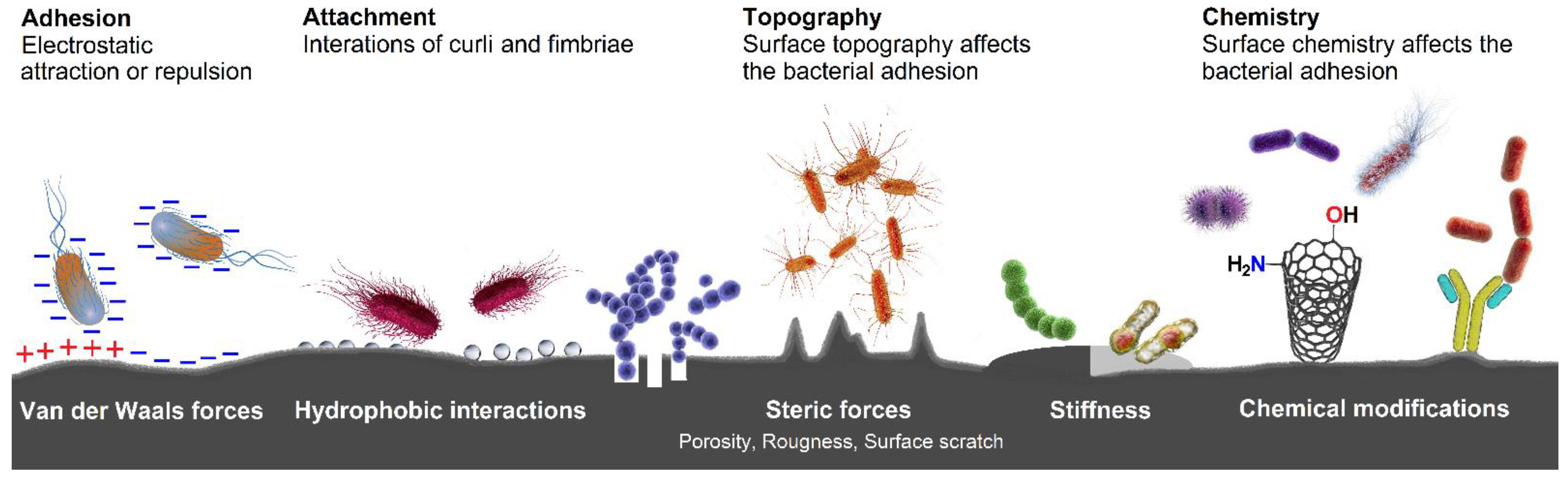
2.4. Bacterial Adhesion and Biofilm Formation

In general, gram-positive (G+) and gram-negative (G−) bacteria differ in wall structure, although their common feature is the presence of peptidoglycan (murein), which can represent up to 90% of the cell wall. The wall of G+ bacteria, with a thickness of approximately 20 nm, is simpler than in the case of G− bacteria. Unlike G− bacteria, almost all N-acetylmuramic acid residues are connected by a peptide bridge. Teichoic acid chains, which have the function of the main surface antigen, run through the peptidoglycan layer. This polysaccharide binds Mg2+ and Ca2+ cations, which are necessary for the integrity of the wall and membrane. With a few exceptions (Mycobacteria, Corynebacteria, and Nocardia), it does not contain any lipids and proteins, except for Streptococci. This is also the reason why they form a microcapsule [83]. The wall of G− bacteria, with a thickness of approximately 15 nm, consists of an outer membrane and the periplasmic space, where a thin layer of peptidoglycan is deposited. The outer membrane then contains a bilayer of phospholipids and proteins attached to the peptidoglycan via lipoproteins. Lipopolysaccharide molecules are present on the outside of the membrane, which are composed of three parts, namely O-polysaccharide, core polysaccharide, and inner lipid A [82,84].
Many bacteria have fine protein fibers outside the cell wall which are commonly referred to as flagella, pili (fimbriae), and curli. The flagella mediate active motion, thus facilitating chemotaxis and phototaxis. In the past, some researchers reserved the term pilus for the appendage required for bacterial conjugation, although all types of pili are primarily composed from pilin proteins, unlike the curli, differing from pilin proteins by representing the coiled surface structures composed of a single type of subunit. They are synthesized in the absence of a cleavable signal peptide [85].
From the point of view of biofilm formation, short pili (“attachment pili”), generally known as fimbriae, are required for the formation of biofilm because they are responsible for the attachment of bacteria to host surfaces for colonization during infection. Curli belong to one of the unique amyloid fibers produced by certain bacteria of the family Enterobacteriaceae and are involved in adhesion to surfaces, cell aggregation, and biofilm formation. It has been proven that curli also mediate host cell adhesion and invasion, and they are potent inducers of the host inflammatory response [86].
Naturally, the first step of biofilm formation starts with the reversible adhesion of bacteria to a surface through nonspecific interactions (physical forces) between the bacterial wall and the substrate. It is assumed that if bacteria cells are to be located more than 50 nm from the surface, they will be affected only by weak electrostatic interaction (van der Waals forces). After that, irreversible adhesion can occur through the effects of specific (short-range) interactions (distance less than 5 nm from the surface) with the involvement of hydrogen bonding, ionic and dipole interactions, hydrophobic interactions, and bacterial structural adhesins [87]. The bacterial adhesion with subsequent attachment might be affected by several factors, which are illustrated below in Figure 2.

3. Biofilms in Food Industry

Firstly, it is important to realize that biofilm-growing cells are fundamentally different from free-floating cells. In particular, they are highly resistant to external influences and therefore represent a source of serious complication in medicine. This is the main area where biofilm-induced diseases are registered, as opposed to food-borne diseases, where the origin is difficult to trace. For this reason, the chapter dealing with this topic is included.
Biofilm in food processing plants is a major source of spoilage and food-borne microorganisms. The improper detection of biofilm formation or its subsequent removal can cause rapid deterioration of food and its quality during processing, retailing, or storing. In Europe and North America, per-capita food losses range from 280 to 330 kg/year [88]. The relative contribution of processing to overall food waste varies across food commodities. For instance, the beginning and end of the food supply chain (agriculture and consumption) is the main source of food waste for fruits and vegetables, while a relatively high contribution of processing has been observed for meat, tuber, or oilseed and pulses. In 2011, the European Commission estimated that the processing sector has released almost 88 million tons of food waste. This corresponds to 33 kg per person per year, including both the edible and inedible parts of processed food products [89]. A year later, when applying the language of money, the cost of EUR 13 billion was estimated for EU-28 (9.1% of overall costs). Biofilm control, or its successful eradication from food processing plant environments, is a great challenge for the food industry to decrease the costs as well as the health implication for consumers.
Biofilms are deposited on the surface of processing equipment and can be the source of spoilage or pathogenic microorganisms. Once the food product is in contact with the processing tool on which the biofilm is present, the cells can be transferred from the biofilm to the food product. It was recently proven that biofilm-bacteria were transferred from freshly cut vegetable surfaces at a higher rate than planktonic bacteria [90]. There is no direct evidence that biofilm was the main cause of food spoilage or alimentary disease; however, it is very likely. Xu et al. found that 80% of the microbial flora isolated from vegetable processing plants has the potential to form biofilm [91]. Food spoilage microorganisms were present in 70–80% of the biofilms collected from meat processing facilities [92]. In the meat industry, approximately 90% of microorganisms form biofilms and are responsible for the loss of USD 150 million per year [93].

3.1. Health Aspects

Biofilm formation in food processing environments can be a source of various kinds of microorganisms, including pathogens [94,95] and toxigenic bacteria [96]. Campylobacteriosis was the most prevalent zoonosis in the EU in 2017, representing nearly 70% of all reported cases [97]. Hospitalization ranged from 30.5% to 42.5% for Campylobacter, Yersinia, and Salmonella infection diseases, with around 150 reported deaths. Strong-evidence outbreaks have been caused by food vehicles, such as milk, broiler meat, eggs, and bakery products. Obviously, food-borne bacteria can be transferred into the food via contact with unclean solid surfaces. Although there is no direct evidence that biofilm-associated bacteria were responsible for these particular outbreaks, such bacteria were consequently screened for their ability to form biofilm. Some studies showed that the majority of Salmonella enteritidis SE86 isolates (96.3%) from outbreaks produced biofilm, which was like those isolated from poultry [98]. On the other hand, one of 14 Yersinia pseudotuberculosis outbreak strains had characteristics favorable for the formation of biofilm in vitro [99] which was in accordance with the literature data [100], where only one Salmonella outbreak strain had biofilm-forming potential. However, the enhanced ability to attach to the polystyrene plates at 37 °C was recognized as an adaptive advantage of cells under adverse conditions, and no evidence of increased virulence was found in their research. The Y. pseudotuberculosis outbreak strain that formed biofilm contained both a biofilm-associated gene and virulence plasmid [99]. An interesting outcome has been published by Jaakkonen in 2020 [101], who had found that the same outbreak type of Campylobacter isolated in two different Finnish dairy farms (milk tank) demonstrated the opposite biofilm-forming ability. Similar findings were also obtained for E. coli O157:H7 outbreak strains which did not form biofilm in vitro [102]. It may suggest that there is no simple relation between an outbreak and biofilm-forming abilities. Despite this fact, the presence of biofilm in food processing environments (devices, pipeline, membranes etc.) represents a serious problem with all its financial consequences and health implications.

3.2. Biofilm Control Methods in Food Industry

3.2.1. Surface Modification of Contact Material

Several approaches have been examined to avoid biofilm formation in the food industry. The modification of food contact surfaces seemed to be the first mode of action against the attachment of bacterial cells. The complexity of particular parts of processing equipment (joints) or bacterial cells “hidden” in cracks and scratches appearing on the surface do not guarantee complete sanitization [103]. Therefore, it is a valuable strategy to have the initial bacterial attachment on the surface of materials under control. According to the Biocidal Products Regulation (EU 528/2012), compounds containing copper or silver are approved for use in food and feed areas. In particular, in food processing plants (brewery, water pipes), copper surfaces are still in use. It has undeniable anti-biofilm properties due to the interaction of copper ions with the cell membrane, causing the formation of reactive oxygenic species, DNA damage, and the impairment of DNA integrity [104]. However, the application of copper material is limited due to the high cost, possible leakage of copper ions, and corrosion. Innovative approaches are needed to embed functional copper ions into food contact surfaces [105,106]. Since stainless steel (SS) is a widely used material in the food industry, a lot of effort has been devoted to the research of SS modification. The environmental factors in food processing environments and the properties of stainless steel enabling the attachment of bacterial cells on the surface of SS have been intensively reviewed [107]. Silver ions are known to interact with thiol groups of cysteine, resulting in the disruption of the permeability of membranes, and the release of a reactive oxygen species causing oxidative stress. Ag-doped stainless-steel surfaces were prepared using various technological processes including sol-gel [108], immersion in nanoparticles suspension [109], ion beam technology [110], spray coating [111], or simple Ag+ adsorption from water disinfectant [112]. Inspired by the peristome of carnivorous pitcher plants (e.g., Nepenthes sp.) which excrete a lubricant liquid making the surface slippery for insects [113], food-contact surfaces have been treated by various lubricants to restrict the adhesion of bacterial cells [114]. However, coating procedures are often too complicated, comprising a number of organic substances, solvents, and accompanying chemical reactions. Although antifouling properties were found to be promising in vitro, their use in food processing environments should be extensively examined to ensure food safety.

3.2.2. Natural Compounds as Biofilm Inhibitors

Various antimicrobial agents were assessed to prevent the adhesion of bacterial cells onto stainless steel surfaces via interrupting their metabolic pathways that lead to membrane damage, protein and cell wall binding, enzyme inactivation etc. [115]. It is important to say that antimicrobial and anti-biofilm formation properties must be distinguished. It was previously described that tea polyphenols did not affect planktonic growth of Shewanella putrefaciens but were more effective in the inhibition of their initial attachment and the metabolic activity of the biofilm [116]. On the contrary, the most potent compounds towards planktonic cells were not always the most potent towards biofilm [117]. The efficiency of chemical compounds against biofilm formation varies depending on other environmental factors (pH, velocity of liquid, concentration etc.) and targeting bacterial species. Plant-based extracts and essential oils [118,119], as well as individual phenolic constituents [120], are very attractive as antibiofilm-forming agents. The mode of action against bacterial cells can be antimicrobial (disrupting cell membrane), anti-QS activity (downregulate the transcription of genes involved in various metabolic pathways), altering the hydrophobicity of cells’ surface, or their combined effect.

3.2.3. Microorganisms for Pathogen Biofilm Control

It is known that various fungi, yeasts or bacteria successfully inhibit the formation of biofilm. Such microorganisms are producers of natural compounds which act as antimicrobial agents against both planktonic cells and can also penetrate into the biofilm matrix. Nisin, a bacteriocin produced by some strains of Lactococcus lactis, being recognized by the FDA as GRAS substance (CRC 184.1538), is widely used as an antimicrobial agent in the food industry. The anti-biofilm properties of nisin have been successfully examined against Listeria monocytogenes on SS surfaces, where significant inhibition (4.6 log CFU/cm2) has been observed [121]. However, re-growth occurred after 24 h. Generally, the adaptation of biofilm-forming microorganisms towards sanitizing agents is the impetus for the search for new anti-biofilm products [122]. After growing various microorganisms in culture media, cell-free supernatant was obtained and used against biofilm-forming bacterial species. For instance, crude extracts of Actinomycetes isolates inhibited the biofilm formed by Bacillus cereus and Shewanella putrefaciens on a SS surface [123]. Bacillus sp. cell-free supernatant exhibited anti-biofilm and anti-QS activity against important fish pathogens [124]. In some papers, detailed analysis revealed particular compounds responsible for anti-QS activity [125] or for changing the surface characteristic of bacterial cells [126] in cell-free supernatants. The artificially developed biofilm of non-pathogenic species introduce another strategy of how to deal with the adhesion of pathogenic bacteria onto the surface of stainless steel. Soil bacterial species were frequently applied in laboratory experiments, where they showed promising outcomes [127,128]. However, there is a possibility that such protective biofilm can be the cause of food spoilage or the source of toxic metabolites. Extensive research is still needed to overcome these problems.

3.3. Detection of Biofilm in Food Processing Plants

The detection of the presence of a biofilm is more important in closed circuits, i.e., tubes, cooking tanks, filling machines, or other hidden places where biofilm may grow without being observed by the naked eye. Prior to cleaning, the manufacturing process has to be stopped, and all the closed circuits have to be emptied. The early detection of biofouling may save on the cost of frequent cleaning. In the literature, 23 various biofilm detection techniques have been identified, including those based on physical, chemical, microscopical, and biological principles [129]. However, most of them are either not applicable to in-line monitoring of biofilm formation or do not provide the results in real time. Conventional methods for detecting bacterial pathogens in biofilm are still based on culturing the microorganisms on agar plates after swabbing biological material from the food contact surface [130]. In order to overcome the problem with bacteria in viable but nonculturable states, direct epifluorescence microscopy, enzyme-linked immunosorbent assays, or PCR have been proposed as more sensitive, albeit more expensive and time-consuming, methods for the detection of pathogens in biofilm [131].

3.4. Conventional Approaches to Biofilm Removal

A common and readily available solution for biofilm eradication is the use of chemicals. According to the European Chemicals Agency, a few active compounds used as biocides have been approved for application in food and feed areas [132]. Biocides are usually a mixture of reagents with antimicrobial properties (active chlorine, aldehydes, peroxides) and surfactants (quaternary ammonium salts). The high efficiency of biofilm removal from the surface is only guaranteed under specific conditions, such as pH [133], temperature [134], velocity of liquid, shear stress, or the composition of the biofilm [65]. The combined application of chemical disinfection with other anti-biofilm practices has been tested in experiments, where the synergistic effect of the active chlorine with enzymes [135], essential oils [136], or ultrasounds [137] was observed. Other interesting disinfection procedures were examined in lab-scale experiments, such as the application of saturated steam [138], plasma-activated water [139], ozone [140], or LED light [141].

4. Electrochemical Biofilm Control

Some research suggests that electrochemistry could be a suitable solution of how to elegantly control biofilm formation, as well as map biofilm location [142]. Several electrochemical approaches have already been designed for these purposes. In general, it can be stated that they represent several electroanalytical tools, whose principles are described in more detail in the following sections.

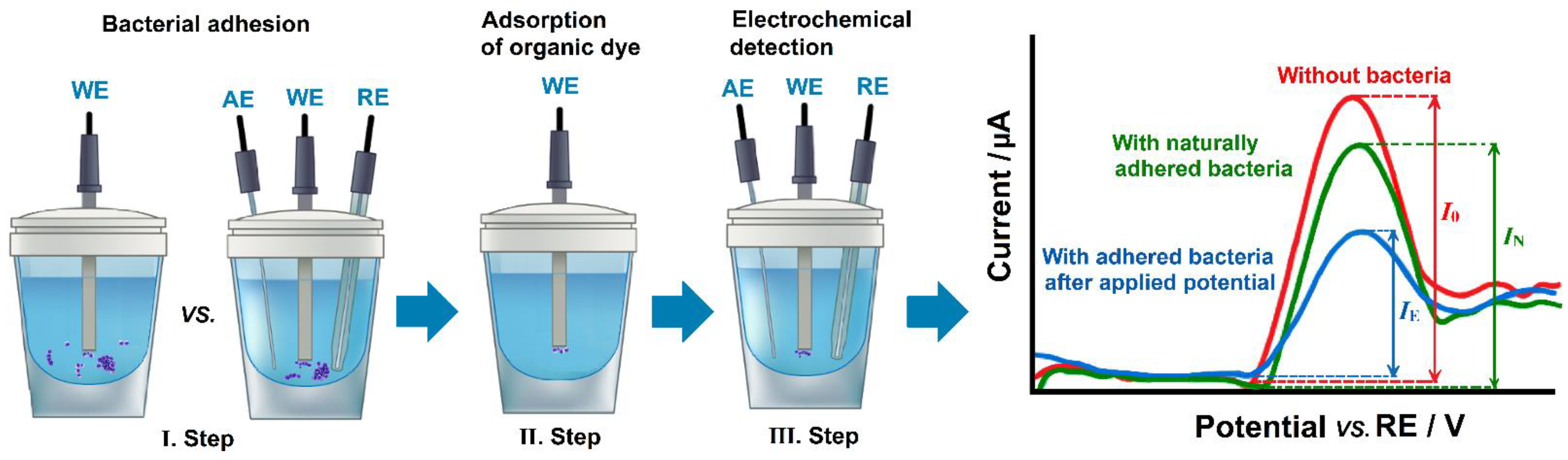
4.1. Electrochemical Control of Bacterial Adhesion

As mentioned above, the bacterial cell wall is formed by peptidoglycan, consisting of sugars and amino acids that form a mesh-like layer outside the plasma membrane, where the sugar component contains alternating residues of β-(1,4) linked N-acetylglucosamine (NAG), and N-acetylmuramic acid (NAM). It is evident that the individual organic molecules of peptidoglycan will undergo protonation and deprotonation at different pH of the environment. Due to the negative charge of the bacterial cell, the isoelectric point occurs at relatively low pH values because the negative charge is compensated by protons, namely Mycobacterium 4.15, Alcaligenes 3.25, Clostridium 2.75, Proteus 2.67, Azotobacter 2.07, and Streptococcus 1.9.
On the verge of the new millennium, Morisaki et al. [143] introduced a simple indirect voltammetric method to determine the number of bacterial cells (Pseudomonas syringae pv. atropurpurea NIAES 1309) that had been attached onto the surface of a carbon paste electrode (CPE). A simplified principle of this method is explained in Figure 3. At first, the CPE is immersed into a bacterial cell suspension for a certain time to allow the cells to attach to its surface, where the process takes place spontaneously (in open circle) or by applying an accumulation potential (Edep) depending on the pH of the environment. In the second step, the CPE with accumulated bacteria cells is transferred into a solution of an organic dye (Hoechst) to adsorb onto the remaining free sites on the electrode surface. Finally, the CPE with the presence of bacteria cells and the adsorbed organic dye is subsequently immersed in detection media. There was a calculated difference in the peak heights corresponding to anodic oxidation signals of absorbed organic dye with (IN) or without spontaneously adhered bacterial cells (I0).
Eleven years later, similar experiments were repeated with Staphylococcus epidermidis in neutral media of phosphate buffer using Amido Black 10B dye. Herein, it was found that the cells of G+ Staphylococcus epidermidis are negatively charged because positive potentials applied during the accumulation have enhanced their adsorption onto the surface of the CPE in comparison with that which occurred during the spontaneous adsorption process. Otherwise, the negative potentials from −0.1 to −0.4 V did not have any significant effect on the adsorption of Staphylococcus epidermidis cells. Only the application of accumulation potentials lower than −0.6 V resulted in the repulsion of bacterial cells from the CPE surface [144].
In 2012, this study was extended to clarify the effect of pH, incubation time, and solid-medium type on the adhesion of Staphylococcus epidermidis cells onto the surface of a CPE. The spontaneous adhesion of Staphylococcus epidermidis to the CPE was not observed in an alkaline environment. This phenomenon is in accordance with the previously reported data confirming an inhibition effect of alkaline media (with pH 8.5) on the adhesion of Staphylococcus epidermidis and Staphylococcus aureus cells [145]. In contrast, the adhesion of Staphylococcus epidermidis cells to the surface of a CPE was slightly enhanced in an acidic environment (pH 5) [146]. However, it should be noted that the above-mentioned knowledge cannot be used in general because a decrease in G+ Listeria monocytogenes adhesion was found in acidic conditions (pH 5) compared to a physiological pH of approximately 7 [147].

Effect of Divalent Ions on Biofilm Formation

Some recent research suggests that the presence of divalent metal ions can significantly affect biofilm formation, either to increase the adhesion of motile cells [148] or to inhibit biofilm formation [149]. For example, it was experimentally confirmed that the presence of Ca2+ and Mg2+ ions increased the biofilm formation of Sphingomonas paucimobilis [150] isolated from an industrial environment. It can be assumed that these ions probably serve as the ion bridges between the negatively charged bacterial cells and the surface. However, not every divalent metal ion has a similar positive effect, where Zn2+ represents a typical example. Even at low concentrations of 500 µmol L−1 Zn2+, the growth of Streptococcus pyogenes and Escherichia coli biofilms was inhibited up to 1.5 and 4.6 times, respectively, in comparison with the positive control [151]. Similar results were found for biofilm formation by bacteria of the genus Bacillus, namely for Bacillus cereus and Bacillus subtilis [152]. A minimal or insignificant inhibitory effect on the biofilm formation of yeast Candida parapsilosis [153] and bacteria Escherichia coli was observed for divalent ions of copper, cobalt, nickel, and manganese [154]. The above-mentioned studies have indicated the fact that electrochemical methods could be used in the continuous monitoring of concentration levels of the metal ions in an aqueous solution [155] during biofilm formation.

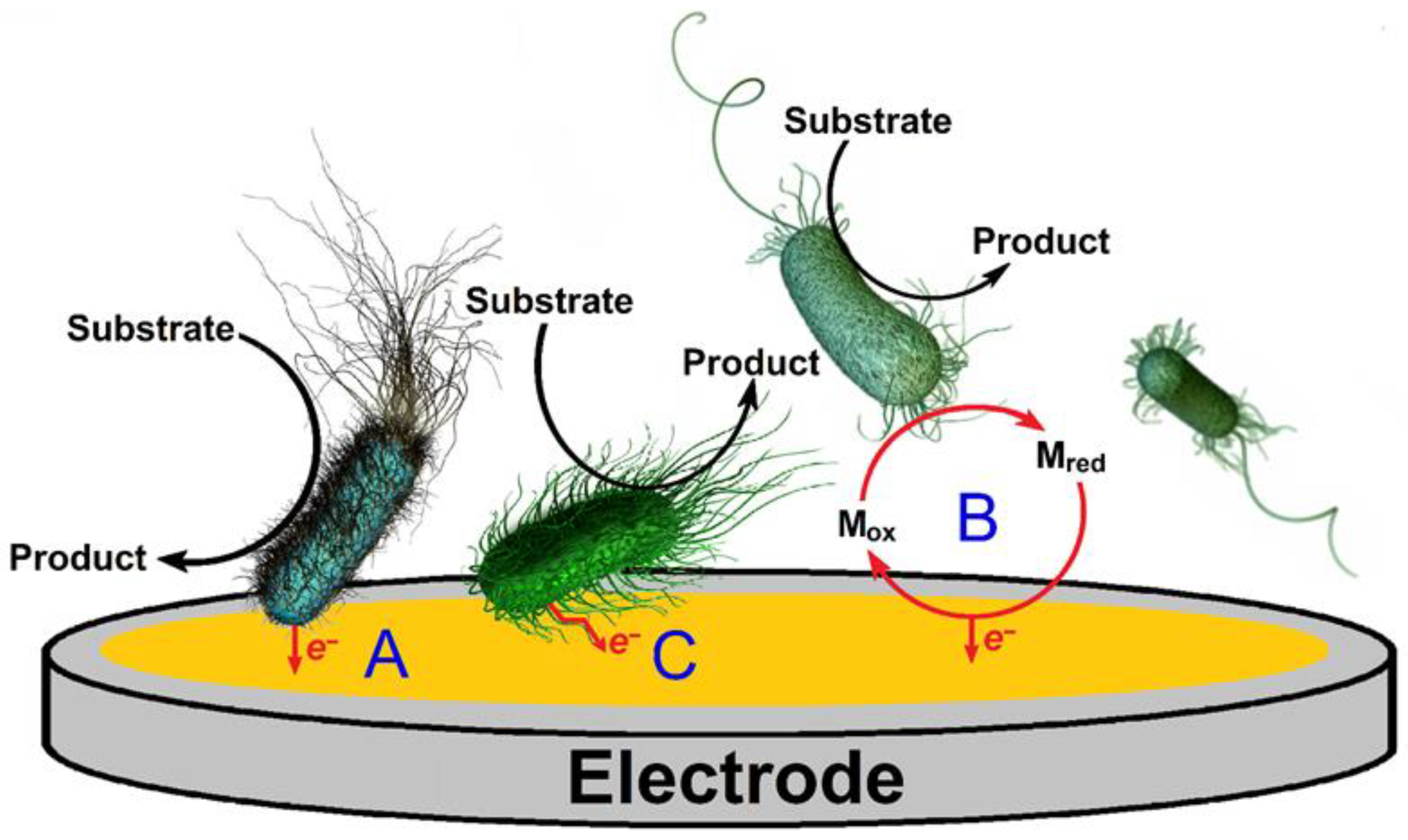
4.2. Electrochemical Communication during Biofilm Formation

In general, all bacterial biofilms can be defined as microbial communities within which the individual bacterial cells communicate with each other. This communication affects the rise of biofilm, the coordination of biofilm growth depending on nutrient availability and environmental conditions, and interspecies interactions as well.
As can be predicted, microbial communication is associated with the transfer of chemical substances secreted by cells during quorum sensing. However, it is evident that chemicals are diluted out when they are secreted into the environment, thus decreasing the concentration of chemicals with distance from the cell reflecting the decrease in the signal. Moreover, it is important to keep in mind that these signals can only be perceived by specific receptors.
In the second half of the previous century, it was found out that intercellular communication is mediated through potassium (K+) channels, which were discovered in Escherichia coli in 1994 [156]. The secretion of K+ ions increases the movement of motile cells toward the biofilm. The K+ ions on the surface of the motile cells cause their depolarization via opening the K+ channels, which results in hyperpolarization and an increase in proton motive force [157]. The use of a microfluidic approach helped to reveal an interesting finding that the biofilm of bacteria Bacillus subtilis was able to attract distant motile cells of Pseudomonas aeruginosa attached and subsequently incorporated into the biofilm structure. Therefore, it seems that bacteria are capable of cross-species interactions by using K+ ion channel-mediated electrical signalization [158,159].
In addition to the mentioned concentration gradient of K+ ions, bacteria cells of Myxococcus xanthus can perform intercellular communication via outer membrane vesicle chains and membrane tubes that interconnect the individual cells. These sophisticated structures range between 30 and 60 nm in width and up to 5 μm in length. There is an assumption that the resulting network enables the transfer of specific molecules between cells, helping the coordination of their social activities [160].
In 2019, Stekolshchikova et al. demonstrated a biocompatibility of an ion-selective sensing platform for potentiometric measurements of potassium in Escherichia coli biofilms [161]. They showed the possibility of how to electrochemically detect biofilm formation through the determination of potassium concentration.

4.2.1. Electrochemical Communication between Microbial Cells and Conductive Surfaces

Only some bacteria from family Shewanellaceae and phylum Proteobacteria, especially Geobacter, are characterized by their capacity of extracellular electron transfer (EET) to the electrode. They are known as exoelectrogens and have aroused great interest in biotechnology, namely in bioelectrochemical systems (BES), for the generation of energy in microbial fuel cells (MFCs). Geobacter sulfurreducens [162], Geobacter metallireducens [163], Shewanella oneidensis [164], Klebsiella quasipneumoniae sp. 203 [165] and Desulfovibrio desulfuricans [166] are able to utilize polarized electrodes as the final electron acceptor of their respiratory chains. For biofilm of Geobacter sulfurreducens, a combination of pilli (nanowires) action and cytochromes, referred to as a “stepping stone” mechanism, was proposed for long range electron transport (>50 µm) through the biofilm [167,168]. In addition to the direct electron transfer, such a process can be mediated using endogenous redox mediators, as shown in Figure 4. For example, Os(III)/Osmium(V), Fe(II)/Fe(III), and quinoid redox couples can be considered as typical examples of endogenous redox mediators [169,170,171,172].
In addition to direct electron transfer, microbial cells can indirectly communicate with conductive surfaces via the electroactive products of their metabolism and/or extracellular polymeric substances (EPS) [168]. Herein, it is necessary to accomplish that the microbial cells inside biofilm do not utilize only one type of electron transfer, but their different combinations. The whole system is more complicated than it may seem at first glance, because of the additional ability of microbial cells to communicate with each other via diffusible intermediates, direct electron exchange between physically connected cells, and conductive particles [173].

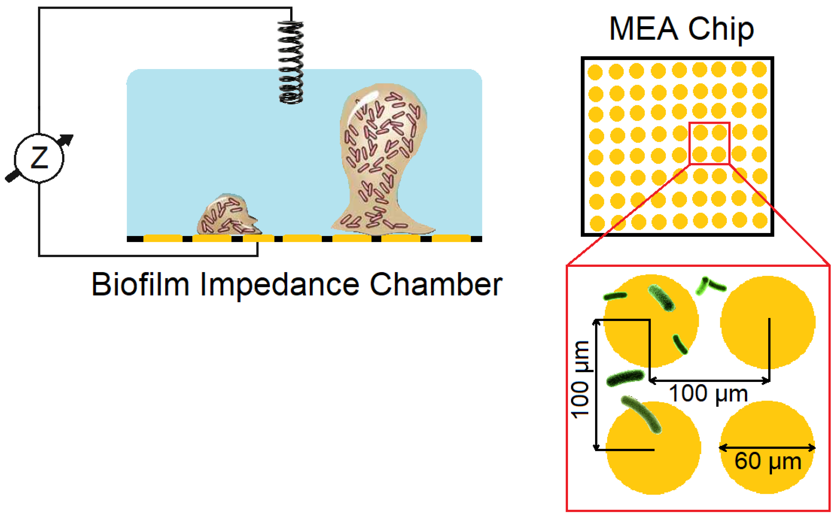
4.2.2. Electrochemical Mapping of Biofilm Location

The previous chapter suggests that bacterial cells are capable of exhibiting electrical activity which can be electrochemically monitored. In addition to the predominant spectral and microscopic methods [81], electrochemical approaches can also find their application in the mapping of biofilm location. Scanning electrochemical microscopy (SECM) [174] with its complementary technique of soft-probe-scanning electrochemical microscopy (Soft-Probe-SEM) [175] have proved themselves in the characterization of biofilms. Although SECM is being recommended as a suitable method for studying interactions in biofilms, it is still a desktop and large device not very suitable for the online monitoring of biofilm formation.
Fortunately, an electrochemical impedance spectroscopy (EIS) can be chosen instead, representing a nearly ideal tool for the non-contact electrochemical evaluation of biofilms [176]. In 2013, it was confirmed that it is capable of being operated in real time for the non-destructive monitoring of Pseudomonas aeruginosa biofilm, when its growth and metabolic activity was investigated using a combination of multi-channel impedance and amperometric sensors, respectively [177]. Numerous reports have shown that the electrochemical mapping of biofilm location can be performed using a multi-electrode array (MEA) system [178,179,180,181] integrated in the biofilm impedance chamber, as shown in Figure 5. MEA chips designed in this way represent promising devices for the control of microbiologically affected corrosion (MIC) related to sulfate-reducing bacteria (SRB). In 2015, a multi-electrode array (MEA) system was used for the monitoring of spatiotemporal electrical activity during the development of Bacillus licheniformis and Pseudomonas alcaliphila biofilms. Based on the data obtained, it was demonstrated that the intensity of the electrical activity did not linearly depend on the bacterial density, but it was instead correlated with biofilm formation [181]. Unfortunately, such a real-time electrochemical monitoring system was examined only in small reactors under laboratory conditions. In the near future, it can be assumed that the MEA chip will be directly installed into food industry processing equipment to verify its applicability.
In addition to the above-mentioned approach utilizing the impedance measurement, several studies demonstrated the usefulness of cyclic voltammetry [182,183] and Tafel analysis [182] at standard disc electrodes, with both these methods providing valuable information about bacterial attachment and biofilm formation [184]. For this purpose, the electrochemical reversibility of a redox marker is monitored over a period of time.
Biofilm location can be also determined using nano-engineered microbial electrochemical systems (MESs) based on voltammetric measurement with nanostructured electrodes that contain hyperbranched chitosan nanoparticles (HBCs) and reduced graphene oxide (rGO) nanosheets. In this case, the shapes of cyclic voltammograms obtained (increase in background current response) have indicated the individual phases of biofilm formation [183].

4.3. Electrochemical Approaches to Biofilm Removal

Up until now, a few electrochemical approaches for the removal of biofilm from conductive surfaces have been designed and tested [185]. Two of them utilize the cathodic evolution of hydrogen [186,187] and the potential pulse/reverse pulse technique [188]. The principles of these two approaches are described below and demonstrated in Figure 6.
Cathodic hydrogen evolution can be considered as one of the possible ways to effectively remove biofilm from various metal surfaces, such as titanium dental implants [186] and stainless-steel substrates [187]. It has been shown that the application of potentials lower than −1.5 V vs. Ag/AgCl within seconds is sufficient for complete removal of 10-day old Pseudomonas aeruginosa biofilm formed on a 316L stainless-steel substrate [187].
Besides this, the bacterial biofilm removal can be carried out using a potential pulse/reverse pulse technique employing a periodic waveform that consists of anodic and cathodic pulses, usually lower than 30 s. A regular alternation of pulses then results in an effective stripping of thin metal layers with adhered biofilm. In addition, this regenerates the original mirror-like shiny surface of stainless-steel substrates [188].
Although these approaches are very effective, they have not found a wider use. From a practical point of view, serious technological problems can be expected for biofilm removal from large areas, especially from pipelines. Thus, electrochemical approaches should at least be verified for the smaller components of equipment used in the food industry.

5. Conclusions

From the literature discussed in this review, it is evident that electrochemical methods can find a wider applicability in food technology due to their ability to prevent the adhesion of microbial cells onto conductive surfaces, to determine the location and activity of biofilm, and to remove biofilm via the vigorous evolution of hydrogen or by using electrochemical stripping.
Generally, biofilm is usually removed mechanically or, if possible, the entire component is replaced with a new one. Nowadays, a great emphasis is placed on material innovation and the optimization of operational conditions, which would lead to the prevention of biofilm formation. It is surprising that methods for electrochemical biofilm control have not come yet into practical use, even though they offer the apparent benefit to increase biofilm removal. Although numerous studies suggest that applying a potential or electrical current to a metal surface can effectively increase biofilm removal and be a more environment-friendly approach than the conventional chemical methods currently used in biofilm control, they still have not found their practical use. Thus, it can be predicted that an application of a constant voltage to the conductive surfaces (most often of a stainless-steel nature) of food processing equipment can decrease the cost associated with the conventional approaches to biofilm removal, thus opening a way for more effective prevention of biofilm formation.
At the beginning, it would be a good idea to at least try the combination of electrochemical approaches with already established procedures, for example in a narrow stainless-steel drinking-water pipe, where a reference electrode could be installed. At the same time, an analogical experiment would have to take place without the presence of this electrochemical contribution, which would serve as a blank for control.

Author Contributions

Chapter: Biofilms, I.B. and P.M.; Chapter: Biofilms in Food Industry, L.Č. and M.F.; Chapter: Electrochemical Biofilm Control, R.M. and M.S.; final revision and language correction, I.Š.; supervision, M.S. All authors have read and agreed to the published version of the manuscript.

Funding

This research received no external funding.

Institutional Review Board Statement

Not applicable.

Informed Consent Statement

Not applicable.

Data Availability Statement

All the relevant data are only provided in the present paper.

Acknowledgments

Financial support from the Faculty of Chemical Technology, University of Pardubice (project No. SGS-2022-002) is gratefully acknowledged.

Conflicts of Interest

The authors declare no conflict of interest.

References

  1. Vestby, L.K.; Grønseth, T.; Simm, R.; Nesse, L.L. Bacterial biofilm and its role in the pathogenesis of disease. Antibiotics 2020, 9, 59. [Google Scholar] [CrossRef] [Green Version]
  2. Kostakioti, M.; Hadjifrangiskou, M.; Hultgren, S.J. Bacterial biofilms: Development, dispersal, and therapeutic strategies in the dawn of the postantibiotic era. Cold Spring Harb. Perspect. Med. 2013, 3, a010306. [Google Scholar] [CrossRef] [Green Version]
  3. Muhammad, M.H.; Idris, L.A.; Fan, X.; Guo, Y.; Yu, Y.; Jin, X.; Qiu, J.; Guan, X.; Huang, T. Beyond risk: Bacterial biofilms and their regulating approaches. Front. Microbiol. 2020, 11, 928. [Google Scholar] [CrossRef]
  4. Billings, N.; Millan, M.; Caldara, M.; Rusconi, R.; Tarasova, Y.; Stocker, R.; Ribbeck, K. The extracellular matrix component Psl provides fast-acting antibiotic defense in Pseudomonas aeruginosa biofilms. PLoS Pathog. 2013, 9, e1003526. [Google Scholar] [CrossRef] [Green Version]
  5. Bjarnsholt, T. The role of bacterial biofilms in chronic infections. J. Pathol. Microbiol. Immunol. 2013, 121, 1–58. [Google Scholar] [CrossRef]
  6. Donlan, R.M.; Costerton, J.W. Biofilms: Survival mechanisms of clinically relevant microorganisms. Clin. Microbiol. Rev. 2002, 15, 167–193. [Google Scholar] [CrossRef] [Green Version]
  7. Stoodley, P.; Hall-Stoodley, L. Evolving concepts in biofilm infections. Cell. Microbiol. 2009, 11, 1034–1043. [Google Scholar] [CrossRef]
  8. Gebreyohannes, G.; Nyerere, A.; Bii, C.; Sbhatu, D.B. Challenges of intervention, treatment, and antibiotic resistance of biofilm-forming microorganisms. Heliyon 2019, 5, e02192. [Google Scholar] [CrossRef] [Green Version]
  9. Flemming, H.C.; Wuertz, S. Bacteria and archaea on Earth and their abundance in biofilms. Nat. Rev. Microbiol. 2019, 17, 247–260. [Google Scholar] [CrossRef]
  10. Coughlan, L.M.; Cotter, P.D.; Hill, C.; Alvarez-Ordonez, A. New weapons to fight old enemies: Novel strategies for the (Bio)control of bacterial biofilms in the food industry. Front. Microbiol. 2016, 7, 1641. [Google Scholar] [CrossRef] [Green Version]
  11. Dobretsov, S.; Dahms, H.U.; Qian, P.Y. Inhibition of biofouling by marine microorganisms and their metabolites. Biofouling 2006, 22, 43–54. [Google Scholar] [CrossRef]
  12. Bi, Y.; Xia, G.; Shi, C.; Wan, J.; Liu, L.; Chen, Y.; Wu, Y.; Zhang, W.; Zhou, M.; He, H.; et al. Therapeutic strategies against bacterial biofilms. Fundam. Res. 2021, 1, 193–212. [Google Scholar] [CrossRef]
  13. Galie, S.; Garcia-Gutierrez, C.; Miguelez, E.M.; Villar, C.J.; Lombo, F. Biofilms in the food industry: Health aspects and control methods. Front. Microbiol. 2018, 9, 898. [Google Scholar] [CrossRef]
  14. Yan, J.; Bassler, B.L. Surviving as a community: Antibiotic tolerance and persistence in bacterial biofilms. Cell Host Microbe 2019, 26, 15–21. [Google Scholar] [CrossRef]
  15. Abee, T.; Kovács, A.T.; Kuipers, O.P.; van der Veen, S. Biofilm formation and dispersal in Gram-positive bacteria. Curr. Opin. Biotechnol. 2011, 22, 172–179. [Google Scholar] [CrossRef] [Green Version]
  16. Bowler, P.; Murphy, C.; Wolcott, R. Biofilm exacerbates antibiotic resistance: Is this a current oversight in antimicrobial stewardship? Antimicrob. Resist. Infect. Control 2020, 9, 162. [Google Scholar] [CrossRef]
  17. Han, N.; Mizan, M.F.R.; Jahid, I.K.; Ha, S.D. Biofilm formation by Vibrio parahaemolyticus on food and food contact surfaces increases with rise in temperature. Food Control 2016, 70, 161–166. [Google Scholar] [CrossRef]
  18. Jahid, I.K.; Mizan, M.F.R.; Ha, A.J.; Ha, S.D. Effect of salinity and incubating time of planktonic cells on biofilm formation, motility, exoprotease production, and quorum sensing of Aeromonas hydrophila. Food Microbiol. 2015, 49, 142–151. [Google Scholar] [CrossRef]
  19. Khatoon, Z.; McTiernan, C.D.; Suuronen, E.J.; Mah, T.F.; Alarcon, E.I. Bacterial biofilm formation on implantable devices and approaches to its treatment and prevention. Heliyon 2018, 4, e01067. [Google Scholar] [CrossRef] [Green Version]
  20. Karygianni, L.; Ren, Z.; Koo, H.; Thurnheer, T. Biofilm matrixome: Extracellular components in structured, microbial communities. Trends Microbiol. 2020, 28, 668–681. [Google Scholar] [CrossRef]
  21. Mizan, M.F.R.; Jahid, I.K.; Park, S.Y.; Silva, J.L.; Kim, T.J.; Myoung, J.; Ha, S.D. Effects of temperature on biofilm formation and quorum sensing of Aeromonas hydrophila. Ital. J. Food Sci. 2018, 30, 456–466. [Google Scholar]
  22. Guzmán-Soto, I.; McTiernan, C.; Gonzalez-Gomez, M.; Ross, A.; Gupta, K.; Suuronen, E.J.; Mah, T.F.; Griffith, M.; Alarcon, E.I. Mimicking biofilm formation and development: Recent progress in in vitro and in vivo biofilm models. iScience 2021, 24, 102443. [Google Scholar] [CrossRef]
  23. Huang, Y.; Flint, S.H.; Palmer, J.S. Bacillus cereus spores and toxins—The potential role of biofilms. Food Microbiol. 2020, 90, 103493. [Google Scholar] [CrossRef]
  24. Korber, D.R.; Mangalappalli-Illathu, A.K.; Vidoviæ, S. Chapter 6—Biofilm formation by food spoilage microorganisms in food processing environments. In Biofilms in the Food and Beverage Industries; Fratamico, P.M., Annous, B.A., Gunthereds, N.W., Eds.; Woodhead Publishing: Sawston, UK, 2009; pp. 166–199. [Google Scholar]
  25. Lin, Y.; Briandet, R.; Kovács, Á.T. Bacillus cereus sensu lato biofilm formation and its ecological importance. Biofilm 2022, 4, 100070. [Google Scholar] [CrossRef]
  26. Park, E.-J.; Hussain, M.S.; Wei, S.; Kwon, M.; Oh, D.-H. Genotypic and phenotypic characteristics of biofilm formation of emetic toxin producing Bacillus cereus strains. Food Control 2019, 96, 527–534. [Google Scholar] [CrossRef]
  27. Wirtanen, G.; Salo, S. Chapter 5—Biofilm risks. In Handbook of Hygiene Control in the Food Industry, 2nd ed.; Lelieveld, H., Holah, J., Gabriæ, D., Eds.; Woodhead Publishing: San Diego, CA, USA, 2016; pp. 55–79. [Google Scholar]
  28. Liu, D.; Ge, S.; Wang, Z.; Li, M.; Zhuang, W.; Yang, P.; Chen, Y.; Ying, H. Identification of a sensor histidine kinase (BfcK) controlling biofilm formation in Clostridium acetobutylicum. Chin. J. Chem. Eng. 2021; in press. [Google Scholar] [CrossRef]
  29. Pantaléon, V.; Bouttier, S.; Soavelomandroso, A.P.; Janoir, C.; Candela, T. Biofilms of Clostridium species. Anaerobe 2014, 30, 193–198. [Google Scholar] [CrossRef]
  30. Pantaléon, V.; Soavelomandroso, A.P.; Bouttier, S.; Briandet, R.; Roxas, B.; Collignon, M.A.; Janoir, C.; Vedantam, G.; Candela, T. The Clostridium difficile protease Cwp84 modulates both biofilm formation and cell-surface properties. PLoS ONE 2015, 10, e0124971. [Google Scholar] [CrossRef]
  31. Hu, L. Prevalence of curli genes among Cronobacter species and their roles in biofilm formation and cell-cell aggregation. Int. J. Food Microbiol. 2018, 265, 65–73. [Google Scholar] [CrossRef]
  32. Ling, N.; Forsythe, S.; Wu, Q.; Ding, Y.; Zhang, Y.; Zeng, H. Insights into Cronobacter sakazakii biofilm formation and control strategies in the food industry. Engineering 2020, 6, 393–405. [Google Scholar] [CrossRef]
  33. Naziri, Z.; Kilegolan, J.A.; Moezzi, M.S.; Derakhshandeh, A. Biofilm formation by uropathogenic Escherichia coli: A complicating factor for treatment and recurrence of urinary tract infections. J. Hosp. Infect. 2021, 117, 9–16. [Google Scholar] [CrossRef]
  34. Makled, A.F.; Salem, E.H.; Elbrolosy, A.M. Biofilm formation and antimicrobial resistance pattern of uropathogenic E. coli: Comparison of phenotypic and molecular methods. Egypt. J. Med. Microbiol. 2017, 26, 37–45. [Google Scholar] [CrossRef]
  35. Sharma, G.; Sharma, S.; Sharma, P.; Chandola, D.; Dang, S.; Gupta, S.; Gabrni, R. Escherichia coli biofilm: Development and therapeutic strategies. J. Appl. Microbiol. 2016, 121, 309–319. [Google Scholar] [CrossRef] [Green Version]
  36. Tajbakhsh, E.; Ahmadi, P.; Abedpour-Dehkordi, E.; Arbab-Soleimani, N.; Khamesipour, F. Biofilm formation, antimicrobial susceptibility, serogroups and virulence genes of uropathogenic E. coli isolated from clinical samples in Iran. Antimicrob. Resist. Infect. Control 2016, 5, 11. [Google Scholar] [CrossRef] [Green Version]
  37. Zamani, H.; Salehzadeh, A. Biofilm formation in uropathogenic Escherichia coli: Association with adhesion factor genes. Turk. J. Med. Sci. 2018, 48, 162–167. [Google Scholar] [CrossRef]
  38. Bonsaglia, E.C.R.; Silva, N.C.C.; Fernades Júnior, A.; Araújo Júnior, J.P.; Tsunemi, M.H.; Rall, V.L.M. Production of biofilm by Listeria monocytogenes in different materials and temperatures. Food Control 2013, 35, 386–391. [Google Scholar] [CrossRef]
  39. Gandra, T.K.V.; Volcan, D.; Kroning, I.S.; Marini, N.; de Oliveira, A.C.; Bastos, C.P.; da Silva, W.P. Expression levels of the agr locus and prfA gene during biofilm formation by Listeria monocytogenes on stainless steel and polystyrene during 8 to 48 h of incubation 10 to 37 °C. Int. J. Food Microbiol. 2019, 300, 1–7. [Google Scholar] [CrossRef]
  40. Ramires, T.; Kleinubing, N.R.; Iglesias, M.A.; Vitola, H.R.S.; Núncio, A.S.P.; Kroning, I.S.; Moreira, G.M.S.G.; Fiorentini, Â.M.; da Silva, W.P. Genetic diversity, biofilm and virulence characteristics of Listeria monocytogenes in salmon sushi. Food Res. Int. 2021, 140, 109871. [Google Scholar] [CrossRef]
  41. Wang, Y.; Sun, L.; Hu, L.; Wang, Z.; Wang, X.; Dong, Q. Adhesion and kinetics of biofilm formation and related gene expression of Listeria monocytogenes in response to nutritional stress. Food Res. Int. 2022, 156, 111143. [Google Scholar] [CrossRef]
  42. Abd El Galil, K.; Abdelghani, S.M.; Sebak, M.A.; El-Naggar, W. Detection of biofilm genes among clinical isolates of Pseudomonas aeruginosa recovered from some Egyptian hospitals. New Egypt. J. Microbiol. 2013, 36, 86–101. [Google Scholar]
  43. Abdelraheem, W.M.; Abdelkader, A.E.; Mohamed, E.S.; Mohammed, M.S. Detection of biofilm formation and assessment of biofilm genes expression in different Pseudomonas aeruginosa clinical isolates. Meta Gene 2020, 23, 100646. [Google Scholar] [CrossRef]
  44. Ambutsi, M.; Okoth, P. Comparative genomic analysis of gene clusters of Pseudomonas aeruginosa that define specific biofilm formation in deciphering target regions for novel treatment options. Sci. Afr. 2021, 13, e00910. [Google Scholar] [CrossRef]
  45. Ghazalibina, M.; Morshedi, K.; Farahani, R.K.; Babadi, M.; Khaledi, A. Study of virulence genes and related with biofilm formation in Pseudomonas aeruginosa isolated from clinical samples of Iranian patients: A systematic review. Gene Rep. 2019, 17, 100471. [Google Scholar] [CrossRef]
  46. Kamali, E.; Jamali, A.; Ardebili, A.; Ezadi, F.; Mohebbi, A. Evaluation of antimicrobial resistance, biofilm forming potential, and the presence of biofilm-related genes among clinical isolates of Pseudomonas aeruginosa. BMC Res. Notes 2020, 13, 27. [Google Scholar] [CrossRef] [PubMed]
  47. Laverty, G.; Gorman, S.; Gilmore, B. Biomolecular mechanisms of Pseudomonas aeruginosa and Escherichia coli biofilm formation. Pathogens 2014, 3, 596–632. [Google Scholar] [CrossRef] [PubMed] [Green Version]
  48. Møretrø, T.; Langsrud, S. Residential bacteria on surfaces in the food industry and their implications for food safety and quality. Compr. Rev. Food Sci. Food Saf. 2017, 16, 1022–1041. [Google Scholar] [CrossRef] [PubMed] [Green Version]
  49. Giacomodonato, M.N.; Sarnacki, S.H.; Aya Castañeda, M.D.R.; Garófalo, A.N.; Betancourt, D.M.; Cerquetti, M.C.; Llana, M.N. Salmonella enterica se.rovar Enteritidis biofilm lifestyle induces lower pathogenicity and reduces inflammatory response in a murine model compared to planktonic bacteria. Rev. Argent. Microbiol. 2021; in press. [Google Scholar] [CrossRef]
  50. Obe, T.; Nannapaneni, R.; Schilling, W.; Zhang, L.; Kiess, A. Antimicrobial tolerance, biofilm formation, and molecular characterization of Salmonella isolates from poultry processing equipment. J. Appl. Poult. Res. 2021, 30, 100195. [Google Scholar] [CrossRef]
  51. Tsai, M.-H.; Liang, Y.-H.; Chen, C.-L.; Chiu, C.-H. Characterization of Salmonella resistance to bile during biofilm formation. J. Microbiol. Immunol. Infect. 2020, 53, 518–524. [Google Scholar] [CrossRef]
  52. Ghaioumy, R.; Tabatabaeifar, F.; Mozafarinia, K.; Mianroodi, A.A.; Isaei, E.; Monores-Ramírez, J.R.; Afshari, S.A.K.; Kalantar-Neyestanaki, D. Biofilm formation and molecular analysis of intercellular adhesion gene cluster (icaABCD) among Staphylococcus aureus strains isolated from children with adenoiditis. Iran. J. Microbiol. 2021, 13, 458–463. [Google Scholar] [CrossRef]
  53. Idrees, M.; Sawant, S.; Karodia, N.; Rahman, A. Staphylococcus aureus biofilm: Morphology, genetics, pathogenesis and treatment strategies. Int. J. Environ. Res. Public Health 2021, 18, 7602. [Google Scholar] [CrossRef]
  54. Khoramrooz, S.S.; Mansouri, F.; Marashifard, M.; Hosseini, S.A.A.M.; Chenarestane-Oli, A.F.; Ganavehei, B.; Charibpour, F.; Shahbazi, A.; Mirzaii, M.; Darban-Sarokhalil, D. Detection of biofilm related genes, classical enterotoxin genes and agr typing among Staphylococcus aureus isolated from bovine with subclinical mastitis in southwest of Iran. Microb. Pathog. 2016, 97, 45–51. [Google Scholar] [CrossRef]
  55. Nourbakhsh, F.; Namvar, A.E. Detection of genes involved in biofilm formation in Staphylococcus aureus isolates. GMS Hyg. Infect. Control 2016, 11, Doc07. [Google Scholar] [PubMed]
  56. Shahmoradi, M.; Faridifar, P.; Shapouri, R.; Mousavi, S.F.; Ezzedin, M.; Mirzaei, B. Determining the biofilm forming gene profile of Staphylococcus aureus clinical isolates via multiplex colony PCR method. Rep. Biochem. Mol. Biol. 2019, 7, 181–188. [Google Scholar] [PubMed]
  57. Bezek, K.; Nipič, D.; Torkar, K.G.; Oder, M.; Dražić, G.; Abram, A.; Žibert, J.; Raspor, P.; Bohinc, K. Biofouling of stainless steel surfaces by four common pathogens: The effects of glucose concentration, temperature and surface roughness. Biofouling 2019, 35, 273–283. [Google Scholar] [CrossRef] [PubMed]
  58. Kim, B.-B.; Bae, Y.-M.; Lee, S.-Y. Effect of environmental conditions on biofilm formation and related characteristics of Staphylococcus aureus. J. Food Saf. 2016, 36, 412–422. [Google Scholar] [CrossRef]
  59. Fan, Y.; Qiao, J.; Lu, Z.; Fen, Z.; Tao, Y.; Lv, F.; Zhao, H.; Zhang, C.; Bie, X. Influence of different factors on biofilm formation of Listeria monocytogenes and the regulation of cheY gene. Food Res. Int. 2020, 137, 109405. [Google Scholar] [CrossRef]
  60. Got, F. Staphylococcus and biofilms. Mol. Microbiol. 2002, 43, 1367–1378. [Google Scholar]
  61. Moraes, J.O.; Cruz, E.A.; Souza, E.G.; Oliveira, T.C.; Alvarenga, V.O.; Peña, W.E.; Sant’Ana, A.S.; Magnani, M. Predicting adhesion and biofilm formation boundaries on stainless steel surfaces by five Salmonella enterica strains belonging to different serovars as a function of pH, temperature and NaCl concentration. Int. J. Food Microbiol. 2018, 281, 90–100. [Google Scholar] [CrossRef]
  62. Shi, X.; Zhu, X. Biofilm formation and food safety in food industries. Trends Food Sci. Technol. 2009, 20, 407–413. [Google Scholar] [CrossRef]
  63. Yin, B.; Zhu, L.; Zhang, Y.; Dong, P.; Mao, Y.; Liang, R.; Niu, L.; Luo, X. The characterization of biofilm formation and detection of biofilm-related genes in Salmonella isolated from beef processing plants. Foodborne Pathog. Dis. 2018, 15, 660–667. [Google Scholar] [CrossRef]
  64. Lee, K.H.; Lee, J.Y.; Roy, P.K.; Mizan, M.F.R.; Hossain, M.I.; Park, S.H.; Ha, S.D. Viability of Salmonella Typhimurium biofilms on major food-contact surfaces and eggshell treated during 35 days with and without water storage at room temperature. Poult. Sci. 2020, 99, 4558–4565. [Google Scholar] [CrossRef]
  65. Gomes, I.B.; Lemos, M.; Fernandes, S.; Borges, A.; Simões, L.C.; Simões, M. The effects of chemical and mechanical stresses on Bacillus cereus and Pseudomonas fluorescens single- and dual-species biofilm removal. Microorganisms 2021, 9, 1174. [Google Scholar] [CrossRef] [PubMed]
  66. Chisti, Y.; Moo-Young, M. Clean-in-place systems for industrial bioreactors: Design, validation and operation. J. Ind. Microbiol. Biotechnol. 1994, 13, 201–207. [Google Scholar] [CrossRef]
  67. Joseph, B.; Otta, S.K.; Karunasagar, I.; Karunasagar, I. Biofilm formation by Salmonella spp. on food contact surfaces and their sensitivity to sanitizers. Int. J. Food Microbiol. 2001, 64, 367–372. [Google Scholar] [CrossRef]
  68. Poimenidou, S.V.; Chrysadakou, M.; Tzakoniati, A.; Bikouli, V.C.; Nychas, G.J.; Skandamis, P.N. Variability of Listeria mono-cytogenes strains in biofilm formation on stainless steel and polystyrene materials and resistance to peracetic acid and quater-nary ammonium compounds. Int. J. Food Microbiol. 2016, 237, 164–171. [Google Scholar] [CrossRef] [PubMed]
  69. Rodríguez, A.; McLandsborough, L.A. Evaluation of the transfer of Listeria monocytogenes from stainless steel and high-density polyethylene to Bologna and American cheese. J. Food Protect. 2007, 70, 600–606. [Google Scholar] [CrossRef] [PubMed]
  70. Coulier, L.; Orbons, H.G.M.; Rijk, R. Analytical protocol to study the food safety of (multiple) recycled high-density polyeth-ylene (HDPE) and polypropylene (PP) crates: Influence of recycling on the migration and formation of degradation products. Polym. Degrad. Stab. 2007, 92, 2016–2025. [Google Scholar] [CrossRef]
  71. Moerman, F.; Partington, E. Materials of construction for food processing equipment and services: Requirements, strengths and weaknesses. J. Hyg. Eng. Des. 2014, 6, 10–37. [Google Scholar]
  72. Srey, S.; Jahid, I.K.; Ha, S.D. Biofilm formation in food industries: A food safety concern. Food Control 2013, 31, 572–585. [Google Scholar] [CrossRef]
  73. Song, B.; Leff, L.G. Influence of magnesium ions on biofilm formation by Pseudomonas fluorescens. Microbiol. Res. 2006, 161, 355–361. [Google Scholar] [CrossRef]
  74. Danilova, T.A.; Danilina, G.A.; Adzhieva, A.A.; Vostrova, E.I.; Zhukhovitskii, V.G.; Cheknev, S.B. Inhibitory effect of copper and zinc ions on the growth of Streptococcus pyogenes and Escherichia coli biofilms. Bull. Exp. Biol. Med. 2020, 169, 648–652. [Google Scholar] [CrossRef]
  75. Simbine, E.O.; Rodrigues, L.C.; Lapa-Guimaraes, J.; Kamimura, E.S.; Corassin, C.H.; Oliveira, C.A.F. Application of silver nanoparticles in food packages: A review. Food Sci. Technol. 2019, 39, 793–802. [Google Scholar] [CrossRef] [Green Version]
  76. Ashrafi, A.M.; Sýs, M.; Sedláčková, E.; Farag, A.S.; Adam, V.; Přibyl, J.; Richtera, L. Application of the enzymatic electrochemical biosensors for monitoring non-competitive inhibition of enzyme activity by heavy metals. Sensors 2019, 19, 2939. [Google Scholar] [CrossRef] [Green Version]
  77. Coenye, T.; De Prijck, K.; De Wever, B.; Nelis, H.J. Use of the modified Robbins device to study the in vitro biofilm removal efficacy of NitrAdine™, a novel disinfecting formula for the maintenance of oral medical devices. J. Appl. Microbiol. 2008, 105, 733–740. [Google Scholar] [CrossRef]
  78. Silvestry-Rodriguez, N.; Bright, K.R.; Slack, D.C.; Uhlmann, D.R.; Gerba, C.P. Silver as a residual disinfectant to prevent biofilm formation in water distribution systems. Appl. Environ. Microbiol. 2008, 74, 1639–1641. [Google Scholar] [CrossRef] [PubMed] [Green Version]
  79. Petala, M.; Tsiridis, V.; Mintsouli, I.; Pliatsikas, N.; Spanos, T.; Rebeyre, P.; Darakas, E.; Patsalas, P.; Vourlias, G.; Kostoglou, M.; et al. Silver deposition on stainless steel container surfaces in contact with disinfectant silver aqueous solutions. Appl. Surf. Sci. 2017, 396, 1067–1075. [Google Scholar] [CrossRef]
  80. Hong, S.-H.; Gorce, J.-B.; Punzmann, H.; Francois, N.; Shats, M.; Xia, H. Surface waves control bacterial attachment and formation of biofilms in thin layers. Sci. Adv. 2020, 6, eaaz9386. [Google Scholar] [CrossRef] [PubMed]
  81. Azeredo, J.; Azevedo, N.F.; Briandet, R.; Cerca, N.; Coenye, T.; Costa, A.R.; Desvaux, M.; Di Bonaventura, G.; Hébraud, M.; Jaglic, Z.; et al. Critical review on biofilm methods. Crit. Rev. Microbiol. 2017, 43, 313–351. [Google Scholar] [CrossRef] [PubMed] [Green Version]
  82. Brindhadevi, K.; LewisOscar, F.; Mylonakis, E.; Shanmugam, S.; Verma, T.N.; Pugazhendhi, A. Biofilm and quorum sensing mediated pathogenicity in Pseudomonas aeruginosa. Process Biochem. 2020, 96, 49–57. [Google Scholar] [CrossRef]
  83. Brown, S.; Santa Maria, J.P., Jr.; Walker, S. Wall teichoic acids of gram-positive bacteria. Annu. Rev. Microbiol. 2013, 67, 313–336. [Google Scholar] [CrossRef] [Green Version]
  84. Silhavy, T.J.; Kahne, D.; Walker, S. The bacterial cell envelope. Cold Spring Harb. Perspect. Biol. 2010, 2, a000414. [Google Scholar] [CrossRef]
  85. Vega-Hernández, R.; Ochoa, S.A.; Valle-Rios, R.; Jaimes-Ortega, G.A.; Arellano-Galindo, J.; Aparicio-Ozores, G.; Ibarra, J.A.; Hernández-Castro, R.; Cruz-Córdova, A.; Xicohtencatl-Cortes, J. Flagella, Type I fimbriae and curli of uropathogenic Escherichia coli promote the release of proinflammatory cytokines in a coculture system. Microorganisms 2021, 9, 2233. [Google Scholar] [CrossRef]
  86. Barnhart, M.M.; Chapman, M.R. Curli biogenesis and function. Annu. Rev. Microbiol. 2006, 60, 131–147. [Google Scholar] [CrossRef] [Green Version]
  87. Song, F.; Koo, H.; Ren, D. Effects of material properties on bacterial adhesion and biofilm formation. J. Dent. Res. 2015, 94, 1027–1034. [Google Scholar] [CrossRef]
  88. FAO. Global Food Losses and Food Waste-Extent, Causes and Prevention, Rome. 2011. Available online: www.fao.org/3/i2697e/i2694e.pdf (accessed on 16 May 2022).
  89. Stenmarck, A.; Jensen, C.; Quested, T.; Moates, G. Estimates of European Food Waste Levels; IVL Swedish Environmental Research Institute: Stockholm, Sweden, 2016; pp. 1–79. [Google Scholar]
  90. Kwok, T.; Ma, Y.; Chua, S.L. Biofilm dispersal induced by mechanical cutting leads to heightened foodborne pathogen dissemination. Food Microbiol. 2022, 102, 103914. [Google Scholar] [CrossRef]
  91. Xu, J.G.; Huang, X.N.; Meng, J.; Chen, J.Y.; Han, B.Z. Characterization and comparison of the bacterial community on environmental surfaces through a fresh-cut vegetables processing line in China. Int. Food Res. J. 2022, 155, 111075. [Google Scholar] [CrossRef]
  92. Wagner, E.M.; Pracser, N.; Thalguter, S.; Fischel, K.; Rammer, N.; Pospíšilová, L.; Alispahic, M.; Wagner, M.; Rychli, K. Identification of biofilm hotspots in a meat processing environment: Detection of spoilage bacteria in multi-species biofilms. Int. J. Food Microbiol. 2020, 328, 108668. [Google Scholar] [CrossRef]
  93. Rather, M.A.; Gupta, K.; Bardhan, P.; Borah, M.; Sarkar, A.; Eldiehy, K.S.H.; Bhuyan, S.; Mandal, M. Microbial biofilm: A matter of grave concern for human health and food industry. J. Basic Microbiol. 2021, 61, 380–395. [Google Scholar] [CrossRef]
  94. Lai, H.; Tang, Y.; Ren, F.; Li, Z.; Li, F.; Cu, C.; Jiao, X.; Huang, J. An investigation into the critical factors influencing the spread of Campylobacter during chicken handling in commercial kitchens in China. Microorganisms 2021, 9, 1164. [Google Scholar] [CrossRef]
  95. Ashrafudoulla, M.; Na, K.W.; Byun, K.-H.; Kim, D.H.; Yoon, J.W.; Mizan, M.F.R.; Kang, I.; Ha, S.-D. Isolation and characterization of Salmonella spp. From food and food conctat surfaces in chicken processing factory. Poult. Sci. 2021, 100, 101234. [Google Scholar] [CrossRef]
  96. Visvalingam, J.; Ells, T.C.; Yang, X. Impact of persistent and nonpersistent generic Escherichia coli and Salmonella sp. recovered from a beef packing plant on biofilm formation by E. coli O157. J. Appl. Microbiol. 2017, 123, 1512–1521. [Google Scholar] [CrossRef]
  97. European Food Safety Authority; European Centre for Disease Prevention and Control. The European Union summary report on trends and sources of zoonozes, zoonotc agents and food-borne outbreaks in 2017. EFSA J. 2018, 16, 5500. [Google Scholar]
  98. Borges, K.A.; Furian, T.Q.; de Souza, S.N.; Menezes, R.; Salle, C.T.P.; de Souza Moraes, H.L.; Tondo, E.C.; do Nascimento, V.P. Phenotypic and molecular characterization of Salmonella Enteritidis SE86 isolated from poultry and salmonellosis outbreaks. Foodborne Pathog. Dis. 2017, 14, 742–754. [Google Scholar] [CrossRef]
  99. Castro, H.; Jaakkonen, A.; Hakakorpi, A.; Hakkinen, M.; Isidro, J.; Korkeala, H.; Lindström, M.; Hallanvuo, S. Genomic epidemiology and phenotyping reveal on-farm persistent and cold adaptation of raw milk outbreak-associated Yersinia pseudotuberculosis. Front. Microbiol. 2019, 10, 1049. [Google Scholar] [CrossRef]
  100. Etter, A.J.; West, A.M.; Burnett, J.L.; Wu, S.T.; Veenhuizen, D.R.; Ogas, R.A.; Oliver, H.F. Salmonella enterica subsp. enterica serovar Heidelberg food isolates associated with a samonellosis outbreak have enhanced stress tolarence capabilities. Appl. Environ. Microbiol. 2019, 85, e01065-19. [Google Scholar]
  101. Jaakkonen, A.; Kivistö, R.; Aarnio, M.; Kalekivi, J.; Hakkinen, M. Persistent contamination of raw milk by Campylobacter jejuni ST-883. PLoS ONE 2020, 15, e0231810. [Google Scholar] [CrossRef]
  102. Uhlich, G.A.; Sinclair, J.R.; Warren, N.G.; Chmielecki, W.A.; Frantamico, P. Characterization of shiga toxin-producing Escherichia coli isolates associated with two multistate food-borne outbreaks that occurred in 2006. Appl. Environ. Microbiol. 2008, 74, 1268–1272. [Google Scholar] [CrossRef] [Green Version]
  103. Sinde, E.; Carballo, J. Attachment of Salmonella spp. and Listeria monocytogenes on stainless steel, rubber and polytetrafluorethylene: The influence of free energy and the effect of commercial sanitizer. Food Microbiol. 2000, 17, 439–447. [Google Scholar] [CrossRef]
  104. Gomes, I.B.; Simões, M.; Simões, L.C. Copper surfaces in biofilm control. Nanomaterials 2020, 10, 2491. [Google Scholar] [CrossRef]
  105. Zhang, X.; Liu, X.; Yang, C.; Xi, T.; Zhao, J.; Liu, L.; Yang, K. New strategy to delay food spoilace: Application of new food contact material with antibacterial function. J. Mater. Sci. Technol. 2021, 70, 59–66. [Google Scholar] [CrossRef]
  106. Pontin, K.P.; Borges, K.A.; Furian, T.Q.; Darvalho, D.; Wilsmann, D.E.; Cardoso, H.R.P.; Alves, A.K.; Chitolina, G.Z.; Salle, C.T.P.; de Souza Moraes, H.L.; et al. Antimicrobial activity of copper surfaces against biofilm formation by Salmonella Enteritidis its potential application in the poultry industry. Food Microbiol. 2021, 94, 103654. [Google Scholar] [CrossRef]
  107. Dula, S.; Ajayeoba, T.A.; Ijabadeniyi, O.A. Bacterial biofilm formation on stainless steel in the food processing environment and its health implications. Folia Microbiol. 2021, 66, 293–302. [Google Scholar] [CrossRef]
  108. Soule, L.D.; Chomorro, N.P.; Chuong, K.; Mellott, N.; Hammer, N.; Hankenson, K.D.; Chatzistavrou, X. Sol-gel-derived bioactive and antibacterial multi-component thin films by the spin-coating technique. ACS Biomater. Sci. Eng. 2020, 6, 5549–5562. [Google Scholar] [CrossRef]
  109. Araújo, E.M.; de Andrade, N.J.; da Silva, L.H.M.; Bernardes, P.C.; de Carvalho Teixeira, A.V.N.; Júnior, J.F.Q.F.; de Sá, J.P.N.; Fernandes, P.É. Modification of stainless steel surface hydrophobicity by silver nanoparticles: Strategies to prevent bacterial adhesion in the food processing. J. Adhes. Sci. Technol. 2013, 27, 2686–2695. [Google Scholar] [CrossRef]
  110. Fang, F.; Kennedy, J.; Dhillon, M.; Flint, S. Antibacterial effect of silver nanofilm modified stainless steel surface. Int. J. Mod. Phys. B 2015, 29, 1540013. [Google Scholar] [CrossRef]
  111. Shi, L.; Santhanakrishnan, S.; Cheah, Y.S.; Li, M.; Chai, C.L.L.; Neoh, K.G. One-pot UV-triggered o-nitrobenzyl dopamine polymerization and coating for surface antibacterial application. ACS Appl. Mater. Interfaces 2016, 8, 33131–33138. [Google Scholar] [CrossRef]
  112. Li, N.; Li, X.; Yang, Y.; Liu, Y.; Zhao, L.; Zhou, Z. Adsorption-desorption behaviour of silver ions on stainless steel as a proxy for dissinfection of domestic hot water. Desalin. Water Treat. 2019, 151, 230–241. [Google Scholar] [CrossRef]
  113. Wong, T.S.; Kang, S.H.; Tang, S.K.; Smythe, E.K.; Hatton, B.D.; Grinthal, A.; Aizenberg, J. Bioinsipired self-repairing slippery surfaces with pressure-stable omniphobicity. Nature 2011, 477, 443–447. [Google Scholar] [CrossRef]
  114. Awad, T.S.; Asker, D.; Hatton, B.D. Food-safe modification on stainless steel food-processing surfaces to reduce bacterial biofilms. ACS Appl. Mater. Interfaces 2018, 10, 22902–22912. [Google Scholar] [CrossRef]
  115. Mishra, R.; Panda, A.K.; de Mandal, S.; Shakeel, M.; Bisht, S.S.; Khan, J. Natural anti-biofilm agents: Strategies to control biofilm-forming pathogens. Front. Microbiol. 2020, 11, 566325. [Google Scholar] [CrossRef]
  116. Zhang, F.; Zhu, J.; Wang, H. Inhibitory activity of tea polyphenols on biofilm development of Shewanella putrefaciens. J. Food Proces. Preserv. 2016, 40, 910–917. [Google Scholar] [CrossRef]
  117. Walsh, D.J.; Livinghouse, T.; Durling, G.M.; Chase-Bayless, Y.; Arnold, A.D.; Stewart, P.S. Sulfenate esters of simple phenols exhibit enhanced activity against biofilm. ACS Omega 2020, 5, 6010–6020. [Google Scholar] [CrossRef] [PubMed] [Green Version]
  118. Nazareth, M.S.; Shreelakshmi, S.V.; Shetty, N.P. Identification and characterization of polyphenols from Carissa spinarum fruit and evaluation of their antioxidant and anti-quorum sensing activity. Curr. Microbiol. 2021, 78, 1277–1285. [Google Scholar] [CrossRef] [PubMed]
  119. Molina, R.D.I.; Campos-Silva, R.; Díaz, M.A.; Macedo, A.J.; Blázquez, M.A.; Alberto, M.R.; Arena, M.E. Laurel extracts inhibit quorum sensing, virulence factors and biofilm of foodborne pathogens. Lebensm. Wiss. Technol. 2020, 134, 109899. [Google Scholar] [CrossRef]
  120. Walczak, M.; Michalska-Sionkowska, M.; Olkiewicz, D.; Tarnawska, P.; Warżyńska, O. Potential of carvacrol and thymol in reducing biofilm formation on technical surfaces. Molecules 2021, 26, 2723. [Google Scholar] [CrossRef]
  121. Minei, C.C.; Gomes, B.C.; Ratti, R.P.; D´Angelis, C.E.M.; de Martinis, E.C.P. Influence of peroxyacetic acid and nisin and coculture with Enterococcus faecium on Listeria monocytogenes biofilm formation. J. Food Prot. 2008, 71, 634–638. [Google Scholar] [CrossRef]
  122. Seo, H.-J.; Kang, S.-S. Inhibitory effect of bacteriocin produced by Pediococcus acidilactici on the biofilm formation of Salmonella Typhimurium. Food Control 2020, 117, 107361. [Google Scholar] [CrossRef]
  123. Mulya, E.; Waturangi, D.E. Screening and quantification of anti-quorum sensing and antibiofilm activity of Actinomycetes isolates against food spoilage biofilm-forming bacteria. BMC Microbiol. 2021, 21, 1. [Google Scholar] [CrossRef]
  124. Santos, R.A.; Oliva-Teles, A.; Pousão-Ferreira, P.; Jerusik, R.; Saavedra, M.J.; Enes, P.; Serra, C.R. Isolation and characterization of fish-gut Bacillus spp. as source of natural antimicrobial compounds to fight aquaculture bacterial diseases. Mar. Biotechnol. 2021, 23, 276–293. [Google Scholar] [CrossRef]
  125. Alexpandi, R.; Ponraj, J.G.; Swasthikka, R.P.; Abirami, G.; Ragupathi, T.; Jayakumar, R.; Ravi, A.V. Anti-QS mediated anti-infection efficacy of probiotic culture-supernatant against Vibrio campbelli infection and the identification of active compounds through in vitro and in silico analyses. Bioacatal. Agric. Biotechnol. 2021, 35, 102108. [Google Scholar] [CrossRef]
  126. De Araujo, L.V.; Guimarães, C.R.; da Silva Marquita, R.L.; Santiago, V.M.J.; de Souza, M.P.; Nitschke, M.; Freire, D.M.G. Rhamnolipid and surfactin: Anti-adhesion/antibiofilm and antimicrobial effects. Food Control 2016, 63, 171–178. [Google Scholar] [CrossRef]
  127. Sterniša, M.; Klančnik, A.; Možina, S.S. Spoilage Pseudomonas biofilm with Escherichia coli protection in fish meat at 5 °C. J. Sci. Food Agric. 2019, 99, 4635–4641. [Google Scholar] [CrossRef] [PubMed]
  128. Hascoët, A.-S.; Ripolles-Avila, C.; Cervantes-Huamán, B.R.H.; Rodríguez-Jerez, J.J. In vitro performed biofilm of Bacillus safensis inhibit the adhesion and subsequent development of Listeria monocytogenes on stainless-steel surfaces. Biomolecules 2021, 11, 475. [Google Scholar] [CrossRef] [PubMed]
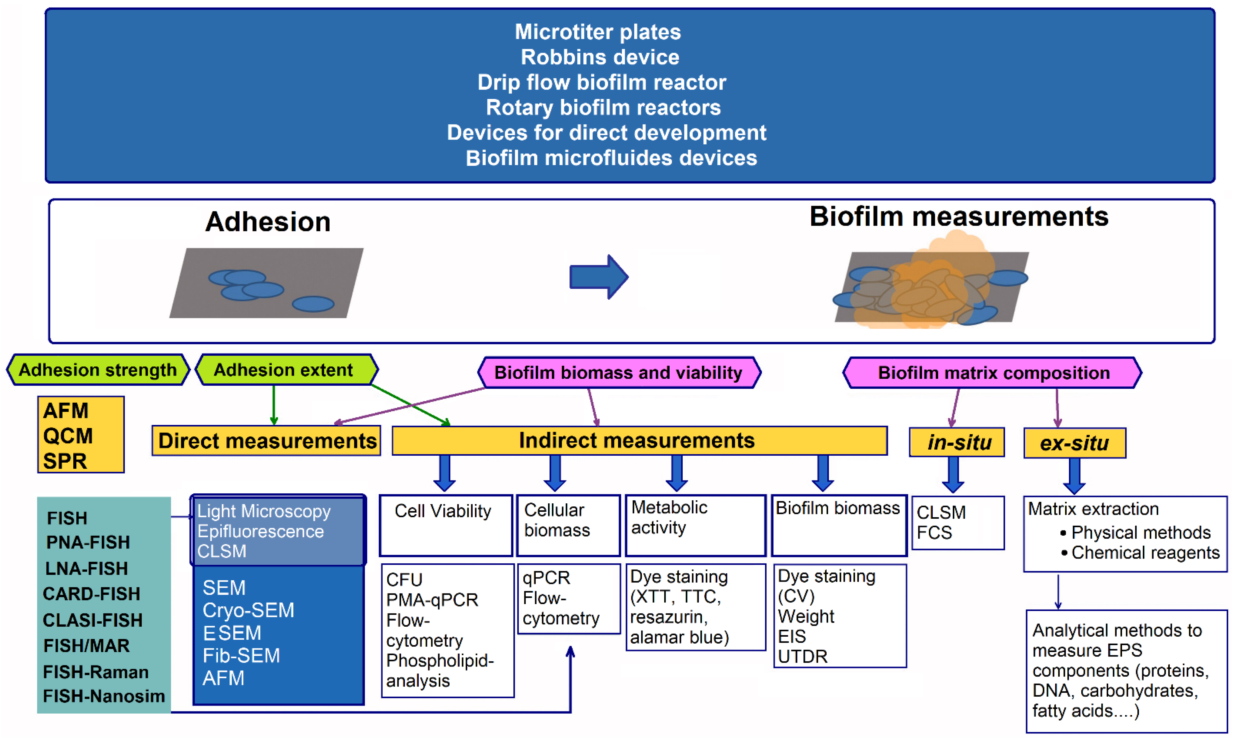
  129. Achinas, S.; Yska, S.K.; Charalampogiannis, N.; Krooneman, J.; Euverink, G.J.W. A technological understanding of biofilm detection techniques: A review. Materials 2020, 13, 3147. [Google Scholar] [CrossRef] [PubMed]
  130. Dygico, L.K.; Gaham, C.G.M.; Grogan, H.; Burgess, C.M. Examining efficacy of mushroom industry biocides on Listeria monocytogenes biofilm. J. Appl. Microbiol. 2020, 130, 1106–1116. [Google Scholar] [CrossRef] [PubMed]
  131. Ríos-Castillo, A.G.; Ripolles-Avila, C.; Rodríguez-Jerez, J.J. Detection of Salmonella Typhimurium and Listeria monocytogenes biofilm cells exposed to different drying and pre-enrichment times using conventional and rapid methods. Int. J. Food Microbiol. 2020, 324, 108611. [Google Scholar] [CrossRef] [PubMed]
  132. European Chemical Agency, Helsinki, Finland. Information on Biocides. Available online: https://echa.europa.eu/cs/information-on-chemicals/biocidal-active-substances (accessed on 16 May 2022).
  133. Castro, M.S.R.; da Silva Fernandes, M.; Kabuki, D.Y.; Kuaye, A.Y. Biofilm formation of Enterococcus faecium on stainless steel surfaces: Modelling and control by disinfection agents. J. Food Process. Eng. 2018, 41, e12663. [Google Scholar] [CrossRef]
  134. Speranza, B.; Monacis, N.; Sinigaglia, M.; Corbo, M.R. Approaches to removal and killing of Salmonella spp. biofilms. J. Food Process. Preserv. 2017, 41, e12758. [Google Scholar] [CrossRef]
  135. Iñiguez-Moreno, M.; Gutiérrez-Lomelí, M.; Avila-Novoa, M.G. Removal of mixed-species biofilm developed on food contact surfaces with a mixture of enzymes and chemical agents. Antibiotics 2021, 10, 931. [Google Scholar] [CrossRef]
  136. Vázquez-Sánchez, D.; Galvão, J.A.; Ambrosio, C.M.S.; Gloria, E.M.; Oetterer, M. Sinlge and binary applications of essential oils effectively control Listeria monocytogenes biofilms. Ind. Crops Prod. 2018, 121, 452–460. [Google Scholar] [CrossRef]
  137. Yu, H.; Liu, Y.; Yang, F.; Xie, Y.; Guo, Y.; Cheng, Y.; Yao, W. Synergistic efficacy of high-intensity ultrasound and chlorine dioxide combination for Staphylococcus aureus biofilm control. Food Control 2021, 122, 107822. [Google Scholar] [CrossRef]
  138. Hua, Z.; Younce, F.; Tang, J.; Ryu, D.; Rasco, B.; Hanrahan, I.; Zhu, M.-J. Efficacy of saturated steam against Listeria innocua biofilm on common food-contact surfaces. Food Control 2021, 125, 107988. [Google Scholar] [CrossRef]
  139. Tan, J.; Karwe, M.V. Inactivation and removal of Enterobacter aerogenes biofilm in a model piping system using plasma-activated water (PAW). Innov. Food Sci. Emerg. Technol. 2021, 69, 102664. [Google Scholar] [CrossRef]
  140. Marino, M.; Maifreni, M.; Baggio, A.; Innocente, N. Inactivation of foodborne bacteria biofilms by aqueous and gaseous ozone. Front. Microbiol. 2018, 9, 2024. [Google Scholar] [CrossRef] [PubMed] [Green Version]
  141. Yang, Y.; Ma, S.; Xie, Y.; Wang, M.; Cai, T.; Li, J.; Guo, D.; Zhao, L.; Xu, Y.; Liang, S.; et al. Inactivation of Pseudomonas aeruginosa biofilms by 405-nanometer-light-emitting diode illumination. Appl. Environ. Microbiol. 2020, 86, e00092-20. [Google Scholar] [CrossRef]
  142. Bellin, D.L.; Sakhtah, H.; Zhang, Y.; Price-Whelan, A.; Dietrich, L.E.P.; Shepard, K.L. Electrochemical camera chip for simultaneous imaging of multiple metabolites in biofilms. Nat. Commun. 2016, 7, 10535. [Google Scholar] [CrossRef] [Green Version]
  143. Morisaki, H.; Sugimoto, M.; Shiraishi, H. Attachment of bacterial cells to carbon electrodes. Bioelectrochemistry 2000, 51, 21–25. [Google Scholar] [CrossRef]
  144. Vu, D.L.; Sýs, M.; Červenka, L. The effect of various potentials on the attachment of Saccharomyces cerevisiae and Staphylococcus epidermidis to carbon paste electrodes. Int. J. Electrochem. Sci. 2011, 6, 5265–5274. [Google Scholar]
  145. Nostro, A.; Cellini, L.; Di Giulio, M.; D’Arrigo, M.; Marino, A.; Blanco, A.R.; Favaloro, A.; Cutroneo, G.; Bisignano, G. Effect of alkaline pH on staphylococcal biofilm formation. APMIS 2012, 120, 733–742. [Google Scholar] [CrossRef]
  146. Vu, D.L.; Červenka, L.; Vavřičková, J. The attachment of Staphylococcus epidermidis on the surface of a carbon paste electrode at various positive potentials: The effect of pH, incubation time, and solid-medium type. J. Biomed. Sci. Eng. 2012, 5, 699–704. [Google Scholar] [CrossRef] [Green Version]
  147. Tresse, O.; Lebret, V.; Benezech, T.; and Faille, C. Comparative evaluation of adhesion, surface properties, and surface protein composition of Listeria monocytogenes strains after cultivation at constant pH of 5 and 7. J. Appl. Microbiol. 2006, 101, 53–62. [Google Scholar] [CrossRef]
  148. Steiger, E.L.; Muelli, J.R.; Braissant, O.; Waltimo, T.; Astasov-Frauenhoffer, M. Effect of divalent ions on cariogenic biofilm formation. BMC Microbiol. 2000, 20, 287. [Google Scholar] [CrossRef] [PubMed]
  149. Falcón García, C.; Kretschmer, M.; Lozano-Andrade, C.N.; Schönleitner, M.; Dragoŝ, A.; Kovács, Á.T.; Lieleg, O. Metal ions weaken the hydrophobicity and antibiotic resistance of Bacillus subtilis NCIB 3610 biofilms. NPJ Biofilms Microbiomes 2020, 6, 1. [Google Scholar] [CrossRef] [Green Version]
  150. Guvensen, N.C.; Demir, S.; Ozdemir, G. Effects of magnesium and calcium cations on biofilm formation by Sphingomonas paucimobilis from an industrial environment. Curr. Opin. Biotechnol. 2013, 24, S68. [Google Scholar] [CrossRef]
  151. Nan, L.; Yang, K.; Ren, G. Anti-biofilm formation of a novel stainless steel against Staphylococcus aureus. Mater. Sci. Eng. C Mater. Biol. Appl. 2015, 51, 356–361. [Google Scholar] [CrossRef] [PubMed] [Green Version]
  152. Hutchings, C.; Rajasekharan, S.K.; Reifen, R.; Shemesh, M. Mitigating milk-associated bacteria through inducing zinc ions antibiofilm activity. Foods 2020, 9, 1094. [Google Scholar] [CrossRef]
  153. Harrison, J.J.; Ceri, H.; Yerly, J.; Rabiei, M.; Hu, Y.; Martinuzzi, R.; Turner, R.J. Metal ions may suppress or enhance cellular differentiation in Candida albicans and Candida tropicalis biofilms. Appl. Environ. Microbiol. 2007, 73, 4940–4949. [Google Scholar] [CrossRef] [Green Version]
  154. Perrin, C.; Briandet, R.; Jubelin, G.; Lejeune, P.; Mandrand-Berthelot, M.A.; Rodrigue, A.; Dorel, C. Nickel promotes biofilm formation by Escherichia coli K-12 strains that produce curli. Appl. Environ. Microbiol. 2009, 75, 1723–1733. [Google Scholar] [CrossRef] [Green Version]
  155. Ferrari, A.G.M.; Carrington, P.; Rowley-Neale, S.J.; Banks, C.E. Recent advances in portable heavy metal electrochemical sensing platforms. Environ. Sci. Water Res. Technol. 2020, 6, 2676–2690. [Google Scholar] [CrossRef]
  156. Milkman, R. An Escherichia coli homologue of eukaryotic potassium channel proteins. Proc. Natl. Acad. Sci. USA 1994, 91, 3510–3514. [Google Scholar] [CrossRef] [Green Version]
  157. Humphries, J.; Xiong, L.; Liu, J.; Prindle, A.; Yuan, F.; Arjes, H.A.; Tsimring, L.; Süel, G.M. Species-independent attraction to biofilms through electrical signaling. Cell 2017, 168, 200–209. [Google Scholar] [CrossRef] [Green Version]
  158. Majumdar, S.; Pal, S. Cross-species communication in bacterial world. J. Cell Commun. Signal 2017, 11, 187–190. [Google Scholar] [CrossRef] [PubMed] [Green Version]
  159. Binepal, G.; Gill, K.; Crowley, P.; Cordova, M.; Brady, L.J.; Senadheera, D.B.; Cvitkovitch, D.G. Trk2 potassium transport system in Streptococcus mutans and its role in potassium homeostasis, biofilm formation, and stress tolerance. J. Bacteriol. 2016, 198, 1087–1100. [Google Scholar] [CrossRef] [PubMed] [Green Version]
  160. Remis, J.P.; Wei, D.; Gorur, A.; Zemla, M.; Haraga, J.; Allen, S.; Witkowska, H.E.; Costerton, J.W.; Berleman, J.E.; Auer, M. Bacterial social networks: Structure and composition of Myxococcus xanthus outer membrane vesicle chains. Environ. Microbiol. 2014, 16, 598–610. [Google Scholar] [CrossRef] [PubMed] [Green Version]
  161. Stekolshchikova, A.A.; Radaev, A.V.; Orlova, O.Y.; Nikolaev, K.G.; Skorb, E.V. Thin and flexible ion sensors based on polyelectrolyte multilayers assembled onto the carbon adhesive tape. ACS Omega 2019, 4, 15421–15427. [Google Scholar] [CrossRef] [PubMed] [Green Version]
  162. Steidl, R.; Lampa-Pastirk, S.; Reguera, G. Mechanistic stratification in electroactive biofilms of Geobacter sulfurreducens mediated by pilus nanowires. Nat. Commun. 2016, 7, 12217. [Google Scholar] [CrossRef] [Green Version]
  163. Kashima, H.; Regan, J.M. Facultative nitrate reduction by electrode-respiring Geobacter metallireducens biofilms as a competitive reaction to electrode reduction in a bioelectrochemical system. Environ. Sci. Technol. 2015, 49, 3195–3202. [Google Scholar] [CrossRef]
  164. Kitayama, M.; Koga, R.; Kasai, T.; Kouzuma, A.; Watanabe, K. Structures, compositions, and activities of live Shewanella biofilms formed on graphite electrodes in electrochemical flow cells. Appl. Environ. Microbiol. 2017, 83, e00903-17. [Google Scholar] [CrossRef] [Green Version]
  165. Guo, Y.; Wang, G.; Zhang, H.; Wen, H.; Li, W. Effects of biofilm transfer and electron mediators transfer on Klebsiella quasipneumoniae sp. 203 electricity generation performance in MFCs. Biotechnol. Biofuels 2020, 13, 162. [Google Scholar] [CrossRef]
  166. Kanga, C.S.; Eaktasang, N.; Kwon, D.Y.; Kim, H.S. Enhanced current production by Desulfovibrio desulfuricans biofilm in a mediator-less microbial fuel cell. Bioresour. Technol. 2014, 165, 27–30. [Google Scholar] [CrossRef]
  167. Saratale, G.D.; Saratale, R.G.; Shahid, M.K.; Zhen, G.; Kumar, G.; Shin, H.S.; Choi, Y.G.; Kim, S.H. A comprehensive overview on electro-active biofilms, role of exo-electrogens and their microbial niches in microbial fuel cells (MFCs). Chemosphere 2017, 178, 534–547. [Google Scholar] [CrossRef]
  168. Choi, O.; Sang, B.I. Extracellular electron transfer from cathode to microbes: Application for biofuel production. Biotechnol. Biofuels 2016, 9, 11. [Google Scholar] [CrossRef] [PubMed] [Green Version]
  169. Pankratova, G.; Gorton, L. Electrochemical communication between living cells and conductive surfaces. Curr. Opin. Electrochem. 2017, 5, 193–202. [Google Scholar] [CrossRef]
  170. Hasan, K.; Patil, S.A.; Leech, D.; Hägerhäll, C.; Gorton, L. Electrochemical communication between microbial cells and electrodes via osmium redox systems. Biochem. Soc. Trans. 2012, 40, 1330–1335. [Google Scholar] [CrossRef] [Green Version]
  171. Angelaalincy, M.J.; Navanietha, K.R.; Shakambari, G.; Ashokkumar, B.; Kathiresan, S.; Varalakshmi, P. Biofilm engineering approaches for improving the performance of microbial fuel cells and bioelectrochemical systems. Front. Energy Res. 2018, 6, 63. [Google Scholar] [CrossRef]
  172. Rau, J.; Knackmuss, H.J.; Stolz, A. Effects of different quinoid redox mediators on the anaerobic reduction of azo dyes by bacteria. Environ. Sci. Technol. 2002, 36, 1497–1504. [Google Scholar] [CrossRef] [PubMed]
  173. Paquete, C.M.; Rosenbaum, M.A.; Bañeras, L.; Rotaru, E.A.; Puig, S. Let’s chat: Communication between electroactive microorganisms. Bioresour. Technol. 2022, 347, 126705. [Google Scholar] [CrossRef]
  174. Darch, S.E.; Koley, D. Quantifying microbial chatter: Scanning electrochemical microscopy as a tool to study interactions in biofilms. Proc. R. Soc. A 2018, 474, 20180405. [Google Scholar] [CrossRef] [Green Version]
  175. Darvishi, S.; Pick, H.; Oveisi, E.; Girault, H.H.; Lesch, A. Soft-probe-scanning electrochemical microscopy reveals electrochemical surface reactivity of E. coli biofilms. Sens. Actuators B Chem. 2021, 334, 129669. [Google Scholar] [CrossRef]
  176. Turick, C.E.; Colon-Mercado, H.; Bagwell, C.E.; Greenway, S.D.; Amoroso, J.W. Non-contact electrochemical evaluation of biofilms. SN Appl. Sci. 2020, 2, 389. [Google Scholar] [CrossRef] [Green Version]
  177. Pires, L.; Sachsenheimer, K.; Kleintschek, T.; Waldbaur, A.; Schwartz, T.; Rapp, B.E. Online monitoring of biofilm growth and activity using a combined multi-channel impedimetric and amperometric sensor. Biosens. Bioelectron. 2013, 47, 157–163. [Google Scholar] [CrossRef]
  178. Song, J.; Li, Y.; Ke, D.; Wang, D.; Zhang, X.E. In situ graphene-modified carbon microelectrode array biosensor for biofilm impedance analysis. Electrochim. Acta 2022, 403, 139570. [Google Scholar] [CrossRef]
  179. Wang, W.; Zhang, X.; Wang, J. Heterogeneous electrochemical characteristics of biofilm/metal interface and local electrochemical techniques used for this purpose. Mater. Corros. 2009, 60, 957–962. [Google Scholar] [CrossRef]
  180. Lebeaux, D.; Chauhan, A.; Rendueles, O.; Beloin, C. From in vitro to in vivo models of bacterial biofilm-related infections. Pathogens 2013, 2, 288–356. [Google Scholar] [CrossRef] [Green Version]
  181. Masi, E.; Ciszak, M.; Santopolo, L.; Frascella, A.; Giovannetti, L.; Marchi, E.; Viti, C.; Mancuso, S. Electrical spiking in bacterial biofilms. J. R. Soc. Interface 2015, 12, 20141036. [Google Scholar] [CrossRef] [Green Version]
  182. Kang, J.; Kim, T.; Tak, Y.; Lee, J.H.; Yoon, J. Cyclic voltammetry for monitoring bacterial attachment and biofilm formation. J. Ind. Eng. Chem. 2012, 18, 800–807. [Google Scholar] [CrossRef]
  183. Sedki, M.; Hassan, R.Y.A.; Andreescu, S.; El-Sherbiny, I.M. Online-monitoring of biofilm formation using nanostructured electrode surfaces. Mater. Sci. Eng. C 2019, 100, 178–185. [Google Scholar] [CrossRef]
  184. Tursun, H.; Liu, R.; Li, J.; Abro, R.; Wang, X.; Gao, Y.; Li, Y. Carbon material optimized biocathode for improving microbial fuel cell performance. Front. Microbiol. 2016, 7, 6. [Google Scholar] [CrossRef] [Green Version]
  185. Sultana, S.T.; Babauta, J.T.; Beyenal, H. Electrochemical biofilm control: A review. Biofouling 2015, 31, 745–758. [Google Scholar] [CrossRef]
  186. Schneider, S.; Rudolph, M.; Bause, V.; Terfort, A. Electrochemical removal of biofilms from titanium dental implant surfaces. Bioelectrochemistry 2018, 121, 84–94. [Google Scholar] [CrossRef]
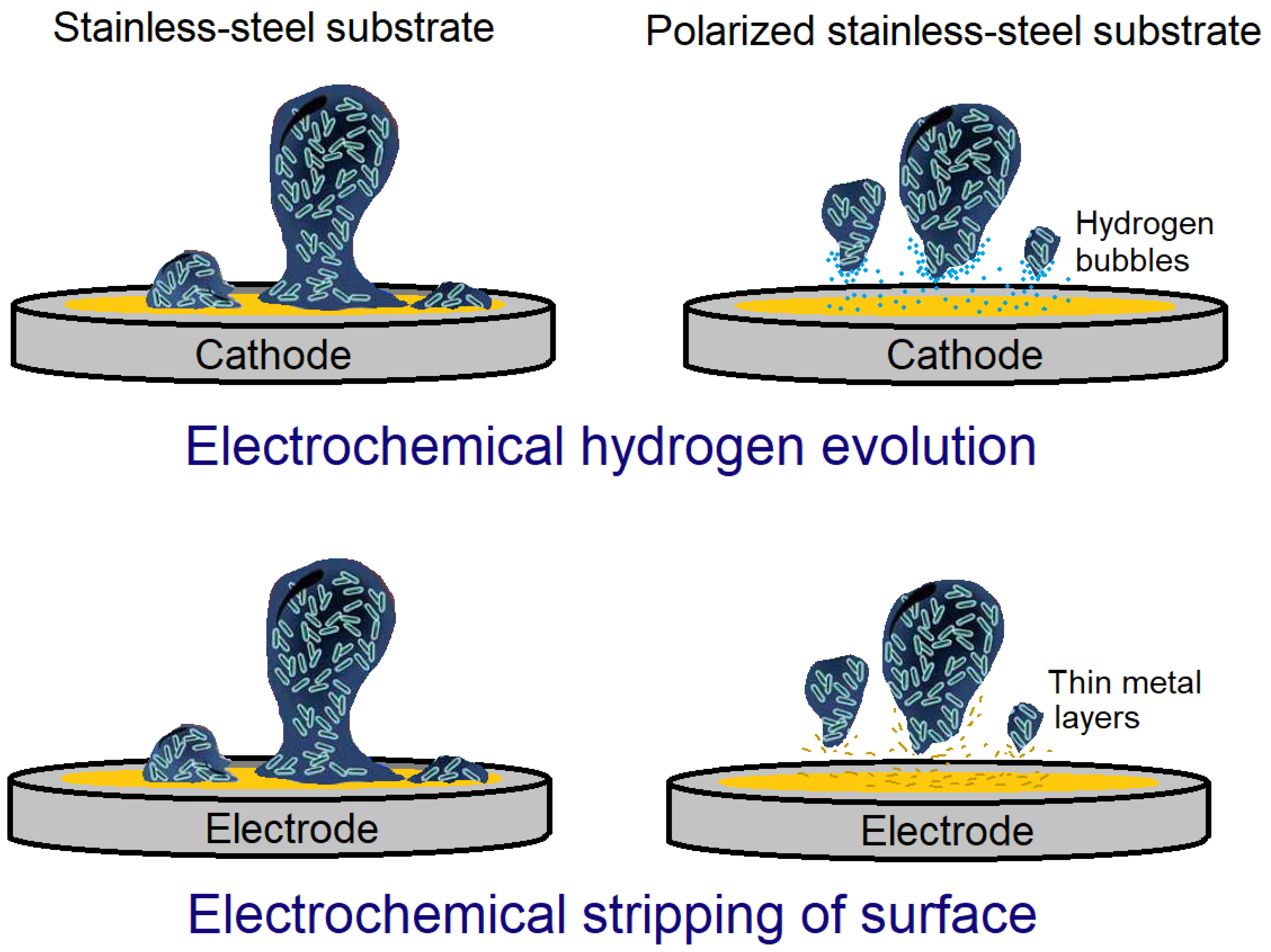
  187. Dargahi, M.; Hosseinidoust, Z.; Tufenkji, N.; Omanovic, S. Investigating electrochemical removal of bacterial biofilms from stainless steel substrates. Colloids Surf. B 2014, 117, 152–157. [Google Scholar] [CrossRef] [Green Version]
  188. Wijesinghe, M.S.; Wen, J.; Oh, J.M.; Chow, K.F.; Sun, Y. Demonstration of biofilm removal from type 304 stainless steel using pulsed-waveform electropolishing. Biofouling 2018, 34, 731–739. [Google Scholar] [CrossRef] [PubMed]
Figure 1. Overview of methods enabling the growth and characterization of biofilms [81], which includes different biofilm devices, methods to assess adhesion extent and strength, and techniques to measure biofilm biomass, viability, and matrix composition.
Applsci 12 06320 g001
Figure 2. The effects of material properties in complex environments on reversible bacterial adhesion.
Applsci 12 06320 g002
Figure 3. Principle of indirect voltammetric control of bacterial adhesion.
Applsci 12 06320 g003
Figure 4. Direct electron transfer mechanisms via membrane-attached cytochrome (short distance electron transfer) (A), soluble redox mediator (B), and conductive pilli (long distance electron transfer) (C).
Applsci 12 06320 g004
Figure 5. Schematic representation of biofilm impedance chamber with incorporated MEA chip.
Applsci 12 06320 g005
Figure 6. Principles of electrochemical methods developed for biofilm removal from stainless-steel substrates.
Applsci 12 06320 g006
Table 1. Representatives of foodborne bacteria that form biofilms.
Foodborne Bacteria Growing SubstrateSpoiled FoodGenes Related to Biofilm FormationReferences
Bacillus
(Bacillus cereus)
Stainless steel, plastic, soil, and glass woolSprouted seeds, fruit juices, fried rice, pasta dishes, meat products, vegetables, and milk productstasA, galE, eps2, mogR, comER, plcR, rpoN, codY, spo0A, abrB, sinI, sinR and others[23,24,25,26,27]
ClostridiumMulti-species biofilmDairy products, fish, cattle meat, poultry, vegetables, honey, and canned foodluxS, spo0A, pilC, pilT, and others[27,28,29,30]
Cronobacter spp.Powder service and powder
packaging rooms, spray-drying areas, and evaporator rooms
Dairy products, vegetables, grains, bread, herbs,
sausages, spices, and meat
bcsR, csgA, csgB and others [27,31,32]
Escherichia coliStainless steel surfaces, food contact surfacesDairy products, fermented meat sausage, meat, poultry, fish products, drinks,
and vegetables
fim, pap, bfp, scg, sfa, foc, afa, flu, pgaABCD, bcsABZC, uvrY, csrA and others[27,33,34,35,36,37]
Listeria
monocytogenes
Wastewater pipes, floors, conveyor belts, rubber seals, elastomers,
and stainless steel
Dairy products, melons, coleslaw, ready-to-eat meat
products, and ready-to-eat
fish products
luxS, agr (agrABCD), inlA, actA, prfA
and others
[27,38,39,40,41]
Pseudomonas spp.
(Pseudomonas
aeruginosa)
Conveyor belts, floors, drains, slicing and milking machinesDairy products, red meat, and poultrypsl (pslA–pslO), pel (pelA–pelG), algD, algU, algL, ppyR, lasR, lecA, rhlI, pilA, pilT and others[4,24,42,43,44,45,46,47,48]
SalmonellaStainless steel, elastomers,
concrete, glass, and food surfaces (such as
lettuce and tomato)
Poultry, pig, cow meats, and dairy productsbapA, csgB, csgD, csgBA, adrA, bcs, fimA, fimH, luxS, flgE and others[27,49,50,51]
Staphylococcus
(Staphylococcus
aureus)
Stainless steel, plastics
(such as polystyrene and polypropylene),
and glass
Dairy products, ready-to-eat meat products, ready-to-eat fish and seafood products, and ready-to-eat dairy
products
icaA, icaD, icaB, ica, icaR, fib, cna, fnbAB, clfA, clfB, agr (agrA-agrD) and others[27,52,53,54,55,56]
Publisher’s Note: MDPI stays neutral with regard to jurisdictional claims in published maps and institutional affiliations.

