Article

Body’s Center of Mass Motion Relative to the Center of Pressure during Gait, and Its Correlation with Standing Balance in Patients with Lumbar Spondylosis

1
Department of Biomedical Engineering, National Taiwan University, Taipei 106038, Taiwan
2
Department of Orthopaedic Surgery, National Taiwan University Hospital Hsin-Chu Branch, Hsinchu County 302058, Taiwan
3
Department of Orthopaedic Surgery, School of Medicine, National Taiwan University, Taipei 100229, Taiwan
4
Department of Orthopaedic Surgery, National Taiwan University Hospital, Taipei 100229, Taiwan
5
Department of Orthopedic Surgery, Far-Eastern Memorial Hospital, New Taipei City 220216, Taiwan
6
Department of Orthopedic, Shin Kong Wu Huo-Shih Memorial Hospital, Taipei 111045, Taiwan
*
Author to whom correspondence should be addressed.
Appl. Sci. 2022, 12(24), 12915; https://doi.org/10.3390/app122412915
Submission received: 23 October 2022 / Revised: 29 November 2022 / Accepted: 9 December 2022 / Published: 15 December 2022
(This article belongs to the Special Issue Biomechanics and Human Motion Analysis)

Abstract

:
Lumbar spondylosis (LS) is a common degenerative spine disease that often leads to impaired motor control, sensory changes, and imbalance. The current study aimed to compare the dynamic balance control between patients with LS and healthy controls in terms of inclination angles (IA) and the rate of change of IA (RCIA) of the center of mass relative to the center of pressure (COM–COP motion) during walking and to identify the correlation between dynamic balance and standing balance in patients with LS. Eleven patients with LS and eleven healthy controls performed level walking and static standing in a gait laboratory while their whole-body motion and ground reaction forces were measured to calculate the IA and RCIA. Gait temporal–spatial parameters were also recorded. Correlations between the COP motions during standing balance and COM–COP motions during gait were quantified using Pearson’s correlation coefficients (r). In the sagittal plane, the patients increased posterior IA with decreased posterior RCIA during the double-limb support phase of gait and showed decreased anterior RCIA, with small ranges of IA and RCIA during the single-limb support phase (p < 0.05). In the frontal plane, the patients increased medial–lateral ranges of RCIA and medial IA during the double-limb support phase of gait and increased medial RCIA and ranges of IA during the single-limb support phase of gait (p < 0.05). A moderate to strong correlation was found between dynamic balance and standing balance in the patients (p < 0.05). The patients presented a conservative anterior–posterior dynamic balance control but an unstable medial–lateral dynamic balance control during walking, which may be related to the decreased gait speed. The results showed that the greater the postural sway in the patients’ standing balance, the more conservative the dynamic balance control in the sagittal plane, and the greater the risk of imbalance in the frontal plane. It is thus suggested that dynamic balance control deviations during gait in patients with LS cannot be deduced solely from standing balance test data, and should thus be monitored via dynamic approaches in clinical applications.

1. Introduction

Spondylosis refers to the chronic degenerative changes, such as bony osteophytes and excrescences [1] within the region of the vertebral bodies, and the associated abnormalities, including segmental motion deviations, loss of vertebral disc height, and arthrosis of the facet joints [2]. Spondylosis is frequently found in middle-aged and older adults, with approximately 50% of individuals over forty years old and 85% of individuals over sixty years old being diagnosed with some degree of spondylosis [3,4]. Depending on the level of the lesion, spondylosis may result in different alterations in the gait patterns and posture control [5,6]. Compared to the other parts of the spine, the lumbar spine transmits the largest forces which are several times body weight during gait; thus, there is a higher risk of developing spondylosis [7,8] in this region of the spine. Lumbar spondylosis (LS), mostly at L4–L5, may change not only the mechanics of the motion segment, but may also compress the cauda equina and lumbosacral nerve roots [9]. Both would lead to severe radicular pain of the lower back and/or down to one or both lower extremities [10] with restricted mobility, absent reflex, sensory changes, and muscle weakness [9,11]. As the disease progresses, patients with LS may develop motor disturbances [12,13], altered gait patterns, loss of balance [14], and increased risks of falling [15,16]. Therefore, quantitative measurement of the static postural balance and dynamic gait balance in patients with LS will be useful for a more complete assessment of the progression of the disease and the risk of falls in this population.
Balance control has been defined as the control of the body’s position in space for the purpose of balance and orientation [17,18]. The effectiveness of the ability of an individual in maintaining standing balance is usually assessed using variables derived from trajectories of the center of pressure (COP) of the ground reaction forces (GRF) [19,20]. Those COP variables provide critical information which is helpful for understanding how the human body controls and maintains standing balance [21,22,23,24]. Typically, the COP sway during quiet standing has been well accepted as an index to evaluate balance: the greater the COP sway, the worse the balance ability. The COP total distance is also frequently used to quantify the standing balance control [25,26]. However, these variables are not directly applicable to the dynamic balance during walking as the COP moves along in the direction of the progression of gait.
Dynamic balance control during motion is best evaluated through the description of the motion of the body’s center of mass (COM) relative to the COP. During dynamic conditions, the COM can be much further away from the COP without loss of balance, as long as the COM is controlled at an appropriate velocity relative to the COP [27]. The velocity of the COM relative to the COP, as well as the influence of the body height or leg length, should be considered [28,29]. This can be evaluated by the inclination angles (IA) of the line connecting the COM and the COP in both sagittal and frontal planes [30], and the rate of change of IA (RCIA), enabling a more complete description of the control of the body’s posture [31]. Generally, the further the COM diverges from the COP (i.e., greater IA) the more difficult it becomes, and more effort (e.g., joint moments) is needed to achieve an RCIA appropriate for dynamic balance [32]. A previous study has shown that the IA and RCIA of patients with scoliosis were associated with the severity of the spinal deformity [33]. Therefore, COM–COP IA and RCIA are now becoming the typical parameters for evaluating balance control in patients with impaired motor or balance control diseases during locomotion [34]. Identifying the correlations between standing balance performance and dynamic balance control during gait would be helpful for evaluating the risk of imbalance during locomotion from standing balance evaluations alone, which are easier to obtain in a clinical setting.
Patients with degenerative lumbar disease present poor postural control ability and tend to rely on visual feedback and a wide-base standing posture [12]. Previous studies also showed that postural sway was increased in patients with spondylosis and other degenerative lumbar diseases [12,35,36]. Wong et al. [13] found that the root-mean-squared distance of the COP in anteroposterior and mediolateral directions was decreased in patients with LS after lumbar surgery. These findings may be indicative of impairment of neuromuscular feedback loops at a different level of motor activation within the central nervous system [37], which could be evaluated through static balance using force plates. However, it remains unclear how LS may affect the dynamic balance control in terms of the body’s motion relative to the center of pressure during level walking, and what the relationship is between dynamic balance control during gait and static standing balance in such patients.
The purpose of this study was to quantify the dynamic balance control in patients with LS in terms of COM–COP IA and RCIA in comparison with healthy controls and to identify the correlations of IA and RCIA between dynamic gait balance and static standing balance in LS. It was hypothesized that, compared to the controls, patients with LS would show compromised balance control with increased IA and RCIA during gait and increased COP total distance and sway area during quiet standing, and that the IA and RCIA during walking would have a significant correlation with COP total distance and COP sway area during quiet standing in patients with LS.

2. Materials and Methods

2.1. Subjects

Eleven patients with LS (LS group; age: 59.7 ± 12.9 years; height: 162.9 ± 6.7 cm; mass: 64.7 ± 12.1 kg), and eleven healthy adults (control group; age: 53.5 ± 6.1 years; height: 158.1 ± 6.0 cm; mass: 59.5 ± 13.3 kg) participated in the current study with informed written consent, as approved by the Institutional Review Board. All the patients in the LS group had a clinical diagnosis of LS and stenosis supported by radiological findings. The inclusion criteria for the patients were (1) age above 40 years, (2) ability to walk without assistive devices, (3) mild myelopathy, and (4) motor assessment scale (MAS) for walking >grade 5 [38]. The exclusion criteria were (1) previous back surgery, (2) age < 40 years or >80 years, or (3) moderate or severe disability and/or difficulty in ambulation. The healthy controls were all free from any neuromusculoskeletal dysfunction, with normal or corrected vision, and had the ability to walk without an assistive device.

2.2. Experimental Protocol
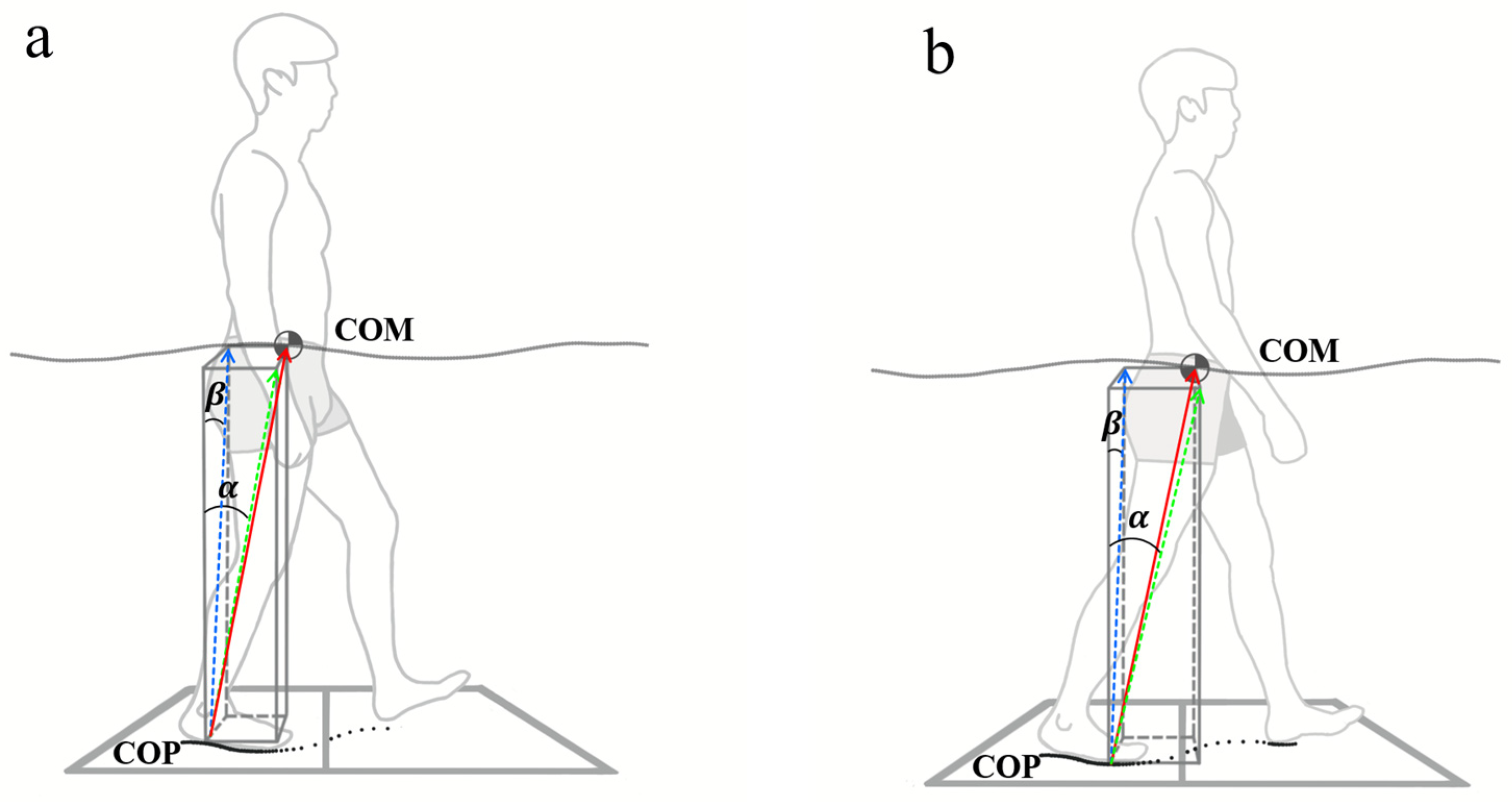
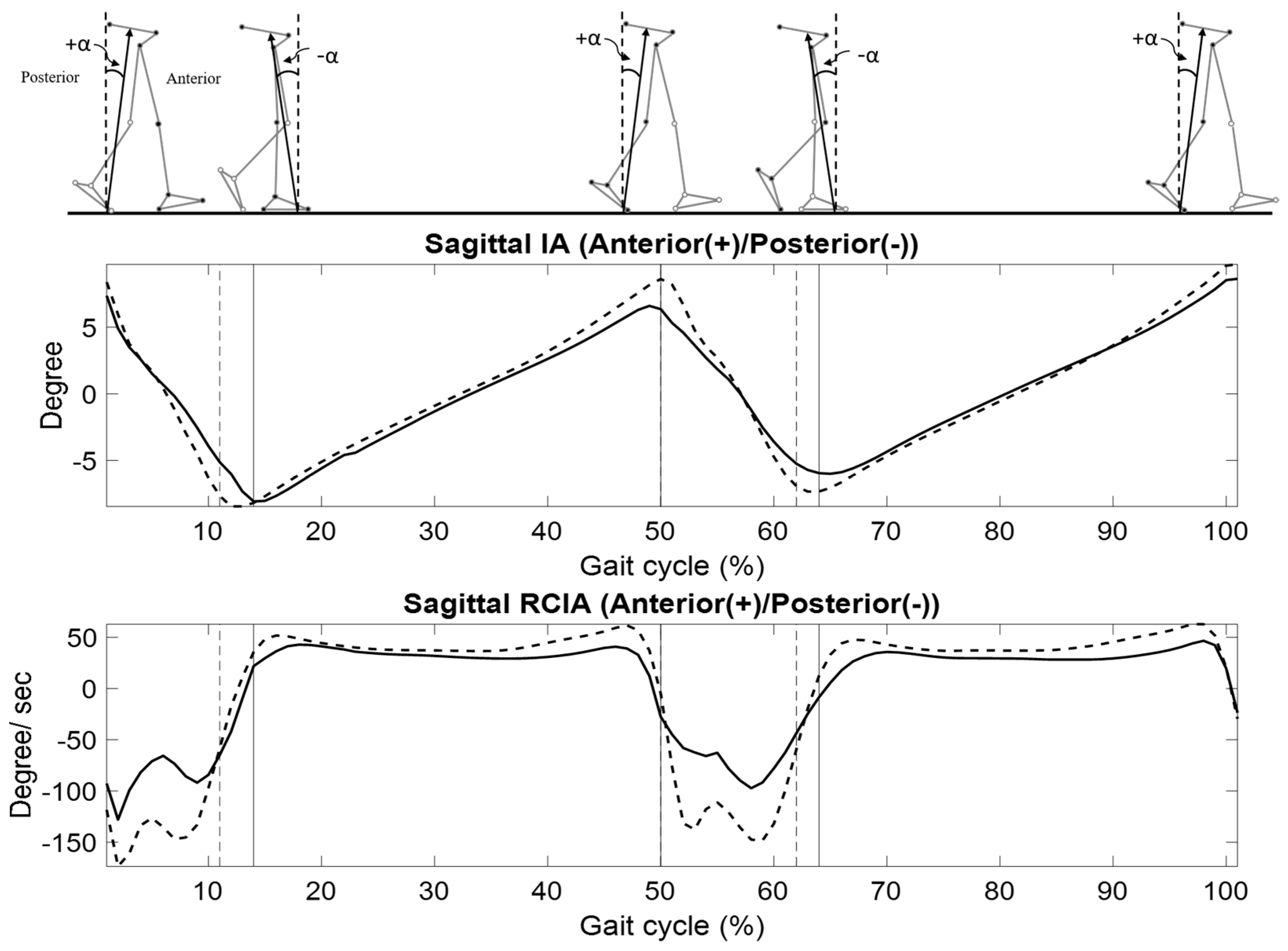
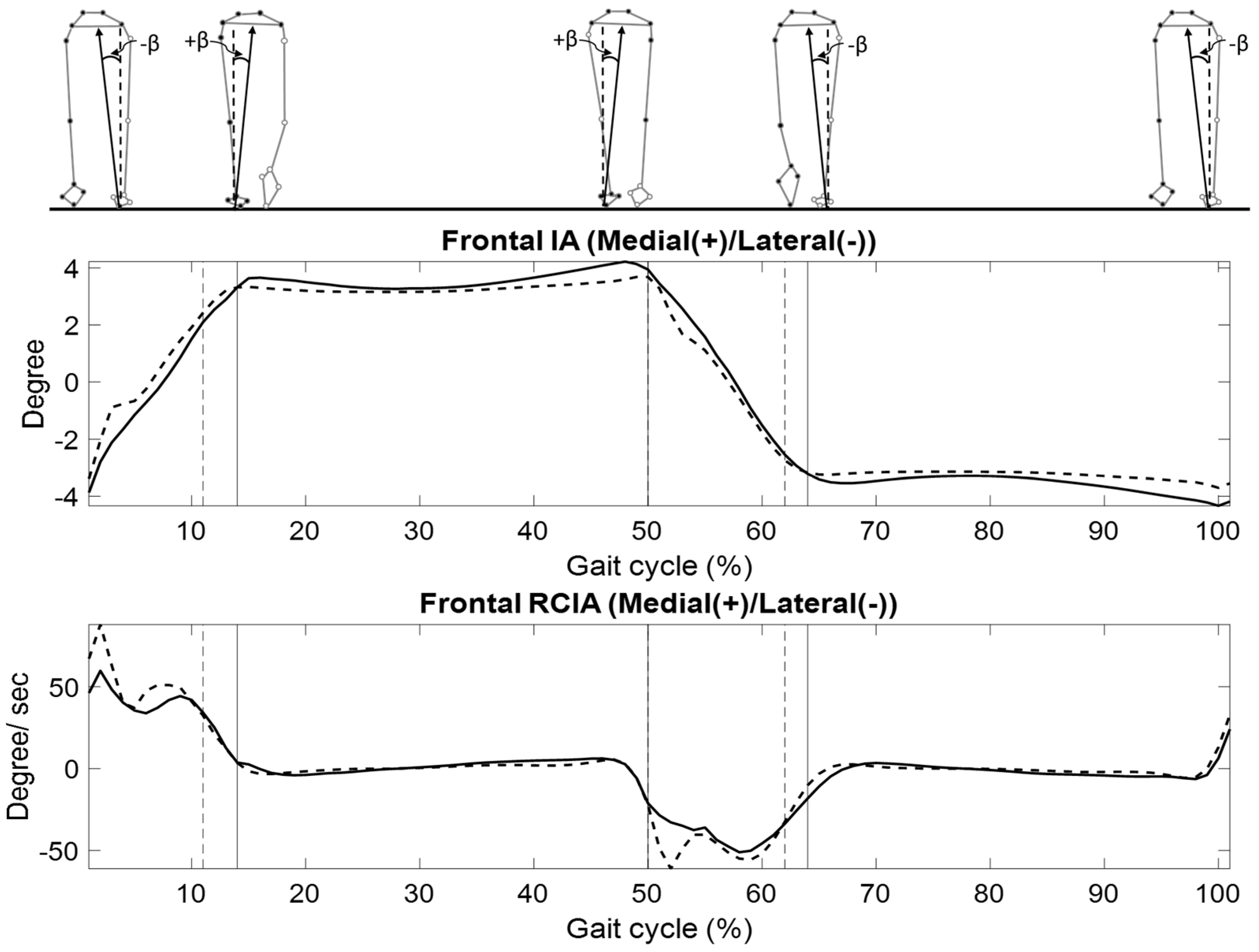
In a university hospital gait laboratory, each subject walked at his/her preferred speed on a 10 m walkway while the motions of all the body segments were measured via 41 infrared retroreflective markers placed on specific anatomical landmarks [29] using a 7-camera motion capture system at a sampling rate of 120 Hz (Vicon 512, OMG, Oxford UK). The ground reaction forces (GRF) were also measured using three force plates at 1080 Hz (AMTI, Watertown, MA, USA). Before the gait trials, the subjects were allowed to walk on the walkway several times to familiarize themselves with the experimental environment. Data from six complete gait cycles for each subject were collected for subsequent analysis. The subjects also performed quiet standing balance tests on a force plate with their feet close together and eyes open for 30 s while the GRF data were measured [12,39].

2.3. Data Analysis

The positions of the center of pressure (COP) of the GRF were calculated using forces and moments measured by the force plates [40]. For standing balance tests, the total distance traveled by the COP over the course of the trial duration (DCOP) was calculated [25]. The COP sway area (ACOP) was calculated as that of an equivalent ellipse covering 95% of the samples, and the major and minor axes of the ellipse were determined using principal component analysis [41].
For gait balance analysis, all body segments, namely the head/neck, trunk, pelvis, arms, forearms, thighs, shanks, and feet, were modeled as rigid bodies. Using the measured marker data, each body segment was embedded with an orthogonal coordinate system with the origin at the segment’s COM and the positive x-axis directed anteriorly, the positive y-axis superiorly, and the positive z-axis to the right. The 13-body segment model of the body was then used to calculate the position of the body’s COM ( C ) as the weighted sum of the segmental COMs of all body segments as follows [29]:
C = i = 1 13 m i   c i B M
where m i and c i were the mass and position of the COM of the i th body segment calculated using the marker data and an optimization-based method [17,42]. B M was the total body mass of the subject. During the double stance phase, a resultant COP was calculated from the COP and GRF of each foot [40]. A global optimization method was used to reduce the effects of soft tissue artifacts of the skin markers on the pelvis-leg apparatus by minimizing the weighted sum of squared distances between measured and calculated marker positions [43].
For the description of the motions of the COM relative to the COP, the line of progression was defined as that which bisected the mediolateral range of motion of the COM during bilateral single-limb support (SLS) and double-limb support (DLS), a zero value being on the bisection line and a positive value being on the right side of the bisection line. The IAs in the sagittal plane and frontal plane were calculated as follows [44]:
t = ( Z × P C O M C O P | P C O M C O P | )
Sagittal   IA = sin 1 ( t Y )
Frontal   IA = { sin 1 ( t X ) , f o r   t h e   r i g h t   l i m b sin 1 ( t X ) , f o r   t h e   l e f t   l i m b
where P COM COP was the vector pointing from the COP to the COM, Z was the unit vector of the vertical axis of the global coordinate system, and X was the unit vector pointing in the direction of progression. The RCIA was also calculated by smoothing and differentiating the trajectories of sagittal and frontal IAs using the generalized cross-validatory spline method [45]. Positive values of the sagittal and frontal IAs indicated that the COM was anterior to and away from the COP toward the contralateral limb, respectively (Figure 1). The force plate data were also used to determine the gait events of heel-strike and toe-off [46]. The values of the IA and RCIA at heel-strike and toe-off were obtained from the gait cycles of both sides. The ranges and time-averaged values of IA and RCIA over DLS were also obtained. Spatiotemporal parameters of gait were also obtained, namely stride length, step length, step width, cadence, gait speed, and stride time.

2.4. Statistical Analysis

For all variables, an independent t-test was used to compare the differences between the LS and the control group for the walking and quiet standing tests. Pearson’s correlation coefficient (r) was used to evaluate the association between the IA and RCIA during gait, and DCOP and ACOP during quiet standing for the patient group. Absolute values of r less than 0.2 indicate a very weak correlation, while 0.2–0.39, 0.40–0.59, 0.6–0.79, and 0.8–1 indicate a weak, moderate, strong, and very strong correlation, respectively. A significance level of 0.05 was set for all the tests. All the statistical analyses were performed using SPSS version 20.0 (SPSS Inc., IBM, Armonk, NY, USA).
An a priori power analysis for a two-group independent sample t-test for the comparison of IA and RCIA between LS and healthy controls based on pilot data using GPOWER [47] determined that a projected sample size of seven subjects for each group would be needed with a power of 0.8 and a large effect size (Cohen’s d = 1.67) at a significance level of 0.05. Therefore, eleven subjects per group were considered adequate for the purpose of the current study.

3. Results

The patients with LS showed significantly decreased gait speed, cadence, and SLS duration, but increased stride time and DLS duration during walking when compared to the control (p < 0.05, Table 1). No significant between-group differences were found in stride length, step length, and step width during walking (Table 1). During quiet standing, the patients showed increased DCOP and ACOP when compared to the control (p < 0.05, Table 1).
In the sagittal plane, compared to the control, the patients showed decreased anterior IA at heel-strike and decreased ranges of IA during SLS, but increased time-averaged posterior IA during DLS (p < 0.05, Figure 2 and Table 2). The patients also showed decreased time-averaged posterior RCIA during DLS, decreased time-averaged anterior RCIA during SLS, and reduced ranges of RCIA during SLS (p < 0.05, Figure 2 and Table 3). In the patient group, the IA at toe-off, and the time-averaged IA during SLS and DLS were found to have strong negative correlations with DCOP during quiet standing, while the ranges of IA during DLS showed strong positive correlations with DCOP (p < 0.05; Table 4). The patients also showed a strong negative correlation between the ranges of IA during SLS and ACOP during quiet standing (p < 0.05, Table 4).
In the frontal plane, compared to the control, the patients showed increased ranges of IA (p < 0.05, Figure 3 and Table 2) with increased time-averaged medial RCIA during SLS, and unaltered IA with increased ranges of RCIA during DLS (p < 0.05, Figure 2 and Table 3). The patients showed a strong negative correlation between IA at HS and ACOP during quiet standing (p < 0.05, Table 5). The patients also showed a strong positive correlation between the ranges of RCIA during DLS and ACOP during quiet standing (p < 0.05, Table 5).

4. Discussion

The current study aimed to quantify the effects of LS on the whole-body balance control during level walking by comparing the IA and RCIA with those of healthy controls, and to identify the possible correlations between the IA and RCIA during gait and the DCOP and ACOP during quiet standing in the LS group. The patients with LS were found to walk with a conservative and cautious strategy for dynamic balance control in the sagittal plane, maintaining a more posterior COM position relative to the COP with reduced ranges and speed of movement throughout the gait cycle, as well as a slower body weight transfer during DLS. In the frontal plane, the patients moved the COM more medially with greater velocity changes during weight transfer, indicating less stable transfer of the body weight in the medial–lateral direction. Standing balance parameters were found to predict IA and RCIA deviations during DLS in the frontal plane, possibly because the standing balance tests were also performed in the double-limb support posture. However, standing balance parameters failed to predict the IA and RCIA deviations during SLS in the frontal plane and the RCIA deviations throughout the gait cycle in the sagittal plane. These correlational results suggest that dynamic balance control deviations during gait in patients with LS could not be deduced solely from data gathered from standing balance tests, and thus patients should be monitored via dynamic approaches in clinical applications.
In the sagittal plane, during SLS the COM travels from a trailing position towards the stance foot, over the stance foot, and then to a leading position, over a much greater distance than that of the COP underneath the stance foot. Compared to the healthy controls, the patients showed a smaller anterior RCIA and smaller ranges of IA and RCIA, indicating that their COM traveled a shorter distance more slowly when moving over the stance foot [18]. This strategy is helpful for a smooth inverted pendulum motion of the body in the sagittal plane [27,48]. In contrast, during DLS, the COM movement was smaller than during SLS, and the changes of the IA and RCIA were largely accounted for by the COP which traveled from the trailing weight-release limb to the leading weight-acceptance limb. Increasing the DLS time and the stride time would help to smooth the weight transfer, and thus stabilize the whole-body balance control, but this will reduce the gait speed. The patients in the current study decreased the posterior RCIA during DLS to ensure a smoother travel of the COP but this resulted in a more posterior IA. This indicates that the patients tended to keep their body mass on the trailing weight-release foot longer, allowing a more cautious weight acceptance on the contralateral limb.
In the frontal plane, during DLS, it showed increased medial–lateral RCIA and ranges of RCIA during the DLS phase, which indicated a great deceleration of IA in the frontal plane during weight transfer, resulting in a contralateral lean of the body. During SLS, however, a greater medial RCIA and greater ranges of IA in the patients during SLS still reflect a greater and faster sway of the body in the frontal plane, which could also be seen in the trend of increased step width, yet it was without statistical significance (p = 0.052). These results showed that the patients presented a conservative anterior–posterior balance control strategy but an unstable medial–lateral balance control. These findings seemed to match a previous finding [13], which showed that patients with lumbar degenerative spine disease may present a weaker medial–lateral balance control due to weak hip abductors during quiet standing [49], while the back muscles compensated for the imbalance in the anterior–posterior direction [50]. Hence, it might be important to establish the relationship between dynamic balance and static balance of quiet standing.
Previous studies reported that patients with degenerative spine disease would present greater postural sway during static standing [12,13]. The current study also found that while standing with their feet close together and their eyes open for 30 s, the patients showed greater COP total distance and sway area when compared to the healthy controls. The greater postural sway could result from nerve compression, as well as impaired sensory and motor function in patients with degenerative spine disease [51]. The greater postural sway is often interpreted as poor postural control or imbalance during static standing [52,53,54], which theoretically would also occur during locomotion [55].
Standing balance parameters failed to predict RCIA deviations in the sagittal plane, as well as the IA and RCIA deviations during SLS in the frontal plane. In the frontal plane, standing balance parameters were able to predict deviations during DLS but not during SLS. The patients showed a strong positive correlation between medial–lateral ranges of RCIA and ACOP during DLS, indicating that poor postural control would lead to a greater velocity of body sway in the medial–lateral direction during walking, and might result in an imbalance. However, standing balance parameters were unable to predict deviations in the IA and RCIA during SLS, possibly because the standing balance tests were performed only in the double-limb support posture. In the sagittal plane, moderate to strong negative correlations between IA during the entire stance phase and DCOP and ACOP indicate that the patients adopted a conservative strategy control of the COM relative to the COP in the anterior–posterior direction, which relied on walking more slowly while having a poorer postural control. The patients also showed decreased time-averaged posterior RCIA during DLS, decreased time-averaged anterior RCIA during SLS, and decreased ranges of RCIA during SLS, which had no significant correlation with standing balance parameters. Since the dynamic balance control should consider both IA and RCIA, the failure to predict RCIA deviations in the sagittal plane indicates that standing balance parameters are not able to predict dynamic balance in the sagittal plane. In the frontal plane, a strong positive correlation between the ranges of RCIA during DLS and ACOP indicates that the patients would have risks of imbalance in the medial/lateral direction during walking if they showed greater posture sway during quiet standing. The patients also showed increased ranges of IA with increased time-averaged medial RCIA during SLS, which had no significant correlation with standing balance parameters. Since the balance control during SLS relies on body sway based on a stationary foot, failure to predict IA and RCIA deviations during SLS indicates that standing balance parameters could better predict the performance of weight-shifting between limbs. These dynamic balance control deviations without significant correlations with standing balance parameters in the patient group suggest that deviations in these variables during walking could not be deduced from data collected from standing balance tests, and thus patients should be monitored in clinical applications.
The current study represented the first attempt to quantify the dynamic balance control during level walking in patients with LS in terms of IA and RCIA of the body’s COM motion relative to the COP. The correlations of balance control variables between walking and standing in patients with LS were also quantified for the first time in the literature. For the current patient group, the COM–COP control was shown to have a significant difference compared to the healthy controls, and the associated risks have been identified. Nonetheless, the current study was limited to patients with lumbar spondylosis. Thus, the generalization of the current findings to other patient groups should be made with caution. Further studies are needed to identify the dynamic balance control strategies and risk factors of imbalance in patients with other degenerative spine diseases. Balance control in other more challenging daily activities such as obstacle-crossing, slope-walking, or stair-locomotion may also be included in future studies.

5. Conclusions

The dynamic balance control during gait in patients with LS was evaluated through the analysis of COM–COP IA and RCIA, and through a correlational analysis between walking balance and standing balance in the patient group. The patients with LS were found to walk with a conservative and cautious strategy for dynamic balance control in the sagittal plane, maintaining a more posterior COM position relative to the COP with reduced ranges and speed of movement throughout the gait cycle, as well as a slower body weight transfer during DLS. In the frontal plane, the patients moved the COM more medially with greater velocity changes during weight transfer, indicating a less stable transfer of the body weight in the medial–lateral direction. In the sagittal plane, the patients decreased the posterior RCIA with increased posterior IA during DLS, and decreased anterior RCIA and ranges of IA and RCIA during SLS. In the frontal plane, the patients increased medial–lateral RCIA and ranges of RCIA, as well as medial IA during DLS, and increased medial RCIA and ranges of IA during SLS. Standing balance parameters were able to predict IA and RCIA deviations during DLS in the frontal plane, possibly because the standing balance tests were also performed in the double-limb support posture. However, standing balance parameters failed to predict the IA and RCIA deviations during SLS in the frontal plane and the RCIA deviations throughout the gait cycle in the sagittal plane. These correlational results suggest that dynamic balance control deviations during gait in patients with LS could not be deduced solely from data from standing balance tests and, thus, patients should be monitored via dynamic approaches in clinical applications.

Author Contributions

Conceptualization, T.-C.H. and T.-W.L.; Methodology, T.-C.H., H.-P.H., and T.-W.L.; Software, H.-P.H. and T.-W.L.; Validation, H.-P.H., T.-M.W., and T.-W.L.; Formal analysis, T.-C.H. and H.-P.H.; Investigation, T.-C.H., H.-P.H., K.-W.W., J.-L.P., C.-K.C., T.-M.W., and T.-W.L.; Resources, T.-C.H., K.-W.W., T.-M.W., and T.-W.L.; Data curation, T.-C.H., H.-P.H., K.-W.W., T.-M.W., and T.-W.L.; Writing—original draft preparation, T.-C.H., H.-P.H., K.-W.W., and T.-W.L.; Writing—review and editing, T.-C.H., H.-P.H., K.-W.W., J.-L.P., C.-K.C., T.-M.W., and T.-W.L.; Visualization, T.-C.H., H.-P.H., and T.-W.L. All authors have read and agreed to the published version of the manuscript.

Funding

This research received no external funding.

Institutional Review Board Statement

The study protocol was reviewed and approved by the committee of the Institutional Review Board of National Taiwan University Hospital (201702025RIND).

Informed Consent Statement

Informed consent was obtained from all subjects involved in the study.

Data Availability Statement

The datasets used and/or analyzed during the current study are available from the corresponding author upon reasonable request.

Acknowledgments

The authors express gratitude towards Wei-Ching Lo for her assistance during the early stage of data collection.

Conflicts of Interest

The authors declare no conflict of interest.

References

  1. Prescher, A. Anatomy and pathology of the aging spine. Eur. J. Radiol. 1998, 27, 181–195. [Google Scholar] [CrossRef] [PubMed]
  2. Shedid, D.; Benzel, E.C. Cervical spondylosis anatomy: Pathophysiology and biomechanics. Neurosurgery 2007, 60, 7–13. [Google Scholar] [CrossRef] [PubMed]
  3. Irvine, D.H.; Foster, J.B.; Newell, D.J.; Klukvin, B.N. Prevalence of Cervical Spondylosis in a General Practice. Lancet 1965, 1, 1089–1092. [Google Scholar] [CrossRef]
  4. Rao, R.D.; Currier, B.L.; Albert, T.J.; Bono, C.M.; Marawar, S.V.; Poelstra, K.A.; Eck, J.C. Degenerative cervical spondylosis: Clinical syndromes, pathogenesis, and management. J. Bone Jt. Surg.-Am. 2007, 89A, 1360–1378. [Google Scholar] [CrossRef] [PubMed]
  5. Rao, R.D.; Currier, B.L.; Albert, T.J.; Bono, C.M.; Marawar, S.V.; Poelstra, K.A.; Eck, J.C. Analysis of gait in cervical myelopathy. Gait Posture 1999, 9, 184–189. [Google Scholar]
  6. Papadakis, N.; Christakis, D.; Tzagarakis, G.; Chlouverakis, G.; Kampanis, N.; Stergiopoulos, K.; Katonis, P. Gait variability measurements in lumbar spinal stenosis patients: Part B. Preoperative versus postoperative gait variability. Physiol. Meas. 2009, 30, 1187. [Google Scholar] [CrossRef] [PubMed]
  7. Edwards, W.C.; Larocca, S.H. The Developmental Segmental Sagittal Diameter in Combined Cervical and Lumbar Spondylosis. Spine 1985, 10, 42–49. [Google Scholar] [CrossRef] [PubMed]
  8. Seidler, A.; Bolm-Audorff, U.; Heiskel, H.; Henkel, N.; Roth-Küver, B.; Kaiser, U.; Bickeböller, R.; Willingstorfer, W.; Beck, W.; Elsner, G. The role of cumulative physical work load in lumbar spine disease: Risk factors for lumbar osteochondrosis and spondylosis associated with chronic complaints. Occup. Environ. Med. 2001, 58, 735–746. [Google Scholar] [CrossRef] [Green Version]
  9. Epstein, J.A. Diagnosis and Treatment of Painful Neurological Disorders Caused by Spondylosis of the Lumbar Spine. J. Neurosurg. 1960, 17, 991–1001. [Google Scholar] [CrossRef]
  10. Sato, K.; Kikuchi, S. Clinical analysis of two-level compression of the cauda equina and the nerve roots in lumbar spinal canal stenosis. Spine 1997, 22, 1898–1903. [Google Scholar] [CrossRef]
  11. Kirkaldy-Willis, W.H.; Wedge, J.H.; Yong-Hing, K.; Reilly, J. Pathology and pathogenesis of lumbar spondylosis and stenosis. Spine 1978, 3, 319–328. [Google Scholar] [CrossRef] [PubMed]
  12. Lin, Y.C.; Niu, C.C.; Nikkhoo, M.; Lu, M.L.; Chen, W.C.; Fu, C.J.; Cheng, C.H. Postural stability and trunk muscle responses to the static and perturbed balance tasks in individuals with and without symptomatic degenerative lumbar disease. Gait Posture 2018, 64, 159–164. [Google Scholar] [CrossRef] [PubMed]
  13. Wong, W.J.; Lai, D.M.; Wang, S.F.; Wang, J.L.; Hsu, W.L. Changes of balance control in individuals with lumbar degenerative spine disease after lumbar surgery: A longitudinal study. Spine J. 2019, 19, 1210–1220. [Google Scholar] [CrossRef]
  14. Easton, J.D. Cervical spondylosis-the overlooked cause of impaired gait. Calif. Med. 1971, 115, 51. [Google Scholar] [PubMed]
  15. Muraki, S.; Akune, T.; Oka, H.; En-yo, Y.; Yoshida, M.; Nakamura, K.; Kawaguchi, H.; Yoshimura, N. Prevalence of falls and the association with knee osteoarthritis and lumbar spondylosis as well as knee and lower back pain in Japanese men and women. Arthritis Care Res. 2011, 63, 1425–1431. [Google Scholar] [CrossRef] [PubMed]
  16. Ito, T.; Sakai, Y.; Nishio, R.; Ito, Y.; Yamazaki, K.; Morita, Y. Relationship between postural stability and fall risk in elderly people with lumbar spondylosis during local vibratory stimulation for proprioception: A retrospective study. Somatosens. Mot. Res. 2020, 37, 133–137. [Google Scholar] [CrossRef] [PubMed]
  17. Patla, A.; Frank, J.; Winter, D. Assessment of balance control in the elderly: Major issues. Physiother. Can. 1990, 42, 89–97. [Google Scholar] [CrossRef]
  18. Kuo, A.D. An optimal control model for analyzing human postural balance. IEEE Trans. Biomed. Eng. 1995, 42, 87–101. [Google Scholar] [CrossRef]
  19. Salavati, M.; Hadian, M.R.; Mazaheri, M.; Negahban, H.; Ebrahimi, I.; Talebian, S.; Jafari, A.H.; Sanjari, M.A.; Sohani, S.M.; Parnianpour, M. Test-retest reliability of center of pressure measures of postural stability during quiet standing in a group with musculoskeletal disorders consisting of low back pain, anterior cruciate ligament injury and functional ankle instability (vol 29, pg 460, 2009). Gait Posture 2009, 30, 126. [Google Scholar]
  20. Bauer, C.; Groger, I.; Rupprecht, R.; Gassmann, K.G. Intrasession Reliability of Force Platform Parameters in Community-Dwelling Older Adults. Arch. Phys. Med. Rehabil. 2008, 89, 1977–1982. [Google Scholar] [CrossRef]
  21. Liu, H.; McGee, M.; Prati, V.; Garrison, K.; Fu, Q. Comparison of Standing Balance Parameters Between Rolling Walker Users and Potential Rolling Walker Users: A Pilot Study. Phys. Occup. Ther. Geriatr. 2009, 27, 298–309. [Google Scholar] [CrossRef]
  22. Garland, S.J.; Ivanova, T.D.; Mochizuki, G. Recovery of standing balance and health-related quality of life after mild or moderately severe stroke. Arch. Phys. Med. Rehabil. 2007, 88, 218–227. [Google Scholar] [CrossRef] [PubMed]
  23. Gerbino, P.G.; Griffin, E.D.; Zurakowski, D. Comparison of standing balance between female collegiate dancers and soccer players. Gait Posture 2007, 26, 501–507. [Google Scholar] [CrossRef] [PubMed]
  24. Schieppati, M.; Hugon, M.; Grasso, M.; Nardone, A.; Galante, M. The Limits of Equilibrium in Young and Elderly Normal Subjects and in Parkinsonians. Electroencephalogr. Clin. Neurophysiol. 1994, 93, 286–298. [Google Scholar] [CrossRef] [PubMed]
  25. Hufschmidt, A.; Dichgans, J.; Mauritz, K.H.; Hufschmidt, M. Some Methods and Parameters of Body Sway Quantification and Their Neurological Applications. Arch. Psychiatry Nerve Dis. 1980, 228, 135–150. [Google Scholar] [CrossRef]
  26. Palmieri, R.M.; Ingersoll, C.D.; Stone, M.B.; Krause, B.A. Center-of-pressure parameters used in the assessment of postural control. J. Sport Rehabil. 2002, 11, 51–66. [Google Scholar] [CrossRef] [Green Version]
  27. Paul, J.C.; Patel, A.; Bianco, K.; Godwin, E.; Naziri, Q.; Maier, S.; Lafage, V.; Paulino, C.; Errico, T.J. Gait stability improvement after fusion surgery for adolescent idiopathic scoliosis is influenced by corrective measures in coronal and sagittal planes. Gait Posture 2014, 40, 510–515. [Google Scholar] [CrossRef]
  28. Hong, S.-W.; Leu, T.-H.; Wang, T.-M.; Li, J.-D.; Ho, W.-P.; Lu, T.-W. Control of body’s center of mass motion relative to center of pressure during uphill walking in the elderly. Gait Posture 2015, 42, 523–528. [Google Scholar] [CrossRef]
  29. Huang, S.-C.; Lu, T.-W.; Chen, H.-L.; Wang, T.-M.; Chou, L.-S. Age and height effects on the center of mass and center of pressure inclination angles during obstacle-crossing. Med. Eng. Phys. 2008, 30, 968–975. [Google Scholar] [CrossRef]
  30. Lee, H.-J.; Chou, L.-S. Detection of gait instability using the center of mass and center of pressure inclination angles. Arch. Phys. Med. Rehabil. 2006, 87, 569–575. [Google Scholar] [CrossRef]
  31. Lu, H.-L.; Kuo, M.-Y.; Chang, C.-F.; Lu, T.-W.; Hong, S.-W. Effects of Gait Speed on the Body’s Center of Mass Motion Relative to the Center of Pressure During Over-Ground Walking; Human Movement Science: Amsterdam, The Netherlands, 2017; pp. 354–362. [Google Scholar]
  32. Pai, Y.C.; Patton, J. Center of mass velocity-position predictions for balance control. J. Biomech. 1997, 30, 347–354. [Google Scholar] [CrossRef] [PubMed]
  33. Wu, K.-W.; Lu, T.-W.; Lee, W.-C.; Ho, Y.-T.; Wang, J.-H.; Kuo, K.N.; Wang, T.-M. Whole body balance control in Lenke 1 thoracic adolescent idiopathic scoliosis during level walking. PLoS ONE 2020, 15, e0229775. [Google Scholar] [CrossRef] [PubMed]
  34. Wu, K.W.; Lu, T.W.; Lee, W.C.; Ho, Y.T.; Huang, T.C.; Wang, J.H.; Wang, T.M. Altered balance control in thoracic adolescent idiopathic scoliosis during obstructed gait. PLoS ONE 2020, 15, e0228752. [Google Scholar] [CrossRef] [PubMed]
  35. Nardone, A.; Galante, M.; Grasso, M.; Schieppati, M. Stance ataxia and delayed leg muscle responses to postural perturbations in cervical spondylotic myelopathy. J. Rehabil. Med. 2008, 40, 539–547. [Google Scholar] [CrossRef] [PubMed] [Green Version]
  36. Karlberg, M.; Persson, L.; Magnusson, M. Reduced postural control in patients with chronic cervicobrachial pain syndrome. Gait Posture 1995, 3, 241–249. [Google Scholar] [CrossRef]
  37. Hong, S.L.; Manor, B.; Li, L. Stance and sensory feedback influence on postural dynamics. Neurosci. Lett. 2007, 423, 104–108. [Google Scholar] [CrossRef]
  38. Carr, J.H.; Shepherd, R.B.; Nordholm, L.; Lynne, D. Investigation of a new motor assessment scale for stroke patients. Phys. Ther. 1985, 65, 175–180. [Google Scholar] [CrossRef] [Green Version]
  39. Ho, T.-J.; Chen, S.-C.; Hong, S.-W.; Lu, T.-W.; Lin, J.-G. Influence of Long-Term Tai-Chi Chuan Training on Standing Balance in the Elderly. Biomed. Eng.-Appl. Basis Commun. 2012, 24, 17–25. [Google Scholar] [CrossRef]
  40. Hsieh, H.-J.; Lu, T.-W.; Chen, S.-C.; Chang, C.-M.; Hung, C. A new device for in situ static and dynamic calibration of force platforms. Gait Posture 2011, 33, 701–705. [Google Scholar] [CrossRef]
  41. Quijoux, F.; Nicolaï, A.; Chairi, I.; Bargiotas, I.; Ricard, D.; Yelnik, A.; Oudre, L.; Bertin-Hugault, F.; Vidal, P.P.; Vayatis, N. A review of center of pressure (COP) variables to quantify standing balance in elderly people: Algorithms and open-access code. Physiol. Rep. 2021, 9, e15067. [Google Scholar] [CrossRef]
  42. Chen, S.-C.; Hsieh, H.-J.; Lu, T.-W.; Tseng, C.-H. A method for estimating subject-specific body segment inertial parameters in human movement analysis. Gait Posture 2011, 33, 695–700. [Google Scholar] [CrossRef] [PubMed]
  43. Lu, T.-W.; O’Connor, J.J. Bone position estimation from skin marker co-ordinates using global optimisation with joint constraints. J. Biomech. 1999, 32, 129–134. [Google Scholar] [CrossRef] [PubMed]
  44. Chien, H.-L.; Lu, T.-W.; Liu, M.-W. Effects of long-term wearing of high-heeled shoes on the control of the body’s center of mass motion in relation to the center of pressure during walking. Gait Posture 2014, 39, 1045–1050. [Google Scholar] [CrossRef]
  45. Woltring, H.J. A fortran package for generalized, cross-validatory spline smoothing and differentiation. Adv. Eng. Softw. Workstn. 1986, 8, 104–113. [Google Scholar] [CrossRef]
  46. Hansen, A.H.; Childress, D.S.; Meier, M.R. A simple method for determination of gait events. J. Biomech. 2002, 35, 135–138. [Google Scholar] [CrossRef] [PubMed]
  47. Erdfelder, E.; Faul, F.; Buchner, A. GPOWER: A general power analysis program. Behav. Res. Methods Instrum. Comput. 1996, 28, 1–11. [Google Scholar] [CrossRef]
  48. Gatev, P.; Thomas, S.; Kepple, T.; Hallett, M. Feedforward ankle strategy of balance during quiet stance in adults. J. Physiol. 1999, 514, 915–928. [Google Scholar] [CrossRef]
  49. Cooper, N.A.; Scavo, K.M.; Strickland, K.J.; Tipayamongkol, N.; Nicholson, J.D.; Bewyer, D.C.; Sluka, K.A. Prevalence of gluteus medius weakness in people with chronic low back pain compared to healthy controls. Eur. Spine J. 2016, 25, 1258–1265. [Google Scholar] [CrossRef]
  50. Wang, T.Y.; Pao, J.L.; Yang, R.S.; Jang, J.S.R.; Hsu, W.L. The adaptive changes in muscle coordination following lumbar spinal fusion. Hum. Mov. Sci. 2015, 40, 284–297. [Google Scholar] [CrossRef]
  51. Allum, J.H.J.; Bloem, B.R.; Carpenter, M.G.; Hulliger, M.; Hadders-Algra, M. Proprioceptive control of posture: A review of new concepts. Gait Posture 1998, 8, 214–242. [Google Scholar] [CrossRef]
  52. Mauritz, K.H.; Dietz, V.; Haller, M. Balancing as a Clinical-Test in the Differential-Diagnosis of Sensory-Motor Disorders. J. Neurol. Neurosurg. Psychiatry 1980, 43, 407–412. [Google Scholar] [CrossRef] [PubMed] [Green Version]
  53. Winter, D.A.; Patla, A.E.; Frank, J.S. Assessment of Balance Control in Humans. Med. Prog. Through Technol. 1990, 16, 31–51. [Google Scholar]
  54. Berg, K.O.; Maki, B.E.; Williams, J.I.; Holliday, P.J.; L, S. Wooddauphinee. Clinical and Laboratory Measures of Postural Balance in an Elderly Population. Arch. Phys. Med. Rehabil. 1992, 73, 1073–1080. [Google Scholar]
  55. Tinetti, M.E.; Speechley, M.; Ginter, S.F. Risk factors for falls among elderly persons living in the community. N. Engl. J. Med. 1988, 319, 1701–1707. [Google Scholar] [CrossRef] [PubMed]
Figure 1. The sagittal and frontal COM–COP inclination angles (IA, denoted α and β, respectively) during level walking (a) at the end of single-limb support at contralateral heel-strike; and (b) during terminal double-limb support. The trajectories of the COM and COP are also shown.
Applsci 12 12915 g001
Figure 2. Ensemble-averaged curves of the COM–COP inclination angle (IA) and rate of change of IA (RCIA) in the sagittal plane during level walking in patients with lumbar spondylosis (solid line) and healthy controls (dashed line).
Applsci 12 12915 g002
Figure 3. Ensemble-averaged curves of the COM–COP inclination angle (IA) and rate of change of IA (RCIA) in the frontal plane during level walking in patients with lumbar spondylosis (solid line) and healthy controls (dashed line).
Applsci 12 12915 g003
Table 1. Means (standard deviations) of the spatiotemporal parameters during walking, and the COP total distance (DCOP) and sway area (ACOP) during 30 s of quiet standing in the lumbar spondylosis group (LS) and healthy controls (Control). DLS: double-limb support; SLS: single-limb support; *: significant group effect (p < 0.05).
VariablesLSControlp-Value
Stride length (mm)1043.5 (88.8)1048.8 (37.9)0.857
Step length (mm)511.3 (47.3)507.0 (15.7)0.777
Step width (mm)105.3 (34.6)78.9 (24.5)0.052
Cadence (steps/min)92.6 (14.5)111.6 (8.5)0.001 *
Gait speed (mm/s)811.0 (174.7)983.5 (91.6)0.009 *
Stride time (s)1.3 (0.2)1.1 (0.1)0.001 *
DLS duration (%)29.2 (6.1)23.5 (3.1)0.012 *
SLS duration (%)36.3 (3.0)39.9 (1.7)0.002 *
DCOP (mm)3104.0 (870.6)2426.7 (518.9)0.038 *
ACOP (mm2)532.4 (619.8)124.7 (72.4)0.042 *
Table 2. Means (standard deviations) of the IA during walking in the lumbar spondylosis (LS) and healthy control (control) groups. HS: heel-strike; TO: toe-off; DLS: double-limb support; SLS: single-limb support; *: significant group effect (p < 0.05).
COM–COP Inclination Angle (IA, °)
VariablesLSControlp-Value
Sagittal Plane: Anterior (+)/Posterior (−)
HS7.2 (1.0)8.4 (0.7)0.004 *
TO−8.1 (1.6)−7.6 (1.8)0.492
Time-averaged values
DLS−1.8 (2.1)0.5 (2.4)0.027 *
SLS−0.2 (0.7)−0.2 (0.4)0.994
Ranges
DLS15.7 (2.7)16.3 (1.6)0.521
SLS16.1 (2.3)18.5 (1.8)0.012 *
Frontal Plane: Medial (+)/Lateral (−)
HS−3.7 (0.7)−3.6 (0.9)0.312
TO−3.6 (0.8)−3.1 (0.9)0.238
Time-averaged values
DLS0.1 (0.6)−0.1 (0.7)0.487
SLS3.7 (0.7)3.3 (0.8)0.304
Ranges
DLS8.0 (1.3)6.9 (1.8)0.120
SLS1.5 (0.3)0.9 (0.2)<0.001 *
Table 3. Means (standard deviations) of the RCIA during walking in the lumbar spondylosis (LS) and healthy control (control) groups. HS: heel-strike; TO: toe-off; DLS: double-limb support; SLS: single-limb support; *: significant group effect (p < 0.05).
COM–COP Inclination Angle (RCIA, °/s)
VariablesLSControlp-Value
Sagittal Plane: Anterior (+)/Posterior (−)
HS−124.7 (57.0)−147.7 (64.5)0.386
TO−21.8 (76.6)−53.7 (34.1)0.221
Time-averaged values
DLS−90.0 (33.0)−136.2 (37.7)0.006 *
SLS32.6 (8.1)40.3 (4.6)0.013 *
Ranges
DLS275.4 (69.8)264.9 (105.4)0.787
SLS166.5 (71.2)234.5 (66.0)0.031 *
Frontal Plane: Medial (+)/Lateral (−)
HS60.0 (23.9)65.8 (35.7)0.661
TO−23.8 (32.7)−46.3 (51.9)0.238
Time-averaged
DLS47.0 (17.5)56.8 (14.9)0.172
SLS1.0 (1.2)−0.2 (1.3)0.037 *
Ranges
DLS109.3 (29.2)69.9 (8.8)0.001 *
SLS76.1 (41.0)99.7 (72.7)0.358
Table 4. Pearson’s correlation coefficient (r) between IA during gait, COP total distance (DCOP), and sway area (ACOP) during quiet standing in the lumbar spondylosis group (LS). HS: heel-strike; TO: toe-off; DLS: double-limb support; SLS: single-limb support; *: significant correlation (p < 0.05).
DCOPp-ValueACOPp-Value
Sagittal Plane
HS0.350.297−0.470.142
TO−0.660.027 *0.430.193
Time-averaged values
DLS−0.630.038 *−0.080.819
SLS−0.610.048 *0.530.094
Ranges
DLS0.670.024 *−0.460.158
SLS0.460.157−0.730.011 *
Frontal Plane
HS−0.080.820−0.690.019 *
TO0.020.961−0.400.222
Time-averaged
DLS0.370.2630.100.762
SLS−0.010.9800.280.397
Ranges
DLS0.120.7250.550.078
SLS0.330.3200.430.182
Table 5. Pearson’s correlation coefficient (r) between IA during gait, COP total distance (DCOP), and sway area (ACOP) during quiet standing in the lumbar spondylosis group (LS). HS: heel-strike; TO: toe-off; DLS: double-limb support; SLS: single-limb support; *: significant correlation (p < 0.05).
COM–COP Inclination Angle (RCIA, °/s) of LS
VariablesDCOPp-ValueACOPp-Value
Sagittal Plane: Anterior (+)/Posterior (-)
HS0.190.5830.090.798
TO0.460.1560.140.687
Time-averaged
DLS−0.080.8180.500.115
SLS0.480.132−0.390.234
Ranges
DLS0.380.2560.070.841
SLS−0.570.065−0.180.607
Frontal Plane: Medial (+)/Lateral (-)
HS−0.340.3130.210.531
TO0.420.2030.130.710
Time-averaged
DLS−0.110.754−0.080.821
SLS0.510.1110.000.999
Ranges
DLS0.040.9080.780.005 *
SLS−0.530.0940.220.509
Publisher’s Note: MDPI stays neutral with regard to jurisdictional claims in published maps and institutional affiliations.

