Article

The Effect of the Digital Manufacturing Technique, Preparation Taper, and Cement Type on the Retention of Aged Anterior Provisional Crowns: An In Vitro Study

by
Honey Lunkad
1,*,†,
Mohammed E. Sayed
1,*,†,
Abdullah Essa Alhazmi
2,
Bandar Alwadani
2,
Ameen Marwei Shafei
2,
Muath Naji Ayoub
2,
Maan Mohammed A. Shabi
2,
Sara Ahmad Mesawa
2,
Basmah Alhassan Abdulfatah
3,
Hatem Alqarni
4,5,
Saeed M. Alqahtani
6,
Ahmed Alamoudi
7,
Mohammed Salman Almalki
8,
Ankur Jethlia
9 and
Saurabh Jain
1
1
Department of Prosthetic Dental Sciences, College of Dentistry, Jazan University, Jazan 45142, Saudi Arabia
2
College of Dentistry, Jazan University, Jazan 45142, Saudi Arabia
3
Ministry of Health, Jazan 45142, Saudi Arabia
4
Restorative and Prosthetic Dental Science Department, College of Dentistry, King Saud Bin Abdulaziz University for Health Sciences, National Guard Health Affairs, Riyadh 14611, Saudi Arabia
5
King Abdullah International Medical Research Center, National Guard Health Affairs, Riyadh 14611, Saudi Arabia
6
Department of Prosthetic Dentistry, College of Dentistry, King Khalid University, Abha 62529, Saudi Arabia
7
Department of Oral Biology, Faculty of Dentistry King Abdulaziz University, Jeddah 22254, Saudi Arabia
8
Department of Prosthodontics, Ministry of Health, Abha 8936, Saudi Arabia
9
Department of Maxillofacial Surgery and Diagnostic Sciences, Diagnostic Division, College of Dentistry, Jazan University, Jazan 45142, Saudi Arabia
*
Authors to whom correspondence should be addressed.
These authors contributed equally to this work.
Appl. Sci. 2022, 12(24), 12714; https://doi.org/10.3390/app122412714
Submission received: 17 October 2022 / Revised: 6 December 2022 / Accepted: 9 December 2022 / Published: 11 December 2022

Abstract

:
A well-made provisional fixed prosthesis must present as a preview of the future prosthesis and may also augment the health of the abutments and periodontium. Provisional restorations have been prepared chairside with polymethyl methacrylate (PMMA) since time immemorial. CAD/CAM additive and subtractive technologies have revolutionized the fabrication of interim restorations in dental clinics. The current literature lacks substantial data about retention of provisional crowns manufactured using Computer-Aided Design/Computer-Aided Manufacturing (CAD/CAM) additive and subtractive techniques with various temporary cements. This in vitro study aims to assess and compare the retention of temporary/provisional anterior crowns based on the combined effect of different digital manufacturing techniques, preparation tapers, and the temporary cements used for cementation. Two maxillary right central incisor typodont teeth were prepared to receive all-ceramic crowns, one with a 10-degree taper and the other with a 20-degree taper. Forty 3D-printed working models with the 10° taper and forty working models with the 20° taper were prepared to receive the temporary crowns. Forty temporary crowns were 3D-printed and forty crowns were milled (20 from each taper group). Kerr Temp-Bond NE conventional cement and Kerr Temp-Bond clear cement were used for cementation in the two groups. The number of samples per test group was 10. All samples were thermocycled and subjected to a universal testing machine to measure the pull-off force until retention loss (N) under tension with a crosshead speed of 5 mm/min. The pull-off force was highest for group 8, i.e., 3D-printed crowns with a 20° taper and cemented with Kerr Temp-Bond clear cement, followed by groups 6, 7, 4, 5, 3, and 2. Group 1, i.e., milled crowns with 10° taper cemented with Kerr Temp-Bond NE conventional cement, exhibited the lowest pull-off retentive force. The clinical selection of long-term provisional crowns fabricated using 3D-printing technology, prepared with 10° or 20° tapers, and cemented with clear cement, is the most favorable in terms of the retention of provisional crowns. 3D-printed provisional crowns can be used as an alternative to conventional and CAD/CAM-milled crowns for long-term provisionalization.

1. Introduction

Provisional restoration is alternatively referred to as “interim” or “transitional” restoration. The Glossary of Prosthetic Terms (GPT) defines it as a “fixed or removable dental prosthesis that is designed in order to improve esthetics, stabilization and/or function for a specified period of time, after which it must be replaced by a permanent dental prosthesis” [1].
Provisional restoration is an essential element of procedures with fixed prostheses, playing a vital role during tooth preparation and until final steps such as fitting and luting of the permanent restoration [2]. These restorations are significant, particularly in cases where they are expected to function for an unmitigated time period while the final prostheses is being fabricated or when additional treatment is also required before the final rehabilitation is accomplished [3].
A good provisional restoration supports optimal tissue health and may offer vital diagnostic information pertaining to occlusion and esthetics, specifically in complex cases where occlusal and/or esthetic changes are projected [4,5]. Provisional restorations prepared with polymethyl methacrylate (PMMA) via direct or indirect techniques have been associated with many limitations, such as pulpal irritation initiated by exothermic reactions and polymerization shrinkage resulting in distortion, color instability, and a lack of marginal integrity [6].
Computer-aided design/computer-aided manufacturing (CAD/CAM) has become a well-established technology in dentistry, resulting in increased digitalization and automation of the production process of dental restorations. The advent of digitalization in dentistry has facilitated the fabrication of a variety of prostheses using different materials. CAD/CAM additive and subtractive technology has revolutionized the fabrication of interim restorations in dental clinics [7].
CAD/CAM provisional crowns are semi-translucent, innate, wear-resistant, extremely durable, and esthetically pleasing [8,9]. The subtractive technique is when the resin blocks are ground to accomplish the desired geometry of the crown, framework, etc. However, this technique causes substantial wastage of raw material and the accuracy is reliant on the size of the cutting burs of the milling machine [10]. 3D printing, also referred to as an additive/layer-upon-layer technique, enables the large-scale production of wear-resistant models, reproduces undercuts and complex internal anatomy, and helps deliver fast, accurate, and economical prostheses with negligible wastage [11].
Retention form involves the features that prevent dislodgment of the prosthesis in the direction along the path of insertion. The taper or convergence angle is the angle formed between the opposing walls of the tooth preparation. As per the literature, the suggested ideal taper should be 2–7° per axial wall [12]. A lower taper may lead to unnecessary undercut, whereas a higher taper may have compromised retention [13].
Temporary cements are used for the cementation of provisional crowns, fixed partial dentures, inlays, onlays, and the temporary cementation of permanent restorations. These cements must provide an efficient seal between the tooth and restoration by mechanical, micromechanical, or chemical means, and resist wash out, marginal leakage, bacterial infiltration, and caries [14].
Ideal cement should have an adequate working time, facilitate easy removal from the tooth preparation and internal surfaces of the restoration, be biocompatible, and possess a long shelf life [15]. Temporary luting cement is essential for protecting the tooth from microleakage and sensitivity [16]. The seal of the temporary cement must not suffer any changes due to thermal changes in the oral cavity. It is also crucial that upon debonding, the cement must stay on the restoration rather than on the tooth, making it less cumbersome to remove the residual cement extraorally, thus avoiding obliteration of the dentinal tubules [17].
First-generation luting cements were water-based with their retentive features being dependent on the geometry of the preparation along with their ability to create mechanical interlocking into surface irregularities. Newer generation cements create an adhesive bond, which in turn augments retention, and these cements possess a greater tensile strength than water-based cements [18].
The current literature is lacking with regard to retention testing of provisional crowns fabricated using different modalities (subtractive vs. additive) in varying clinical scenarios and with different temporary cement options. Therefore, the purpose of the present study was to assess and compare the retention of provisional anterior crowns based on the combined effect of different digital manufacturing techniques, preparation tapers, and the temporary cements used. The null hypothesis was that the retention of temporary crowns was independent of the manufacturing type, degree of taper, and type of temporary cement used.

2. Materials and Methods

2.1. Fabrication of Test Samples

Two standardized all-ceramic tooth preparations were achieved on maxillary right central incisor typodont teeth (Practicon, Greenville, NC, USA) according to the following guidelines: an incisal reduction of 2 mm, an axial reduction of 1.2–1.4 mm, a shoulder width of 1 mm, and 2 plane reductions of the labial surface. The first model was prepared with a 10° taper, whereas the second model was prepared with a 20° taper. The preparation was smoothened and prepared free of any sharp points or line angles (Figure 1).
These models were scanned using a Bench Top Scanner (3 Shape, Copenhagen, Denmark, Model No. 4). After scanning the models, the scan data were retrieved using the design software, and the working model from each group was 3D-printed using the corresponding resin material (DentaModel, Asiga, Alexandria, Australia, Lot: MO/16020), and a 3D printing machine (Asiga 3D printer, Alexandria, Australia, Serial Number 70B3D5362C6A, Model Number PN01233) with a layer thickness of 50 micrometer, exposure time of 2.975 s, and an orientation of printing of −0 degrees. The design of the base was made compatible to the size and configuration of the universal testing machine holding clamp.
Many previous studies using standardized metal dies similar to the 3D-printed models in the current study have evaluated the retentive strength of crowns based on different parameters [19,20].
A computer system consisting of a stereomicroscope with a connected USB CCD camera (Amscope, Irvine, CA, USA), a personal computer, and compatible measurement software (Version No. 3.7.12924) was used to measure and verify the taper before printing the working models (Figure 2B).
G* Power version 3.1.9.7 (2020) software (Heinrich Heine University Düsseldorf, Düsseldorf, Germany) was used to estimate the sample size. The effect size was computed as 1.23, alpha was set at 5%, and a power of 80% was used to calculate the sample size group. A total of 80 specimens were used in the study, with 10 samples in each group.
Forty working models were 3D printed with the 10-degree taper and forty models were printed with the 20-degree taper. All of the samples were evaluated for integrity and suitability before being included in the study. The models were scanned individually to develop separate STL files, in which each file was processed individually. After setting the margin with a cement space of 50 μm, as well as designing an incisal ring with a 4 mm external diameter and a 2 mm internal diameter, each STL file was saved and used to produce 40 samples of CAD/CAM-milled temporary crowns (twenty from each taper group) using a five-axis milling machine (DG SHAPE, Roland DGA, Irvine, CA, USA, Model DWX-52D) and PMMA temporary crowns blocks (CopraTemp Shade A1, WhitePeaks Dental Solutions GmbH, Wesel, Germany). Similarly, 40 samples of 3D-printed temporary crowns (twenty from each taper group) were produced using the same printing system on the working models with the corresponding resin material (DentaTooth Shade A1, Asiga, Alexandria, Australia, Lot MO/08782) (Figure 3A,B). All of the temporary crowns were checked for fit on the corresponding working models.

2.2. Cementation of Provisional Crowns

Two different types of provisional cements, Kerr Temp-Bond NE conventional cement (Kerr, Romulus, MI, USA, Lot 8205180) and Kerr Temp-Bond clear cement (Kerr, Romulus, MI, USA, Lot 8224958), were used. Half of the crowns from each subgroup (n = 10) were cemented on the respective model with Temp-Bond NE conventional cement and the other half with Kerr Temp-Bond clear cement. A description of each test group is listed in Table 1. The crowns were cemented on the models until a complete set of temporary cement was obtained. An approximate force of 2.5 kg for 5 min was used for cementation [21]. Excess cement was then removed using an explorer after the initial setting (Figure 4A,B).

2.3. Aging and Pull-Off Testing with Universal Testing Machine

The cemented provisional crowns were thermocycled for 5000 cycles between baths held at 5 °C and 55 °C with a dwell time of 30 s and a transfer time of 5 s using a thermocycling machine (Model 1100, SD Mechatronik, Bayern, Germany) to represent 6 months in the oral environment [22] (Figure 5).
Subsequently, all samples were subjected to pull-off testing using a universal testing machine(Instron System ID 5967L1040; Model No 5967 Norwood, MA, USA) to measure retention under tension with a crosshead speed of 5 mm/min. All 80 samples underwent pull-off testing until retention was lost (Figure 6).
The retention in newtons (N) was measured by separating the crowns from the prepared teeth under tension on the universal testing machine. The retention of crowns using a similar crown pull-off test with a universal testing machine was conducted in previous studies [23,24].

2.4. Statistical Analysis

Analysis of variance (one-way ANOVA) was used to test the retentive strength of the different temporary crowns, cements, and crown tapers. Intergroup comparisons between groups 1–8 were performed using the post-hoc Bonferroni test (Figure 7). Additionally, the 95% confidence interval was calculated (Figure 8).

3. Results

The present study was conducted on a total of eighty 3D-printed working models of a maxillary right central incisor with two different tapers. Forty CAD/CAM-milled provisional crowns and forty 3D-printed crowns were fabricated on working models.
Eight study groups were considered with 10 samples in each group (Table 2).
There was a significant difference between groups 1 and 8. The retentive force was highest for group 8, i.e., 3D-printed crowns with a 20° taper and cemented with Kerr Temp-Bond clear cement, followed by groups 6, 7, 4, 5, 3, and 2. Group 1, i.e., milled crowns with 10° taper and cemented with Kerr Temp-Bond NE conventional cement, exhibited the lowest retentive force.
There was a significant difference in the retention of crowns fabricated using 3D printing and milling. The retentive values were statistically higher for samples cemented with clear cement than those cemented with non-eugenol cement.
Multiple comparisons between the groups revealed that the 3D-printed temporary crowns cemented with clear cement possessed statistically significant values of retention, as determined by the pull-off test (Table 3).

4. Discussion

This study assessed and compared the retention of temporary/provisional anterior crowns based on the combined effect of different digital manufacturing techniques, preparation tapers, and the temporary cements used. A significant difference in retentive force was observed among the eight groups. Therefore, the hypothesis can be rejected.
The production of prostheses by digitalization may be achieved by two methods: subtractive and additive manufacturing technologies. Subtractive manufacturing is a method used to obtain a designed shape by grinding the materials of the block or disc, whereas additive manufacturing involves the consecutive piling of powder and liquid material [25].
The results of the present study indicated that the retention of 3D-printed crowns is superior to that of CAD/CAM-milled crowns. The mean pull-off force value (in newtons) was higher for group 5, i.e., 3D-printed crowns with 10-degree taper and non-eugenol cement (49.47 N), than for group 1, i.e., milled crowns with 10-degree taper and non-eugenol cement (43.01 N). When comparing the mean pull-off values between groups 2 and 6, an exponentially higher value (158.38 N) was recorded for the 3D-printed group than for the milled group. Additionally, the outcome pertaining to group 7 (65.48 N) was found to be higher than that for group 3 (49.25 N). Further, the highest outcome was obtained for group 8, i.e., 3D-printed crowns with 20-degree taper and clear cement (178.74 N).
Our results were in concordance with a large number of previously conducted studies. A study conducted by Lee et al. evaluated the internal fit of crowns manufactured using the CAD/CAM milling and 3D printing methods. It was concluded that the marginal and internal fit of the provisional restoration/prostheses created using the 3D-printing method was superior to those created using CAD/CAM milling [21]. Additionally, in a study carried out by Aldahian et al. to assess the influence of fabrication techniques (conventional, CAD/CAM, and 3D printing) on the marginal fit, adaptation, surface roughness, and wear of the interim restorations of crowns, it was highlighted that 3D-printed interim specimens demonstrated superior fit, adaptation, and wear properties compared to the other groups [26].
Prostheses fabricated using 3D printing technology have favorable fracture strength during compression compared to self-cured or CAD/CAM-milled crowns, which could be attributed to the increased flexural strength, since an increase in flexural strength is associated with bending at the onset of compression [27]. It has also been showcased that the 3D printing system makes use of a self-heated tray and an ultraviolet layer-by-layer polymerization technique to manufacture long-term provisional restorations for intraoral use, resulting in acceptable biocompatible properties and a lower tendency of plaque build-up, thus causing fewer chances of gingival irritation [28]. Additionally, 3D-printed crowns have shown superior efficacy to CAD/CAM-milled and conventionally fabricated crowns in terms of marginal fit, adaptation, and surface wear.
Retention and resistance forms are the features of a preparation that prevent prostheses from becoming uncemented, debonded, or cement failures [29]. Occlusal convergence may be described as the converging angle of two opposite axial walls in a given plane. The literature suggests that parallel axial walls offer maximum retention and resistance, whereas highly converging or tapering walls offer the least. The permissible degree of preparation taper in the current literature recommends the taper within the range of 2° to 6° [30]. However, this is not applicable in clinical situations.
An adequate taper in a preparation makes up for any imprecisions that may transpire during the fabrication of the prostheses, facilitates a favorable path of insertion, and permits precise seating during cementation. An excessive taper may result in reduced retention, cement failure, and pulp devitalization, whereas an inadequate taper jeopardizes the structural durability, esthetics, and existing occlusion [31].
The present study was carried out to compare the retention of the preparation with 10° and 20° tapers. It was found that the crowns with the 20° taper had superior retention. The results from our study showed a higher mean pull-off retentive force (49.77 N) in group 3, i.e., milled crowns with 20-degree taper cemented with non-eugenol cement, than in group 1, i.e., milled crowns with 10-degree taper cemented with non- eugenol (43.01 N). The mean pull-off force (N) was significantly less for group 2, i.e., milled crowns with 10-degree taper and cemented with clear cement (43.57 N), than for group 4, i.e., milled crowns with 20-degree taper and cemented with clear cement (60.77 N). Comparisons between groups 5 (49.47 N) and 7(65.48 N) and between groups 6 (158.38 N) and 8 (178.74 N) also highlighted similar findings with higher retentive forces for the 20-degree taper.
Research carried out by Zidan et al. [18] evaluated the retention of full crowns prepared with three different tapers and cemented with two conventional and two adhesive resin cements. The retentive values of crowns cemented with adhesive resin and the 24° taper were 20% higher than those cemented with the conventional cement and the 6° taper.
A study carried out by Mack et al. reflected that that a minimum taper of 5° was required to corroborate the absence of undercuts during preparation. However, in clinical investigations, an average taper of 22° was achieved, showing no correspondence to theoretical recommendations. This study concluded that a taper of 5° is difficult to achieve clinically [32].
Many studies conducted in the past quote the favorability of a greater degree of taper, since it is more likely to resist lateral dislodging forces than parallel forces. Many dentists prefer making preparations with taper values over 20° [33,34,35].
Our results contradicted the results reported by Tripathi et al. on cement lute stress. These authors found the highest stress fields in crowns cemented on abutments having 5 mm height and prepared with 30° taper, while the lowest stress fields were found in abutments having 5 mm height and prepared with 10° taper. They concluded that a lower taper of 10° is more biomechanically acceptable than a higher taper of 30° [36].
According to studies, the marginal fit and internal adaptation are better with 3D-printed temporary crowns than those prepared using the milled technique [37,38]. The retention difference between crowns prepared with 10- and 20-degree tapers can be explained by the fact that CAD/CAM crowns, whether milled or 3D printed, have better internal adaptation and marginal fit at increased tapers, especially when crowns are cemented [39]. Iwai et al. concluded that the internal adaptation of the 20-degree convergence angle group exhibited statistically smaller internal spaces than those of the 6-degree convergence angle group, regardless of the computer-fixed cement space. These findings would indicate that the internal spaces are reduce as the convergence angle of abutments is increased [40]. Gumus et al. showed that temporary cement dissolution was significantly affected by thermal cycling for both cements used in the study, despite the fact that Temp-Bond clear cement was less affected than Temp-Bond NE conventional temporary cement. The improved marginal fit of the crown prepared with the 20-degree taper may have shielded the clear temporary cement from aging during thermal cycling, thus maintaining the physical and mechanical properties of the cement and resulting in the highest retention in comparison with the other groups [41].
Temporary cements must provide sufficient retention to the prostheses and provide an efficient seal between the tooth and restoration. Our study indicated that the retention was superior with Kerr Temp-Bond clear cement than with Kerr Temp-Bond NE conventional cement. The result in the present study for the retentive force in group 1, i.e., milled crowns with 10-degree taper and cemented with non-eugenol cement (43.01 N), was slightly less than that in group 2, i.e., milled crowns with 10-degree taper and cemented with clear cement (43.57 N). However, the outcome pertaining to group 3, i.e., milled crowns with 20-degree taper and cemented with non-eugenol cement (49.25 N) was lower than that of group 4, i.e., milled crowns with 20-degree taper and cemented with clear cement (60.77 N). Significantly higher outcomes were achieved when clear cement was used along with 3D-printed crowns, as shown in the comparison of group 5, i.e., 3D-printed crowns with 10-degree taper and cemented with non-eugenol cement (49.47 N), and group 6, i.e., 3D-printed crowns with 10-degree taper and cemented with clear cement (158.38 N). The highest retentive pull-off force was recorded in group 8, i.e., 3D-printed crowns with 20-degree taper and cemented with clear cement (178.74 N).
Temp-Bond clear cement is a dual-cure, eugenol-free, transparent, non-adhesive, resin-based cement for temporary and provisional restorations. It offers dual-curing, ease of handling, excellent bond strength, and easy retrievability when desired. Kerr Temp-Bond NE conventional cement is a non-eugenol, zinc-oxide-based temporary cement that may be used in patients allergic to eugenol [42].
A vigilant selection of temporary cement is as significant as the fabrication of the provisional prostheses, since the cement must retain the prostheses during function, provide an efficient seal at the margins, and decrease post-operative sensitivity [43].
Arwatchanakan et al. found that temporary crowns cemented with zinc oxide-based eugenol and non-eugenol cements had better retention than those cemented with resin-based temporary cement [44]. Sarfaraz et al. found that the non-eugenol, resin-based temporary cement had the highest tensile strength, followed by the non-eugenol zinc oxide-based cement, and the least retentive strength was observed in the resin-based acrylic urethane cement [45]. In the present study, the samples were thermocycled for 5000 cycles between baths held at 5° and 55° with a dwell time of 60 s and a transfer time of 5 s using a thermocycling machine to represent 6 months in the oral environment.
The clinical selection of long-term provisional crowns fabricated with 3D printing technology, prepared with 10° or 20° tapers, and cemented with clear cement would be most favorable in terms of the retention of crowns. 3D-printed provisional crowns can be used as an alternative to conventional and CAD/CAM-milled crowns for long-term provisionalization.
However, the present study had some limitations. Occlusal forces acting on cemented restorations in the mouth are dynamic in nature, in contrast to the monotonic static loading used in this in vitro study. Experimental conditions can be carried out with cyclic loading for closer simulations of the oral environment in clinical situations. Further studies can also be conducted with different preparation designs to assess their influence on the mechanical properties of milled and 3D-printed provisional restorations, such as wear resistance, flexural strength, fracture toughness, and color stability [46]. Lastly, to standardize the tooth preparation, 3D-printed resin dies were used to cement the provisional crowns instead of natural teeth. The data obtained may overestimate the pull-off retentive force, because in clinical situations, the dentine cement interface may be weaker than the interface between the 3D-printed resin and the cement. Further in vivo studies should be conducted to confirm the results of this study.
A variety of studies also state that the efficiency of 3D printing is related to many parameters, such as layer thickness, laser intensity, laser speed, build angle, the geometry of the supporting structures, and printing technology. Layer thickness, which affects the printing speed and accuracy, ranges between 20 to 150 μm [46]. However, in the present study, emphasis was not placed on the various 3D printing parameters and can be added in future research.

5. Conclusions

In the present study, 3D-printed crowns (bismethacrylate) with a 20° taper and cemented with Kerr Temp-Bond clear cement required the maximum pull-off retentive force to dislodge the crown in comparison to all the other groups.
It may thus be concluded that 3D-printed crowns have a significantly greater retention than CAD/CAM-milled crowns (PMMA).

Author Contributions

Conceptualization, M.E.S., H.L., A.J. and S.J.; methodology, M.E.S., H.L., A.E.A., B.A., A.M.S., M.N.A., S.M.A., A.A. and M.S.A.; software, M.E.S., H.L., M.M.A.S., S.A.M. and B.A.A.; validation, M.E.S., H.L., A.E.A., B.A., A.M.S., H.A., S.M.A. and A.A.; formal analysis, M.E.S., H.L., S.A.M., B.A.A., H.A., M.S.A. and A.J.; investigation, M.E.S., H.L., A.E.A., B.A., A.M.S., M.N.A., M.M.A.S., M.S.A. and S.J.; resources, M.E.S., H.L., S.A.M., B.A.A., H.A., S.M.A. and A.A.; data curation, M.E.S., H.L., A.J., S.J. and M.S.A.; writing—original draft preparation, M.E.S., H.L., M.N.A. and S.J.; writing—review and editing, M.E.S., H.L., A.E.A., B.A., A.M.S., M.N.A., M.M.A.S., S.A.M., B.A.A., H.A., S.M.A., A.A., M.S.A., A.J. and S.J.; visualization, M.E.S., H.L., S.A.M., B.A.A., S.M.A., A.J. and S.J.; supervision, B.A.A., M.N.A., M.M.A.S. and A.A.; project administration, A.E.A., B.A., A.M.S., M.M.A.S. and H.A. All authors have read and agreed to the published version of the manuscript.

Funding

This research received no external funding.

Institutional Review Board Statement

The research protocol was approved by the scientific research committee of the College of Dentistry, Jazan University (Reference number: CODJU-2127I).

Informed Consent Statement

Not applicable.

Data Availability Statement

The data that support the findings of this study are available from the corresponding author upon reasonable request.

Conflicts of Interest

The authors declare no conflict of interest.

References

  1. Abad-Coronel, C.; Carrera, E.; Córdova, N.M.; Fajardo, J.I.; Aliaga, P. Comparative Analysis of Fracture Resistance between CAD/CAM Materials for Interim Fixed Prosthesis. Materials 2021, 14, 7791. [Google Scholar] [CrossRef] [PubMed]
  2. Mârțu, I.; Murariu, A.; Baciu, E.-R.; Savin, C.-N.; Foia, I.; Tatarciuc, M.; Diaconu-Popa, D. An Interdisciplinary Study Regarding the Characteristics of Dental Resins Used for Temporary Bridges. Medicina 2022, 58, 811. [Google Scholar] [CrossRef] [PubMed]
  3. Pantea, M.; Ciocoiu, R.C.; Greabu, M.; Ripszky Totan, A.; Imre, M.; Țâncu, A.M.C.; Sfeatcu, R.; Spînu, T.C.; Ilinca, R.; Petre, A.E. Compressive and Flexural Strength of 3D-Printed and Conventional Resins Designated for Interim Fixed Dental Prostheses: An In Vitro Comparison. Materials 2022, 15, 3075. [Google Scholar] [CrossRef] [PubMed]
  4. Skorulska, A.; Piszko, P.; Rybak, Z.; Szymonowicz, M.; Dobrzyński, M. Review on Polymer, Ceramic and Composite Materials for CAD/CAM Indirect Restorations in Dentistry—Application, Mechanical Characteristics and Comparison. Materials 2021, 14, 1592. [Google Scholar] [CrossRef] [PubMed]
  5. Güth, J.F.; e Silva, J.A.; Edelhoff, D. Enhancing the predictability of complex rehabilitation with a removable CAD/CAM-fabricated long-term provisional prosthesis: A clinical report. J. Prosthodont. Dent. 2021, 107, 1–6. [Google Scholar] [CrossRef]
  6. Sampaio, C.S.; Niemann, K.D.; Schweitzer, D.D.; Hirata, R.; Atria, P.J. Microcomputed tomography evaluation of cement film thickness of veneers and crowns made with conventional and 3D printed provisional materials. J. Esthet. Restor. Dent. 2021, 33, 487–495. [Google Scholar] [CrossRef]
  7. Mayer, J.; Stawarczyk, B.; Vogt, K.; Hickel, R.; Edelhoff, D.; Reymus, M. Influence of cleaning methods after 3D printing on two-body wear and fracture load of resin-based temporary crown and bridge material. Clin. Oral Investig. 2021, 25, 5987–5996. [Google Scholar] [CrossRef]
  8. Karaman, T.; Eser, B.; Altintas, E.; Atala, M.-H. Evaluation of the effects of finish line type and width on the fracture strength of provisional crowns. Odontology 2021, 109, 76–81. [Google Scholar] [CrossRef]
  9. Hensel, F.; Koenig, A.; Doerfler, H.-M.; Fuchs, F.; Rosentritt, M.; Hahnel, S. CAD/CAM Resin-Based Composites for Use in Long-Term Temporary Fixed Dental Prostheses. Polymers 2021, 13, 3469. [Google Scholar] [CrossRef]
  10. Jain, S.; Sayed, M.E.; Shetty, M.; Alqahtani, S.M.; Al Wadei, M.H.D.; Gupta, S.G.; Othman, A.A.A.; Alshehri, A.H.; Alqarni, H.; Mobarki, A.H.; et al. Physical and Mechanical Properties of 3D-Printed Provisional Crowns and Fixed Dental Prosthesis Resins Compared to CAD/CAM Milled and Conventional Provisional Resins: A Systematic Review and Meta-Analysis. Polymers 2022, 14, 2691. [Google Scholar] [CrossRef]
  11. Sidhom, M.; Zaghloul, H.; Mosleh, I.E.-S.; Eldwakhly, E. Effect of Different CAD/CAM Milling and 3D Printing Digital Fabrication Techniques on the Accuracy of PMMA Working Models and Vertical Marginal Fit of PMMA Provisional Dental Prosthesis: An In Vitro Study. Polymers 2022, 14, 1285. [Google Scholar] [CrossRef] [PubMed]
  12. Parize, H.; Tardelli, J.D.C.; Bohner, L.; Sesma, N.; Muglia, V.A.; dos Reis, A.C. Digital versus conventional workflow for the fabrication of physical casts for fixed prosthodontics: A systematic review of accuracy. J. Prosthet. Dent. 2022, 128, 25–32. [Google Scholar] [CrossRef] [PubMed]
  13. Al-Moaleem, M.M.; AlMakhloti, E.; Porwal, A.; Shariff, M.; Tikare, S. Evaluation of the degree of taper and convergence angle of full ceramo-metal crown preparations by different specialists centers at Assir Region, Saudi Arabia. Saudi J. Med. Med. Sci. 2015, 3, 198. [Google Scholar] [CrossRef]
  14. Alabdulkader, M.A.; Habib, S.R. Effect of cement application techniques on the adaptation and retention of provisional crowns. Technol. Health Care 2018, 6, 945–955. [Google Scholar] [CrossRef] [PubMed]
  15. Narula, S.; Punia, V.; KhaNdelWal, M.; Sharma, V.; Pamecha, S. Retention in Conventional Fixed Partial Dentures: A Review. J. Clin. Diagn. Res. 2011, 5, 1128–1133. [Google Scholar]
  16. Rodríguez, J.L.R.; Martínez, D.M.; Font, A.F.; Panadero, R.A.; Fernandez-Estevan, L. Traction test of temporary dental cements. J. Clin. Exp. Dent. 2017, 9, e564–e568. [Google Scholar] [CrossRef] [Green Version]
  17. Lewinstein, I.; Stoleru-Baron, J.; Block, J.; Kfir, A.; Matalon, S.; Ormianer, Z. Antibacterial activity and tensile strength of provisional cements modified with fluoridecontaining varnish. Quintessence Int. 2013, 44, 107–112. [Google Scholar] [CrossRef]
  18. Zidan, O.; Ferguson, G.C. The retention of complete crowns prepared with three different tapers and luted with four different cements. J. Prosthet. Dent. 2003, 89, 565–571. [Google Scholar] [CrossRef]
  19. Chan, D.C.; Wilson, A.H., Jr.; Barbe, P.; Cronin, R.J., Jr.; Chung, C.; Chung, K. Effect of preparation convergence on retention and seating discrepancy of complete veneer crowns. J. Oral. Rehabil. 2005, 32, 58–64. [Google Scholar] [CrossRef]
  20. Ayad, M.F.; Johnston, W.-M.; Rosenstiel, S.-F. Influence of tooth preparation taper and cement type on recementation strength of complete metal crowns. J. Prosthet. Dent. 2009, 10, 354–361. [Google Scholar] [CrossRef]
  21. Lepe, X.; Bales, D.J.; Johnson, G.H. Retention of provisional crowns fabricated from two materials with the use of four temporary cements. J. Prosthet. Dent. 1999, 81, 469–475. [Google Scholar] [CrossRef] [PubMed]
  22. Reeponmaha, T.; Angwaravong, O.; Angwarawong, T. Comparison of fracture strength after thermo-mechanical aging between provisional crowns made with CAD/CAM and conventional method. J. Adv. Prosthodont. 2020, 12, 218–224. [Google Scholar] [CrossRef] [PubMed]
  23. Heintze, S.D. Crown pull-off test (crown retention test) to evaluate the bonding effectiveness of luting agents. Dent. Mater. 2010, 26, 193–206. [Google Scholar] [CrossRef] [PubMed]
  24. Graf, T.; Erdelt, K.-J.; Güth, J.-F.; Edelhoff, D.; Schubert, O.; Schweiger, J. Influence of Pre-Treatment and Artificial Aging on the Retention of 3D-Printed Permanent Composite Crowns. Biomedicines 2022, 10, 2186. [Google Scholar] [CrossRef]
  25. Sathish, K.; Kumar, S.S.; Magal, R.T.; Selvaraj, V.; Narasimharaj, V.; Karthikeyan, R.; Sabarinathan, G.; Tiwari, M.; Kassa, A.E. A Comparative Study on Subtractive Manufacturing and Additive Manufacturing. Adv. Mater. Sci. Eng. 2022, 2022, 6892641. [Google Scholar] [CrossRef]
  26. Aldahian, N.; Khan, R.; Mustafa, M.; Vohra, F.; Alrahlah, A. Influence of Conventional, CAD-CAM, and 3D Printing Fabrication Techniques on the Marginal Integrity and Surface Roughness and Wear of Interim Crowns. Appl. Sci. 2021, 11, 8964. [Google Scholar] [CrossRef]
  27. Barazanchi, A.; Li, K.C.; Al-Amleh, B.; Lyons, K.; Waddell, J.N. Additive Technology: Update on Current Materials and Applications in Dentistry. J. Prosthodont. 2017, 26, 156–163. [Google Scholar] [CrossRef]
  28. Al-Halabi, M.N.; Bshara, N.; Nassar, J.A.; Comisi, J.C.; Rizk, C.K. Clinical Performance of Two Types of Primary Molar Indirect Crowns Fabricated by 3D Printer and CAD/CAM for Rehabilitation of Large Carious Primary Molars. Eur. J. Dent. 2021, 15, 463–468. [Google Scholar] [CrossRef]
  29. Muruppel, A.M.; Muruppel, A.M.; Nair, D.; Thomas, J.; Saratchandran, S.; Gladstone, S.; Rajeev, M.M. Assessment of Retention and Resistance Form of Tooth Preparations for All Ceramic Restorations using Digital Imaging Technique. J. Contemp. Dent. Pract. 2018, 19, 143–149. [Google Scholar] [CrossRef]
  30. Tiu, J.; Al-Amleh, B.; Waddell, J.N.; Duncan, W.J. Clinical tooth preparations and associated measuring methods: A systematic review. J. Prosthet. Dent. 2015, 113, 175–184. [Google Scholar] [CrossRef]
  31. Naidoo, N.; Moipolai, P.D.; Motloba, P. A Comparison of Convergence Angles of Crown preparations in an undergraduate programme at a Tertiary Institution. S. Afr. Dent. J. 2022, 76, 602–606. [Google Scholar] [CrossRef]
  32. Mack, P.J. A theoretical and clinical investigation into the taper achieved on crown and inlay preparations. J. Oral. Rehabil. 1980, 7, 255–265. [Google Scholar] [CrossRef] [PubMed]
  33. El-Mubarak, N.; Abu-Bakr, N.; Omer, O.; Ibrahim, Y. Assessment of undergraduate students’ tooth preparation for full veneer cast restorations. Open J. Stomatol. 2014, 4, 43–48. [Google Scholar] [CrossRef] [Green Version]
  34. Rafeek, R.N.; Marchan, S.M.; Seymour, K.G.; Zou, L.F.; Samarawickrama, D.Y. Abutment taper of full cast crown preparations by dental students in the UWI School of Dentistry. Eur. J. Prosthodont. Restor. Dent. 2006, 14, 63–66. [Google Scholar] [PubMed]
  35. Dorriz, H.; Nokar, S.; Naini, R.B.; Madadi, A. The convergence angle of full-coverage crown preparations made by dental students. J. Dent. Med. Tehran Univ. Med. Sci. 2008, 5, 37–41. [Google Scholar]
  36. Tripathi, S.; Amarnath, G.S.; Muddugangadhar, B.C.; Sharma, A.; Choudhary, S. Effect of Preparation Taper, Height and Marginal Design Under Varying Occlusal Loading Conditions on Cement Lute Stress: A Three Dimensional Finite Element Analysis. J. Indian Prosthodont. Soc. 2014, 14, 110–118. [Google Scholar] [CrossRef]
  37. Lee, W.-S.; Lee, D.-H.; Lee, K.-B. Evaluation of internal fit of interim crown fabricated with CAD/CAM milling and 3D printing system. J. Adv. Prosthodont. 2017, 9, 265–270. [Google Scholar] [CrossRef] [Green Version]
  38. Al Wadei, M.H.D.; Sayed, M.E.; Jain, S.; Aggarwal, A.; Alqarni, H.; Gupta, S.G.; Alqahtani, S.M.; Alahmari, N.M.; Alshehri, A.H.; Jain, M.; et al. Marginal Adaptation and Internal Fit of 3D-Printed Provisional Crowns and Fixed Dental Prosthesis Resins Compared to CAD/CAM-Milled and Conventional Provisional Resins: A Systematic Review and Meta-Analysis. Coatings 2022, 12, 1777. [Google Scholar] [CrossRef]
  39. Nakamura, T.; Dei, N.; Kojima, T.; Wakabayashi, K. Marginal and internal fit of Cerec 3 CAD/CAM all-ceramic crowns. Int. J. Prosthodont. 2003, 16, 244–248. [Google Scholar]
  40. Iwai, T.; Komine, F.; Kobayashi, K.; Saito, A.; Matsumura, H. Influence of convergence angle and cement space on adaptation of zirconium dioxide ceramic copings. Acta Odontol. Scand. 2008, 4, 214–218. [Google Scholar] [CrossRef]
  41. Gumus, H.-O.; Kurtulus, I.-L.; Kuru, E. Evaluation and comparison of the film thicknesses of six temporary cements before and after thermal cycling. Niger. J. Clin. Pract. 2018, 21, 1656–1661. [Google Scholar] [CrossRef] [PubMed]
  42. Peixoto, R.F.; De Aguiar, C.R.; Jacob, E.S.; Macedo, A.P.; Mattos, M.D.G.C.D.; Antunes, R.P.D.A. Influence of Temporary Cements on the Bond Strength of Self-Adhesive Cement to the Metal Coronal Substrate. Braz. Dent. J. 2015, 26, 637–641. [Google Scholar] [CrossRef] [PubMed] [Green Version]
  43. Sound, S. ADA Professional Product Review. Tempor. Cem. 2011, 6, 13–16. [Google Scholar]
  44. Arwatchanakan, S.; Phatomworachad, S.; Kosuwon, P.; Phetpranomporn, S.; Luansritisakul, P. The Comparison of Temporary Crown Retention Among Three Temporary Cements. In Proceedings of the Continental European Division Meeting, Florence, Italy, 9 June 2013. [Google Scholar]
  45. Sarfaraz, H.; Hassan, A.; Shenoy, K.K.; Shetty, M. An in vitro study to compare the influence of newer luting cements on retention of cement-retained implant-supported prosthesis. J. Indian Prosthodont. Soc. 2019, 19, 166–172. [Google Scholar] [CrossRef] [PubMed]
  46. Çakmak, G.; Cuellar, A.R.; Donmez, M.B.; Schimmel, M.; Abou-Ayash, S.; Lu, W.-E.; Yilmaz, B. Effect of Printing Layer Thickness on the Trueness and Margin Quality of 3D-Printed Interim Dental Crowns. Appl. Sci. 2021, 11, 9246. [Google Scholar] [CrossRef]
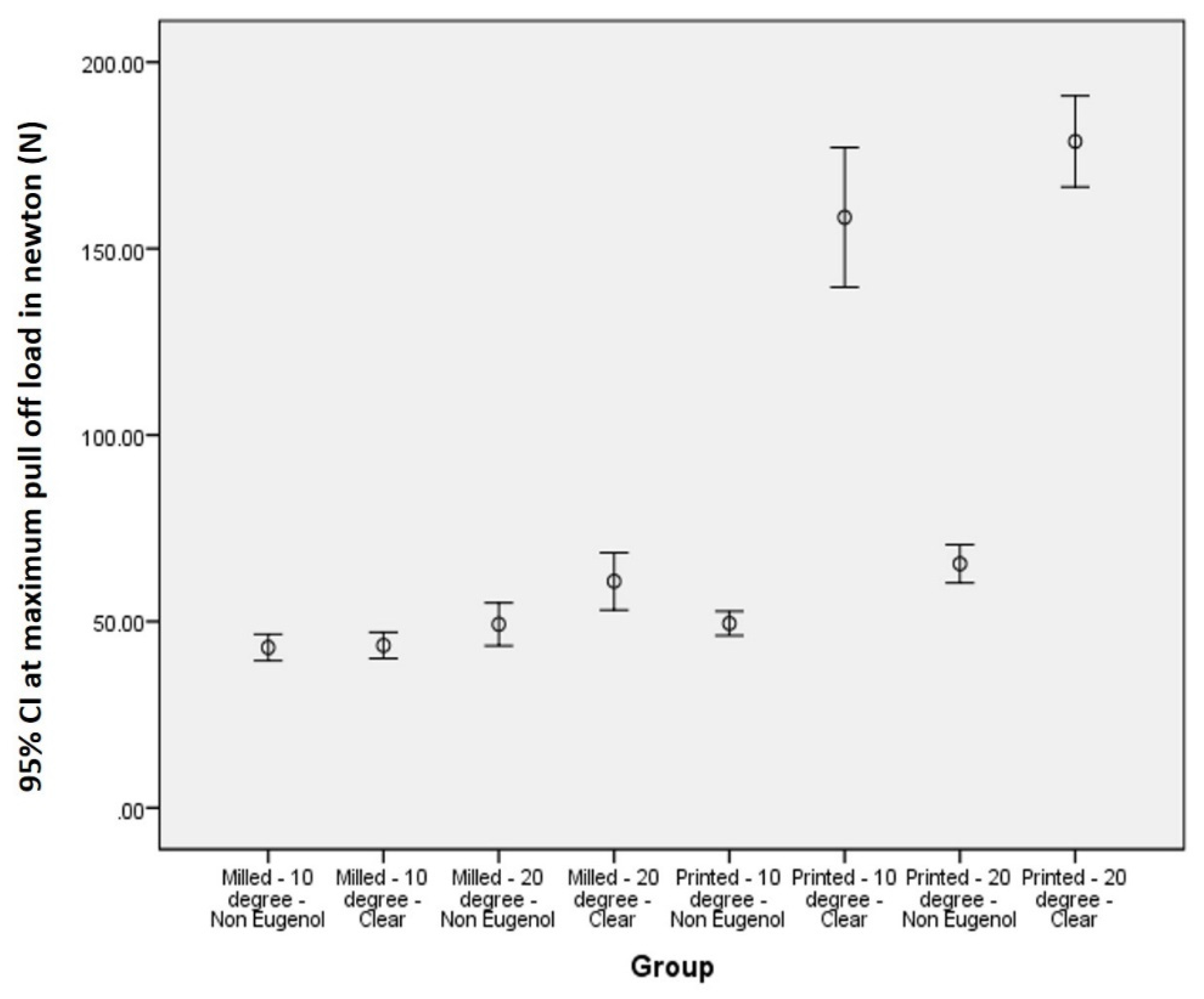
Figure 1. Two standardized all-ceramic tooth preparations with 10-degree and 20-degree total occlusal convergence angles prepared on typhodont teeth.
Applsci 12 12714 g001
Figure 2. (A) Working models made with 3D-printed resin dies and 10° and 20° tapers. (B) Tapers measured using a computer system consisting of a stereomicroscope with a connected USB CCD camera, a personal computer, and compatible measurement software.
Applsci 12 12714 g002
Figure 3. (A) STL file saved with the margin set-up and the cement space set-up. (B) CAD/CAM-milled temporary crowns and 3D-printed crowns on working models.
Applsci 12 12714 g003
Figure 4. Cementation of temporary crowns with Kerr Temp-Bond NE cement and Kerr Temp- Bond clear cement. (A) Temporary crowns and cement used for cementation (B) Crowns after cementation.
Applsci 12 12714 g004
Figure 5. Thermocycling of cemented provisional crowns (5000 cycles between baths held at 5 °C and 55 °C).
Applsci 12 12714 g005
Figure 6. Specimen subjected to the retention test in the universal testing machine.
Applsci 12 12714 g006
Figure 7. Box plot showing pull-off force values (N) from groups 1-8 performed using the post-hoc Bonferroni test.
Applsci 12 12714 g007
Figure 8. Intergroup comparison of 95% CI relative to pull-off force (N) in the test groups. * Statistically significant.
Applsci 12 12714 g008
Table 1. Description of each group in the study.
Group 1 Milled—10°—Non-eugenol conventional cement
Group 2 Milled—10°—Clear cement
Group 3 Milled—20°—Non-eugenol conventional cement
Group 4 Milled—20°—Clear cement
Group 5 Printed—10°—Non-eugenol conventional cement
Group 6 Printed—10°—Clear cement
Group 7 Printed—20°—Non-eugenol conventional cement
Group 8 Printed—20°—Clear cement
Table 2. Group-specific pull-off force values (in Newtons) compared between groups 1, 2, 3, 4, 5, 6, 7 and 8 using one-way ANOVA. The maximum, mean, median, minimum, and standard deviations are shown for the pull-off forces in groups 1–8.
Group 1Group 2Group 3Group 4Group 5Group 6Group 7Group 8
n—number of samples per group1010101010101010
Maximum (N)52.3448.3565.2580.1053.88188.0280.24205.65
Mean (N)43.0143.4849.2460.7649.15158.3765.48178.74
Median (N)41.2745.4648.9456.7950.56153.2362.69180.44
Minimum(N)36.3832.1437.9847.3139.28127.5055.31154.95
Standard deviation (N)4.935.208.0410.774.6426.197.1617.09
Table 3. Intergroup comparison of pull-off force in newton s(N) for groups 1–8 using one-way ANOVA test.
Group12345678
MD/p-ValueMD/p-ValueMD/p-ValueMD/p-ValueMD/p-ValueMD/p-ValueMD/p-ValueMD/p-Value
1X−0.56/0.921−6.23/0.27417.75/0.002 *−6.46/0.257115.36/0.000 *115.36/0.000 *−135.73 */0.000 *
2−0.56/0.921X−5.67/0.319−17.19 */0.003 *−5.90/0.301−114.80 */0.000 *−21.91/0.000 *135.17 */0.000 *
3−6.23/0.274−5.67/0.319X−11.52/0.045 *−0.23/0.968−109.13/0.000 *−16.24 */0.005 *−129.50 */0.000 *
417.75/0.002 *−17.19 */0.003 *−11.52/0.045 *X11.30 */0.050 *−109.13/0.000 *−16.24 */0.005 *−129.50 */0.000 *
5−6.46/0.257−5.90/0.301−0.23/0.96811.30 */0.050 *X−108.91 */0.000 *−16.01 */0.006 *−129.27 */0.000 *
6−115.36/0.000 *−114.80 */0.000 *−109.13/0.000 *−97.61 */0.000 *−108.91 */0.000 *X92.90 */0.000 *−20.36 */0.001 *
7−22.47 */0.000 *−21.91/0.000 *−16.24 */0.005 *−4.72/0.407−16.01 */0.006 *92.90 */0.000 *X−113.26 */0.000 *
8−135.73 */0.000 *135.17 */0.000 *−129.50 */0.000 *−117.98 */0.000 *−129.27 */0.000 *−20.36 */0.001 *−113.26 */0.000 *X
Publisher’s Note: MDPI stays neutral with regard to jurisdictional claims in published maps and institutional affiliations.

