Article

Mandibular Second Molar Impaction-Part II: Etiology and Role of the Third Molar

1
The Maurice and Gabriela Goldshleger School of Dental Medicine, Tel Aviv University, Tel Aviv 69978, Israel
2
Department of Orthodontics, New York University College of Dentistry, New York, NY 10010, USA
*
Author to whom correspondence should be addressed.
Appl. Sci. 2022, 12(22), 11520; https://doi.org/10.3390/app122211520
Submission received: 3 October 2022 / Revised: 6 November 2022 / Accepted: 10 November 2022 / Published: 13 November 2022
(This article belongs to the Special Issue Present and Future of Orthodontics)

Abstract

:
(1) Introduction: Mandibular second molar (MM2) impaction is a relatively rare dental anomaly, but its early detection is imperative for orthodontic diagnosis and treatment. This study evaluates early radiographic characteristics of MM2 in the process of its impaction and the role of the third molar (MM3) in this process. (2) Materials and Methods: Panoramic radiographs of orthodontic patients of Chinese-American origin (n = 5575), age 9–15 years (mean 13.13) were examined for MM2 impactions and the presence of MM3. (3) Results: A total of 151 patients with 212 impacted MM2s were detected in the Chinese-American study group. Unilateral impactions were found more than bilateral and were distributed equally between the left and right sides. Mesial angulation of the impacted teeth was present in 92.7% of the cases, and MM3s were found adjacent to the impacted MM2s in 97.4% of the cases. (4) Conclusions: MM2 differential root developments with deficient mesial root lengths appeared to be the primary cause for MM2 impaction. Posterior crowding and arch length deficiency were contributing factors causing MM2 impaction. This impaction occurred early, before the third molar bud was developed. It sometimes occurred even when the third molar was congenitally missing. Nevertheless, the possible involvement of the third molar in MM2 impaction needs further investigation.

1. Introduction

The process of non-orthodontic movement of a tooth, occurring from the time of its initial formation to its occupying place in a functional occlusal table, can be divided and described as pre- and post-eruptive. For the purpose of this work, the term “eruption” shall mean the tooth’s penetration of the alveolar mucosa, or the stage when some part of a tooth can be seen by a naked eye. This is also termed the emergence of a tooth, however this later term, although very well defined by various anthropologists [1], is not routinely used. Consequently, various disturbances in the tooth’s progression from its place of initial formation to the so-called “anatomically correct position in the dental arch” can happen pre- and post-eruptively. Better terms, and by the anthropologists preferred ones, would be the pre- and post-emergence phases of tooth eruption. Eruption disturbances can occur in any stage by an obstacle in the eruptive path, such as a supernumerary tooth or odontoma, ectopic position of the tooth germ, and failure of the eruption mechanism [2].
This paper will address both of these phases, with a particular emphasis on the pre-eruptive disturbances.
The cessation of eruption of a normally developed tooth germ before emergence without identification of a physical barrier in the eruption path is defined as primary retention [3]. The post-emergence cessation of eruption of a tooth without a physical barrier in the path of its eruption is considered secondary retention [4]. The cessation of eruption of a tooth caused by a physical barrier in the path of eruption, as well as a lack of space and the abnormal anatomical position of the tooth, is termed impaction. In a case of a clinically observed or a post-emergence mandibular molar impaction, often times a tooth presents with an obvious mesial angulation. Similarly, the great majority of pre-emergence impacted mandibular second molars (MM2), typically detected radiographically, have been reported with a marked mesial angulation [5].
MM2 impaction is usually associated with crowding and arch length deficiency in the posterior region [6,7]. In addition, it may also be a result of an ectopic germ position or an ectopic eruption path. It is often diagnosed as a secondary finding during examinations for pediatric or orthodontic diagnosis and treatment planning. It is rarely the main reason for referral to an orthodontic clinic. It is a relatively rare dental anomaly with a reported incidence of 0.03% to 0.6% in the general population [8], and 1–3% in orthodontic patients [5,6]. MM2 impactions can be classified as mesio-angular, disto-angular, vertical, or horizontal. The mesio-angular position has been reported for most of the MM2 impactions [5,9]. Unilateral impaction of the MM2s is more common than bilateral. They have been reported more often in males than in females and with a right side predominance [6].
Various treatment options have been suggested in the literature, depending on the position and the depth of the impacted tooth in the bone, including surgical exposure and orthodontic uprighting [9,10], surgical repositioning with or without removal of the third molar [11,12], extraction of the second molar with substitution with the third molar [11], and early prophylactic removal of the third molar germ when it presents an obstacle to the erupting second molar [13]. The optimal time for treatment of MM2 impaction cases coincides with the expected time of the MM2 eruption, from 11 to 14 years.
This paper evaluates radiographic characteristics of the MM2 and MM3 and the possible association in the process of their impaction. It discusses the early diagnosis and progression in the process of MM2 impaction, including the possible role of the third molars in this process.

2. Materials and Methods

In a retrospective study of 151 patients, 73 males and 78 females, 212 impacted MM2s were detected in 5575 panoramic radiographs of consecutively treated orthodontic patients of Chinese-American origin in New York City.
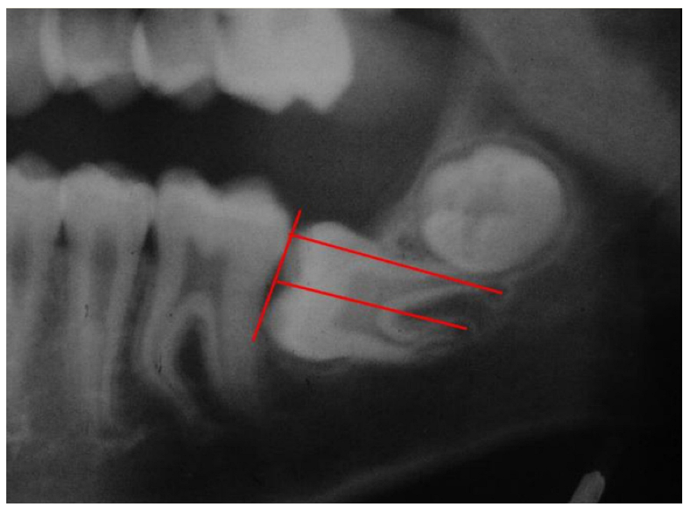
At the time of impaction detection, their age range was 9–15 years with a mean age of 13.13 years. The angle between the long axis of the mandibular first and second molars [MM1 and MM2] was measured with a cephalometric protractor (Figure 1). The presence or absence of third molars and the stage of their development was noted and registered for every patient.
The inclusion criteria were:
(1) Full eruption of the MM2 on one side, while the MM2 in the contralateral side had not emerged, even though three quarters of one root was developed.
(2) The impacted MM2, both unilateral and bilateral, had an abnormal mesial inclination with its crown arrested under the distal height of contour of the first molar crown. The University Ethics Committee approved the study.

3. Results

In the Chinese-American group, a total of 212 MM2s detected in 151 patients were diagnosed as impacted, in 73 males and in 78 females. Unilateral impactions were found in 90 patients and bilateral in 61 patients. (Table 1). The unilateral impactions were distributed equally between the left and right sides. Third molars in various stages of development, from early bud formation to complete root development, were found adjacent to the impacted second molar in all (97.4%) except for four patients (2.6%), where third molars were missing. Mesial angulation of the impacted MM2s was present in 92.7% of patients, with a notably increased angle between the long axis of the mandibular first and second molars (Figure 1), ranging between 30 to 70 degrees (mean 47 degrees).

4. Discussion

An impacted molar remains unerupted in the alveolar bone or in the body of the mandible (corpus mandibularis) beyond the time at which it normally erupts. Crowding and arch length deficiency in the posterior region of the mandibular arch have been suggested as important local factors causing MM2 impaction [6,14]. Our findings, however, do not fully support that theory, as will be explained later in the discussion.
Tooth size may be affected by genetic factors that control dental morphogenesis [15]. In addition, the tooth size in ethnic Chinese people has been reported to be larger than in Caucasians [16,17]. This may partially explain the higher prevalence of MM2 impactions in the Chinese population.
The presence of third molars in 97.4% of our cases was in agreement with a previous study reporting that all patients with arrested MM2 had the germ of the third molar [18]. Paradoxically, according to Gorlin and Goldman [19], the prevalence of developing third molars in the general population is 63.4% to 77.5%, which is significantly less than our study’s observations. This may suggest a different role of the developing MM3 in MM2 impactions. While the presence of MM3 may contribute to the space shortage for the MM2, equally, or even more importantly, it may contribute to the arrested eruption of the MM2, often leading to its impaction. This data suggests that the third molar might be involved in MM2 impaction. However, the mechanism of such an effect is still unknown.
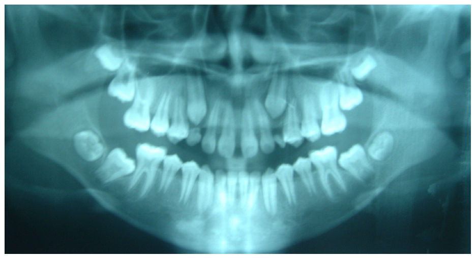
It has been previously reported that the second molar eruption may be unpredictable and, for unknown reasons, its inclination changes during its eruption, becomes mesially inclined and resulting in impaction [9]. This has been observed in an 8-year-old boy with excess space between his fully erupted first molar and the developing crown of the second molar. However, 3.5 years later, the tooth became mesially angulated and almost horizontally impacted, while the bud of the third molar had just began its development [9]. A similar phenomenon occurred even where the third molar was missing, as seen in the panoramic radiograph of an 11-year-old girl with a mesially angulated right MM2 (Figure 2). Similarly, in cases where the developing third molar crown appears to be far distal to the right second molar, as was present in an 11-year-old boy (Figure 3) and in a 10-year-old girl, where excess space appeared between the bilaterally developing buds of the third molars and the mesially angulated and impacted MM2s (Figure 4).
It appears that, at least in these and the similar cases, radiographic evidence-based treatment suggests insignificant association between developmental paths of the MM2 and MM3. In an abnormal eruption pathway of the second molar, the role of an ectopic position of the third molar is still questionable. According to previous reports, the third molar position should be considered as a contributory factor and not the primary cause for second molar impaction [20,21]. Instead, we suggest that the mesial angulation of the second molar may provide a better clue or indication of the MM2 eruptive outcome. It is therefore suggested that, in the presented cases, an arch length deficiency in the mandibular posterior segment is not the major contributing factor in causing second molar impaction. We repeatedly observed that the MM2 region had more than adequate arch length for the second molar, yet the tooth became impacted. A similar situation has been reported where the second molar became impacted on the side where a larger space was available [10,22]. In addition, previous reports [7,23] demonstrated mesial inclination among most of the mandibular second molars, suggesting it mainly attributed to their abnormal eruption pathway. This is contrary to several reports, supported by the presented radiographic evidence, that the majority of MM2 impactions are associated with crowding and arch length deficiency in the posterior region of the dental arch [6].
It has also been suggested that excess space between the first molar root and the developing second molar crown may allow the later to incline more mesially and become impacted. This is probably due to the initial mesial angulation of the developing tooth bud [9]. Another explanation has been suggested by which the MM2 may change its angulation due to differential, asymmetrical root growth, with a decrease in the mesial root length and increased length of the distal root (Figure 5). This was demonstrated in an 11-year-old girl, where the mandibular left second molar was horizontally impacted with a shorter mesial root than the distal one (Figure 6). A significantly shorter mesial root has been previously reported in mesially angulated impacted MM2s, whereas a distally angulated MM2 appears to be associated with a shorter distal root [5].
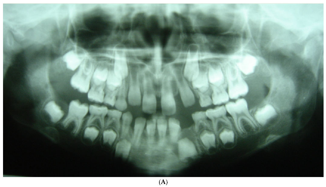
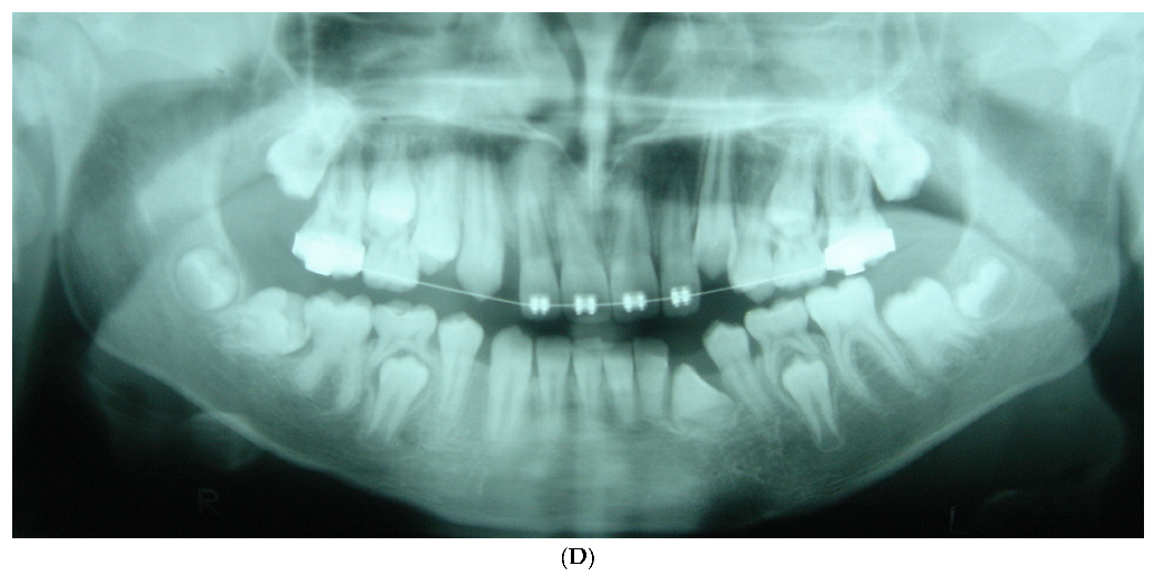
Most studies in the literature report on MM2 impaction in adolescent individuals presenting the impacted MM2 in association with the presence of the third molar. To the best of our knowledge, this is the first report presenting the early stages of the MM2 impaction process. The following longitudinal panoramic radiographs demonstrate an unequal differential growth of the MM2 roots, where the mesial root is shorter than the distal root. As a result, the mesial root serves as a pivot for the MM2 which gradually tips and becomes mesially angulated, often resulting in its impaction distal of the first molar. The panoramic radiograph of an 8-year-old boy in his early mixed dentition is a representative of the developing crowns of the MM2s in normally erupting positions with no signs of the third molar buds (Figure 7A). It is reasonable to predict that their eruption would be uneventful. A panoramic radiograph taken one year later, at the age of nine years, shows the right permanent second molar mesially inclined with its mesial root shorter than the distal. At that stage, the early developing third molar bud can be identified at the anterior rim of the ramus behind and away from the already mesially angulated second molar when comparing this with the contralateral view, where a small space exists between the second molar and the adjacent developing third molar bud (Figure 7B). In a one year interval, at the age of 10 years, the panoramic radiograph revealed the MM2 impacted in a more horizontal position the more clearly showed its shorter mesial root with the developing third molar bud still away from the second molar (Figure 7C). The panoramic radiograph taken two years later at the age of 12 years demonstrates a horizontally impacted right second molar with a shorter mesial root and the third molar crown now mesially inclined close to the second molar’s distal root (Figure 7D).
Figure 7A–D have previously been published in the article “Rootless eruption of a mandibular permanent canine”. AJO-DO 2011; 139:563–566.
Based on the available recent information, it is not clear what causes the differential root development in certain MM2s [24]. The tooth development undergoes a serious of complex reciprocal interactions between the ectodermally derived dental epithelium and the underlying mesenchymal cells during tooth morphogenesis. It has been reported that the Wnt/betta-catenin signaling process controls the root odontogenesis and cementogenesis during the tooth root development [25,26]. Disruption of the Wntless leads to the inhibition of odontoblast maturation and root elongation, thus resulting in short roots [27]. Experiments on the mandibular first molars in mice have demonstrated that deficient Wnt/betta–catenin signaling effectively blocked odontoblasts and cementoblasts differentiation during the root development, resulting in their arrested development [28]. An association between root anomalies and eruption disorders of MM2s has also been reported [18]. Why this phenomenon affects the mesial root more than the distal root of the MM2 is not yet clear and should be further investigated.
This may also support our theory [5,9] that MM2 mesial angulation occurs early, before the third molar bud is developed and even when the third molar is congenitally missing, as presented in the panoramic radiograph of a 12-year-old girl with a mesio-angular impacted right MM2 having a shorter mesial root (Figure 2). This was also demonstrated in a 10-year-old girl with normal erupting-positioned MM2s (Figure 8A), which four years later, at the age of 14 years, presented her right MM2 as mesially inclined, while the third molars were congenitally missing (Figure 8B). That is, the differential MM2 root development and its mesial angulation could probably be the major cause, while the posterior crowding might have been a secondary contributing factor, occurring later and causing the MM2 to become impacted. It has been shown that patients with congenitally missing third molars have fewer problems of crowding or impacting of the MM2 during treatment. Thus, early removal of the third molar relieves the congestion at the end of the lower arch. An early prophylactic approach for interceptive removal of the third molar germ has been suggested. Such extraction of the mandibular third molar germ, before or during orthodontic treatment, could be beneficial because it may facilitate the eruption of the second molar [15]. In 1934, Henry [29] described the enucleation technique at the bud stage of the third molar at an early age (7–9 years) as a simple and relatively atraumatic procedure. This is contrasted by the often difficult extraction of deeply impacted third molars in adults. Similarly, in 1970, Ricketts [30] revisited the enucleation of third molar buds as a prophylactic measure in young children, using electrosurgery.
An interesting analogy could be postulated by speculating that the developing second molar “needs” the close guidance of the first molar distal root for its normal eruption. This would be similar to the “guidance theory” related to the maxillary canine that requires the guidance of the lateral incisor’s root for its normal eruption [31]. Although a careful study of panoramic radiographs may reveal the orientation of the mandibular second molars, CBCT scans can supply us with more accurate information. However, good radiation hygiene should be practiced, whereby all patients should not be routinely referred for screening CBCT scans.

5. Conclusions

The early diagnosis and detection of a potentially developing MM2 impaction is imperative to initiate adequate corrective measures. MM2 mesial angulation and impaction appears to be a result of differential growth of its roots, where the shorter mesial root becomes a pivot for its mesial angulation. This usually occurs early, before the third molar bud is developed, and even when the third molar is congenitally missing. Differential root development is suggested to be the major cause, while posterior crowding and arch length deficiency seem to play a secondary role in causing MM2 impactions. Nevertheless, the differential root development and the possible involvement of the third molar in MM2 impaction needs further investigation.

Author Contributions

Conceptualization, Y.S.; Investigation, S.S. and T.F.; Methodology, N.S.; Validation, Y.L.; Writing—review & editing, Y.S. All authors have read and agreed to the published version of the manuscript.

Funding

This research received no external funding.

Institutional Review Board Statement

Not applicable.

Informed Consent Statement

Informed consent was obtained from all subjects involved in the study.

Acknowledgments

The authors wish to thank Amir Shapira, BSc, for his valuable assistance in the preparation of this manuscript.

Conflicts of Interest

The authors declare no conflict of interest. I, Yehoshua Shapira, declare, according to the guidelines for Authors regarding use of previously published materials, and according to the kind permission and instructions received from Elsevier’s Global Rights Department in Oxford, UK, that my Figure 7A–D, which appeared in my article “Rootless eruption of a mandibular permanent canine” AJO-DO 2011; 139:563–566, can be used in the present manuscript entitled “Mandibular Second Molar Impaction–Part II: Etiology and role of the third molar”.

References

  1. Garn, S.M.; Moorrees, C.F.A. Stature, body-build and tooth emergence in Aleutian Aleut children. Child Dev. 1951, 22, 261–270. [Google Scholar] [CrossRef] [PubMed]
  2. Andreasen, J.O. Review of root resorption systems and models. Etiology of root resorption and the homeostatic mechanisms of the periodontal ligament. In The Biological Mechanism of Tooth Eruption and Root Resorption; Davidovitch, Z., Ed.; EBSCO Media: Birmingham, AL, USA, 1998; pp. 9–21. [Google Scholar]
  3. Raghoebar, G.M.; Boeing, G.; Vissink, A.; Stegenga, B. Eruption disturbances of permanent molars: A review. J. Oral Pathol. Med. 1991, 20, 159–166. [Google Scholar] [CrossRef] [PubMed]
  4. Raghoebar, G.M.; Boering, G.; Jansen, H.W.B.; Vissink, A. Secondary retention of permanent molars: A histologic study. J. Oral Pathol. Med. 1989, 18, 427–431. [Google Scholar] [CrossRef] [PubMed]
  5. Shapira, Y.; Finkelstein, T.; Shpack, N.; Lai, Y.H.; Kuftinec, M.M.; Vardimon, A. Mandibular second molar impaction. Part 1: Genetic traits and characteristics. Am. J. Orthod. Dentofac. Orthop. 2011, 140, 32–37. [Google Scholar] [CrossRef]
  6. Varpio, M.; Wellfelt, B. Disturbed eruption of the lower second molar: Clinical appearance, prevalence, and etiology. ASDC J. Dent. Child. 1988, 55, 114–118. [Google Scholar]
  7. Wellfelt, B.; Varpio, M. Disturbed eruption of the permanent lower second molar: Treatment and results. ASDC J. Dent. Child. 1988, 55, 183–189. [Google Scholar]
  8. Grover, P.S.; Lorton, L. The incidence of unerupted permanent teeth and related clinical cases. Oral Surg. Oral Med. Oral Path. 1985, 59, 420–425. [Google Scholar] [CrossRef]
  9. Shapira, Y.; Borell, G.; Nahlieli, O.; Kuftinek, M.M. Uprighting mesially impacted mandibular permanent second molar. Angle Orthod. 1998, 68, 173–178. [Google Scholar]
  10. Ferrazzini, G. Uprighting of a deeply impacted mandibular second molar. Am. J. Orthod. Dentofac. Orthop. 1989, 96, 168–171. [Google Scholar] [CrossRef]
  11. Johnsen, J.V.; Quirk, G.P. Surgical repositioning of impacted mandibular second molar teeth. Am. J. Orthod. Dentofac. Orthop. 1987, 91, 242–251. [Google Scholar] [CrossRef]
  12. McAboy, C.P.; Grumet, J.T.; Siegel, E.B.; Iacopino, A.M. Surgical uprighting and repositioning of severely impacted mandibular second molars. J. Am. Dent. Assoc. 2003, 134, 1459–1462. [Google Scholar] [CrossRef] [PubMed]
  13. Kavadia, S.; Antoniades, K.; Kaklamanos, E.; Antoniades, V.; Markovitsi, E.; Zafiriadis, L. Early extraction of the mandibular third molar in case of eruption disturbances of the second molar. J. Dent. Child. 2003, 70, 29–32. [Google Scholar]
  14. Johnsen, D.C. Prevalence of delayed emergence of permanent teeth as a result of local factors. J. Am. Dent. Assoc. 1977, 94, 100–106. [Google Scholar] [CrossRef] [PubMed]
  15. Radlanski, R.J.; Renz, H. Explainable and critical periods during human dental morphogenesis and their control. Arch. Oral Biol. 2005, 50, 199–203. [Google Scholar] [CrossRef] [PubMed]
  16. Cho, S.Y.; Ki, Y.; Chu, V.; Chan, J. Impaction of permanent mandibular second molars in ethnic Chinese schoolchildren. J. Can. Dent. Assoc. 2008, 76, 521–521e. [Google Scholar]
  17. Ling, J.Y.; Wong, R.W. Tanaka-Johnston mixed dentition analysis for Southern Chinese in Hong Kong. Angle Orthod. 2006, 76, 632–636. [Google Scholar]
  18. Vedtofte, H.; Andreasen, J.O.; Kjaer, I. Arrested eruption of the permanent lower second molar. Euro. J. Orthod. 1999, 21, 31–40. [Google Scholar] [CrossRef] [Green Version]
  19. Gorlin, R.J.; Goldman, H.M. Thoma’s Oral Pathology, 6th ed.; C.V. Mosby Co.: St. Louis, MI, USA, 1970; pp. 103–109, 124–125. [Google Scholar]
  20. Valmaseda-Castellon, E.; de-la-Rosa-Gay, C.; Gay-Escoda, C. Eruption disturbances of the first and second permanent molars: Results of treatment in 43 cases. Am. J. Orthod. Dentofac. Orthop. 1999, 116, 651–658. [Google Scholar] [CrossRef]
  21. Palma, C.; Coelho, A.; Gonzalez, Y.; Cahuana, A. Failure of eruption of first and second permanent molars. J. Clin. Pediatr. Dent. 2003, 27, 239–245. [Google Scholar] [CrossRef] [Green Version]
  22. Freeman, R.S. Mandibular second molar problems. Am. J. Orthod. Dentofac. Orthop. 1988, 94, 19–21. [Google Scholar] [CrossRef]
  23. Shpack, N.; Finkelstein, T.; Lai, Y.H.; Kuftinec, M.M.; Vardimon, A.; Shapira, Y. Mandibular permanent second molar impaction treatment options and outcome. Open J. Dent. Oral Med. 2013, 1, 9–14. [Google Scholar] [CrossRef]
  24. Kim, T.H.; Bae, C.H.; Lee, J.C.; KOSO; Yang, X.; Juang, R.; Cho, E.S. Betta-catenin is required in odontoblasts for tooth root formation. J. Dent. Res. 2013, 92, 215–221. [Google Scholar] [CrossRef]
  25. Bae, C.H.; Lee, J.Y.; Kim, T.H.; Baek, J.A.; Lee, J.C.; Yang, X.; Taketo, M.M.; Jiang, R.; Cho, E.S. Excessive Wnt/betta-catenin signaling disturbs tooth root formation. J. Periodo. Res. 2013, 48, 405–410. [Google Scholar] [CrossRef] [PubMed]
  26. Lim, W.H.; Liu, B.; Hunter, D.J.; Cheng, D.; Mah, S.J.; Helms, J.A. Downregulation of Wnt causes root resorption. Am. J. Orthod. Dentofac. Orthop. 2014, 146, 337–345. [Google Scholar] [CrossRef] [PubMed]
  27. Bae, C.H.; Kim, T.H.; Ko, S.O.; Lee, J.C.; Yang, X.; Cho, E.S. Wntless regulates dentin apposition and root elongation in the mandibular molar. J. Dent. Res. 2015, 94, 439–445. [Google Scholar] [CrossRef] [PubMed] [Green Version]
  28. Zang, R.; Yang, G.; Wu, X.; Xie, J.; Yang, X.; Li, T. Disruption of Wnt/betta-catenin signaling in odontoblasts and cementoblasts arrests tooth root development in postnatal mouse teeth. Int. J. Biol. Sci. 2013, 9, 228–236. [Google Scholar] [CrossRef]
  29. Henry, C.B. Prophylactic enucleation of lower wisdom tooth follicles. Lancent 1936, 18, 921. [Google Scholar] [CrossRef]
  30. Ricketts, R.M.; Turley, P.; Chaconas, S.; Schulhof, R.J. Third molar enucleation: Diagnosis and technique. J. Calif. Dent. Assoc. 1976, 4, 52–57. [Google Scholar]
  31. Becker, A.; Zilberman, Y.; Tsur, B. Root length of lateral incisors adjacent to palatally displaced maxillary cuspids. Angle Orthod. 1984, 54, 218–225. [Google Scholar]
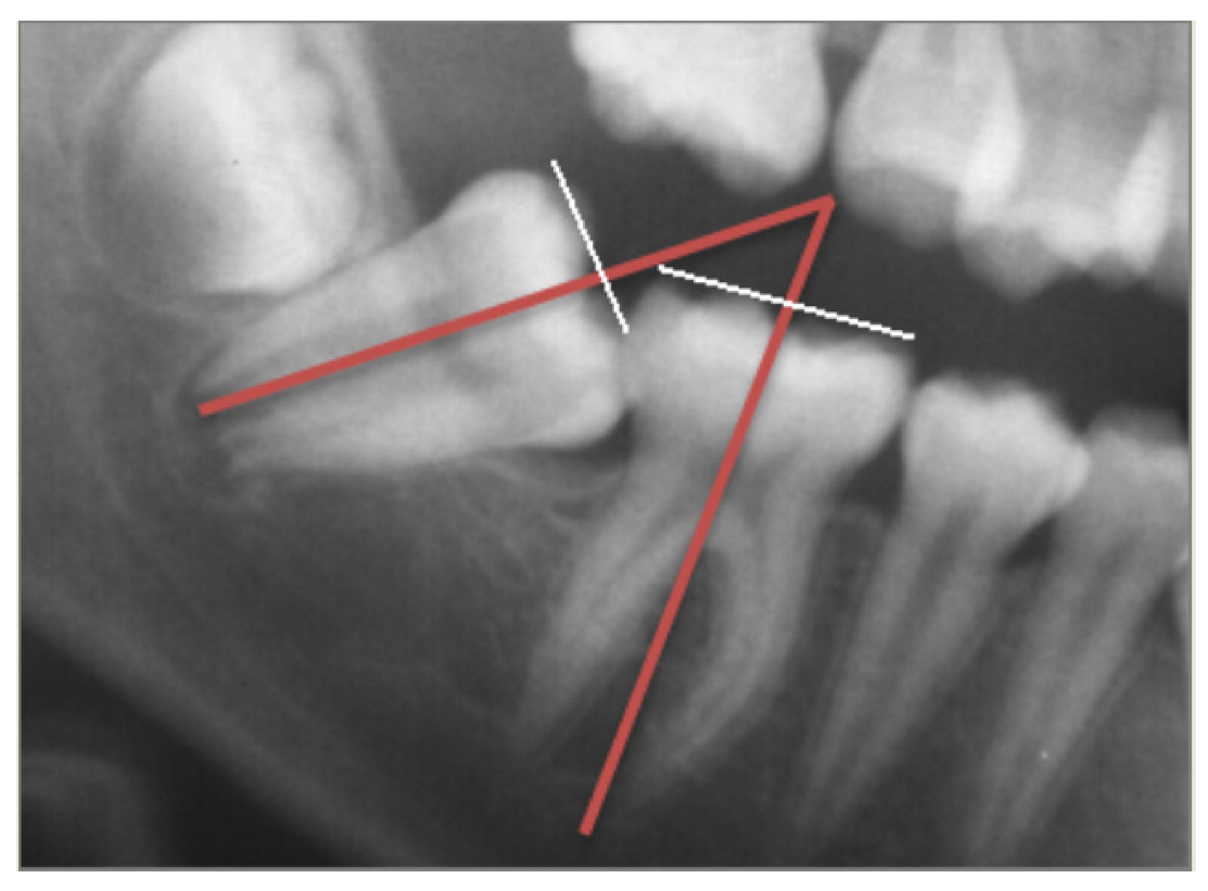
Figure 1. The angle between the long axis of the first and second molars.
Applsci 12 11520 g001
Figure 2. Fully developed mandibular right second molar mesially angulated and impacted with missing third molar behind it.
Applsci 12 11520 g002
Figure 3. Mandibular right second molar mesially angulated and impacted with excess space between the second molar and the third molar developing crown.
Applsci 12 11520 g003
Figure 4. Bilateral mandibular second molar impaction showing excess space between the second molars and the developing buds of the third molars.
Applsci 12 11520 g004
Figure 5. A horizontally impacted left MM2 with shorter mesial than distal root.
Applsci 12 11520 g005
Figure 6. Panoramic radiograph of an 11-year-old girl with a horizontally impacted left MM2. Note its shorter mesial root and the developing MM3 crown on top of the MM2 longer distal root.
Applsci 12 11520 g006
Figure 7. (A) Panoramic radiograph of an 8-year-old boy presents the developing crowns of the mandibular second molars in the normally erupting position with no signs of the third molar buds. (B) Panoramic radiograph of the same boy at the age of nine years, showing the right second molar mesially inclined with a shorter mesial root than the distal. The early developing third molar bud can be identified at the anterior rim of the ramus behind and away from the already mesially angulated second molar. (C) At the age of 10 years, the panoramic radiograph reveals the MM2 impacted in a more horizontal position with its shorter mesial root, and the developing third molar bud still far away from the second molar. (D) At the age of 12 years, the panoramic radiograph demonstrates a horizontally impacted right second molar with shorter mesial root and the third molar crown now mesially inclined close to the second molar distal root.
Applsci 12 11520 g007aApplsci 12 11520 g007bApplsci 12 11520 g007c
Figure 8. (A) Normal erupting-positioned MM2s in a 10-year-old girl. (B) The same girl, at the age of 14 years, presented her right MM2 as mesially inclined, with third molars congenitally missing.
Applsci 12 11520 g008
Table 1. Distribution of MM2 impactions.
EthnicityNo. of PatientsMaleFemaleUnilateralBilateralTotal No. of Impacted MM2
Chinese-Americans15173789061212
Publisher’s Note: MDPI stays neutral with regard to jurisdictional claims in published maps and institutional affiliations.