Article

Streptomyces spp. Biofilmed Solid Inoculant Improves Microbial Survival and Plant-Growth Efficiency of Triticum aestivum

by
Karla Gabriela Domínguez-González
1,
J. Jesús Robledo-Medrano
1,
Juan José Valdez-Alarcón
2,
Orlando Hernández-Cristobal
3,
Héctor Eduardo Martínez-Flores
1,*,
Jorge Francisco Cerna-Cortés
4,
Ma. Guadalupe Garnica-Romo
5 and
Raúl Cortés-Martínez
1,*
1
Facultad de Químico Farmacobiología, Universidad Michoacana de San Nicolás de Hidalgo, Tzintzuntzan 173, Col. Matamoros, Morelia 58240, Michoacán, Mexico
2
Laboratorio de Epidemiología Molecular y Biotecnología de Enfermedades Infecciosas, Centro Multidisciplinario de Estudios en Biotecnología, Facultad de Medicina Veterinaria y Zootecnia, Universidad Michoacana de San Nicolás de Hidalgo, Km 9.5 Carretera Morelia-Zinapécuaro s/n, Posta Veterinaria. La Palma, Tarímbaro, C.P., Morelia 58893, Michoacán, Mexico
3
Escuela Nacional de Estudios Superiores (ENES), Unidad Morelia, UNAM. Antigua Carretera a Pátzcuaro 8701, col. Ex Hacienda San José de la Huerta, Morelia 58190, Michoacán, Mexico
4
Escuela Nacional de Ciencias Biológicas, Instituto Politécnico Nacional, Prolongación de Carpio, Calle Plan de Ayala s/n, Santo Tomás, Miguel Hidalgo, Ciudad de México 11350, Mexico
5
Facultad de Ingeniería Civil, Universidad Michoacana de San Nicolás de Hidalgo, Gral. Francisco J. Mújica 172B, Felicitas del Río, Morelia 58040, Michoacán, Mexico
*
Authors to whom correspondence should be addressed.
Appl. Sci. 2022, 12(22), 11425; https://doi.org/10.3390/app122211425
Submission received: 22 October 2022 / Revised: 3 November 2022 / Accepted: 3 November 2022 / Published: 10 November 2022
(This article belongs to the Special Issue Role of Microbes in Agriculture and Food)

Abstract

:
Streptomyces species have been successfully used in diverse biotechnological processes; however, little is known about actinobacterial biofilm formation and its use as a biofilmed inoculant. The present study assessed and compared the ability of some plant growth-promoting actinobacterial strains to form biofilms on a carrier to improve microbial survival and colonize the rhizosphere and roots of Triticum aestivum, resulting in positive interactions and benefits to the plant. Forty-one actinobacterial isolates from Persea americana organic rhizosphere farms were tested on plant growth and biofilm-forming capacities, showing their potential use as bio-fertilizers in agriculture. Three Streptomyces strains were selected and tested for biofilm formation and plant growth-promoting (PGP) features. Biofilms were induced on the perlite carrier and used to inoculate seven treatments with T. aestivum in pot essays, resulting in a >200% increase in root weight and a >400% increase in total biomass. Endophytic colonization was achieved in all the treatments. Microbial survival ranged between 108 and 109 CFU/g after 12 weeks of treatment, indicating actinobacterial permanence on the carrier. Successful consortia formation was observed for mixed-strain treatments, suggesting long-term plant recolonization.

1. Introduction

An increase in the global population requires greater food production. However, global conflicts, climate variability, and economic recession (exacerbated by the COVID-19 pandemic) are the leading causes of food insecurity [1]. Consequently, several adverse effects have occurred, including low agricultural productivity, reduced agricultural land, salinization and desertification of soils, erosion, and reduced fertility. These effects result from the more intensive use of pesticides and chemical fertilizers, which are currently less accessible because of their high cost and low production [1,2]. Therefore, there is greater interest in soil microorganisms that can improve plant nutrition, health, and soil quality [3,4]. These microorganisms could increase crop production and reduce chemical products, leading to what is called “Sustainable Agriculture” [5,6,7,8,9].
Actinobacteria are ubiquitous in persistent populations in several ecosystems, especially in the soil. These organisms grow profusely at different depths in the soil surrounding the roots of plants, called the rhizosphere [10,11]. Actinobacteria are spore-forming, Gram-positive, filamentous bacteria [12], and possess aerial mycelia that remind the growth of fungi. They appear to play a vital role in organic matter cycling and fertility [13]. The Streptomyces genus is economically and biotechnologically beneficial not only in the pharmaceutical industry producing enzymes, antibiotics, vitamins, plant hormones, amino acids, and other biologically active substances but also in agriculture. It shows potential by interacting with plants and maintaining a healthy rhizosphere [10,13]. Its presence in the rhizosphere has been discussed in agriculture as a biocontrol and plant growth-promoting bacterium (PGP) due to direct mechanisms, including nitrogen fixation (i.e., Streptomyces, Arthrobacter, and Frankia), phosphate solubilization (i.e., Streptomyces and Rhodococcus), potassium solubilization, and phytohormone synthesis, mainly Indol Acetic Acid (i.e., Streptomyces, Actinomyces, and Micrococcus). Some indirect mechanisms, such as antagonism, competition, siderophore production, vitamin production, and antimicrobial compound production, also determine PGP properties [14]. In addition, some actinobacteria can colonize plant roots in an endophytic or epiphytic way, offering extra protection to the plant against pathogens and desiccation and contributing to micronutrient sequestration for plant benefits [8,15,16].
Iron deficiency in plants is one of the most critical problems in fertilization [6]. Some actinobacteria genera are associated with acquiring iron and biocontrol in phytopathogenic fungi. For example, some Streptomyces spp. are antifungal biocontrol agents that inhibit numerous plant pathogenic fungi, such as Phytophthora sp., Fusarium sp., Alternaria sp., and Rhizoctonia sp. through siderophore and HCN production [11,14,17]. Thus, considering such attributes, actinobacteria are potential inoculants in agriculture.
Actinobacteria, especially Streptomyces species, have advantages as bioinoculants because they are less harmful and more eco-friendly when used as fertilizers. Moreover, they affect only specific pathogens and support the colonization of beneficial microorganisms such as mycorrhizae [5,18,19]. In addition, they can help plants balance biotic and abiotic stresses [8,16]. However, the proliferation rate of Actinobacteria is generally slower than that of other bacterial inoculants, and storage and application are not successful in the planktonic manner [11].
Microorganisms can attach to biotic or abiotic surfaces and differentiate into complex multicellular communities called biofilms. A biofilm consists of microbial cells (e.g., bacteria, fungi, or algae) protected by an extracellular polymer that cells produce, providing structure and protection to the community [20,21]. Its use has been investigated to obtain multiple beneficial applications in industry and agricultural and environmental fields [11]. Biofilms play a more critical role in agriculture than previously believed. The most beneficial biofilms developed in vitro have begun to be used as biofertilizers to colonize plant roots and multiply their benefits. Thus they have been called “biofilmed biofertilizers” [6,10,22,23] and were first highlighted a decade ago [24].
Biofilms are more resilient to various environmental stressors, including pH changes, chromium, earthworm predation, UV rays, osmotic shock, desiccation, and pathogen defense [7,21,25]. Despite the abundance of Actinobacteria, very few reports are available on biofilm formation regarding this phylum [25]. A few studies have shown that a biofilmed biofertilizer used as monoculture or as a consortium significantly increases N2 fixation in legumes (by ca. 30%). In addition, plant growth promotion (by approximately 25%) increased rice plant dry weight in its early growth stage compared to conventional bioinoculants; for example, P. fluorescens biofilm, which increased endophytic colonization of tomato, showed a biocontrol activity of over 1000% compared to inoculation with a planktonic culture [7,21,26]. Compared with those examples, successful fertilization has been reported in rice, wheat, soybean, corn, tea, sorghum, rubber crops, and some other cereals and vegetables under greenhouse conditions, offering multiple benefits but highlighting the 50% reduction in chemical fertilization in field crop conditions [6,27,28,29,30,31,32]. Though not all microorganisms can form a biofilm, a few actinobacteria can. It is believed that actinobacteria are not capable of forming biofilms by themselves, that is, without the help of another microorganism or consortium [12,22].
A key issue with conventional inoculant technology (planktonic cells) is the low survival of introduced microorganisms in the soil as a result of numerous environmental stress factors; thus, the need to use carriers has been increasing [7]. A carrier is the main component that transports a substantial quantity of PGP into the soil in agriculture and whose function, in addition to transport, is to maintain the viability of the microorganism for a prolonged period [33,34]. Although many options are commercially available depending on the application method or selected microorganisms, peat is the most popular carrier. However, other carriers such as perlite, vermiculite, bentonite, sepiolite, attapulgite, compost, calcium alginate, biochar, or polymers have been used with different results and are not always positive [35,36,37]. A carrier is considered ideal if it meets specific characteristics, such as high water retention capacity, high surface area, inertness, ease of handling and sterilization, environmental friendliness, neutral or adjustable pH, chemical and physical stability, and low cost [33,34,35].
The present study investigated the role of PGP actinobacteria strains in a novel biofilmed inoculant regarding the growth-promoting efficiency of T. aestivum through greenhouse experiments. Furthermore, its introduction using perlite was assessed to determine microbial viability and survival owing to its ideal characteristics as a carrier.

2. Materials and Methods

2.1. Actinobacteria from Avocado Rhizosphere

Forty-one actinobacterial strains from the rhizosphere of Persea americana obtained from organic avocado farms in the state of Michoacan, Mexico, were selected and identified by a classical approach using macroscopic and microscopic morphology and biochemical tests. First, the isolates were labeled as ARI (avocado rhizosphere isolate), followed by the avocado farm number and an internal identification number. The isolates were then analyzed for mycelial organization and sporulation under a light microscope. Finally, the colors of the substrate and aerial mycelium (including mature spores) were determined using the ISCC-NBS Centric Color Charts [38], as well as the melanoid pigments, reverse side, and soluble pigments in the culture, cultural characteristics in yeast-malt extract agar (ISP2) (BD Difco™), oatmeal agar (ISP3) (HiMEDIA® M358), BHI agar (Bioxon™) and oatmeal-yeast extract glycerol (OYG), and tested with 20 carbohydrate compounds (data not shown) [39,40]. They were grown for 7 to 10 days at 30 °C and 110 rpm in a 50 mL glass flask with malt extract broth (MEB) designed in our laboratory: malt extract (Merck, Kenilworth, NJ, USA) 10 g, dextrose (Sigma-AldrichTM, St. Louis, MO, USA) 2 g, yeast extract (Sigma-AldrichTM) 5 g, casein peptone (BioxonTM) 6 g, meat peptone (BioxonTM) 2 g, tryptone (Sigma-AldrichTM, St. Louis, MO, USA) 1 g, 1-L final volume, pH 7.0. This study used the same formulations but with different presentations, solid supplemented with agar (Merck®) 1.5% (MEA) or liquid (MEB).

2.2. Molecular Identification

Molecular identification was performed for the actinobacteria selected for pot assays. The DNAzol® reagent (Invitrogen, Cat. 10503-027) was used for DNA extraction following the manufacturer’s instructions of the commercial house, and 10 µL of Proteinase-K (40 µg/mL; Invitrogen™, Cat. AM2542), and 3 µL RNase A Pure Link (Invitrogen, cat. 12091-021), and incubated for 15 min at 37 °C. The mixture was then centrifuged for 5 min at 4000× g. The supernatant was then carefully transferred to a clean tube.
The presence and quality of the extracted DNA were verified by electrophoresis on a 0.8% agarose gel. For 1500 bp, amplification of the 16S rRNA region was performed using primers 27F (5′-AGAGTTTGATCCTGGCTCAG-3′) and 1492R (5′-TACGGYTACCTTGTTACGACTT-3′) [41,42] obtained from OligoT4® (Irapuato, Mexico). Reactions (50 µL) were performed in 1 × Taq PCR Master Mix containing 1.5 Mg2+, dNTP mix (2.5 mM each), 50 ng DNA, 1.0 µM 27F primer (final concentration), 1.0 µM 1492R primer (final concentration). Amplifications were performed in an Applied Biosystems Veriti 9902® AB thermocycler under the following conditions: 5 min at 94 °C; 30 cycles of exponential amplification (60 s at 94 °C, 60 s at 54 °C, 2 min at 72 °C), and 10 min at 72 °C [42]. To ensure a fragment of the right size was amplified, PCR products were electrophoresed on 1% agarose gels stained with ethidium bromide.
The PCR products were purified using the gel QIAquick PCR Purification Kit® (Qiagen®, Hilden, Germany), Cat. 28104. The 16S rRNA fragments were sequenced (Macrogen Genome Center, Seoul, Korea). The sequences were assembled using Ugene® (Unipro, Atlanta, GA, USA) software and identified using the NBCI Taxonomy Browser https://blast.ncbi.nlm.nih.gov/Blast.cgi (accessed on 20 October 2022), and BLAST analysis was performed. The complete PCR sequences were registered in GenBank under accession numbers ARI_A20-3 OP364748, ARI_A20-5 OP364749, and ARI_A30-32 OP364750.

2.3. Plant Growth-Promoting Assays

The development of the actinobacterial isolates in a nitrogen-free environment, the detection of aerial mycelia, and the existence of diazotrophic structures as a component of the nitrogen-fixing capacity were the tests taken into consideration to assess the plant growth promotion of the isolates. In addition, the production of siderophores, indol acetic acid, and phosphate-solubilizing activity was also evaluated.

2.3.1. Phosphates Solubility

Two colonies of actinobacteria isolates were inoculated into glass phosphate-free essay tubes filled with 7 mL of NBRIP broth medium. The samples were shaken at 125 rpm in an incubator at 30 °C for 14 days. This media contained: dextrose 10 g, Ca3(PO4)2 5 g, MgCl2·6H2O 5 g, MgSO4·7H2O 0.25 g, KCl 0.2 g, (NH4)2SO4 0.1 g; quantities per litter, final pH 7.0 ± 0.2 [43]. After incubation, the cultures were harvested and centrifuged at 5000 rpm for 20 min. The supernatant was collected and filtered through 0.45 μ pore size PVDF membrane Whatman® GD/XP syringe filters (Cat. WHA69732504. The filtrate was used for phosphorus estimation using the paramolybdate blue method [44,45,46,47], and a UV/Vis spectrophotometer (UNICO 2100+, Unico systems) was used at 880 nm [48].

2.3.2. IAA Test

Actinobacteria isolates were grown in the B broth supplemented with tryptone. The composition of this broth was: dextrose 5.0 g, K2HPO4 1 g, MgSO4·7H2O 0.4 g, NH4NO3 0.4 g, NaCl 0.2 g, tryptone (Sigma-AldrichTM, St. Louis, MO, USA) 20 g, FeCl3 0.1 g, quantities per litter, final pH 7.0 ± 0.2. Actinobacteria were grown in triplicate conical tubes containing 7 mL of the medium in a 125 rpm shaking incubator at 30 °C for 14 days. After an incubation period, 5 mL of each culture was centrifuged at 3000 rpm for 15 min, 100 μL of supernatant was collected, and 200 µL of Salkowsky reagent (1 mL of 0.5 M FeCl3 in 50 mL of 35% HClO4) [49] was added to reveal the reaction, during 15 min darkness, in a 96 well, flat-bottom Falcon™ microplate 08-772-2C. A VARIOSKAN™ Lux multimode microplate reader (Thermo Scientific™, Waltham, MA, USA, ELISA spectrophotometer) at 530 nm was used for the absorbance measurements. IAA-producing Pseudomonas aeruginosa was used as a positive control [50].

2.3.3. Growth of Isolates in Nitrogen-Free Medium

Actinobacteria isolates were grown in a nitrogen-free medium (NFB). The composition of this culture media was: KH2PO4 0.2 g, K2HPO4 0.8 g, Mg SO4·7H2O 0.2 g, CaSO4 0.1 g, Na2MoO4 0.001 g, FeSO7H2O 0.04 g, sucrose 5 g, Bromothymol Blue (Sigma-AldrichTM, St. Louis, MO, USA) solution (5%) in KOH (2N) 2 ml L−1. These media were supplemented with 1.5% bacteriological agar free from microbial inhibitors without a nitrogen source (Merck®, Kenilworth, NJ, USA); quantities were per liter, and the final pH was 7.6 ± 0.1 [5,51]. The presence of aerial mycelia has also been previously reported. The growth characteristics of the cultures were examined after 14 days of incubation at 30 °C and analyzed every three days for diazotrophic structures under a light microscope [52].

2.3.4. Siderophore Assay

Actinobacteria were inoculated into Simon and Tessman [53] medium under severe iron limitations. First, actinobacteria were grown in triplicate conical tubes with 7 mL of medium in a shaking incubator at 120 rpm and 30 °C for 12 days. This medium contained: Sodium succinate 10 g, CaCl2·2H2O 0.15 g, Tris 12.1 g, MgCl·7H2O 0.1 g, NH4Cl 1.1 g, Na2SO3 0.142 g, K2HPO4 0.272 g; the quantities were per litter, and the final pH was 6.8 ± 0.2 [5,54]. After the incubation period, 5 mL of each culture was centrifuged at 4000 rpm for 6 min. Then, 100 μL of supernatant was carefully collected and mixed with 100 μL of 5 mM ferric perchlorate in 0.14 M perchloric acid solution to reveal the reaction (during 5 min in darkness) in a 96-well, flat-bottom microplate (Falcon™ Cat. 08-772-2C). A VARIOSKAN™ Lux multimode microplate reader ELISA spectrophotometer (Thermo Scientific™, Waltham, MA, USA) at 480 nm was used for the quantitative test method [55]; this wavelength was selected because it is its maximum absorbance peak and has been used in previous studies [5]. Desferrioxamine mesylate salt C25H48N6O8·CH4O3S or Desferal® (Sigma-Aldrich, St. Louis, MO, USA Cat. D9533) was used as a positive control for the patron curve.

2.4. Biofilm Formation and Induction on Perlite as a Carrier

Actinobacteria were previously evaluated for their biofilm formation capacity by crystal violet (CV) [56,57,58,59], Congo red [60,61], and sliding test [62,63] methods and are reported below.
The carrier selected was perlite, an inert amorphous volcanic rock, because its characteristics make it ideal for this inoculant. A 2 mm particle size was chosen after the perlite was sieved using an 8″-FR-BR-BR-US-10 Tyler® sieve. Then, perlite (0.5 g) was added to an essay glass tube containing 10 mL of EM broth and autoclaved for 20 min at 121 °C and 15 lb. Two actinobacteria colonies were inoculated in the medium and incubated at 30 °C and 120 rpm for 10 days. Then, the tubes were placed in a static incubator at 30 °C for 8 to 10 days until biofilm formation. The inocula were adjusted before they were used in the pot assays. Scanning electron microscopy (SEM; JEOL™ JSM IT300LV) before and after the pot assay confirmed the biofilm formation. Samples were fixed in 2.5% glutaraldehyde in phosphate-buffered saline solution for 24 h and then washed with PBS (pH = 7.2). The biofilm samples were then dehydrated in a methanol gradient (30%, 50%, 70%, 80%, 90%, and absolute methanol). Subsequently, the samples were dried with liquid CO2 grade II for 15 min in a Toussimis Autosamdri®-815, Series A, incorporated into aluminum caps, and sputter-coated with a 10 nm gold layer using spray coating (Denton Desk V sputter coater). The scans were performed at an accelerating voltage of 18–20 kV.

2.5. Biofilmed Inoculant Pot Essays

Three actinobacteria isolates were selected for featured PGP attributes explained in the Results section: ARI_A20-3, ARI_A20-5, and ARI_A30-32 were tested to investigate their plant growth promotion effects. The specific features of each isolate considered to be selected were growth in N-free media, diazotrophic structures or aero-mycelium presence, high siderophore production, AIA production, phosphate solubilizing activity, and biofilm formation capacity. Pot experiments were performed to test the plant promotion effects of these actinobacteria using wheat (T. aestivum) seeds. Seeds were germinated in sterilized eroded soil (400 mL per pot) in the presence of biofilmed inoculants produced with seven combinations of actinobacteria or treatments (T). The treatments were carried out with five replicates each. The eroded soil was collected from a conventional 50 years old avocado farm in Uruapan, Michoacan, Mexico. It was triple autoclave sterilized for periods of 1.5 h, 121 °C, and 15 lb, leaving 24 h between sterilization periods. According to national and international standards, soil analysis was performed before use (Table S1). In addition, 0.2 g of avocado biochar was added to each treatment as a witness to interactions in the rhizosphere, and three controls without bacteria were also used (Table 1). Biochar was prepared by the slow pyrolysis of avocado shell feedstock waste using a laboratory-scale tube furnace at 400 °C. The pyrolysis experiment was performed at a heating rate of 10 °C/min, a holding time of 30 min, and an N2 flow rate of 0.5 L/min.
The biofilms of three actinobacteria isolates were induced on perlite separately, measured, and mixed before each treatment. Then, the biofilmed inoculants were added to each pot at one centimeter deep in contact with surface-sterilized seeds at a concentration of 106 cells/pot, using two seeds per pot. The pots were watered as needed and grown for 12 weeks under greenhouse conditions. The biofilmed inoculant viability was tested at 0, 6, and 12 weeks using the method described by Janssen et al. [64]. After the growth period, the plants were harvested, dried at 60 °C for one day, and weighed.
After the treatment period, the roots were analyzed for endophytic colonization. First, the roots were surface-sterilized according to Coombs and Franco’s methodology [65], modified by a subsequent washing process for 1 min using a 10%(w/w) NaHCO3 solution to prevent fungal contamination. It was then rinsed with sterile distilled water. Then, the roots were put on the surface of agar EM for 4 to 10 days at 30 °C or until colonies were observed on the surface of the roots. Finally, the roots were fixed for observation and analysis by SEM.

2.6. Statistical Analysis

Statistical analyses were performed using JPM 11 in SAS™, including one-way analysis of variance (ANOVA) and Tukey’s pairwise comparisons.

3. Results and Discussion

3.1. Molecular Identification

Partial 16S rDNA sequencing was performed to identify selected actinobacterial isolates from the genus and species. The results of the partial 16S rDNA sequencing of the isolates (Table 2) indicated that the actinobacterial strains used in the pot assay belonged to the Streptomyces genus. These strains were identified as ARI_A20-3 Streptomyces griseorubens, ARI_A20-5 S. flaveolus and ARI_A30-32 S. aureus.

3.2. Plant Growth-Promoting Essays

The plant growth promotion test results are summarized in Table 3. Biofilm formation was the main parameter to consider, followed by plant growth-promotion features. Thirteen isolates showed a high capacity for biofilm formation using the crystal violet (CV) method (letter a). Positive PGP characteristics were analyzed to discriminate between these isolates, discarding ARI_A2-2, ARI_A4-11, and ARI_A20-13 isolates and selecting those with outstanding features. Subsequently, ten of the remaining isolates were tested for biofilm formation on the perlite mineral carrier, resulting in the selection of ARI_A20-3, ARI_A20-5, and ARI_A30-32 isolates for the pot essay, which showed excellent biofilm formation. These three isolates presented all the tested PGP characteristics except for the ARI_A20-3 isolate, which was reported to be a low IAA producer. This strain was selected to determine differences in growth compared to the other chosen isolates, as this feature is important for stimulating root elongation [66]. Panneerselvam et al. reported an improvement in plant nutrient uptake, soil microbial properties, and enzymatic activities in the pomegranate rhizosphere against a pathogen using planktonic Streptomyces consortia [67]. Boubekri et al. highlighted the use of planktonic Streptomyces griseorubens and Nocardiopsis as PGP’s in wheat, describing higher activity in root growth and IAA and siderophore producers [68]. The previously reported actinobacteria were similar to those used in this study; however, our strains were isolated from the avocado rhizosphere. In this study, the advantages of biofilm formation on a carrier were assessed.

3.3. Biofilm Formation on the Perlite Carrier

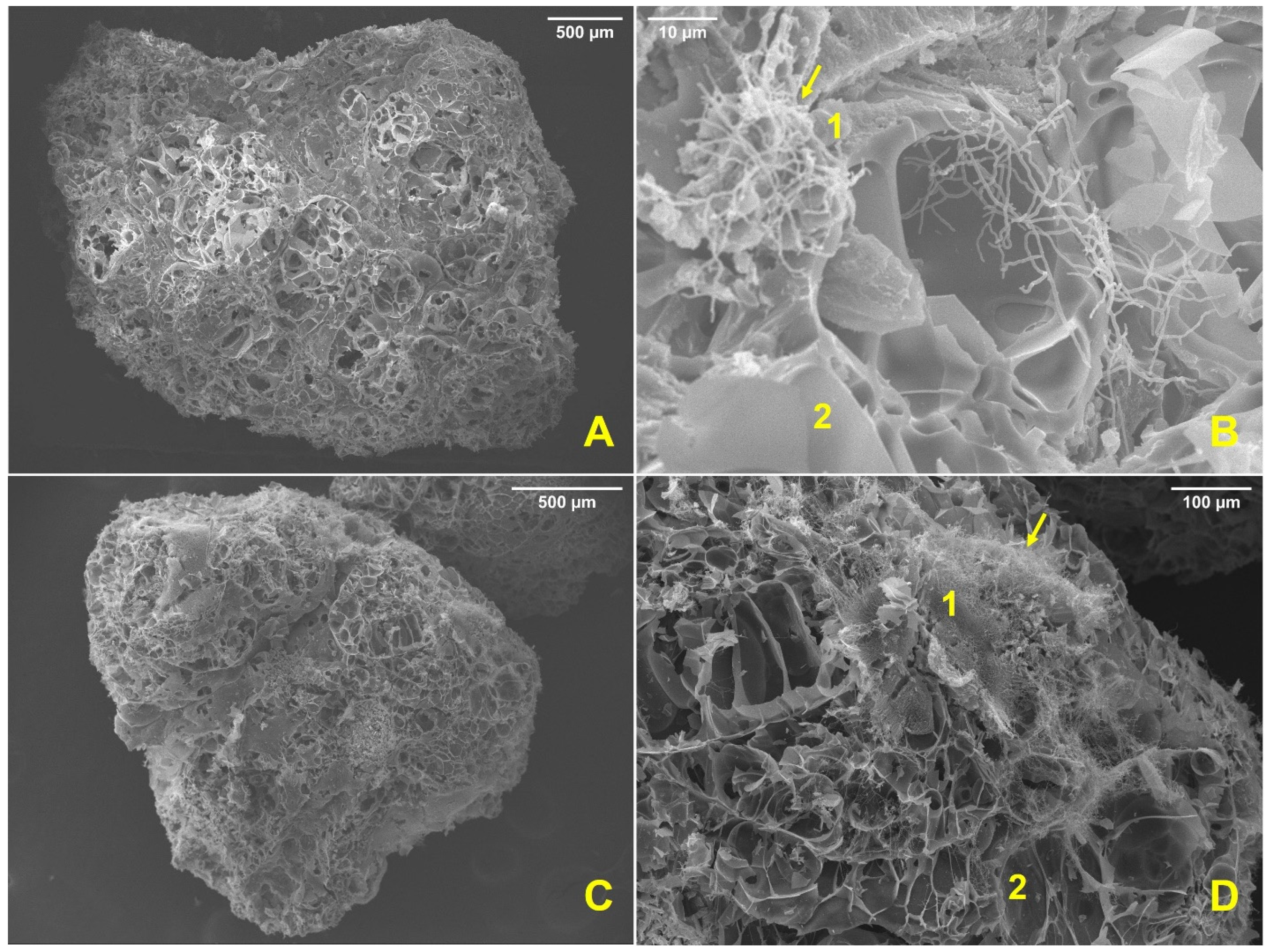
Actinobacteria were visually inspected to ensure they formed biofilms after incubation on perlite. When moving the tube containing perlite beads, it was observed that they adhered to each other through the biofilm and were no longer loose in the liquid medium. These characteristics provide the first evidence of biofilm formation on carriers. Next, random beads were removed from each tube to visualize them under a bright field microscope using a Gram stain, looking for the pellets and hyphae on the perlite crystals. Finally, high-vacuum SEM was used to visualize the biofilm inoculant samples at 18.0 kV (Figure 1) and to corroborate the biofilm formation by actinobacteria before the introduction of the bioinoculant in the pots. Biofilm formation was successful in all the treatments (Figure 1), verified by the viable count. Figure 1A–C shows how the perlite grain is entirely covered by the biofilm (indicated by number 1 in such micrographs), indicating that the biofilm induction process is successful under these experimental conditions. Furthermore, it was also possible to corroborate that the actinobacteria hyphae penetrated the carrier internally and externally in all bioinoculant samples, as exemplified by the strain ARI_A20-5 (Figure 1D). Only small areas on the outer surface of the entire perlite without biofilm coating were observed (indicated on the micrographs of Figure 1 with number 2).
Only nine actinobacteria genera exhibit the ability to produce biofilms, and even though actinobacteria are widely distributed, there are few studies about biofilm development in the phylum [25]. Alonso et al. performed the same CV methodology used in this study [12], testing the biofilm formation with a thermophilic actinobacteria Thermobifida fusca. In their research, the formation of biofilms with this bacterium was examined on microscopic glass slides, plastic coverslips, metal wires, Teflon seal tape, and cellulose surfaces. Cell growth was macroscopically visible as mucoid-attached agglomerates, unlike the planktonic mycelial pellets, which did not bind surfaces. They concluded that T. fusca could produce biofilm on nutritive surfaces such as chitin and cellulose, but on non-nutritive surfaces; it appeared to be molded by filamentous cells in an unconventional “spaghetti-like” form. [12]. Studying biofilm formation in actinobacteria is necessary, especially for the Streptomyces genus. The contributions of the components to matrix integrity are poorly understood at the molecular level, and extract and molecular interactions of extracellular matrix in the actinobacterial biofilm have not been thoroughly described [23,25,69]. However, it has been reported that the Streptomyces biofilm producers in a single species (mono-biofilm) preferred rough surfaces as a substrate for adhesion and were more suitable for forming biofilm than in a smooth surface [25], which is the case of the use of perlite beads in this study. Perlite contributes to the stability of the biofilm. It has been documented that some Streptomyces consortia biofertilizers occur when fungi serve as the biotic surface to which the bacteria attach [21]. In this work, the potential use of a novel biofilmed solid inoculant on perlite as a carrier using Streptomyces as mono-biofilm or as consortia species that were different from those reported previously [67,70], has been exposed as an improved biofilmed biofertilizer for uses in agriculture.
Moreover, to verify the difference with the non-formation of a biofilm on perlite by some actinobacteria reported as non-biofilm-forming in Table 3 (i.e., the ARI_A7-3 and ARI_A13-11 strains), such isolates were randomly induced on perlite and observed using SEM. There was no successful colonization of perlite beads by actinobacteria, leaving almost all the pores exposed (Figure 2A,C). A low actinobacteria population is observed on the perlite surface without covering it with a biofilm. Furthermore, a few bacteria were found at a higher magnification but without evidence of biofilm formation (indicated by number 1 in Figure 2B,D). Most of the outer surface of the perlite was exposed without biofilm formation (indicated by number 2 in the micrographs of Figure 2B,D). Thus, it was confirmed that the CV biofilm formation tests helped to have better orientation and discrimination for the correct selection of isolates that were and were not biofilm formers. It should be emphasized that the materials were different, though, because perlite is a mineral surface, while polypropylene was used for the CV tests. Thus, it cannot be ruled out that some isolates may have an affinity for perlite and have been previously reported to be unfavorable by CV tests. However, in this case, biofilm formation on perlite was consistent with the CV tests. To some Streptomyces species, the formation of the pellets is more common when bacteria have contact with smaller particles, which as a result, can become free and have large pellets with no attachment to any surface [25]. This was the case in this work with some isolates under study. Streptomyces biofilm development appears to be a regular aspect of these bacteria’s life cycle, albeit there exist little data on it [25]. It is also poorly understood why some of them are biofilm-forming species, but it seems to be related to the extracellular surface polymers involved in shaping Streptomyces pellets [25]. According to the results of this work, the nutrition conditions and the carrier could cause no biofilm formation. However, changing the vehicle for a substrate-rich carrier and modifying the growth-nutrition conditions could enhance biofilm formation using these strains.

3.4. Pot Essays

After 12 weeks, the plants (T. aestivum) were transported to the laboratory for corresponding measurements and observations per treatment. The parameters determined for wheat plants were the height of the plant (from the beginning of the stem to the tip of the longest edge of the spike), complete spike, base spike that constitutes the glumes without borders (which we call a single spike), total biomass in dry weight, and root biomass in dry weight. The controls were compared using one-way ANOVA and Tukey–Kramer and Dunnett comparisons of means with p < 0.05. The three control tests appeared to be linked with the same letter in all parameters, and no significant differences were found; thus, the differences in additives such as perlite and biochar did not interfere with the response variable. Therefore, any control is valid for comparison with the treatments.
Similarly, one-way ANOVA and Tukey–Kramer, and Dunnett comparisons of means with p < 0.05 were carried out using C0 for comparison between treatments. Figure 3 clearly shows the significant differences in height between treatments (linked with the letter a) compared to the controls (linked with the letter b). Levels not linked by the same letter are significantly different.
The differences between the control and different treatments with the biofilmed inoculants and their effects on T. aestivum are shown in Figure 3, where the levels not linked by the same letter are significantly different according to a one-way ANOVA analysis and a comparison of means with the Tukey–Kramer control. The evaluated measurements of height, complete spike, base spike, and total biomass were not significantly different between them, but with control, the variable root biomass T2, T1, and T6 were similar between them and different than T3, T4, T5, and T7, all of which are different from the control. This fact means that all treatments improve plant growth compared to plants without biofilmed inoculant treatment. The selected strains in this study showed outstanding results for T. aestivum.
Table 4 shows the percentage of plant growth promotion. The control was used as 100% plant growth without stimulation and was considered as the starting point to determine the percentage increase in plant biomass for later use in comparing the mean values between treatments with the biofilmed inoculant in T. aestivum.
All treatments showed evidence of plant growth promotion, considering the improvement in percentage, and the outstanding results were more evident in T1 and T2 (Figure 4). A 15–30% increase in height and 148% increase in total biomass with T. aestivum compared to the control using 108 spores of Streptomyces species has been reported [66,71]. This study observed a mean increase in the height between 140% and 200% in total biomass, with a root biomass increase of over 300% using Streptomyces flaveolus (ARI_A20-5). While using Streptomyces aureus T3 (ARI_A30-32), an increase over 220% was observed for total biomass; this strain also improved the following parameters: height and complete spike when combined with S. griseorubens T1 (ARI_A20-3) in T5 treatment, compared to the control. Both strains, S. flaveolus and S. aureus, showed significant activity as plant growth promoters even when these strains had only been used mostly for their antimicrobial and antifungal potential against human pathogens [69,72,73,74]. In addition, S. aureus has been used in soil bioremediation [75,76]; this strain was found to be an efficient siderophore producer in this study. Jog et al. (2014) improved the treatment of one Streptomyces species by using 106 cells/biofilm per pot. Highlighting results were reported even from using it as a consortium with another Streptomyces sp., as described by Panneerselvam et al. (2021). They used S. canus in a consortium applied in pomegranate fields. Moreover, S. griseorubens (ARI_A20-3) significantly improved root growth compared to the results reported by Boubekri et al. (2021) using the same planktonic microorganism in wheat. The results in this work are better than those reported by Jog et al. (2014) for treatments with one Streptomyces species, using 106 cells/biofilm per pot [67,68,77].
Even though it is well known that the low doses of the auxin IAA promote the growth of the primary root and induce lateral root development, results showed (Table 4) that ARI_A20-3 from T1 described as no IAA producer (Table 3) [50] showed a higher value in root biomass weight compared to ARI_A30-32 from T3 (described as IAA producer). This fact suggests that direct plant growth promotion can also occur by facilitating nutrients to the root through other well-known mechanisms, such as nitrogen fixation, iron acquisition by siderophores, and phosphate solubilization [13], which were reported in Table 3 as attributes for these strains, among others not reported in this work.
Microbial viability analysis was carried out during the 12 weeks of treatment at 0, 6, and 12 weeks. An exponential increase of 2 or 3 orders of magnitude was observed in all cases (Table 5). The actinobacteria population did not decrease; on the contrary, it multiplied. This behavior is evidence of positive interaction with the plant since most inoculants, reported elsewhere, that are not used as biofilmed inoculants decrease their viable counts during the treatment with the plant [78].
Viable microbial counts higher than those inoculated from the origin indicate successful colonization of the rhizosphere by microorganisms. For example, perlite has been used as a carrier for planktonic cells of Rhizobium leguminosarum bv. phaseoli, R. tropici, Bradyrhizobium japonicum, and Bacillus megaterium, reduced their population to 3 log in 32 days. In our study, the viability increased by 2–3 log using Streptomyces as a biofilm on perlite and keeping the population for 82 days, three times greater than that obtained by Daza et al. [78].
Powdered perlite with organic waste was used by Khabazi et al. as a carrier for B. japonicum to be used as a biofertilizer, achieving good results but indicating a decrease in viability after four weeks [36]. Albareda et al. presented similar results with perlite as the base of biofertilizer in compost and peat, which have been considered the best options for B. japonicum (an Alphaproteobacteria), reaching viability of 109 CFU/g for six months [35]. The viabilities reported by Albareda are quite good, considering that it is planktonic biofertilization. In our study, it was possible to reach similar population levels in a shorter period, considering the permanence of the spores in the perlite vehicle. Therefore, viability is expected to remain longer [79]. In this study, the biomass viability was higher than in other works using other carriers. This behavior could be attributed to the fact that when the biofilm was integrated into the perlite carrier, the hyphae were able to penetrate the material structure, taking advantage of its porosity (Figure 1D) and providing better protection to the remaining hyphae or spores against hostile conditions in the rhizosphere, such as pH, desiccation, predators, and UV radiation [34,56]. This behavior could be explained since perlite is an innocuous material with water retained inside its structure. To the best of our knowledge, no earlier studies using biofilms on this type of material utilized as an actinobacterial carrier have produced such results.
It has been demonstrated in earlier studies and publications that the use of biochar promotes the conservation of the viability of bacterial biomass in a range from 10% [80] to 30% [81], even if it was only included in the trials to witness the rhizosphere’s interactions, so it cannot be ruled out that it could be an aid in the viability of our tests. Among the bacteria that benefited from the increase in their population were the actinobacteria of the genera Mycolisibacteria, Streptosporangium, Actinomadura, Actinoallomurus, and Streptomyces [82].
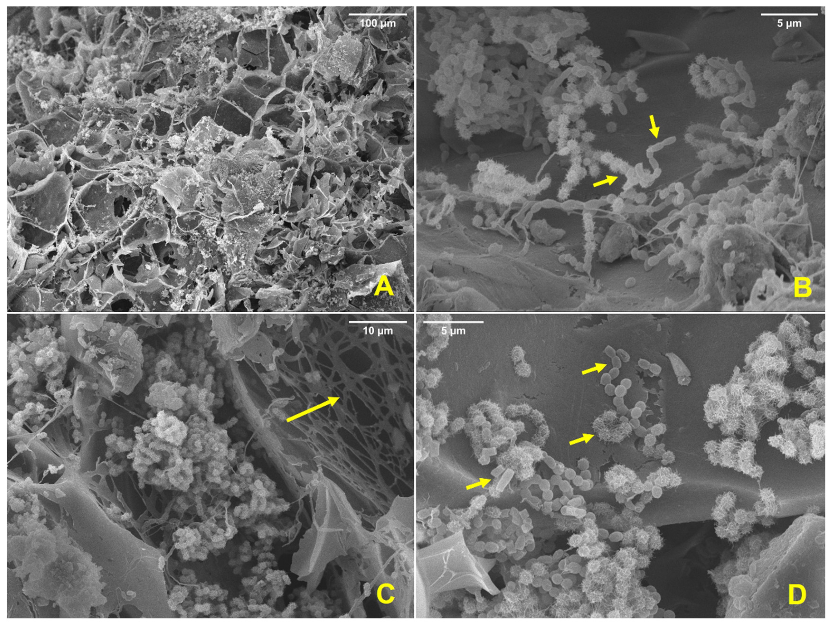
At the end of the pot assay, perlite from the biofilmed inoculant was recovered, fixed, and dehydrated according to the methods mentioned in the 3.2 subsection and analyzed by SEM. In all the cases, the mineral was not biofilm covered (Figure 5A), but in a close-up, it was possible to visualize vast amounts of spores between pores and surface. It was easy to distinguish the difference between spores belonging to the different strains from each treatment (Figure 5B,D), and it was possible to identify the residual components of the biofilm matrix and some spores still attached to it (Figure 5C). Spores are a resistant form of life, and they can remain in this phase for several years until the germination conditions are again suitable [65]. Therefore, their permanence in the rhizosphere between 9 and 12 years could be ensured using this type of material as a vehicle and by its conversion to spores, according to in vitro conservation studies carried out previously [79]. Although the actinobacteria were grown separately, this study provides evidence that a long-lasting reaction was carried out with the formation of a successful consortium between the different species of Streptomyces, as can be seen in the micrograph of Figure 5D, where the different types of spores corresponding to the different species (S. griseorubens, S. flaveolus, and S. aureus) can be clearly observed.
Post-treatment perlites were inoculated randomly in EM agar to ensure their viability and recovery of the original microorganism (Table 5).
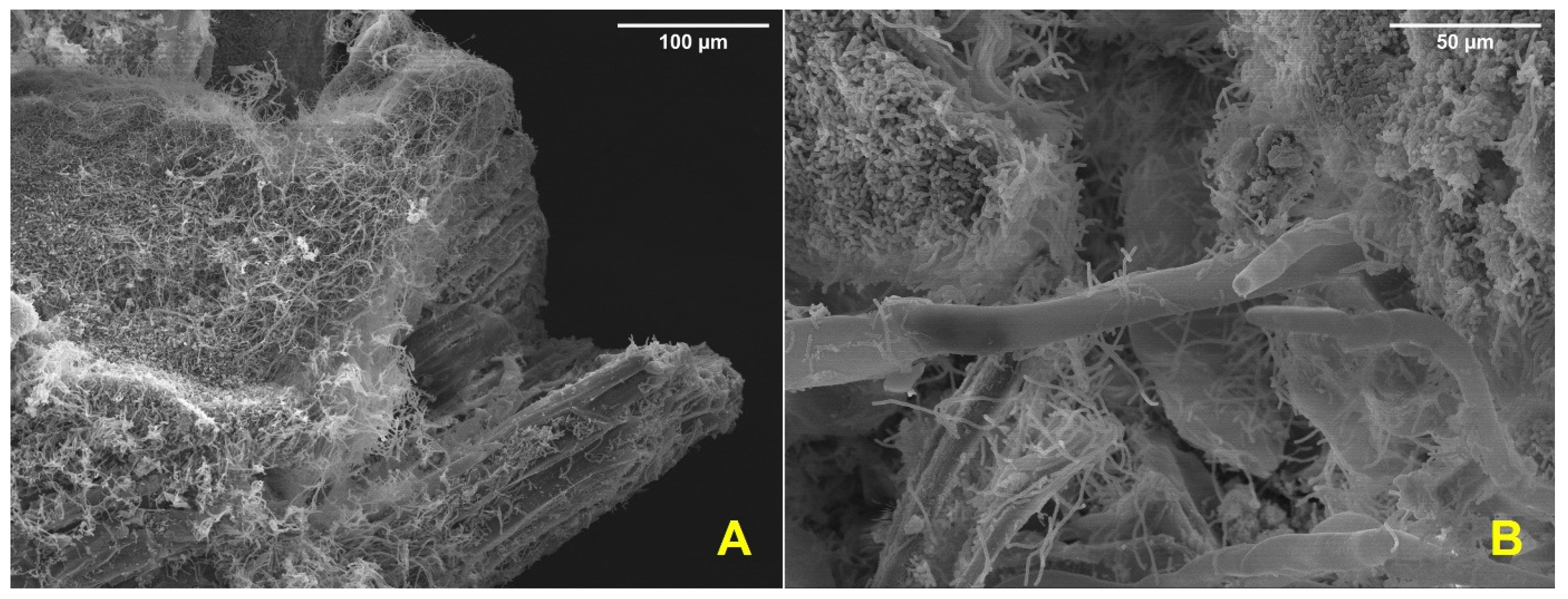
Previously sterilized to avoid superficial microorganisms, wheat roots were analyzed for endophytic colonization using the Coombs and Franco methodology [10,65]. The seven treatments showed positive results for endophytic colonization of the actinobacterial strains tested between 4 and 6 days. Root analysis using SEM was performed (Figure 6), and hyphae protruding from a cross-section of the root covered the surface (Figure 6A) and near the secondary roots (Figure 6B). The colonization of actinobacteria in healthy wheat tissue suggests that the host benefits in some way. Since Streptomyces can produce a wide variety of antibacterial, antifungal, and plant growth-regulating metabolites, the advantage in this situation might be a secondary metabolite produced by them. Not all associations are beneficial; for instance, Streptomyces scabies, S. acidiscabies, and S. turgidiscabies are potato scab-causing [65]. On the other hand, this group of strains can colonize the internal tissue of wheat due to the plant growth-promoting attributes shown in this work, enhancing nutrient uptake and producing secondary metabolites that can inhibit pathogens, which contributes to systemic resistance.
Although it has been shown that actinobacteria inhabit the rhizosphere of different plants in close association and that they can endophytically colonize the roots and internal tissues of healthy plants, this endophytic colonization by actinobacteria is not common (because there are usually pathogenic interactions); however, it has been reported in some species of the genus Streptomyces, Micromonospora, Nocardioides, Nocardia, Streptosporangium, and Frankia [10], acting as the pathogen defense, such as in the case of phytopathogenic fungi, where it has been demonstrated that actinobacteria diminish or eliminate the pathogen’s viability and the ranges of toxin emission in the rhizosphere [83], conferring induced resistance to plants and regulating and improving the growth of their hosts [65].

4. Conclusions

The actinobacterial strains tested in this study showed an outstanding improvement in long-term microbial survival due to their use as a biofilmed inoculant on a perlite mineral as a carrier compared to the general formulations that use planktonic cultures. Furthermore, the plant growth efficiency of Triticum aestivium was significantly improved when using the biofilmed inoculant under greenhouse conditions. The use of actinobacteria as a biofilmed solid inoculant is related to the success of beneficial plant-microorganism interaction shown by the endophytic root colonization. Furthermore, it ensures its permanence in the rhizosphere for a long time by keeping the spores protected in the perlite mineral. In the present study, the use of PGP actinobacteria as a biofilmed biofertilizer using perlite mineral as carrier is proposed as an alternative to improve crop production over a long period and to ensure plant health protection due to its multiple benefits.

Supplementary Materials

The following supporting information can be downloaded at: https://www.mdpi.com/article/10.3390/app122211425/s1, Table S1: Eroded soil analysis for pot essays.

Author Contributions

Conceptualization, K.G.D.-G., R.C.-M. and H.E.M.-F.; methodology, K.G.D.-G., J.J.R.-M., O.H.-C. and J.J.V.-A.; software, K.G.D.-G., J.J.R.-M. and O.H.-C.; validation, K.G.D.-G., R.C.-M. and J.F.C.-C.; formal analysis, K.G.D.-G., M.G.G.-R., J.F.C.-C. and H.E.M.-F.; investigation, K.G.D.-G. and R.C.-M.; resources, R.C.-M., H.E.M.-F. and J.J.V.-A.; data curation, K.G.D.-G., J.J.R.-M., O.H.-C. and J.J.V.-A.; writing—original draft preparation, K.G.D.-G. and R.C.-M.; writing—review and editing, R.C.-M., J.F.C.-C., J.J.V.-A. and H.E.M.-F.; supervision, R.C.-M., H.E.M.-F., M.G.G.-R. and J.J.V.-A.; funding acquisition, R.C.-M., J.J.V.-A., J.F.C.-C. and H.E.M.-F. All authors have read and agreed to the published version of the manuscript.

Funding

This research was funded by Coordinación de la Investigación Científica-UMSNH, grant number CIC-UMSNH-2022, and PRODEP funded the APC.

Institutional Review Board Statement

Not applicable.

Informed Consent Statement

Not applicable.

Data Availability Statement

Data is contained within the article.

Acknowledgments

We appreciate the technical support provided by Itzi L. Ortiz-Godinez and Dafne S. Rivas-Méndez from QFB-UMSNH.

Conflicts of Interest

The authors declare no conflict of interest. The funders had no role in the design of the study; in the collection, analyses, or interpretation of data; in the writing of the manuscript, or in the decision to publish the results.

References

  1. FAO. The State of Food Security and Nutrition in the World 2021: Transforming Food Systems for Food Security, Improved Nutrition and Affordable Healthy Diets for All; The State of Food Security and Nutrition in the World (SOFI); FAO: Rome, Italy, 2021; ISBN 978-92-5-134325-8. [Google Scholar]
  2. Carvalho, F.P. Agriculture, Pesticides, Food Security and Food Safety. Environ. Sci. Policy 2006, 9, 685–692. [Google Scholar] [CrossRef]
  3. Javaid, A.; Bajwa, R. Field Evaluation of Effective Microorganisms (EM) Application for Growth, Nodulation, and Nutrition of Mung Bean. Turk. J. Agric. For. 2011, 35, 443–452. [Google Scholar] [CrossRef]
  4. Sharf, W.; Javaid, A.; Shoaib, A.; Khan, I.H. Induction of Resistance in Chili against Sclerotium Rolfsii by Plant-Growth-Promoting Rhizobacteria and Anagallis Arvensis. Egypt. J. Biol. Pest Control 2021, 31, 16. [Google Scholar] [CrossRef]
  5. Franco-Correa, M.; Quintana, A.; Duque, C.; Suarez, C.; Rodríguez, M.X.; Barea, J.-M. Evaluation of Actinomycete Strains for Key Traits Related with Plant Growth Promotion and Mycorrhiza Helping Activities. Appl. Soil Ecol. 2010, 45, 209–217. [Google Scholar] [CrossRef]
  6. Gupta, G.; Snehi, S.K.; Singh, V. Role of PGPR in Biofilm Formations and Its Importance in Plant Health. In Biofilms in Plant and Soil Health; Ahmad, I., Husain, F.M., Eds.; John Wiley & Sons, Ltd: Chichester, UK, 2017; pp. 27–42. ISBN 978-1-119-24632-9. [Google Scholar]
  7. Rana, K.L.; Kour, D.; Yadav, A.N.; Yadav, N.; Saxena, A.K. Chapter 16—Agriculturally Important Microbial Biofilms: Biodiversity, Ecological Significances, and Biotechnological Applications. In New and Future Developments in Microbial Biotechnology and Bioengineering: Microbial Biofilms; Yadav, M.K., Singh, B.P., Eds.; Elsevier: Amsterdam, The Netherlands, 2020; pp. 221–265. ISBN 978-0-444-64279-0. [Google Scholar]
  8. Yadav, A.N.; Singh, J.; Rastegari, A.A.; Yadav, N. Plant Microbiomes for Sustainable Agriculture; Springer Nature: Berlin/Heidelberg, Germany, 2020; ISBN 978-3-030-38453-1. [Google Scholar]
  9. Zakeel, M.C.M.; Safeena, M.I.S. Biofilmed Biofertilizer for Sustainable Agriculture. In Plant Health Under Biotic Stress: Volume 2: Microbial Interactions; Ansari, R.A., Mahmood, I., Eds.; Springer: Singapore, 2019; pp. 65–82. ISBN 9789811360404. [Google Scholar]
  10. Govindasamy, V.; Franco, C.M.M.; Gupta, V.V.S.R. Endophytic Actinobacteria: Diversity and Ecology. In Advances in Endophytic Research; Verma, V.C., Gange, A.C., Eds.; Springer India: New Delhi, India, 2014; pp. 27–59. ISBN 978-81-322-1575-2. [Google Scholar]
  11. Solanki, M.K.; Malviya, M.K.; Wang, Z. Actinomycetes Bio-Inoculants: A Modern Prospectus for Plant Disease Management. In Plant Growth Promoting Actinobacteria: A New Avenue for Enhancing the Productivity and Soil Fertility of Grain Legumes; Subramaniam, G., Arumugam, S., Rajendran, V., Eds.; Springer: Singapore, 2016; pp. 63–81. ISBN 978-981-10-0707-1. [Google Scholar]
  12. Alonso, A.N.; Pomposiello, P.J.; Leschine, S.B. Biofilm Formation in the Life Cycle of the Cellulolytic Actinomycete Thermobifida fusca. Biofilms 2008, 5, 1–11. [Google Scholar] [CrossRef]
  13. Bhatti, A.A.; Haq, S.; Bhat, R.A. Actinomycetes Benefaction Role in Soil and Plant Health. Microbial. Pathogen. 2017, 111, 458–467. [Google Scholar] [CrossRef]
  14. Amaresan, N.; Kumar, K.; Naik, J.H.; Bapatla, K.G.; Mishra, R.K. Chapter 8—Streptomyces in Plant Growth Promotion: Mechanisms and Role. In New and Future Developments in Microbial Biotechnology and Bioengineering; Singh, B.P., Gupta, V.K., Passari, A.K., Eds.; Elsevier: Amsterdam, The Netherlands, 2018; pp. 125–135. ISBN 978-0-444-63994-3. [Google Scholar]
  15. Prasanna, R.; Triveni, S.; Bidyarani, N.; Babu, S.; Yadav, K.; Adak, A.; Khetarpal, S.; Pal, M.; Shivay, Y.S.; Saxena, A.K. Evaluating the Efficacy of Cyanobacterial Formulations and Biofilmed Inoculants for Leguminous Crops. Arch. Agron. Soil Sci. 2014, 60, 349–366. [Google Scholar] [CrossRef]
  16. Sharma, M.; Dangi, P.; Choudhary, M. Actinomycetes: Source, Identification, and Their Applications. Int. J. Curr. Microbiol. Appl. Sci. 2014, 3, 801–832. [Google Scholar]
  17. Imbert, M.; Béchet, M.; Blondeau, R. Comparison of the Main Siderophores Produced by Some Species of Streptomyces. Curr. Microbiol. 1995, 31, 129–133. [Google Scholar] [CrossRef]
  18. Lasudee, K.; Tokuyama, S.; Lumyong, S.; Pathom-aree, W. Actinobacteria Associated with Arbuscular Mycorrhizal Funneliformis Mosseae Spores, Taxonomic Characterization and Their Beneficial Traits to Plants: Evidence Obtained from Mung Bean (Vigna radiata) and Thai Jasmine Rice (Oryza sativa). Front. Microbiol. 2018, 9, 1247. [Google Scholar] [CrossRef] [Green Version]
  19. Palaniyandi, S.A.; Yang, S.H.; Zhang, L.; Suh, J.-W. Effects of Actinobacteria on Plant Disease Suppression and Growth Promotion. Appl. Microbiol. Biotechnol. 2013, 97, 9621–9636. [Google Scholar] [CrossRef]
  20. Costerton, J.W.; Geesey, G.G.; Cheng, K.-J. How Bacteria Stick. Sci. Am. 1978, 238, 86–95. [Google Scholar] [CrossRef]
  21. Seneviratne, G.; Kecskés, M.; Kennedy, I. Biofilmed Biofertilisers: Novel Inoculants for Efficient Nutrient Use in Plants. ACIAR Proc. 2008, 130, 126–130. [Google Scholar]
  22. Bhattacharya, D.C. Biofilmed Biofertilizer: Promising Technology for Tomorrow. JAM 2015, 4, 19–22. [Google Scholar]
  23. Flemming, H.-C.; Wingender, J. The Biofilm Matrix. Nat. Rev. Microbiol. 2010, 8, 623–633. [Google Scholar] [CrossRef]
  24. Buddhika, U.; Athauda, A.; Seneviratne, G.; Kulasooriya, S.; Abayasekara, C. Emergence of Diverse Microbes on Application of Biofilmed Biofertilizers to a Maize Growing Soil. Ceylon J. Sci. (Biol. Sci.) 2014, 42, 87. [Google Scholar] [CrossRef] [Green Version]
  25. El Othmany, R.; Zahir, H.; Ellouali, M.; Latrache, H. Current Understanding on Adhesion and Biofilm Development in Actinobacteria. Int. J. Microbiol. 2021, 2021, 6637438. [Google Scholar] [CrossRef]
  26. Seneviratne, G.; Jayasinghearachchi, H.S. A Rhizobial Biofilm with Nitrogenase Activity Alters Nutrient Availability in a Soil. Soil Biol. Biochem. 2005, 37, 1975–1978. [Google Scholar] [CrossRef]
  27. Adak, A.; Prasanna, R.; Babu, S.; Bidyarani, N.; Verma, S.; Pal, M.; Shivay, Y.S.; Nain, L. Micronutrient Enrichment Mediated by Plant-Microbe Interactions and Rice Cultivation Practices. J. Plant Nutr. 2016, 39, 1216–1232. [Google Scholar] [CrossRef]
  28. Hettiarachchi, R.P.; Seneviratne, G.; Jayakody, A.N.; De Silva, E.; Gunatilake, P.; Edirimanna, V.; Thewarapperuma, A.; Chandrasiri, J.A.S.; Malawaraarachchi, G.C.; Siriwardana, N.S. Effect of Biofilmed Biofertilizer on Plant Growth and Nutrient Uptake of Hevea Brasiliensis Nursery Plants at Field Condition. J. Rubber Res. Inst. Sri Lanka 2018, 98, 16. [Google Scholar] [CrossRef]
  29. Seneviratne, G.; Weerasekara, M.L.M.A.W.; Seneviratne, K.A.C.N.; Zavahir, J.S.; Kecskés, M.L.; Kennedy, I.R. Importance of Biofilm Formation in Plant Growth Promoting Rhizobacterial Action. In Plant Growth and Health Promoting Bacteria; Microbiology Monographs; Maheshwari, D.K., Ed.; Springer Berlin: Heidelberg/Berlin, Germany, 2010; Volume 18, pp. 81–95. ISBN 978-3-642-13611-5. [Google Scholar]
  30. Seneviratne, G.; Zavahir, J.S.; Bandara, W.M.M.S.; Weerasekara, M.L.M.A.W. Fungal-Bacterial Biofilms: Their Development for Novel Biotechnological Applications. World J. Microbiol. Biotechnol. 2007, 24, 739. [Google Scholar] [CrossRef]
  31. Seneviratne, G.; Jayasinghearachchi, H.S. Mycelial Colonization by Bradyrhizobia and Azorhizobia. J. Biosci. 2003, 28, 243–247. [Google Scholar] [CrossRef] [PubMed]
  32. Tennakoon, P.L.K.; Rajapaksha, R.M.C.P.; Hettiarachchi, L.S.K. Tea Yield Maintained in PGPR Inoculated Field Plants despite Significant Reduction in Fertilizer Application. Rhizosphere 2019, 10, 100146. [Google Scholar] [CrossRef]
  33. Ajeng, A.A.; Abdullah, R.; Ling, T.C.; Ismail, S.; Lau, B.F.; Ong, H.C.; Chew, K.W.; Show, P.L.; Chang, J.-S. Bioformulation of Biochar as a Potential Inoculant Carrier for Sustainable Agriculture. Environ. Technol. Innov. 2020, 20, 101168. [Google Scholar] [CrossRef]
  34. Malusá, E.; Sas-Paszt, L.; Ciesielska, J. Technologies for Beneficial Microorganisms Inocula Used as Biofertilizers. Available online: https://www.hindawi.com/journals/tswj/2012/491206/ (accessed on 28 October 2020).
  35. Albareda, M.; Rodríguez-Navarro, D.N.; Camacho, M.; Temprano, F.J. Alternatives to Peat as a Carrier for Rhizobia Inoculants: Solid and Liquid Formulations. Soil Biol. Biochem. 2008, 40, 2771–2779. [Google Scholar] [CrossRef]
  36. Khavazi, K.; Rejali, F.; Seguin, P.; Miransari, M. Effects of Carrier, Sterilisation Method, and Incubation on Survival of Bradyrhizobium japonicum in Soybean (Glycine max L.) Inoculants. Enzyme Microb. Technol. 2007, 41, 780–784. [Google Scholar] [CrossRef]
  37. Sun, D.; Hale, L.; Crowley, D. Nutrient Supplementation of Pinewood Biochar for Use as a Bacterial Inoculum Carrier. Biol. Fertil. Soils 2016, 52, 515–522. [Google Scholar] [CrossRef]
  38. Kelly, K.L.; Judd, D.B.; Inter-Society Color Council; United States National Bureau of Standards. ISCC-NBS Color-Name Charts Illustrated with Centroid Colors; U.S. National Bureau of Standards: Washington, DC, USA, 1965.
  39. Nonomura, H. Studies on Isolation, Taxonomy and Ecology of Soil Actinomycetes (in Japanese with English Abstract). Actinomycetologica 1989, 3, 45–54. [Google Scholar] [CrossRef]
  40. Goodfellow, M.; Ferguson, E.V.; Sanglier, J.-J. Numerical Classification and Identification of Streptomyces Species—A Review. Gene 1992, 115, 225–233. [Google Scholar] [CrossRef]
  41. Weisburg, W.G.; Barns, S.M.; Pelletier, D.A.; Lane, D.J. 16S Ribosomal DNA Amplification for Phylogenetic Study. J. Bacteriol. 1991, 173, 697–703. [Google Scholar] [CrossRef] [Green Version]
  42. DeLong, E.F. Archaea in Coastal Marine Environments. Proc. Natl. Acad. Sci. USA 1992, 89, 5685–5689. [Google Scholar] [CrossRef] [Green Version]
  43. Nautiyal, C.S. An Efficient Microbiological Growth Medium for Screening Phosphate Solubilizing Microorganisms. FEMS Microbiol. Lett. 1999, 170, 265–270. [Google Scholar] [CrossRef]
  44. Adesanwo, O.O.; Ige, D.V.; Thibault, L.; Flaten, D.; Akinremi, W. Comparison of Colorimetric and ICP Methods of Phosphorus Determination in Soil Extracts. Commun. Soil Sci. Plant Anal. 2013, 44, 3061–3075. [Google Scholar] [CrossRef]
  45. Bowman, R.A. A Rapid Method to Determine Total Phosphorus in Soils. Soil Sci. Soc. Am. J. 1988, 52, 1301–1304. [Google Scholar] [CrossRef]
  46. Fiske, C.H.; Subbarow, Y. The colorimetric determination of phosphorus. J. Biol. Chem. 1925, 66, 375–400. [Google Scholar] [CrossRef]
  47. Olsen, S.R. Sommers 1982 Phosphorus. Methods Soil Anal. 1982, 2, 403–430. [Google Scholar]
  48. Sahu, M.K.; Sivakumar, K.; Thangaradjou, T.; Kannan, L. Phosphate Solubilizing Actinomycetes in the Estuarine Environment: An Inventory. J. Environ. Biol. 2007, 28, 795. [Google Scholar]
  49. Glickmann, E.; Dessaux, Y. A Critical Examination of the Specificity of the Salkowski Reagent for Indolic Compounds Produced by Phytopathogenic Bacteria. Appl. Environ. Microbiol. 1995, 61, 793–796. [Google Scholar] [CrossRef] [Green Version]
  50. Patten, C.L.; Glick, B.R. Role of Pseudomonas Putida Indoleacetic Acid in Development of the Host Plant Root System. Appl. Environ. Microbiol. 2002, 68, 3795–3801. [Google Scholar] [CrossRef] [Green Version]
  51. Balagurunathan, R.; Radhakrishnan, M.; Shanmugasundaram, T.; Gopikrishnan, V.; Jerrine, J. Characterization and Identification of Actinobacteria. In Protocols in Actinobacterial Research; Springer Protocols Handbooks; Balagurunathan, R., Radhakrishnan, M., Shanmugasundaram, T., Gopikrishnan, V., Jerrine, J., Eds.; Springer US: New York, NY, USA, 2020; pp. 39–64. ISBN 978-1-07-160728-2. [Google Scholar]
  52. Valdés, M.; Pérez, N.-O.; los Santos, P.E.; Caballero-Mellado, J.; Peña-Cabriales, J.J.; Normand, P.; Hirsch, A.M. Non-Frankia Actinomycetes Isolated from Surface-Sterilized Roots of Casuarina Equisetifolia Fix Nitrogen. Appl. Environ. Microbiol. 2005, 71, 460–466. [Google Scholar] [CrossRef] [Green Version]
  53. Simon, E.H.; Tessman, I. Thymidine-Requiring Mutants of Phage T4. Proc. Natl. Acad. Sci. USA 1963, 50, 526–532. [Google Scholar] [CrossRef] [PubMed] [Green Version]
  54. Costa, J.M.; Loper, J.E. Characterization of Siderophore Production by the Biological Control Agent Enterobacter cloacae. MPMI—Mol. Plant Microbe Interact. 1994, 7, 440–448. [Google Scholar] [CrossRef]
  55. Baakza, A.; Vala, A.K.; Dave, B.P.; Dube, H.C. A Comparative Study of Siderophore Production by Fungi from Marine and Terrestrial Habitats. J. Exp. Mar. Biol. Ecol. 2004, 311, 1–9. [Google Scholar] [CrossRef]
  56. Anderson, G.G.; O’Toole, G.A. Innate and Induced Resistance Mechanisms of Bacterial Biofilms. In Bacterial Biofilms; Romeo, T., Ed.; Springer: Berlin/Heidelberg, Germany, 2008; pp. 85–105. ISBN 978-3-540-75418-3. [Google Scholar]
  57. Gavín, R.; Merino, S.; Altarriba, M.; Canals, R.; Shaw, J.G.; Tomás, J.M. Lateral Flagella Are Required for Increased Cell Adherence, Invasion and Biofilm Formation by Aeromonas spp. FEMS Microbiol. Lett. 2003, 224, 77–83. [Google Scholar] [CrossRef] [Green Version]
  58. Oriani, A.S.; Gentili, A.R.; Zuñiga, A.E.; Oriani, D.S.; Baldini, M. Composición lipídica de la pared celular de tres especies de micobacterias ambientales y su posible correlación con la formación de biofilms y movilidad por sliding/Lipid composition of cell wall of three species of environmental mycobacteria and its po. Cienc. Vet. 2017, 17, 47–59. [Google Scholar] [CrossRef]
  59. O’Toole, G.; Kaplan, H.B.; Kolter, R. Biofilm Formation as Microbial Development. Ann. Rev. Microbiol. 2000, 54, 49–79. [Google Scholar] [CrossRef]
  60. Freeman, D.J.; Falkiner, F.R.; Keane, C.T. New Method for Detecting Slime Production by Coagulase Negative Staphylococci. J. Clin. Pathol. 1989, 42, 872–874. [Google Scholar] [CrossRef] [Green Version]
  61. Kaiser, T.D.L.; Pereira, E.M.; dos Santos, K.R.N.; Maciel, E.L.N.; Schuenck, R.P.; Nunes, A.P.F. Modification of the Congo Red Agar Method to Detect Biofilm Production by Staphylococcus epidermidis. Diagn. Microbiol. Infect. Dis. 2013, 75, 235–239. [Google Scholar] [CrossRef] [Green Version]
  62. Martínez, A.; Torello, S.; Kolter, R. Sliding Motility in Mycobacteria. J. Bacteriol. 1999, 181, 7331–7338. [Google Scholar] [CrossRef] [Green Version]
  63. Sousa, S.; Bandeira, M.; Carvalho, P.A.; Duarte, A.; Jordao, L. Nontuberculous Mycobacteria Pathogenesis and Biofilm Assembly. Int. J. Mycobacteriol. 2015, 4, 36–43. [Google Scholar] [CrossRef] [Green Version]
  64. Janssen, P.H.; Yates, P.S.; Grinton, B.E.; Taylor, P.M.; Sait, M. Improved Culturability of Soil Bacteria and Isolation in Pure Culture of Novel Members of the Divisions Acidobacteria, Actinobacteria, Proteobacteria, and Verrucomicrobia. Appl. Environ. Microbiol. 2002, 68, 2391–2396. [Google Scholar] [CrossRef] [Green Version]
  65. Coombs, J.T.; Franco, C.M.M. Isolation and Identification of Actinobacteria from Surface-Sterilized Wheat Roots. Appl. Environ. Microbiol. 2003, 69, 5603–5608. [Google Scholar] [CrossRef] [Green Version]
  66. Srinivas, V.; Gopalakrishnan, S.; Kamidi, J.P.; Chander, G. Effect of Plant Growth-Promoting Streptomyces sp. on Plant Growth and Yield of Tomato and Chilli. Andhra Pradesh J. Agril. Sci. 2020, 6, 65–70. [Google Scholar]
  67. Panneerselvam, P.; Selvakumar, G.; Ganeshamurthy, A.N.; Mitra, D.; Senapati, A. Enhancing Pomegranate (Punica granatum L.) Plant Health through the Intervention of a Streptomyces Consortium. Biocontrol Sci. Technol. 2021, 31, 430–442. [Google Scholar] [CrossRef]
  68. Boubekri, K.; Soumare, A.; Mardad, I.; Lyamlouli, K.; Hafidi, M.; Ouhdouch, Y.; Kouisni, L. The Screening of Potassium- and Phosphate-Solubilizing Actinobacteria and the Assessment of Their Ability to Promote Wheat Growth Parameters. Microorganisms 2021, 9, 470. [Google Scholar] [CrossRef]
  69. Hu, Y.; Qi, Y.; Stumpf, S.D.; D’Alessandro, J.M.; Blodgett, J.A.V. Bioinformatic and Functional Evaluation of Actinobacterial Piperazate Metabolism. ACS Chem. Biol. 2019, 14, 696–703. [Google Scholar] [CrossRef]
  70. Alekhya, G.; Gopalakrishnan, S. Biological Control and Plant Growth-Promotion Traits of Streptomyces Species Under Greenhouse and Field Conditions in Chickpea. Agric. Res. 2017, 6, 410–420. [Google Scholar] [CrossRef] [Green Version]
  71. Ferrer, C.M.; Olivete, E.; Orias, S.L.; Rocas, M.R.; Juan, S.; Dungca, J.Z.; Mahboob, T.; Barusrux, S.; Nissapatorn, V. A Review on Streptomyces spp. as Plant-Growth Promoting Bacteria (PGPB). Asian J. Pharmacogn. 2018, 2, 32–40. [Google Scholar]
  72. Naorungrote, S.; Chunglok, W.; Lertcanawanichakul, M.; Bangrak, P. Actinomycetes Producing Anti-Methicillin Resistant Staphylococcus aureus from Soil Samples in Nakhon Si Thammarat. Walailak J. Sci. Technol. (WJST) 2011, 8, 131–138. [Google Scholar]
  73. Bologa, C.G.; Ursu, O.; Oprea, T.I.; Melançon, C.E.; Tegos, G.P. Emerging Trends in the Discovery of Natural Product Antibacterials. Curr. Opin. Pharmacol. 2013, 13, 678–687. [Google Scholar] [CrossRef] [Green Version]
  74. Wang, W.; Feng, M.; Li, X.; Chen, F.; Zhang, Z.; Yang, W.; Shao, C.; Tao, L.; Zhang, Y. Antibacterial Activity of Aureonuclemycin Produced by Streptomyces aureus Strain SPRI-371. Molecules 2022, 27, 5041. [Google Scholar] [CrossRef] [PubMed]
  75. Chen, S.; Lai, K.; Li, Y.; Hu, M.; Zhang, Y.; Zeng, Y. Biodegradation of Deltamethrin and Its Hydrolysis Product 3-Phenoxybenzaldehyde by a Newly Isolated Streptomyces aureus Strain HP-S-01. Appl. Microbiol. Biotechnol. 2011, 90, 1471–1483. [Google Scholar] [CrossRef] [PubMed]
  76. Chen, S.; Luo, J.; Hu, M.; Lai, K.; Geng, P.; Huang, H. Enhancement of Cypermethrin Degradation by a Coculture of Bacillus cereus ZH-3 and Streptomyces aureus HP-S-01. Biores. Technol. 2012, 110, 97–104. [Google Scholar] [CrossRef] [PubMed]
  77. Jog, R.; Pandya, M.; Nareshkumar, G.; Rajkumar, S. Mechanism of Phosphate Solubilization and Antifungal Activity of Streptomyces spp. Isolated from Wheat Roots and Rhizosphere and Their Application in Improving Plant Growth. Microbiology 2014, 160, 778–788. [Google Scholar] [CrossRef] [PubMed] [Green Version]
  78. Daza, A.; Santamaría, C.; Rodríguez-Navarro, D.N.; Camacho, M.; Orive, R.; Temprano, F. Perlite as a Carrier for Bacterial Inoculants. Soil Biol. Biochem. 2000, 32, 567–572. [Google Scholar] [CrossRef]
  79. Filippova, S.N.; Surgucheva, N.A.; Gal’chenko, V.F. Long-Term Storage of Collection Cultures of Actinobacteria. Microbiology 2012, 81, 630–637. [Google Scholar] [CrossRef]
  80. Jeffery, S.; Verheijen, F.G.A.; van der Velde, M.; Bastos, A.C. A Quantitative Review of the Effects of Biochar Application to Soils on Crop Productivity Using Meta-Analysis. Agric. Ecosyst. Environ. 2011, 1, 175–187. [Google Scholar] [CrossRef]
  81. Biederman, L.A.; Harpole, W.S. Biochar and Its Effects on Plant Productivity and Nutrient Cycling: A Meta-Analysis. GCB Bioenergy 2013, 5, 202–214. [Google Scholar] [CrossRef]
  82. Anderson, C.R.; Condron, L.M.; Clough, T.J.; Fiers, M.; Stewart, A.; Hill, R.A.; Sherlock, R.R. Biochar Induced Soil Microbial Community Change: Implications for Biogeochemical Cycling of Carbon, Nitrogen and Phosphorus. Pedobiol.—J. Soil Ecol. 2011, 5–6, 309–320. [Google Scholar] [CrossRef]
  83. Zucchi, T.D.; De Moraes, L.A.B.; De Melo, I.S. Streptomyces sp. ASBV-1 Reduces Aflatoxin Accumulation by Aspergillus parasiticus in Peanut Grains. J. Appl. Microbiol. 2008, 105, 2153–2160. [Google Scholar] [CrossRef]
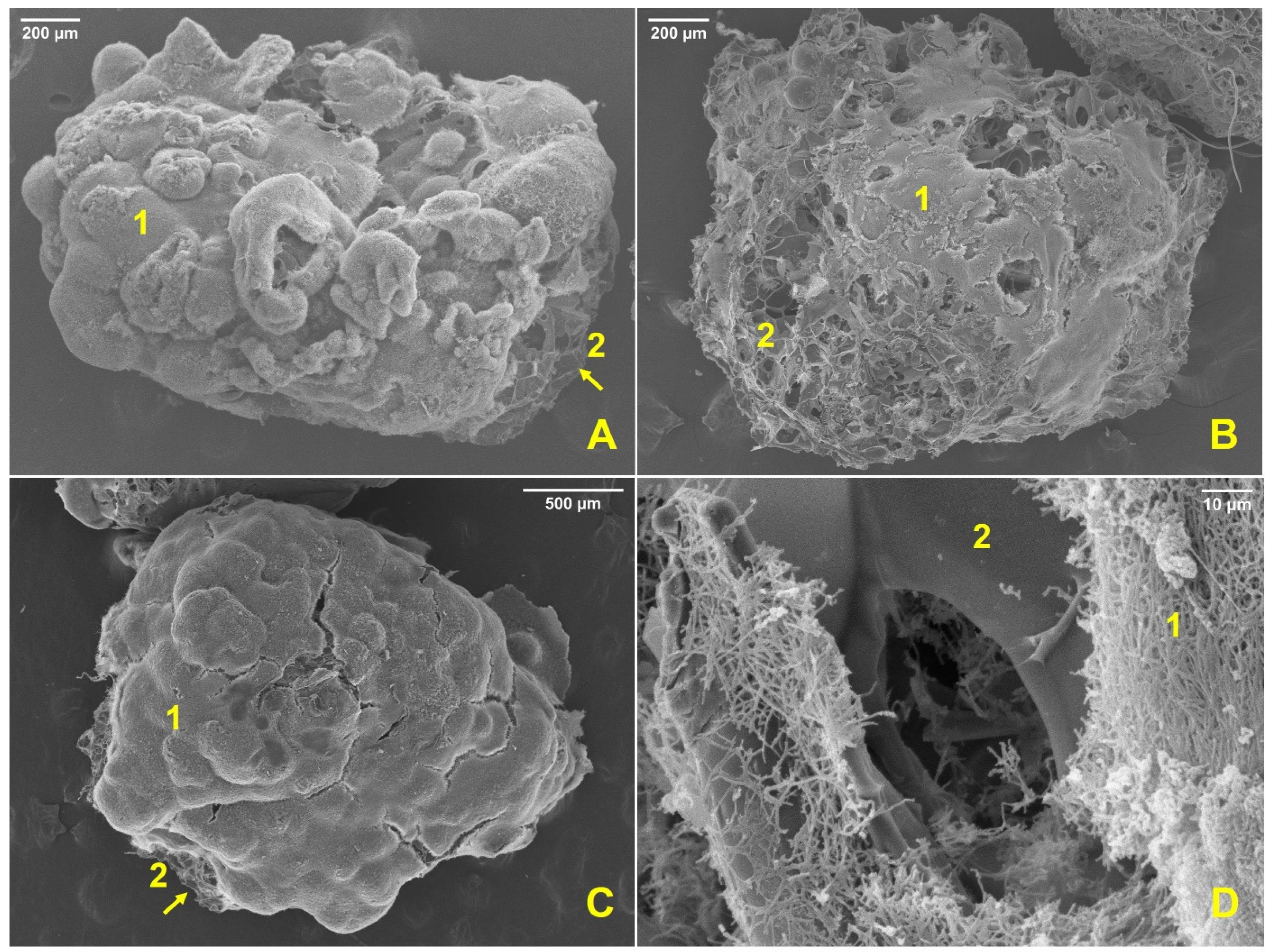
Figure 1. Scanning electron microscopy of biofilm formation on perlite: (A) strain ARI_A20-3 (60×); (B) strain ARI_A20-5 (60×); (C) strain ARI_A30-32 (40×); (D) strain ARI_A20-5 (1000×).
Applsci 12 11425 g001
Figure 2. Scanning electron microscopy of unsuccessful biofilm formation on perlite: (A) strain A7-3 (30×); (B) strain A7-3 (1400×); (C) strain A13-11 (45×); (D) strain A13-11 (160×).
Applsci 12 11425 g002
Figure 3. Effects in plant height with the biofilmed inoculant per treatments in T. aestivum. (C0, and C2 letters refer to control experiments; letters from T1 to T7 refer to experimental treat-ments; a, and b letters indicate similarities between experimental treatments).
Applsci 12 11425 g003
Figure 4. Effects in plant growth with the biofilmed inoculant per treatments in T. aestivum: (A) height (cm); (B) complete spike (cm); (C) base spike (cm); (D) total biomass (g); (E) root biomass (cm).
Applsci 12 11425 g004
Figure 5. Scanning electron microscopy of biofilm on perlite post-pot essay: (A) treatment 4 (180×); (B) treatment 4 (3500×), two different spores of ARI_A20-3 and ARI_A20-5 (yellow arrows); (C) treatment 7 (1800×), residual biofilm (yellow arrow); (D) treatment 7 (3700×), the three different spores of ARI_A20-3, ARI_A20-5 and ARI_A30-32 strains (yellow arrows).
Applsci 12 11425 g005
Figure 6. Scanning electron microscopy of endophytic actinobacterial colonization: (A) treatment 2, wheat root (250×); (B) treatment 5, wheat root (500×).
Applsci 12 11425 g006
Table 1. Biofilmed inoculant pot essay treatments.
TreatmentsFormulation
C0Control, Sterile soil
C1Control, Sterile soil + biochar
C2Control, Sterile soil + perlite + biochar
T1A20-3 + biochar
T2A20-5 + biochar
T3A30-32 + biochar
T4A20-3, A20-5 combination + biochar
T5A20-3, A30-32 combination + biochar
T6A20-5, A30-32 combination + biochar
T7A20-3, A20-5, A30-32 combination + biochar
Table 2. Identification of the actinobacterial isolates based on partial sequencing of the 1500-pb region of the 16S rDNA gene.
StrainBLAST Analysis a% SimilarityAccession Number
ARI_A20-3Streptomyces griseorubens98.16KP718552.1
ARI_A20-5Streptomyces flaveolus99.7KU647253.1
ARI_A30-32Streptomyces aureus99.56EU841615.1
a Nearest match.
Table 3. Results of the PGP attributes of Avocado Rhizosphere Isolates.
Strain ARIBiofilm Formation 1NFB Growth 2Aerial Mycelium Diazotrophic StructuresSiderophore ProductionAIA Production Phosphate Solubilizer ActivityStrain ARIBiofilm Formation 1NFB Growth 2Aerial Mycelium Diazotrophic StructuresSiderophore ProductionAIA Production Phosphate Solubilizer Activity
A1-7+a+++++++A11-30+b+--+-+
A2-2+a+--++++A11-36-+++++++
A2-5-++++-++A11-37-+++++-++
A4-9-++++++-A13-11-+++++-++
A4-11+a++-++-A15-11----+++
A4-15----+--A16-3-+++++++++
A4-18-+++++++A16-4-+++++++
A4-26-++++++++A16-7-+++++-++
A4-31-++-++++A16-52+a++++++++
A4-33+a++++-+A18-14+a+++++-++
A4-49-++++-+A18-15-+++++++++
A6-12+a++++++A20-1+a+++++++++
A7-3-+++++++A20-2+a+++++-+
A7-13----++++A20-3+a+++++-+
A7-17-+++++++A20-5+a++++++++
A7-22-+++++++-A20-6-++-++++
A8-4+b+--++-A20-13+a-+-+++++
A10-3-++-+++A30-7-++-++++
A10-13+b++++-++A30-14-+++++++++
A11-10-+++++-+A30-32+a+++++++++
A11-29-+++++++
1 The mean values followed by the same letter for each experimental variable are not significantly different at p = 0.05 compared to the positive control in the crystal violet essay at 30 °C in EMA media. 2 ++, excellent growth. + good growth. The growth capacity in the NFB medium is related to the ability to fix nitrogen in association with the presence of diazotrophic structures and aerial mycelium.
Table 4. Percentage of PGP and Tukey–Kramer and Dunnett comparisons between treatments in T. aestivum.
TreatmentHeight (%)Complete Spike (%)Base Spike (%)Total Biomass (%)Root Biomass (%)
T1142.96 a,*154.22 a,*174.83 a,*215.93 a,*282.36 a,b,*
T2138.64 a153.35 a,*164.90 a,*214.83 a,*334.71 a,*
T3137.82 a138.77 a161.58 a224.31 a,*246.14 b
T4140.22 a,*143.44 a156.95 a188.92 a228.25 b
T5141.54 a,*146.06 a,*155.62 a186.04 a236.97 b
T6139.29 a141.98 a162.91 a,*212.47 a262.35 a,b,*
T7125.50 a141.39 a159.60 a181.16 a232.22 b
C0100.00 b100.00 b100.00 b100.00 b100.00 c
* corresponding to the first three outstanding positions in Tukey–Kramer and Dunnett comparisons of means with a p < 0.05. a, b, and c letters indicate similarities between treatments.
Table 5. Population dynamics of actinobacteria strains inoculated by treatments using T. aestivum (Ta) (three replicates per treatment).
TreatmentWeekCFU/g Rizospheric SoilTreatmentWeekCFU/g Rizospheric Soil
T1 (Ta)03.0 × 10⁶ dT5 (Ta)03.5 × 10⁶ d
66.5 × 10⁷ c61.6 × 108 b
126.7 × 10⁸ b121.4 × 109 a
T2 (Ta)04.6 × 10⁶ dT6 (Ta)03.0 × 10⁶ d
67.0 × 10⁶ d61.2 × 107 c
123.3 × 10⁸ b121.1 × 109 a
T3 (Ta)02.5 × 10⁶ dT7 (Ta)03.5 × 10⁶ d
61.6 × 10⁸ b61.6 × 108 b
121.3 × 10⁹ a126.7 × 108 b
T4 (Ta)02.0 × 10⁶ dC000
65.4 × 10⁶ d00
122.3 × 10⁸ b00
The mean values followed by the same letter for each experimental variable are not significantly different at p = 0.05. a, b, and c letters indicate similarities between treatments.
Publisher’s Note: MDPI stays neutral with regard to jurisdictional claims in published maps and institutional affiliations.