Featured Application
This manuscript performs an electromyographic analysis of two exercises for conditioning the upper muscle extremities. Thus, a greater knowledge for prescribing these exercises in strengthening or functional recovery programs will be obtained.
Abstract
Pullover and straight arm pulldown exercises are commonly used in resistance exercise programs to improve sports performance or in physical activity health programs. This study aimed to evaluate the individual electromyographic (EMG) activity of the pectoralis major (clavicular, sternal, and costal portions), latissimus dorsi, anterior deltoid, triceps brachii, and rectus abdominis muscles in a barbell pullover exercise at a 100% biacromial width and a straight arm pulldown exercise at a 100% and 150% biacromial width and to compare the EMG activity in these selected muscles and exercises. Twenty healthy and physically active adults performed a set of eight repetitions of each exercise against 30% of their body mass. The barbell pullover exercise presented a higher EMG activity (p ≤ 0.01) than the straight arm pulldown exercise in both biacromial widths in all evaluated muscles except for the latissimus dorsi and the triceps brachii. These muscles showed the highest EMG activity in the straight arm pulldown exercise at both biacromial widths. In all of the exercises and muscles evaluated, the concentric phase showed a greater EMG activity than the eccentric phase. In conclusion, the barbell pullover exercise can highlight muscle activity in the pectoralis major (mainly in the sternal and lower portions), triceps brachii, and rectus abdominis muscles. However, the straight arm pulldown exercise at 100% and 150% biacromial widths could be a better exercise to stimulate the latissimus dorsi and triceps brachii muscles. Moreover, all exercises showed significantly greater EMG activity (p < 0.001) in the concentric phase than in the eccentric phase for all the evaluated muscles.
1. Introduction
Upper limb strength training is essential in sports performance [1,2] and in improving autonomy for the activities of daily living [3,4].
Specifically, pullover exercises are commonly used in resistance exercise programs to improve sports performance, such as in swimming [5], skiing [6,7], handball [7,8], volleyball [9], and ice sledge hockey [10], or for improving health in sedentary people [11]. Additionally, the straight arm pulldown is another frequently performed upper body strengthening exercise [12,13]. In this sense, depending on the training objectives, choosing the most appropriate exercise to stimulate the desired musculature is necessary. This measurement of muscle activity has been extensively evaluated by electromyography [14,15,16].
Thus, some studies have attempted to evaluate the electromyographic (EMG) activity in both pullover and pulldown exercises [17,18,19,20]. Marchetti and Uchida [20] evaluated the EMG activity of the pectoralis major and latissimus dorsi muscles during pullover exercises. These authors reported that barbell pullover exercises produced a greater EMG activity in the pectoralis major than in the latissimus dorsi. However, these authors did not compare the pullover exercise with any other exercise. In this regard, Campos and da Silva [19] compared the EMG activity of the pectoralis major, triceps brachii, anterior deltoid, and latissimus dorsi muscles between the barbell pullover and horizontal bench press exercises. These authors found a higher EMG activity for the triceps brachii and latissimus dorsi muscles in the pullover exercise than in the horizontal bench press. However, the EMG activity for the pectoralis major and anterior deltoid muscles was higher for the bench press exercise. Similar results were found by Rabelo Mota et al. [17]. These authors observed that there were no significant differences in the EMG activity of the pectoralis major between the pullover and horizontal bench press exercises, but there was a greater EMG activity with the anterior deltoid in the bench press when the exercises were performed until concentric failure.
However, it is necessary to highlight that there are many differences in shoulder movement between these exercises [21]. For instance, the horizontal bench press involves the flexion and extension of the elbow and adduction of the shoulder in the horizontal plane, whereas the barbell pullover involves shoulder flexion and extension while maintaining a slight elbow angle. Therefore, it would be interesting to compare the EMG activity of the pullover exercise with another exercise involving a similar upper limb movement, for instance, the pulldown exercise. In this sense, Teixeira et al. [18] compared the EMG activity and peak force between the pullover and pulldown exercises at different shoulder joint positions during the maximum isometric contraction. These authors reported that the pectoralis major and latissimus dorsi muscles showed similar maximal EMG activity in both pullover and pulldown exercises, with higher values between 90° and 135° for the pectorals major and between 0° and 45° for the latissimus dorsi.
Most studies, such as those cited above, have focused on evaluating the EMG activity of the upper extremity musculature. However, no studies have assessed the EMG activity of the abdomen. In the literature, there are different proposed exercises, called pullover passes, oriented toward conditioning the abdominal musculature [22]. In addition, it is known that in exercises such as crunches, when the arms are placed above the head, the EMG activity of the anterior rectus abdominis muscle significantly increases [23]. This position would be similar to that adopted at the end of the eccentric phase and the beginning of the concentric phase in the pullover and pulldown exercises. However, to date, we are not aware of any study that has analyzed the EMG activity of this abdominal muscle in pullover and pulldown exercises.
Therefore, the purpose of this study was to evaluate the EMG activity of the pectoralis major (clavicular -upper-, sternal -middle-, and costal -lower- portion), latissimus dorsi, anterior deltoid, triceps brachii, and rectus abdominis muscles in the barbell pullover at a 100% biacromial width, and straight arm pulldown exercises at a 100% and 150% biacromial width; this study also aims to (1) analyze which exercise generates the highest EMG activity in the selected muscles; (2) compare the EMG activity between the concentric and eccentric phases; and (3) determine the EMG activity of each muscle for each exercise.
2. Materials and Methods
2.1. Participants
Twenty physically active and healthy adults voluntarily participated in this study. Table 1 shows the descriptive characteristics of the participants. To participate in this study, the inclusion criteria were as follows: (1) at least 1 year of resistance training experience with a minimum frequency of twice a week; and (2) no musculoskeletal injuries or physical limitations during the 12 months prior to assessment. The participants were requested not to take stimulants or perform vigorous exercise during the 24 h period prior to the study. If any participant did not comply with any of the instructions above, they were removed from the study sample. This study was approved by the Bioethical Committee of the University of Almería, according to the Declaration of Helsinki. Before the beginning of the measurements, all participants were informed about the study protocol and signed an informed consent form.

Table 1.
General characteristics of the participants.
2.2. Procedures
The procedure began with the determination of the body mass of each participant using an electronic weighing scale (Tanita, BF-350, Tokyo, Japan), their height with a stadiometer (Seca, Hamburg, Germany), and their biacromial distance (Table 1). Prior to the test, each participant completed a 10 min warm-up aerobic activity on an elliptical machine. Then, they performed joint mobility exercises and dynamic stretching exercises of the body segments involved in the pullover and pulldown exercises [24,25]. Afterward, as a specific warm-up, participants performed fifteen repetitions of the pullover exercise using two dumbbells of 5 kg each. These repetitions served to make the participants feel confident and comfortable with the exercise and to ensure that the researchers were satisfied with the technique performed.
Next, the skin was cleaned with cotton and 96% alcohol. According to the manufacturer’s specifications, bipolar Ag/AgCl disposable electrodes (Medico Lead-Lok, Noida, India) were attached in parallel to the muscle fibers. The distance between the electrodes was two centimeters. Moreover, a reference electrode was separated as much as possible from the electrode pair. The placement of all the electrodes followed the surface electromyography for the noninvasive assessment of muscles (SENIAM) recommendations [26] on the dominant side of each participant. In particular, the electrodes were positioned as such with: the clavicular head of the pectoralis major (PMUP), between the first and second rib [27]; the sternal head of the pectoralis major (PMMP) horizontal to the muscle mass of the chest wall (approximately two centimeters from the axillary crease) [27,28]; the costal head of the pectoralis major (PMLP) at the middle clavicular line between the fifth and sixth rib [27]; the latissimus dorsi (LD) at four centimeters under the lower tip of the scapula and half of the distance between the spinal column and the lateral edge of the torso, with an oblique angle of ~25° [29]; the anterior deltoid (AD) at 1.5 cm from the distal end and at the anterior part of the acromion process [30]; the triceps brachii (TB) medial head at the middle point between the posterior part of the acromion and the olecranon protuberance [31]; and the rectus abdominis (RA) placed 3 cm lateral to the middle line and at a middle distance between the xiphoid process and the umbilicus [32]. All the electrodes were covered with adhesive tape to avoid the possibility of the electrodes moving during the execution of the exercises.
Then, in each muscle, the maximum voluntary isometric contraction (MVIC) was recorded to subsequently normalize the electromyographic signal for each exercise and in the grip amplitude. To this end, the MVICs were performed twice, for three seconds each time, with ten seconds of rest between each contraction and two minutes of rest between each MVIC assessment [25]. In particular, the MVIC maneuvers were conducted as follows: for the pectoralis major (clavicular, sternal, and costal portions), a standing posture was assumed, with the shoulders and elbows flexed at 90° (in the horizontal plane), participants had to bring their elbows to the middle line of the body against manual resistance performed in the opposite direction. For the latissimus dorsi, in a standing posture, with the shoulders and elbows flexed to 90° (in the horizontal plane), participants had to perform a scapular-humeral adduction, leading the humerus to the trunk against manual resistance performed in the opposite direction. For the anterior deltoid, in a seated position with an erect posture and no back support, participants had to perform a deltoid flexion at 90° against manual resistance performed in the opposite direction. For the triceps brachii, in a seated position with an erect posture and no back support, participants had to perform a forearm extension with the elbows at 90° against manual resistance performed in the opposite direction. For the rectus abdominis, the participants were positioned in a supine posture with the soles of the feet resting on the floor; they were required to perform a resisted curl-up exercise against manual resistance performed in the opposite direction. All the muscles were randomly tested to avoid fatigue. To keep consistent efforts while the MVIC maneuvers were performed, a tester verbally encouraged each participant.
Later, the participants performed a more specific warm-up that consisted of 20 repetitions, with a relative load of 10% of their body weight, according to the exercise to be evaluated. Finally, after 5 min of rest, the electromyographic activity recording began during the barbell pullover and straight arm pulldown exercises. The barbell pullover started with the participant lying in a supine position on a bench, with their feet on the floor, holding a barbell with a pronated grip, and with their upper limbs perpendicular to their body. Then, with the participant lying down, through a shoulder flexion and keeping the elbows slightly flexed, the arms had to be brought to the earlobe (at the end of the eccentric phase). Subsequently, through shoulder extensions, the arms had to return to the initial position (at the end of the concentric phase) (Figure 1a). In this pullover exercise, the grip distance was determined by the biacromial width of each participant (100% biacromial width) [19]. The straight arm pulldown exercise was performed in a standing posture with the trunk in a vertical position by gripping a bar that was connected by a cable to a pulley [18] (Figure 1b). At the beginning of the exercise, the participants, keeping their elbows extended, had to lower the bar until it reached the level of the navel (at the end of the concentric phase). Subsequently, in a controlled manner, the bar had to be raised to the height of the earlobe (at the end of the eccentric phase). The straight arm pulldown exercise was performed in two situations: (1) with a grip distance of 100% of the biacromial width and (2) with a grip distance of 150% of the biacromial width. Participants performed a set of 8 repetitions of each exercise against 30% of their body mass [20] at a rhythm of 2 s for the eccentric phase and 2 s for the concentric phase. A total of 4 s was used for the movement of 1 repetition [19,25]. The repetition velocity was measured by a metronome (KORG MA-1, Keio Electronic Laboratories, Tokyo, Japan).
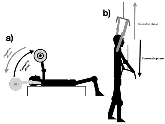
Figure 1.
Phases of the pullover (a) and straight arm pulldown exercises (b).
2.3. Electromyography
To record the electromyographic signals of each muscle, a WBA Mega device (Mega Electronics, Ltd., Kuopio, Finland) was used with a sampling of 1000 Hz. Then, a digital signal was filtered by bandwidth (12–450 Hz) using a fourth-order Butterworth filter with the LabView software program (National Instruments, Austin, TX, USA). The root mean square (RMS) signals in microvolts (µV) were used for further analysis using the MEGAWIN software program (Mega Electronics, Ltd.). Of the eight repetitions recorded, only six were analyzed; the first and last repetitions were discarded to eliminate movement variability due to the initiation and termination of the exercise [24].
2.4. Statistical Analysis
Before the statistical analysis and comparisons among the dependent variables, the normality and homogeneity of the variances were confirmed with the Shapiro–Wilk, and Brown–Forsythe tests. As all variables followed a normal distribution, parametric tests were performed. A 3 × 7 ANOVA design (exercise*muscle) was applied to measure differences in the EMG activity (% MVIC) among the exercises and among the muscles in each exercise. A 3 × 7 × 2 ANOVA design (exercise*muscle*contraction type) was performed to assess differences in the EMG activity according to the different contraction types (concentric and eccentric) in each exercise. Pairwise comparisons were performed using a Bonferroni adjustment in order to observe if a significant main effect was observed. The effect sizes (ES) were calculated by partial eta-squared (ηp2).
To compare the EMG values, expressed in microvolts (µV), during the concentric and eccentric phases, Student’s t-test was performed for the paired samples. In this case, the effect size was calculated through Cohen’s d using the combined standard deviation formula [33].
The IBM SPSS software (v. 28) was used for statistical analyses, and the significance level was set at p < 0.05.
3. Results
The ANOVA showed a significant main effect of exercise on EMG activity (F(4.03,76.65) = 26.06, p < 0.001; ηp2 = 0.58). The barbell pullover exercise, at a 100% biacromial width, presented a statistically higher EMG activity (p ≤ 0.01) than the straight arm pulldown exercises at 100% and 150% biacromial widths in all muscles except for the latissimus dorsi, which reported a significantly lower EMG activity (p ≤ 0.001), and the triceps brachii, which did not show any significant differences among the exercises (p > 0.05) (Figure 2).
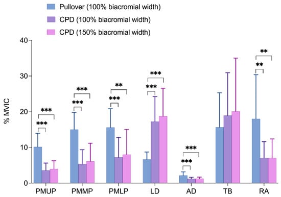
Figure 2.
Comparison of the EMG activity among the evaluated exercises: pullover and straight arm pulldown (expressed in %MVIC). PMUP—pectoralis major upper portion; PMMP—pectoralis major middle portion; PMLP—pectoralis major lower portion; LD—latissimus dorsi; AD—anterior deltoid; TB—triceps brachii; RA—rectus abdominis. ** p < 0.01; *** p < 0.001.
Subsection
Table 2, Table 3 and Table 4 compare the EMG activity between the concentric and eccentric phases in each exercise (the barbell pullover at a 100% biacromial width; and the straight arm pulldown exercise at 100% and 150% biacromial widths). The ANOVAs showed the statistically significant main effects for exercise (F(1.16,22.02) = 4.33, p = 0.044; ηp2 = 0.18), muscle (F(2.01,38.32) = 103.72, p < 0.001; ηp2 = 0.84), and contraction phase (F(1,19) = 111.78, p < 0.001; ηp2 = 0.85), as well as for the exercise*muscle (F(2.41,1.32) = 10.71, p < 0.001; ηp2 = 0.36), exercise*contraction phase (F(1.32,25.08) = 7.80, p = 0.006; ηp2 = 0.29), muscle*contraction phase (F(2.91,55.41) = 48.93, p < 0.001; ηp2 = 0.72), and exercise*muscle*contraction phases (F(3.35,63.78) = 14.12, p < 0.001; ηp2 = 0.42). All the exercises showed a significantly greater EMG activity (p < 0.001) in the concentric phase than in the eccentric phase for all the evaluated muscles (Table 2, Table 3 and Table 4).
Table 2.
Comparison of EMG activity (expressed in µV) during the concentric and eccentric phases in the pullover exercise. Mean ± standard deviation (SD).
Table 3.
Comparison of EMG activity (expressed in µV) during the concentric and eccentric phases in the straight arm pulldown exercise at 100% biacromial width. Mean ± standard deviation (SD).
Table 4.
Comparison of EMG activity (expressed in µV) during the concentric and eccentric phases in the straight arm pulldown exercise at 150% biacromial width. Mean ± standard deviation (SD).
When comparing the EMG activity of each muscle in each exercise, the ANOVA showed that in the barbell pullover exercise, at a 100% biacromial width, the PMMP, PMLP, TB, and RA showed the highest EMG activity. In the straight arm pulldown exercises at 100% and 150% biacromial widths, the greatest EMG activity appeared in the LD and TB muscles. The AD showed the lowest EMG activity in the three evaluated exercises (Figure 3).
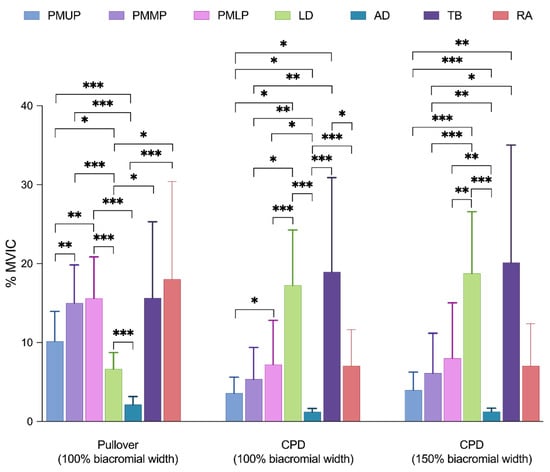
Figure 3.
Mean EMG activity and standard deviation normalized to the maximal voluntary isometric contraction (MVIC) in the barbell pullover at 100% biacromial width and straight arm pulldown exercises at 100% and 150% biacromial widths. * p < 0.05; ** p < 0.01; *** p < 0.001.
4. Discussion
The strength training of the upper limbs is essential, both for sports and for the activities of daily life. In this regard, pullovers and straight-arm pulldowns are two frequently used exercises. Additionally, straight-arm pulldown exercises are sometimes performed with different grip distances. However, to date, little scientific evidence has analyzed the muscular activity of these exercises and their variants with a bar grip distance. Therefore, the main purpose of the current study was to evaluate the EMG activity of the pectoralis major (upper, middle, and lower portion), latissimus dorsi, anterior deltoid, triceps brachii, and rectus abdominis muscles during barbell pullover exercises at a 100% biacromial width and straight-arm pulldown exercises at 100% and 150% biacromial widths.
The current study found that the barbell pullover exercise, at a 100% biacromial width, presented statistically higher EMG activity than the straight arm pulldown exercise at 100% and 150% biacromial widths in all the evaluated muscles except for the latissimus dorsi and the triceps brachii. Recently, Teixeira et al. [18] reported that the pectoralis major and the latissimus dorsi muscles showed similar maximal EMG activity in both pullover and pulldown exercises. However, the differences concerning our results could be because these authors recorded the maximum voluntary isometric contractions at different angles and compared them between exercises. By contrast, in our study, the EMG activity was evaluated dynamically at 30% of the participant’s body mass in both exercises. Similarly, our findings are consistent with those of Marchetti and Uchida [20], who reported that the pectoralis major presented a higher activation than the latissimus dorsi during all movement cycles of a pullover exercise. However, these authors only analyzed and compared the EMG activity of the pectoralis major in the sternal portion versus the latissimus dorsi. Our results show that in addition to the EMG activity of the sternal portion of the pectoralis major, the EMG activity of the upper and lower portions and the EMG activity of the triceps brachii and rectus abdominis were also significantly higher than that of the latissimus dorsi.
Furthermore, it was observed that in the pullover exercise and in the straight arm pulldown at 100% and 150% biacromial widths, the EMG activity was significantly greater in the concentric phase than in the eccentric phase in all the evaluated muscles. These results are in agreement with previous studies [20,27,34].
A novel finding of the present study, which has not been reported to date, is that in the pullover exercise, the rectus abdominis presented the highest EMG activity (≈18% MVIC) of all the muscles evaluated, although without statistically significant differences compared with the triceps brachii or pectoralis major (in its three portions). The EMG activity of the rectus abdominis could be due to its stabilizing function of the lumbar spine in both the concentric and eccentric phases of the pullover exercise. In the eccentric phase, as the barbell descends, the arm resistance increases [35], and with it, there is an increase in lumbar lordosis. In this case, the rectus abdominis would be activated to fix and stabilize the lumbar curvature to maintain the pelvis in retroversion [36]. In the concentric phase, during the lifting of the barbell, the rectus abdominis acts as an auxiliary muscle, performing a light flexion of the trunk (shrinkage) to overcome the generated torque [23,37]. However, in the straight arm pulldown at 100% and 150% biacromial widths, as it was performed with the participant in a standing position pulling the load perpendicular to the position of the trunk, the arm resistance was lower, and therefore, the EMG activity of the rectus abdominis was lower. In this case, the muscles with the greatest EMG activity in the straight arm pulldown exercise were the triceps brachii (≈19% MVIC), to maintain the elbow join in extension [35], and the latissimus dorsi (≈18% MVIC), to perform the shoulder extension with an internal rotation of the humerus [38].
This study had some limitations, including the load intensity that we used to evaluate the EMG activity (30% of the body mass). This load was chosen to individualize and standardize, as much as possible, the load lifted by each participant. This load was also chosen to ensure correct execution by the participants, avoiding possible injuries to the glenohumeral joint [20,39]. Furthermore, this load has been used in previous studies which analyzed EMG activity in the pullover exercise and is therefore considered safe and effective [20]. Future studies could evaluate the EMG activity at different percentages with a 1 repetition maximum (1 RM) to compare whether different loads affect the EMG activity in these exercises. Another limitation is the velocity of the movement, which was performed for 2 s in the concentric phase and 2 s in the eccentric phase. This velocity was chosen to obtain a clearer EMG signal and greater movement control, as has been proposed by previous studies [20,24,25,34]. Further research is required at several velocities of these exercises to determine the influence on EMG activity [40]. A final limitation is that the pullover exercise was only evaluated at 100% of the biacromial width and not 150%. This is because participants indicated that they were uncomfortable with their performance at the widest width in this exercise. For the safety of the participants, it was decided to only evaluate the width shown in the study (100% of the biacromial width).
5. Conclusions
In conclusion, the current findings show that the barbell pullover exercise emphasizes the muscle activity of the pectoralis major (mainly in the sternal and lower portions), the triceps brachii, and the rectus abdominis. However, the straight-arm pulldown exercise at 100% and 150% biacromial widths could be a better exercise to stimulate the latissimus dorsi and triceps brachii muscles. Furthermore, in the straight-arm pulldown exercise, the width of the grip did not significantly influence the EMG activity of the tested musculature. Thus, in both exercises, there was significantly higher EMG activity in the concentric phase than in the eccentric phase.
Author Contributions
Conceptualization, J.M.M. and P.A.L.-M.; methodology, J.M.M. and P.A.L.-M.; validation, J.M.M., P.A.L.-M. and F.A.; formal analysis, J.M.M. and P.A.L.-M.; investigation, J.M.M. and P.A.L.-M.; resources, J.M.M.; data curation, J.M.M. and P.A.L.-M.; writing—original draft preparation, J.M.M.; writing—review and editing, J.M.M., P.A.L.-M. and F.A.; visualization, J.M.M., P.A.L.-M. and F.A.; supervision, J.M.M., P.A.L.-M. and F.A.; funding acquisition, J.M.M. All authors have read and agreed to the published version of the manuscript.
Funding
This research was funded by the University of Almeria Mobility Grants (Reference: EST2022/028).
Institutional Review Board Statement
The study was conducted in accordance with the Declaration of Helsinki and was approved by the Institutional Review Board of the University of Almería.
Informed Consent Statement
Informed consent was obtained from all subjects involved in the study.
Data Availability Statement
Not applicable.
Conflicts of Interest
The authors declare no conflict of interest.
References
- Langer, K.; Simon, C.; Wiemeyer, J. Strength Training in Climbing: A Systematic Review. J. Strength Cond. Res. 2022. [Google Scholar] [CrossRef]
- Zhang, J. Influence of Progressive Upper Limb Strength Training on Table Tennis Athletes. Rev. Bras. Med. Do Esporte 2022, 28, 734–737. [Google Scholar] [CrossRef]
- Kim, M.; Kuruma, H.; Thawisuk, C. Effectiveness of Elongation Band Exercise on the Upper Limb Strength and Range of Motion among Older Adults. J. Exerc. Rehabil. 2022, 18, 110–116. [Google Scholar] [CrossRef] [PubMed]
- da Silva, P.B.; Antunes, F.N.; Graef, P.; Cechetti, F.; de Souza Pagnussat, A. Strength Training Associated with Task-Oriented Training to Enhance Upper-Limb Motor Function in Elderly Patients with Mild Impairment after Stroke. Am. J. Phys. Med. Rehabil. 2015, 94, 11–19. [Google Scholar] [CrossRef]
- Belfry, G.R.; Noble, E.G.; Taylor, A.W. Effects of Two Different Weight Training Programs on Swimming Performance and Muscle Enzyme Activities and Fiber Type. J. Strength Cond. Res. 2016, 30, 305–310. [Google Scholar] [CrossRef] [PubMed]
- Rud, B.; Øygard, E.; Dahl, E.B.; Paulsen, G.; Losnegard, T. The Effect of Resistance Exercise Priming in the Morning on Afternoon Sprint Cross-Country Skiing Performance. Int. J. Sports Physiol. Perform. 2021, 16, 1786–1793. [Google Scholar] [CrossRef]
- Hermassi, S.; Laudner, K.; Schwesig, R. The Effects of Circuit Strength Training on the Development of Physical Fitness and Performance-Related Variables in Handball Players. J. Hum. Kinet. 2020, 71, 191–203. [Google Scholar] [CrossRef]
- Hermassi, S.; Wollny, R.; Schwesig, R.; Shephard, R.J.; Chelly, M.S. Effects of In-Season Circuit Training on Physical Abilities in Male Handball Players. J. Strength Cond. Res. 2019, 33, 944–957. [Google Scholar] [CrossRef]
- Valadés Cerrato, D.; Palao, J.M.; Femia, P.; Ureña, A. Effect of Eight Weeks of Upper-Body Plyometric Training during the Competitive Season on Professional Female Volleyball Players. J. Sport. Med. Phys. Fit. 2018, 58, 1423–1431. [Google Scholar] [CrossRef]
- Sandbakk, Ø.; Hansen, M.; Ettema, G.; Rønnestad, B. The Effects of Heavy Upper-Body Strength Training on Ice Sledge Hockey Sprint Abilities in World Class Players. Hum. Mov. Sci. 2014, 38, 251–261. [Google Scholar] [CrossRef]
- Janyacharoen, T.; Thayon, M.; Bushong, W.; Jaikla, N.; Sawanyawisuth, K. Effects of Resistance Exercise on Cardiopulmonary Factors in Sedentary Individuals. J. Phys. Ther. Sci. 2016, 28, 213–217. [Google Scholar] [CrossRef] [PubMed]
- Liao, C.-N.; Fan, C.-H.; Hsu, W.-H.; Chang, C.-F.; Yu, P.-A.; Kuo, L.-T.; Lu, B.-L.; Hsu, R.W.-W. Twelve-Week Lower Trapezius-Centred Muscular Training Regimen in University Archers. Healthcare 2022, 10, 171. [Google Scholar] [CrossRef] [PubMed]
- Brigatto, F.A.; Bueno de Camargo, J.B.; Benhur Machado, Y.; Germano, M.D.; Saldanha Aoki, M.; Volpi Braz, T.; Lopes, T.R. Does Split-Body Resistance Training Routine Performed Two versus Three Days per Week Induce Distinct Strength and Morphological Adaptations in Resistance-Trained Men? A Randomized Longitudinal Study. Int. J. Strength Cond. 2022, 2, Page. [Google Scholar] [CrossRef]
- Stastny, P.; Gołaś, A.; Blazek, D.; Maszczyk, A.; Wilk, M.; Pietraszewski, P.; Petr, M.; Uhlir, P.; Zajac, A. A Systematic Review of Surface Electromyography Analyses of the Bench Press Movement Task. PLoS ONE 2017, 12, e0171632. [Google Scholar] [CrossRef]
- Oliva-Lozano, J.M.; Muyor, J.M. Core Muscle Activity during Physical Fitness Exercises: A Systematic Review. Int. J. Environ. Res. Public Health 2020, 17, 4306. [Google Scholar] [CrossRef]
- Martín-Fuentes, I.; Oliva-Lozano, J.M.; Muyor, J.M. Electromyographic Activity in Deadlift Exercise and Its Variants. A Systematic Review. PLoS ONE 2020, 15, e0229507. [Google Scholar] [CrossRef]
- Rabelo Mota, M.; Vieira Bogéa, R.; Pardono, E.; Brito, C.J.; Magalhaes Sales, M.; Guimaraes Boia Do Nascimento, M.; Oliveira Silva, I. Activation of Pectoralis Major and Deltoid during Bench Press and Pullover Exercises until the Concentric Failure. J. Phys. Educ. Sport 2017, 17, 2588–2592. [Google Scholar]
- Teixeira, L.F.M.; Gomes, W.A.; da Silva, J.J.; Magalhaes, R.A.; Marchetti, P.H. Differences between Pullover and Pulldown Exercises on Maximal Isometric Force and Myoelectric Activity in Recreationally-Trained Me. Int. J. Exerc. Sci. 2022, 15, 797–807. [Google Scholar]
- Campos, Y.D.A.C.; da Silva, S.F. Comparison of Electromyographic Activity during the Bench Press and Barbell Pullover Exercises. Mot. Rev. Educ. Fis. 2014, 20, 200–205. [Google Scholar] [CrossRef]
- Marchetti, P.H.; Uchida, M.C. Effects of the Pullover Exercise on the Pectoralis Major and Latissimus Dorsi Muscles as Evaluated by Emg. J. Appl. Biomech. 2011, 27, 380–384. [Google Scholar] [CrossRef]
- Gentil, P.; Fisher, J.; Steele, J. A Review of the Acute Effects and Long-Term Adaptations of Single- and Multi-Joint Exercises during Resistance Training. Sport. Med. 2017, 47, 843–855. [Google Scholar] [CrossRef] [PubMed]
- Ratamess, N. ACSM’s Foundations of Strength Training and Conditioning; Lippincott Williams & Wilkins, Wolters Kluswer: Indianápolis, Indiana, 2012. [Google Scholar]
- Rutkowska-Kucharska, A.; Szpala, A. Electromyographic Muscle Activity in Curl-up Exercises with Different Positions of Upper and Lower Extremities. J. Strength Cond. Res. 2010, 24, 3133–3139. [Google Scholar] [CrossRef] [PubMed]
- Rodríguez-Ridao, D.; Antequera-Vique, J.A.; Martín-Fuentes, I.; Muyor, J.M. Effect of Five Bench Inclinations on the Electromyographic Activity of the Pectoralis Major, Anterior Deltoid, and Triceps Brachii during the Bench Press Exercise. Int. J. Environ. Res. Public Health 2020, 17, 7339. [Google Scholar] [CrossRef] [PubMed]
- Muyor, J.M.; Rodríguez-Ridao, D.; Martín-Fuentes, I.; Antequera-Vique, J.A. Evaluation and Comparison of Electromyographic Activity in Bench Press with Feet on the Ground and Active Hip Flexion. PLoS ONE 2019, 14, e0218209. [Google Scholar] [CrossRef]
- Stegeman, D.; Hermens, H.J. Standards for Surface Electromyography: The European Project Surface EMG for Non-Invasive Assessment of Muscles (SENIAM). Enschede Roessingh Res. Dev. 2007, 1, 108–112. [Google Scholar]
- Glass, S.C.; Armstrong, T. Electromyographical Activity of the Pectoralis Muscle during Incline and Decline Bench Presses. J. Strength Cond. Res. 1997, 11, 163–167. [Google Scholar] [CrossRef]
- Park, K.-M.; Cynn, H.-S.; Yi, C.-H.; Kwon, O.-Y. Effect of Isometric Horizontal Abduction on Pectoralis Major and Serratus Anterior EMG Activity during Three Exercises in Subjects with Scapular Winging. J. Electromyogr. Kinesiol. 2013, 23, 462–468. [Google Scholar] [CrossRef]
- Park, S.; Yoo, W. Selective Activation of the Latissimus Dorsi and the Inferior Fibers of Trapezius at Various Shoulder Angles during Isometric Pull-down Exertion. J. Electromyogr. Kinesiol. 2013, 23, 1350–1355. [Google Scholar] [CrossRef]
- Saeterbakken, A.H.; Fimland, M.S. Electromyographic Activity and 6rm Strength in Bench Press on Stable and Unstable Surfaces. J. Strength Cond. Res. 2013, 27, 1101–1107. [Google Scholar] [CrossRef]
- Cogley, R.M.; Archambault, T.A.; Fibeger, J.F.; Koverman, M.M.; Youdas, J.W.; Hollman, J.H. Comparison of Muscle Activation Using Various Hand Positions during the Push-up Exercise. J. Strength Cond. Res. 2005, 19, 628–633. [Google Scholar]
- Workman, J.C.; Docherty, D.; Parfrey, K.C.; Behm, D.G. Influence of Pelvis Position on the Activation of Abdominal and Hip Flexor Muscles. J. Strength Cond. Res. 2008, 22, 1563–1569. [Google Scholar] [CrossRef] [PubMed]
- Cohen, J. Statistical Power Analysis for the Behavioral Sciences, 2nd ed.; Routledge: Hillsdale, NJ, USA, 2013; ISBN 9780203771587. [Google Scholar]
- Muyor, J.M.; Martín-Fuentes, I.; Rodríguez-Ridao, D.; Antequera-Vique, J.A. Electromyographic Activity in the Gluteus Medius, Gluteus Maximus, Biceps Femoris, Vastus Lateralis, Vastus Medialis and Rectus Femoris during the Monopodal Squat, Forward Lunge and Lateral Step-Up Exercises. PLoS ONE 2020, 15, e0230841. [Google Scholar] [CrossRef] [PubMed]
- Hamill, J.; Knutzen, K.M.; Derrick, T.R. Biomechanical Basis of Human Movement; Lippincott Williams & Wilkins: Philadelphia, PA, USA, 2015. [Google Scholar]
- White, A.A.; Panjabi, M.M. Clinical Biomechanics of the Spine, 2nd ed.; J. B. Lippincott Company: Philadelphia, PA, USA, 1990. [Google Scholar]
- David, P.; Mora, I.; Pérot, C. Neuromuscular Efficiency of the Rectus Abdominis Differs with Gender and Sport Practice. J. Strength Cond. Res. 2008, 22, 1855–1861. [Google Scholar] [CrossRef]
- Snyder, B.J.; Leech, J.R. Voluntary Increase in Latissimus Dorsi Muscle Activity during the Lat Pull-down Following Expert Instruction. J. Strength Cond. Res. 2009, 23, 2204–2209. [Google Scholar] [CrossRef] [PubMed]
- Maffulli, N.; Mikhail, H.M.T. Bilateral Anterior Glenohumeral Dislocation in a Weightlifter. Injury 1990, 21, 254–256. [Google Scholar] [CrossRef]
- Gołaś, A.; Maszczyk, A.; Petr, M.; Statsny, P.; Wilk, M.; Wróbel, G. Changes in Bar Velocity and Muscular Activity during the Bench Press in Relation to the Load Lifted. Cent. Eur. J. Sport Sci. Med. 2015, 11, 95–101. [Google Scholar] [CrossRef][Green Version]


Publisher’s Note: MDPI stays neutral with regard to jurisdictional claims in published maps and institutional affiliations. |
© 2022 by the authors. Licensee MDPI, Basel, Switzerland. This article is an open access article distributed under the terms and conditions of the Creative Commons Attribution (CC BY) license (https://creativecommons.org/licenses/by/4.0/).