The Virtual Patient in Daily Orthodontics: Matching Intraoral and Facial Scans without Cone Beam Computed Tomography
Abstract
1. Introduction
2. Technique
3. Discussion and Conclusions
Author Contributions
Funding
Institutional Review Board Statement
Informed Consent Statement
Data Availability Statement
Conflicts of Interest
References
- Conejo, J.; Dayo, A.F.; Syed, A.Z.; Mupparapu, M. The Digital Clone: Intraoral Scanning, Face Scans and Cone Beam Computed Tomography Integration for Diagnosis and Treatment Planning. Dent. Clin. N. Am. 2021, 65, 529–553. [Google Scholar] [CrossRef] [PubMed]
- Mai, H.-N.; Kim, J.; Choi, Y.-H.; Lee, D.-H. Accuracy of Portable Face-Scanning Devices for Obtaining Three-Dimensional Face Models: A Systematic Review and Meta-Analysis. Int. J. Environ. Res. Public Health 2020, 18, 94. [Google Scholar] [CrossRef] [PubMed]
- Joda, T.; Gallucci, G.O. The virtual patient in dental medicine. Clin. Oral. Implants Res. 2015, 26, 725–726. [Google Scholar] [CrossRef] [PubMed]
- Alkhayer, A.; Piffkó, J.; Lippold, C.; Segatto, E. Accuracy of virtual planning in orthognathic surgery: A systematic review. Head Face Med. 2020, 16, 1–9. [Google Scholar] [CrossRef] [PubMed]
- Venezia, P.; Ronsivalle, V.; Rustico, L.; Barbato, E.; Leonardi, R.; Giudice, A.L. Accuracy of orthodontic models prototyped for clear aligners therapy: A 3D imaging analysis comparing different market segments 3D printing protocols. J. Dent. 2022, 124, 104212. [Google Scholar] [CrossRef]
- Mangano, C.; Luongo, F.; Migliario, M.; Mortellaro, C.; Mangano, F.G. Combining Intraoral Scans, Cone Beam Computed Tomography and Face Scans: The Virtual Patient. J. Craniofac. Surg. 2018, 29, 2241–2246. [Google Scholar] [CrossRef]
- Joda, T.; Bragger, U.; Gallucci, G. Systematic Literature Review of Digital Three-Dimensional Superimposition Techniques to Create Virtual Dental Patients. Int. J. Oral Maxillofac. Implant. 2015, 30, 330–337. [Google Scholar] [CrossRef]
- Galhano, G.P.; Pellizzer, E.P.; Mazaro, J.V.Q. Optical Impression Systems for CAD-CAM Restorations. J. Craniofac. Surg. 2012, 23, e575–e579. [Google Scholar] [CrossRef]
- Bohner, L.; Gamba, D.D.; Hanisch, M.; Marcio, B.S.; Neto, P.T.; Laganá, D.C.; Sesma, N. Accuracy of digital technologies for the scanning of facial, skeletal, and intraoral tissues: A systematic review. J. Prosthet. Dent. 2019, 121, 246–251. [Google Scholar] [CrossRef]
- Antonacci, D.; Caponio, V.C.A.; Troiano, G.; Pompeo, M.G.; Gianfreda, F.; Canullo, L. Facial scanning technologies in the era of digital workflow: A systematic review and network meta-analysis. J. Prosthodont. Res. 2022. [Google Scholar] [CrossRef] [PubMed]
- Garib, D.G.; Calil, L.R.; Leal, C.R.; Janson, G. Is there a consensus for CBCT use in Orthodontics? Dent. Press J. Orthod. 2014, 19, 136–149. [Google Scholar] [CrossRef] [PubMed]
- Jaju, P.P.; Jaju, S.P. Cone-beam computed tomography: Time to move from ALARA to ALADA. Imaging Sci. Dent. 2015, 45, 263–265. [Google Scholar] [CrossRef] [PubMed]
- Scarfe, W.C.; Azevedo, B.; Toghyani, S.; Farman, A.G. Cone Beam Computed Tomographic imaging in orthodontics. Aust. Dent. J. 2017, 62 (Suppl. 1), 33–50. [Google Scholar] [CrossRef] [PubMed]
- Giudice, A.L.; Ronsivalle, V.; Gastaldi, G.; Leonardi, R. Assessment of the accuracy of imaging software for 3D rendering of the upper airway, usable in orthodontic and craniofacial clinical settings. Prog. Orthod. 2022, 23, 22. [Google Scholar] [CrossRef] [PubMed]
- Farman, A.G. Image gently: Enhancing radiation protection during pediatric imaging. Oral Surg. Oral Med. Oral Pathol. Oral Radiol. 2014, 117, 657–658. [Google Scholar] [CrossRef]
- Amezua, X.; Iturrate, M.; Garikano, X.; Solaberrieta, E. Analysis of the influence of the facial scanning method on the transfer accuracy of a maxillary digital scan to a 3D face scan for a virtual facebow technique: An in vitro study. J. Prosthet. Dent. 2021. [Google Scholar] [CrossRef] [PubMed]
- Pérez-Giugovaz, M.G.; Park, S.H.; Revilla-León, M. Three-dimensional virtual representation by superimposing facial and intraoral digital scans with an additively manufactured intraoral scan body. J. Prosthet. Dent. 2020, 126, 459–463. [Google Scholar] [CrossRef]
- Revilla-León, M.; Zandinejad, A.; Nair, M.K.; Barmak, A.B.; Feilzer, A.J.; Özcan, M. Accuracy of a patient 3-dimensional virtual representation obtained from the superimposition of facial and intraoral scans guided by extraoral and intraoral scan body systems. J. Prosthet. Dent. 2021. [Google Scholar] [CrossRef]
- Petre, A.; Drafta, S.; Stefanescu, C.; Oancea, L. Virtual facebow technique using standardized background images. J. Prosthet. Dent. 2019, 121, 724–728. [Google Scholar] [CrossRef]
- Ritschl, L.M.; Wolff, K.-D.; Erben, P.; Grill, F.D. Simultaneous, radiation-free registration of the dentoalveolar position and the face by combining 3D photography with a portable scanner and impression-taking. Head Face Med. 2019, 15, 1–9. [Google Scholar] [CrossRef] [PubMed]
- Gallichan, N.; Albadri, S.; Dixon, C.; Jorgenson, K. Trends in CBCT current practice within three UK paediatric dental departments. Eur. Arch. Paediatr. Dent. 2020, 21, 537–542. [Google Scholar] [CrossRef]
- De Grauwe, A.; Ayaz, I.; Shujaat, S.; Dimitrov, S.; Gbadegbegnon, L.; Vannet, B.V.; Jacobs, R. CBCT in orthodontics: A systematic review on justification of CBCT in a paediatric population prior to orthodontic treatment. Eur. J. Orthod. 2019, 41, 381–389. [Google Scholar] [CrossRef] [PubMed]
- Cetmili, H.; Tassoker, M.; Sener, S. Comparison of cone-beam computed tomography with bitewing radiography for detection of periodontal bone loss and assessment of effects of different voxel resolutions: An in vitro study. Oral Radiol. 2019, 35, 177–183. [Google Scholar] [CrossRef] [PubMed]
- Fokas, G.; Vaughn, V.M.; Scarfe, W.C.; Bornstein, M.M. Accuracy of linear measurements on CBCT images related to presurgical implant treatment planning: A systematic review. Clin. Oral Implant. Res. 2018, 29 (Suppl. 16), 393–415. [Google Scholar] [CrossRef] [PubMed]
- Dusseldorp, J.K.; Stamatakis, H.C.; Ren, Y. Soft tissue coverage on the segmentation accuracy of the 3D surface-rendered model from cone-beam CT. Clin. Oral Investig. 2017, 21, 921–930. [Google Scholar] [CrossRef] [PubMed][Green Version]
- Piedra-Cascón, W.; Meyer, M.J.; Methani, M.M.; Revilla-León, M. Accuracy (trueness and precision) of a dual-structured light facial scanner and interexaminer reliability. J. Prosthet. Dent. 2020, 124, 567–574. [Google Scholar] [CrossRef]
- Pellitteri, F.; Brucculeri, L.; Spedicato, G.A.; Siciliani, G.; Lombardo, L. Comparison of the accuracy of digital face scans obtained by two different scanners. Angle Orthod. 2021, 91, 641–649. [Google Scholar] [CrossRef]
- Ghanai, S.; Marmulla, R.; Wiechnik, J.; Mühling, J.; Kotrikova, B. Computer-assisted three-dimensional surgical planning: 3D virtual articulator: Technical note. Int. J. Oral Maxillofac. Surg. 2010, 39, 75–82. [Google Scholar] [CrossRef]
- Ganzer, N.; Feldmann, I.; Liv, P.; Bondemark, L. A novel method for superimposition and measurements on maxillary digital 3D models—Studies on validity and reliability. Eur. J. Orthod. 2018, 40, 45–51. [Google Scholar] [CrossRef] [PubMed]
- Battista, G. Face scanning and digital smile design with an intraoral scanner. J. Clin. Orthod. JCO 2019, 53, 149–153. [Google Scholar] [PubMed]
- Zecca, P.A.; Fastuca, R.; Beretta, M.; Caprioglio, A.; Macchi, A. Correlation Assessment between Three-Dimensional Facial Soft Tissue Scan and Lateral Cephalometric Radiography in Orthodontic Diagnosis. Int. J. Dent. 2016, 2016, 1473918. [Google Scholar] [CrossRef] [PubMed]
- Yuan, L.; Shen, G.; Wu, Y.; Jiang, L.; Yang, Z.; Liu, J.; Mao, L.; Fang, B. Three-Dimensional Analysis of Soft Tissue Changes in Full-Face View After Surgical Correction of Skeletal Class III Malocclusion. J. Craniofac. Surg. 2013, 24, 725–730. [Google Scholar] [CrossRef] [PubMed]
- Fastuca, R.; Campobasso, A.; Zecca, P.A.; Caprioglio, A. 3D facial soft tissue changes after rapid maxillary expansion on primary teeth: A randomized clinical trial. Orthod. Craniofac. Res. 2018, 21, 140–145. [Google Scholar] [CrossRef] [PubMed]
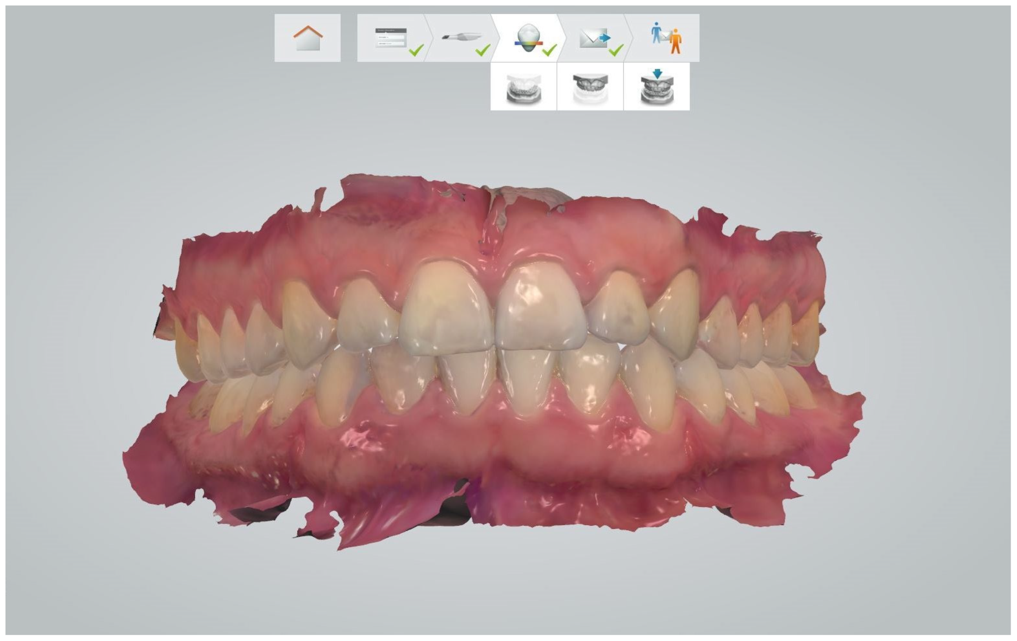
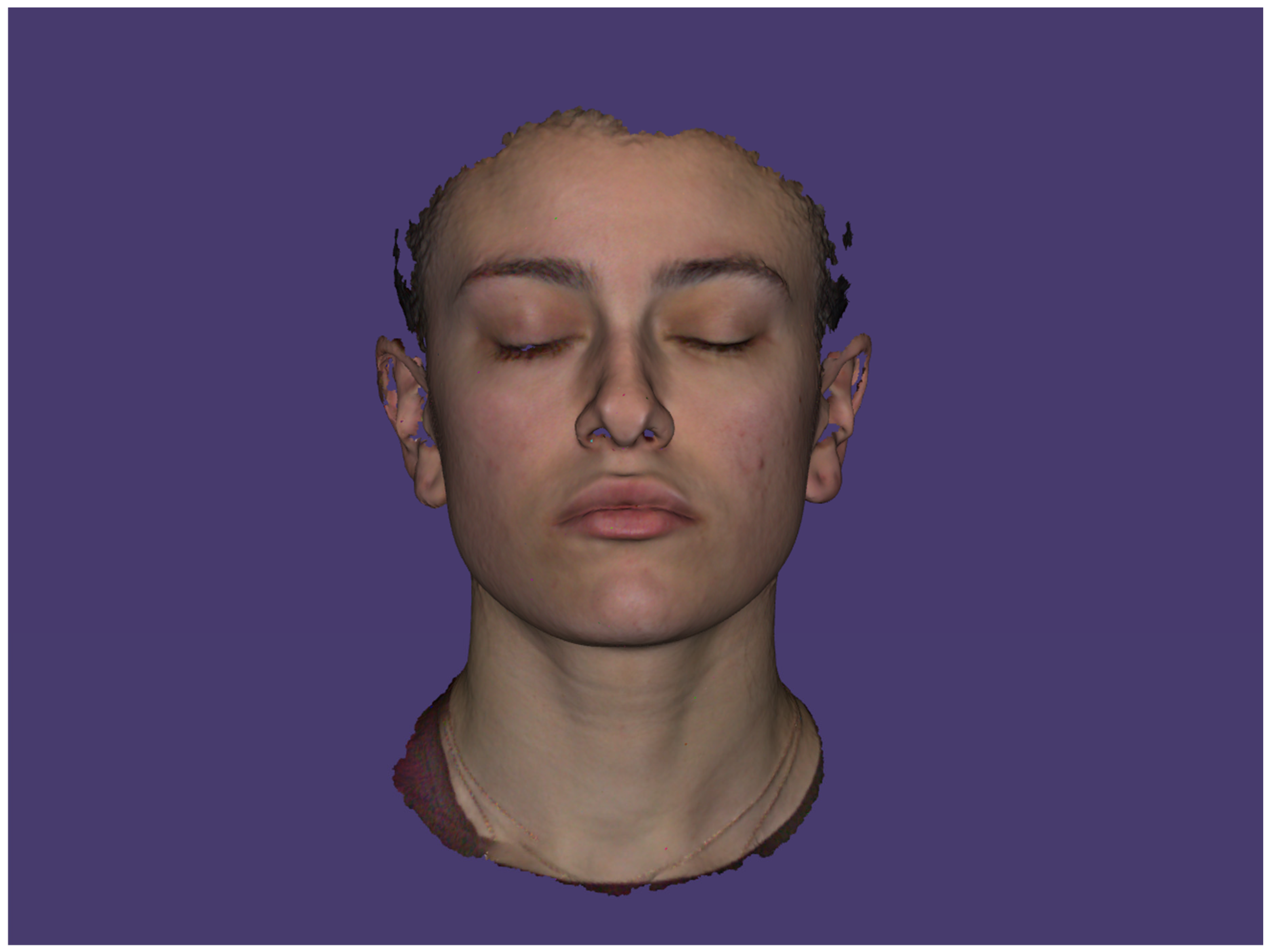
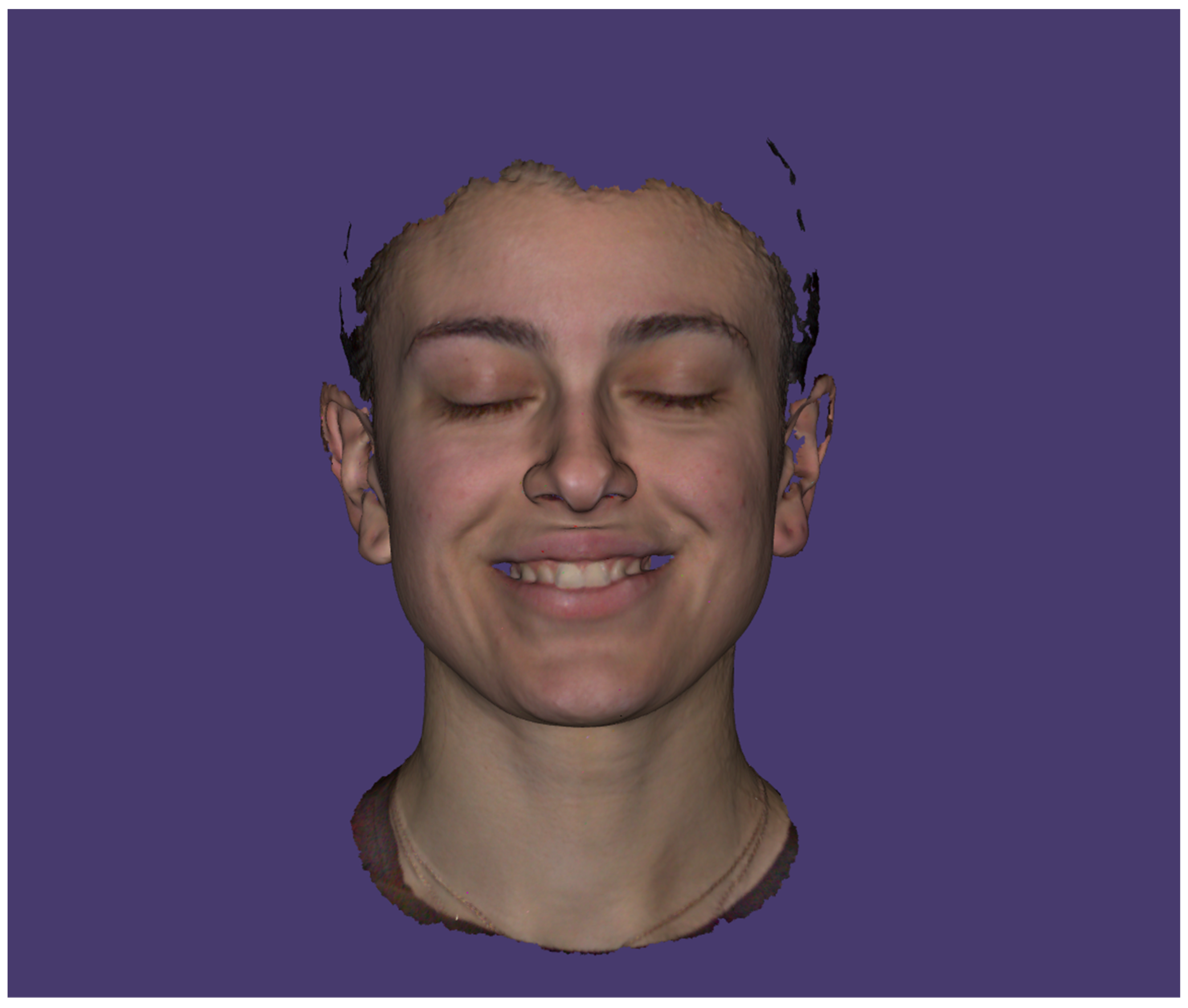
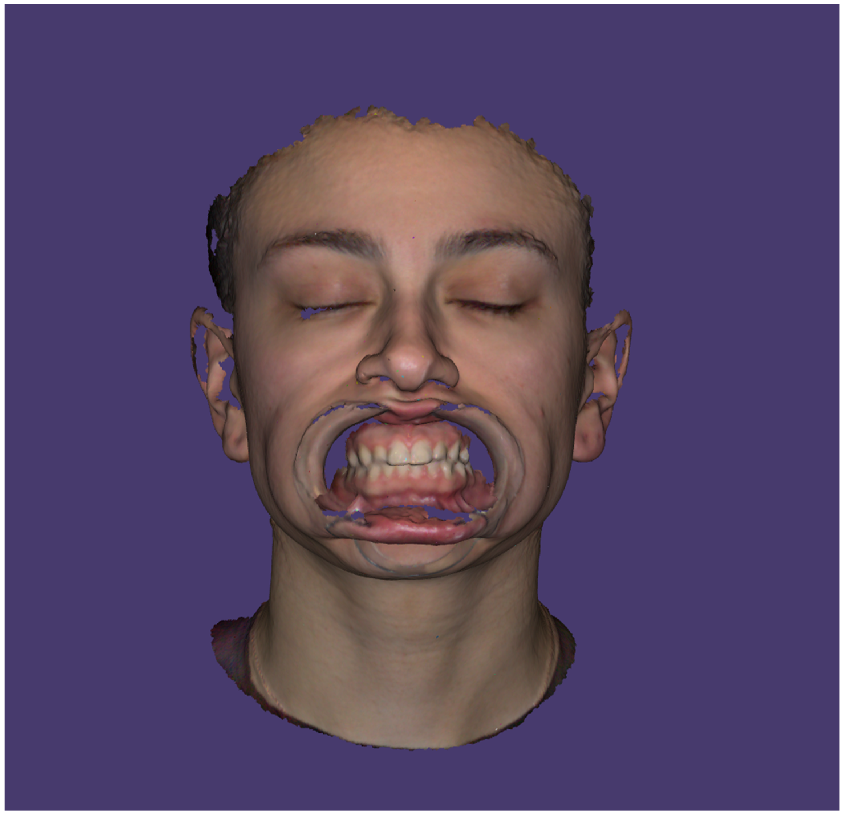

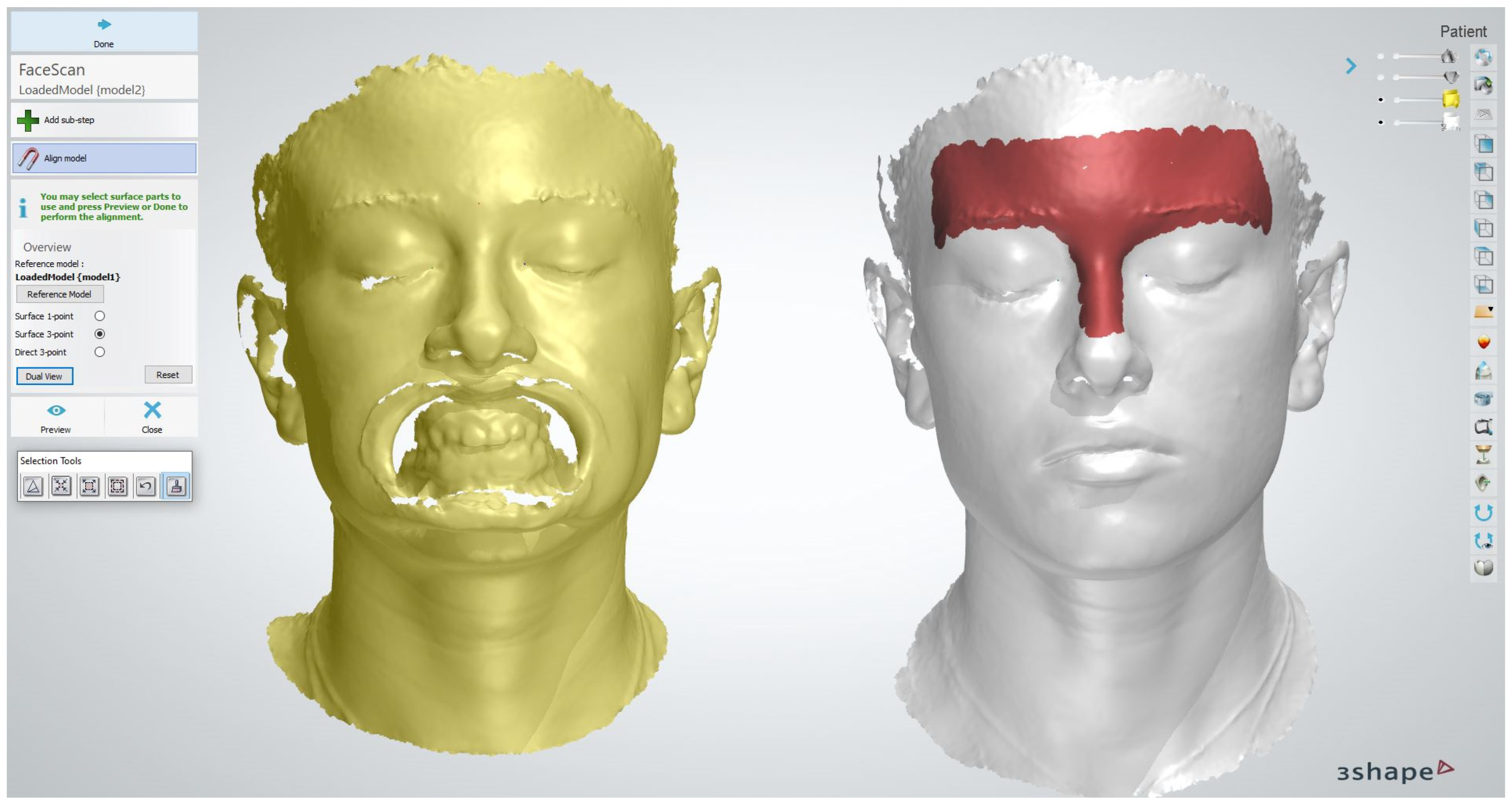
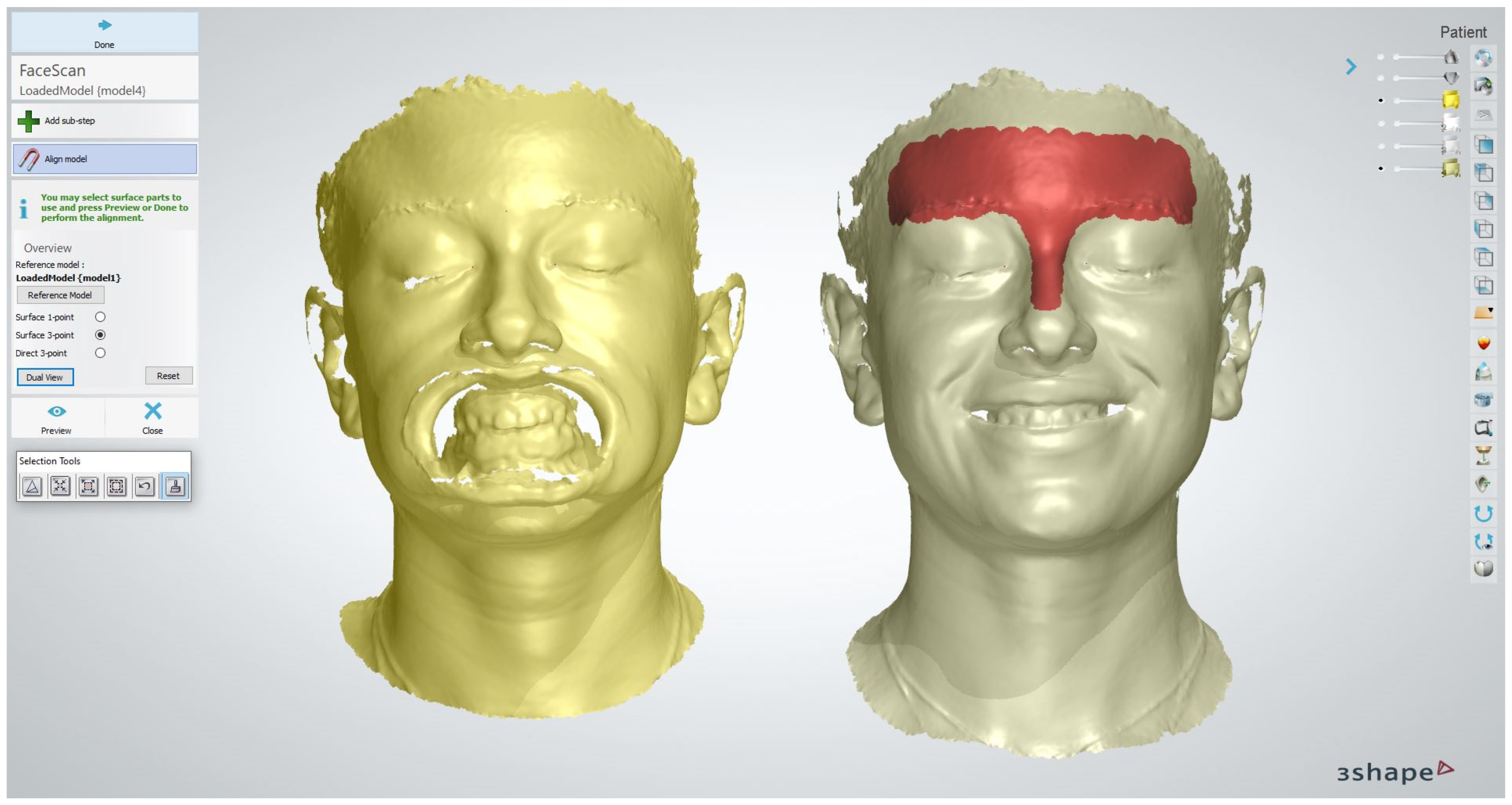
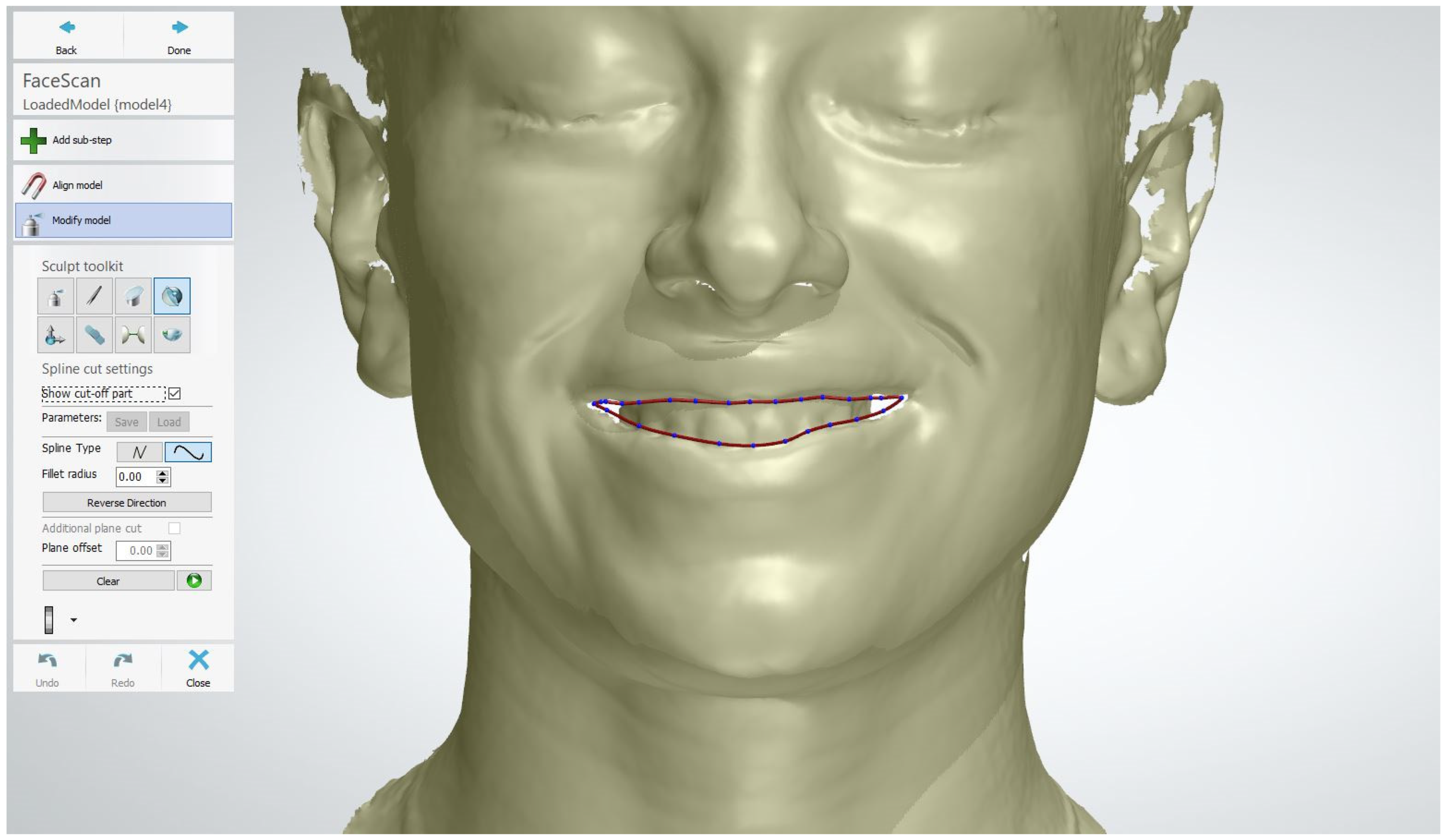
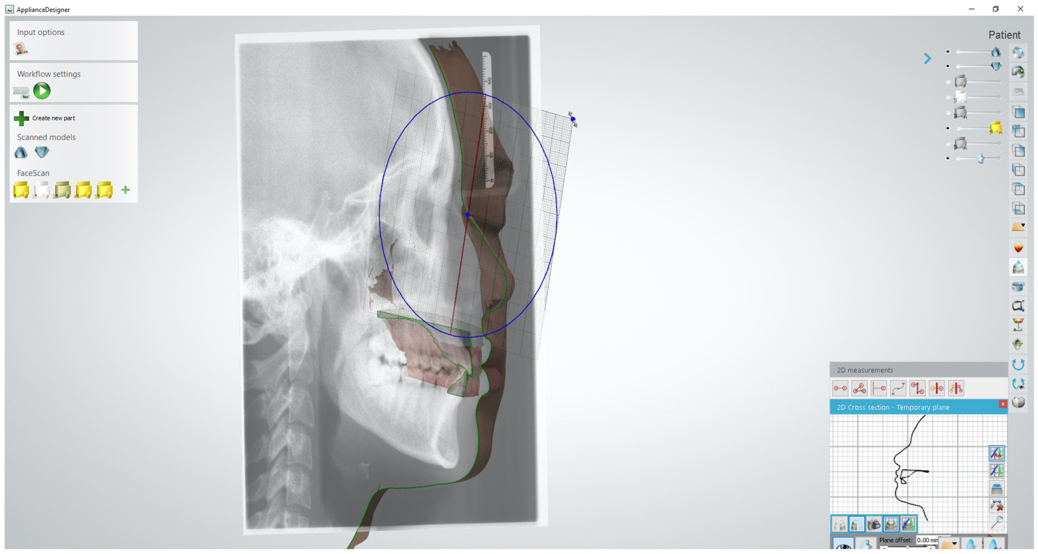
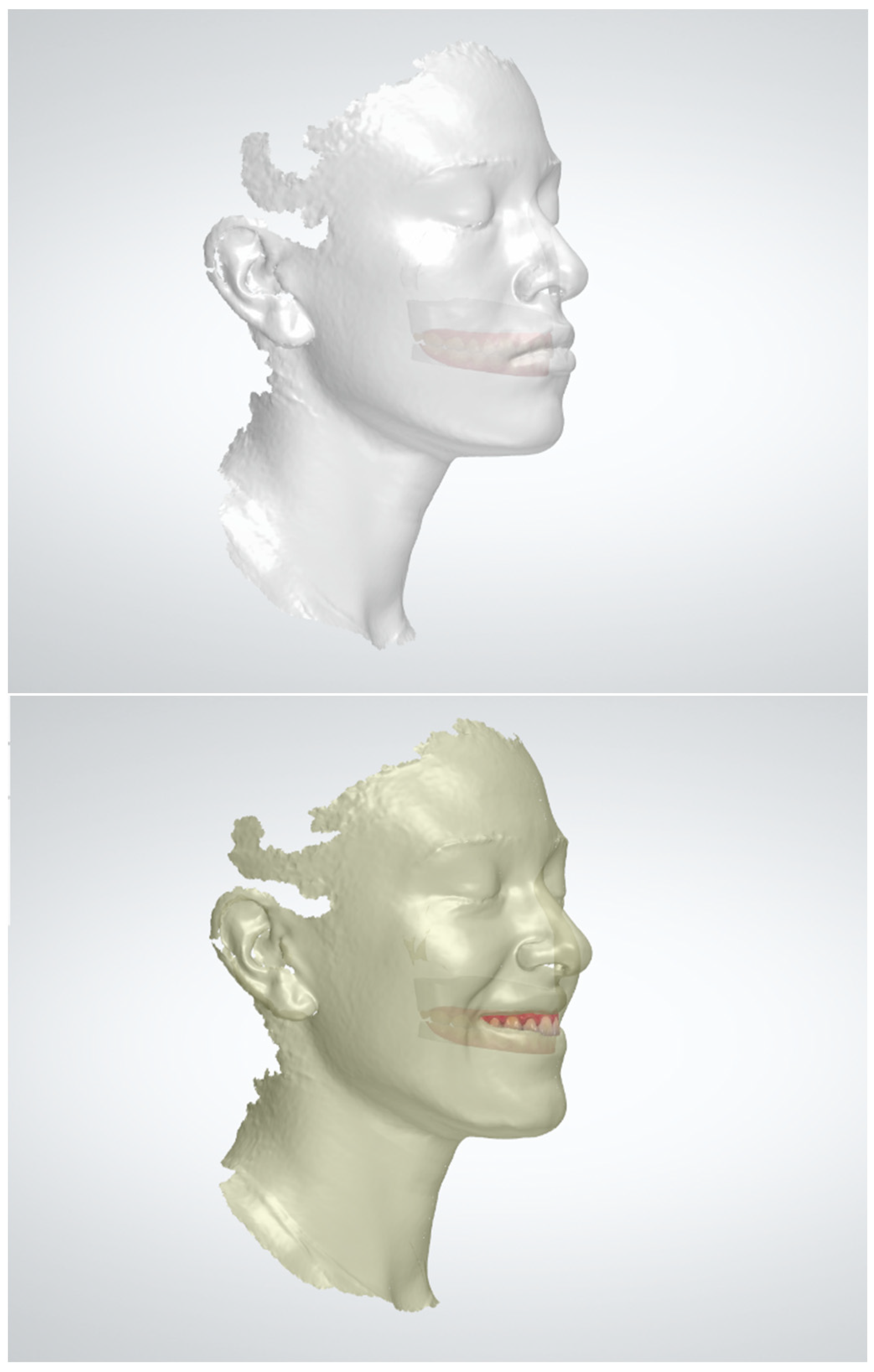
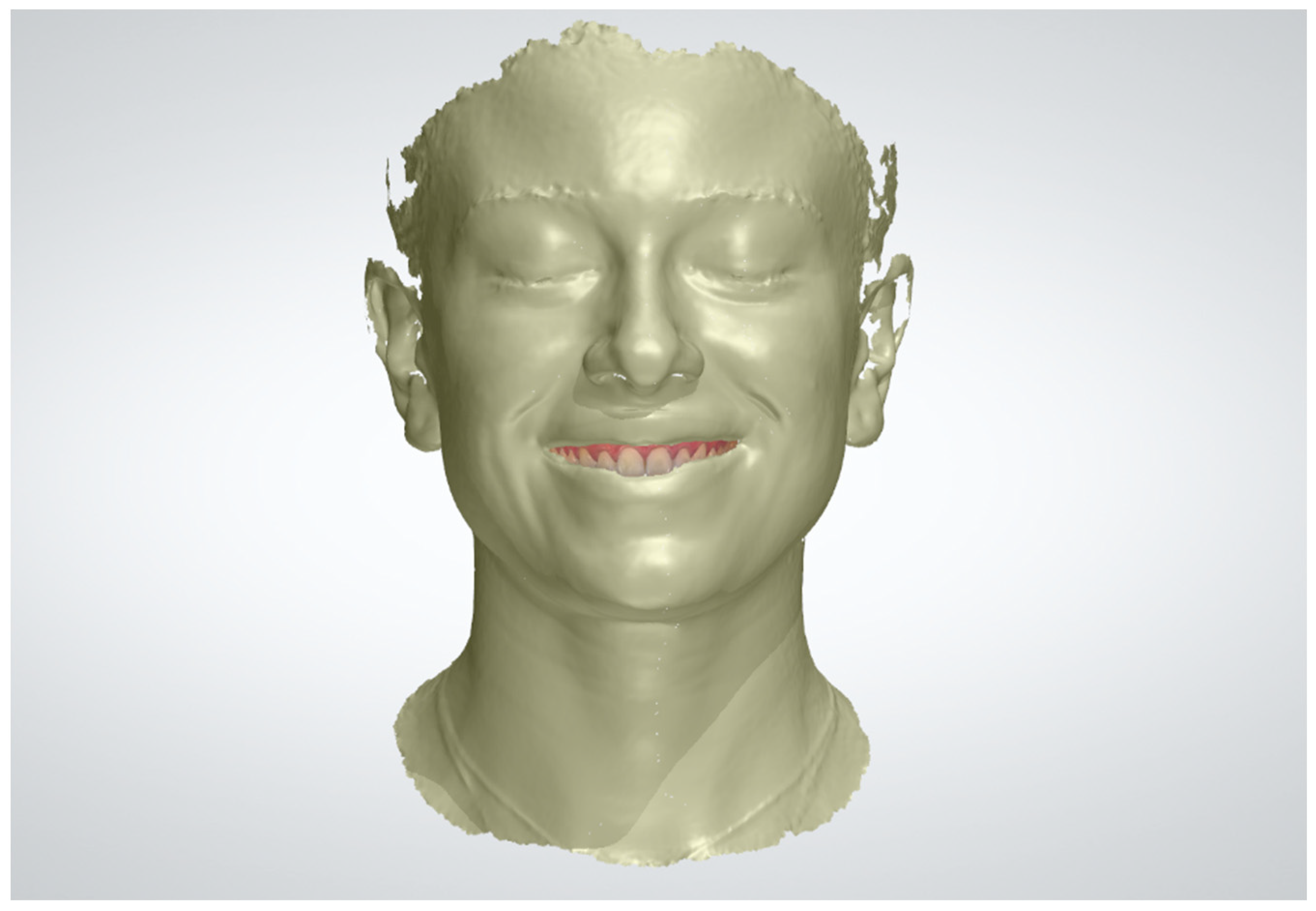
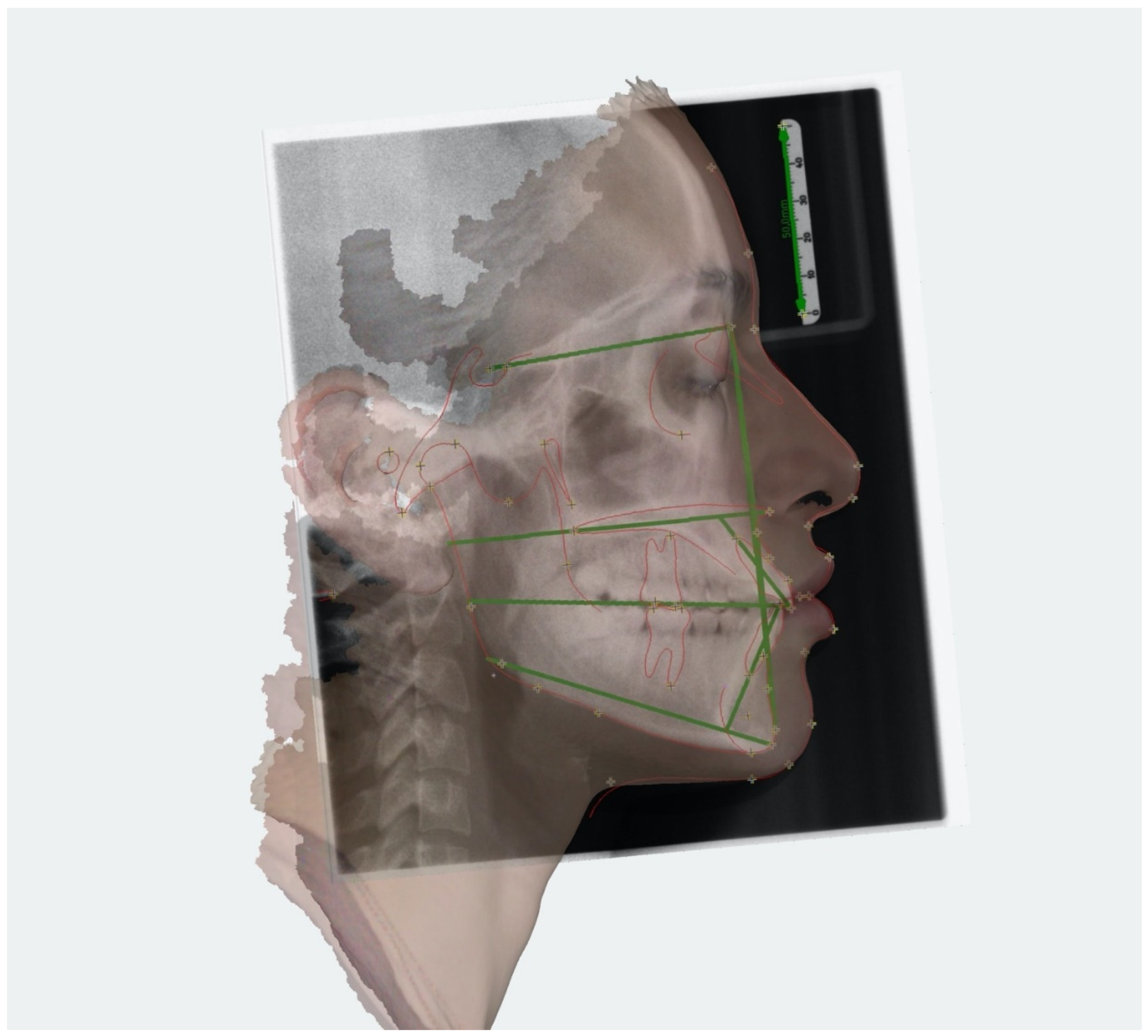
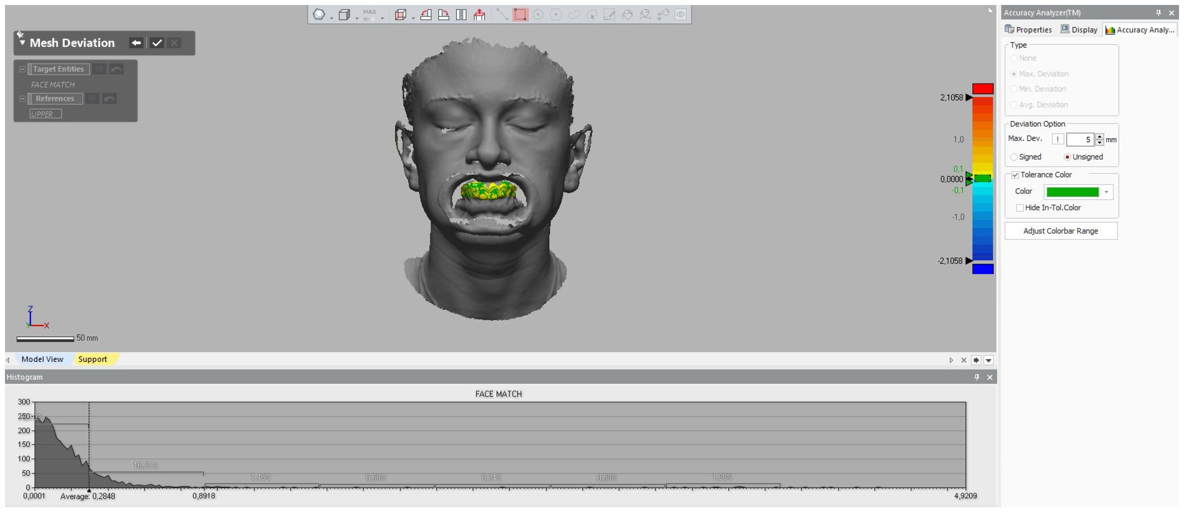
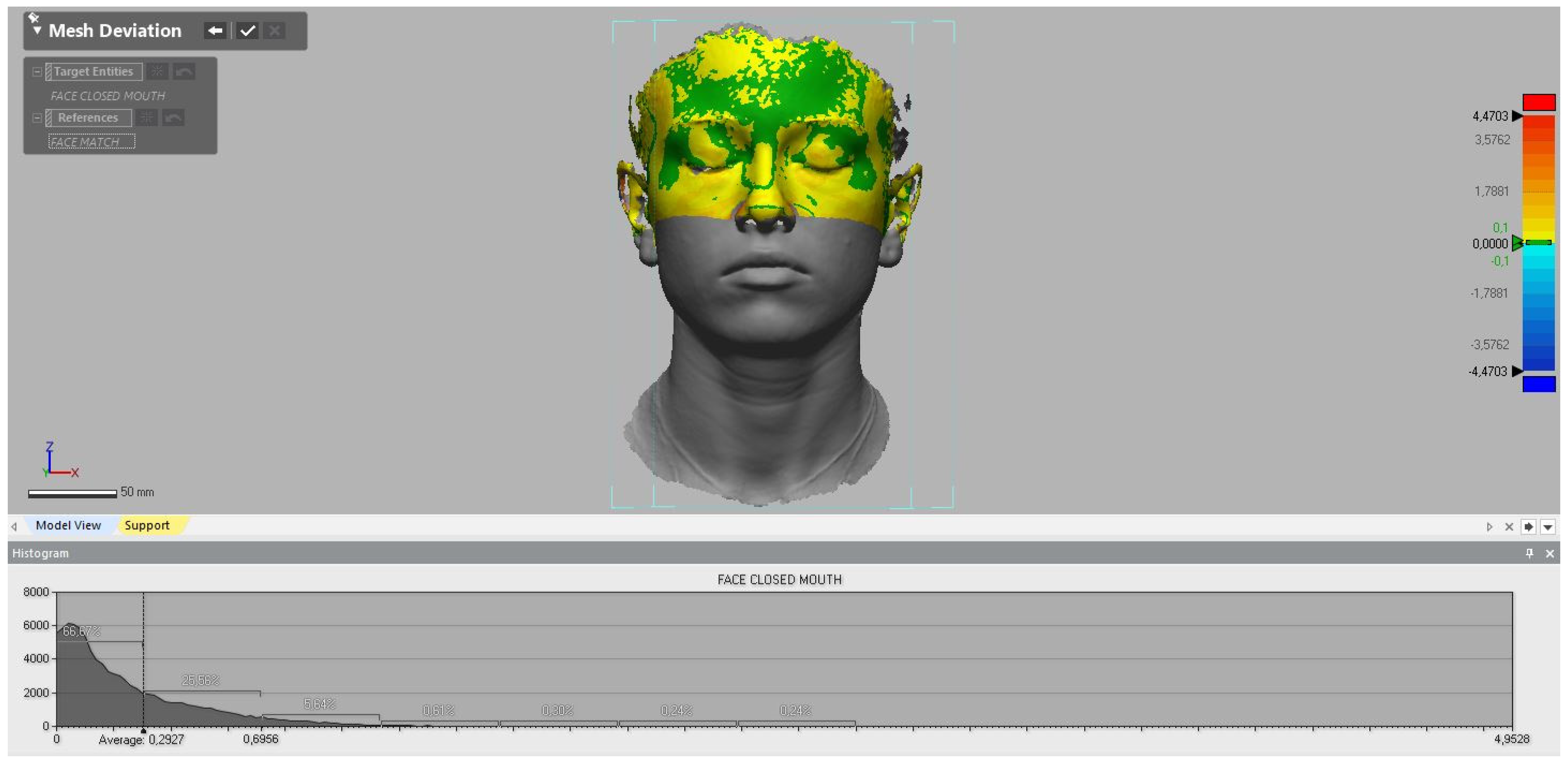
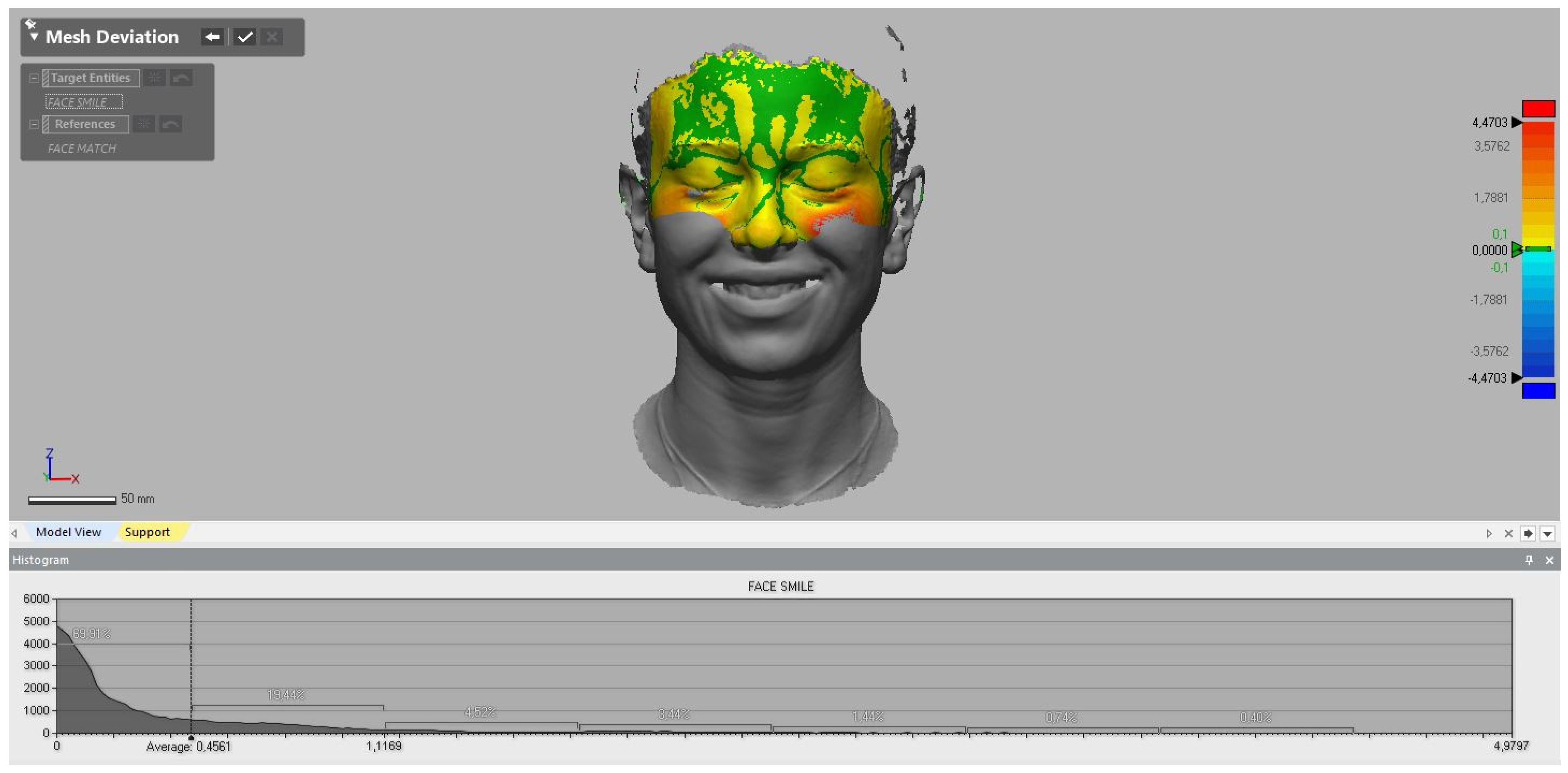
| Light Source | White Light, Visible—Infrared Light, Invisible |
|---|---|
| Safety | LED light (eye-safe)—CLASS I (eye-safe) |
| Scan accuracy | Up to 0.05 mm |
| Volumetric accuracy | 0.05 + 0.1 mm/m |
| Scan and align speed | 1,200,000 points/s, 20 frames per second |
| Align modes | Markers alignment, feature alignment, hybrid alignment, texture alignment |
| Working distance | 470 mm |
| Depth of field | 200–1500 mm |
| Maximum FOV | 420 × 900 mm |
| Point distance | 0.25–3 mm |
| Color scanning | Yes |
| Output formats | OBJ; STL; PLY; P3; 3MF |
| Certifications | CE, FCC, ROHS, WEEE, KC |
Publisher’s Note: MDPI stays neutral with regard to jurisdictional claims in published maps and institutional affiliations. |
© 2022 by the authors. Licensee MDPI, Basel, Switzerland. This article is an open access article distributed under the terms and conditions of the Creative Commons Attribution (CC BY) license (https://creativecommons.org/licenses/by/4.0/).
Share and Cite
Campobasso, A.; Battista, G.; Lo Muzio, E.; Lo Muzio, L. The Virtual Patient in Daily Orthodontics: Matching Intraoral and Facial Scans without Cone Beam Computed Tomography. Appl. Sci. 2022, 12, 9870. https://doi.org/10.3390/app12199870
Campobasso A, Battista G, Lo Muzio E, Lo Muzio L. The Virtual Patient in Daily Orthodontics: Matching Intraoral and Facial Scans without Cone Beam Computed Tomography. Applied Sciences. 2022; 12(19):9870. https://doi.org/10.3390/app12199870
Chicago/Turabian StyleCampobasso, Alessandra, Giovanni Battista, Eleonora Lo Muzio, and Lorenzo Lo Muzio. 2022. "The Virtual Patient in Daily Orthodontics: Matching Intraoral and Facial Scans without Cone Beam Computed Tomography" Applied Sciences 12, no. 19: 9870. https://doi.org/10.3390/app12199870
APA StyleCampobasso, A., Battista, G., Lo Muzio, E., & Lo Muzio, L. (2022). The Virtual Patient in Daily Orthodontics: Matching Intraoral and Facial Scans without Cone Beam Computed Tomography. Applied Sciences, 12(19), 9870. https://doi.org/10.3390/app12199870


