Article

Factors Determining Ticagrelor-Induced Dyspnea in Patients with Acute Coronary Syndrome

by
Vytenis Tamakauskas
1,2,*,
Remigijus Žaliūnas
3,
Vaiva Lesauskaitė
1,
Nora Kupstytė-Krištaponė
1,2,3,
Gintarė Šakalytė
1,3,
Julija Jurgaitytė
2,
Ieva Čiapienė
1 and
Vacis Tatarūnas
1
1
Institute of Cardiology, Medical Academy, Lithuanian University of Health Sciences, Sukilėlių g. 15, LT-50009 Kaunas, Lithuania
2
Cardiovascular Centre, Republican Šiauliai Hospital, V. Kudirkos g. 99, LT-76231 Šiauliai, Lithuania
3
Department of Cardiology, Faculty of Medicine, Medical Academy, Lithuanian University of Health Sciences, Eivenių g. 2, LT-50009 Kaunas, Lithuania
*
Author to whom correspondence should be addressed.
Appl. Sci. 2022, 12(19), 10021; https://doi.org/10.3390/app121910021
Submission received: 6 September 2022 / Revised: 2 October 2022 / Accepted: 3 October 2022 / Published: 6 October 2022

Abstract

:
(1) Background: The aim of this study was to determine clinical and genetic factors predicting the development of dyspnea in patients receiving ticagrelor. (2) Methods: A total of 277 patients with acute myocardial infarction (with and without ST-segment elevation), who underwent coronary angiography and PTCA with stent implantation and treated with antiplatelet drugs (ticagrelor and aspirin), were enrolled in this study. Platelet aggregation (induction with high-sensitivity ADP, ADP HS) testing was performed using a MULTIPLATE analyzer and reagents for the determination of P2Y12 receptor activity. Venous blood samples were collected for genotyping. (3) Results: Patients experiencing ticagrelor-related dyspnea had lower ADP HS. ROC curve analysis showed that an ADP HS cut-off of ≤19.5 U was associated with the development of dyspnea. The ADP HS value of ≤19.5 U and any dose of atorvastatin lower than 80 mg (or no atorvastatin) increased the risk of dyspnea by more than 4 and 2 times, respectively (OR = 4.07, p ≤ 0.001 and OR = 2.25; p = 0.008). (4) Conclusion: A lower ADP HS value possibly indicates greater ticagrelor activity and a higher plasma concentration of this drug. Atorvastatin might have an impact on the occurrence of ticagrelor-related dyspnea by affecting ticagrelor metabolism. No impact of any genetic variant on the development of dyspnea was determined.

1. Introduction

Cardiovascular diseases (CVDs) are the leading cause of morbidity and mortality in Europe [1]. Acute coronary syndrome (ACS) represents a life-threatening manifestation of CVDs [1]. Patients with ACS are treated with an interventional approach by performing immediate coronary angiography and percutaneous transluminal coronary angioplasty (PTCA) with stent implantation [2,3].
Based on the guidelines of the European Society of Cardiology (ESC), dual antiplatelet therapy (DAPT) for up to 12 months is recommended for patients with ACS who underwent PTCA with stent implantation [2,3]. The most recent ESC guidelines recommend ticagrelor in a combination with aspirin as a first-line therapy for patients with ACS irrespective of the initial treatment strategy [2,3]. The findings of the most recent clinical trials are controversial. The study by Adamski et al. showed that patients with ST-segment elevation myocardial infarction (STEMI) at 1–2 h after receiving a loading dose of ticagrelor were at a greater risk of high platelet reactivity, which is considered an independent risk factor for stent thrombosis [4]. Other researchers state that ticagrelor is safe to use even if there is a need for treatment with anticoagulants: a combination of ticagrelor, aspirin, and dabigatran can be administered for patients with ACS and non-valvular atrial fibrillation (AF) who underwent PTCA with stent implantation [5]. However, the administration of dual or triple antiplatelet therapy leads to numerous clinical challenges [6]. The most frequent clinical challenge associated with ticagrelor administration is its premature discontinuation attributed to sudden-onset dyspnea or bleeding [7]. It is well-known that ticagrelor causes dyspnea and can be responsible for bleeding; only the incidence of these adverse events differs. Discontinuation of ticagrelor attributed to these causes leads to an increased risk of stent thrombosis and recurrent ACS [6,7]. Stent thrombosis and recurrent ACS are associated with significantly greater mortality, longer hospital stay, and worse functional status and quality of life in patients [7].
Ticagrelor is a cyclopentyltriazolo-pyrimidine (CPTP) that directly and reversibly binds to P2Y12 receptors. The drug reversibly inhibits the binding of endogenous adenosine diphosphate (ADP) to platelet membrane-bound P2Y12 receptors and blocks platelet aggregation. The maximum plasma concentration of ticagrelor is reached within 1.3–2 h, and the mean elimination half-life is 7–12 h. Ticagrelor is metabolized to its active metabolite, AR-C124910XX. Ticagrelor and its active metabolite are highly bound to plasma proteins (99.8%); the bioavailability of ticagrelor is about 36%. Ticagrelor-related dyspnea of varying severity usually appears suddenly 2–3 h after drug administration and lasts for several hours, days, or the whole period of drug use. It is directly associated with the maximum plasma concentration of the drug [8,9,10].
Drugs of the thienopyridine class (clopidogrel, prasugrel) bind to P2Y12 receptors irreversibly and, therefore, inhibit platelet aggregation longer than ticagrelor (for the entire lifespan of platelets, i.e., 7–10 days) [11]. Clopidogrel is metabolically activated in the liver, and its antiplatelet effect directly depends on a genetic polymorphism. Prasugrel exhibits a more consistent and less metabolism-dependent antiplatelet effect but is associated with a significantly higher risk of bleeding [11]. The results of large-scale PLATO, DISPERSE-2, RESPOND, and ONSET/OFFSET clinical trials showed that ticagrelor in combination with aspirin was superior to a combination of clopidogrel and aspirin in reducing the incidence of cardiovascular death, recurrent myocardial infarction, and stroke [12,13,14,15]. The most recent data indicate that enzymes encoded by CYP2C19 and SLCO1B1 can have an impact on the rate of elimination of M8, a ticagrelor metabolite, and the clinical effect of ticagrelor itself [16]. However, in the PLATO trial, CYP2C19 and ABCB1 variants were not significantly related to the antiplatelet effect of ticagrelor [17].
An analysis of the literature related to ticagrelor has revealed that different enzymes may be involved in determining the therapeutic effect of ticagrelor. Nie et al. showed that CYP4F2 rs2074900 had an effect on the pharmacokinetics of ticagrelor [18]. Our previous studies showed [16,19] that CYP4F2 variants may determine bleeding during ticagrelor therapy. CYP4F2 may participate in the resolution of inflammation after myocardial infarction [20]. The pleiotropic effects of ticagrelor could also be related to beneficial cyclooxygenase-2 (COX2) activation. In contrast, aspirin may reduce the activity of COX2 [21].
Plasminogen activator inhibitor-1 (PAI-1) slows down the activity of the fibrinolysis system and contributes to thrombus formation. The study by Reiner et al. showed that ticagrelor may reduce the activity of PAI-1 [22]. Ticagrelor blocks platelet P2Y12 receptors, inhibits platelet activation, and, finally, aggregation through binding α2β3 receptors, which bind fibrinogen [23]. In addition, the study by Gasecka et al. showed that ticagrelor may reduce the release of extracellular vesicles (EVs) [24]. During acute myocardial infarction, platelets, endothelial cells, and leukocytes release EVs that contain pro-thrombotic enzymes such as fibrinogen. Studies showed that about twice higher EV concentrations were present in patients with acute myocardial infarction as compared to healthy ones [25].
Different studies suggest that 1 in every 20 patients treated with ticagrelor experiences dyspnea of varying intensity [7,26,27]. The causes of ticagrelor-related dyspnea remain unknown. Dyspnea can be mild, moderate, and severe, manifesting in 56.5, 28.3, and 15.2% of patients, respectively [28]. Ticagrelor-related dyspnea can last less than 24 h or can persist over the course of the treatment. Up to 6% of patients with moderate or severe dyspnea discontinue ticagrelor prematurely more frequently or ticagrelor is changed to a less effective combination of clopidogrel and aspirin [6,7].
In clinical practice, dyspnea caused by pulmonary, cardiac, and metabolic diseases is most common [29]. There are several clinical trials during which cardiac and pulmonary function was investigated in patients with ACS or stable angina pectoris, receiving ticagrelor or clopidogrel. The findings of these clinical trials have shown that dyspnea is not related to drug-induced impairment of pulmonary and cardiac function or acidosis [10,30]. The incidence of ticagrelor-related dyspnea was the same in both patients with chronic heart failure or chronic obstructive pulmonary disease and those without these diseases [31]. Up to date, the two most plausible hypotheses explaining the potential mechanisms of ticagrelor-induced dyspnea have been debated [10,32]. In the literature, the greatest attention is paid to a structural similarity between ticagrelor and adenosine, activation of adenosine receptors (A1R), and stimulation of vagal C fibers via receptors. Other authors state that ticagrelor can induce dyspnea via P2Y12 receptors present on vagal C fibers themselves. Some literature sources mention the third cause of dyspnea, i.e., ticagrelor-induced release of adenosine triphosphate (ATP) from erythrocytes; however, this hypothesis is less plausible (Figure 1) [10,32].
The most plausible hypotheses explaining the causes for the occurrence of ticagrelor-related dyspnea are as follows [10,32]:
1.
An increase in extracellular adenosine levels.
One of the possible causes of dyspnea is related to the effect of adenosine on specific receptors in the respiratory system, resulting in the stimulation of specific sensory receptors. Adenosine receptors are abundantly expressed in all tissues, including the myocardium, nervous system, kidneys, and respiratory system. Purine receptors P1 and P2, so-called adenosine receptors, are specific to adenosine. There are four known types of P1 adenosine receptors: A1, A2A, A2B, and A3. Activated P1 receptors interact with vagal receptors in the lungs and airways, which leads to airway smooth muscle contraction and the sensation of dyspnea. Sensory receptors in the airways are innervated by the vagal nerve and consist of myelinated and unmyelinated C fibers and stretch receptors. Unmyelinated C fibers (type J receptors) provide sensory input from the airways to lung structures. Vagal C fibers in the lungs are involved in the development of tachypnea and dyspnea.
The effect of an activated adenosine receptor depends on receptor and tissue systemic innervation. An increase in adenosine levels activates A1R receptors, which mediate airway smooth muscle contraction. Activation of A1R receptors stimulates sensory receptors in lung tissue, causing the sensation of dyspnea and cough. A2B receptors are linked to inflammation and bronchoconstriction due to mast cell degranulation (release of specific mediators), leading to the development of bronchospasm and dyspnea [10,32].
2.
Dyspnea is caused via P2Y12 receptors.
P2Y12 receptors are present not only on the outer membrane of platelets but are also abundant on the cells of other tissues (endothelium, smooth muscles, and neurons) and microglia in the central nervous system. Inhibition of P2Y12 receptors on neurons and glial cells leads to increased signaling of vagal C fibers and glial cells to the central nervous system. Glial cells can stimulate the central chemoreflex system, eliciting the sensation of acute dyspnea [10,32].
3.
Dyspnea is associated with ATP release from erythrocytes.
It is thought that ticagrelor induces a directly dose-dependent ATP release from erythrocytes due to changes in pH, which is greater than physiological ATP levels. However, this hypothesis seems to be unlikely as ticagrelor-mediated, erythrocyte-derived ATP is rapidly degraded and has no influence on the occurrence of dyspnea [10,32].
Up to date, no clinically relevant biomarkers that would allow prediction of the risk of developing dyspnea or bleeding during treatment with ticagrelor are available [10]. Moreover, information on pharmacokinetic characteristics in patients with or without dyspnea is scarce [10]. It is known that aspirin, depending on the consumed amount, exhibits different activities: at low doses, it inhibits platelets; at higher doses, it provides anti-inflammatory effects; and at even higher doses, it causes vasodilation and a toxic effect on the body (respiratory alkalosis, fever, dehydration, acidosis, and shock) [33]. Most of the sustained episodes of dyspnea resolve with the discontinuation of ticagrelor use and a decrease in ticagrelor plasma levels in all patients [31]. Based on the accumulated clinical data and experience of our center, ticagrelor-induced dyspnea is a subjective sensation and does not have any negative impact on the patient’s pulmonary function (respiratory rate, spirometry, total lung capacity, oxygen saturation, and other) [26,31].
The aim of this study was to determine clinical and genetic factors that would allow prediction of the development of dyspnea in patients receiving ticagrelor. This is important to ensure a safe and continuing use of this drug during the entire treatment period and to prevent the occurrence of complications such as stent thrombosis and recurrent ACS.

2. Materials and Methods

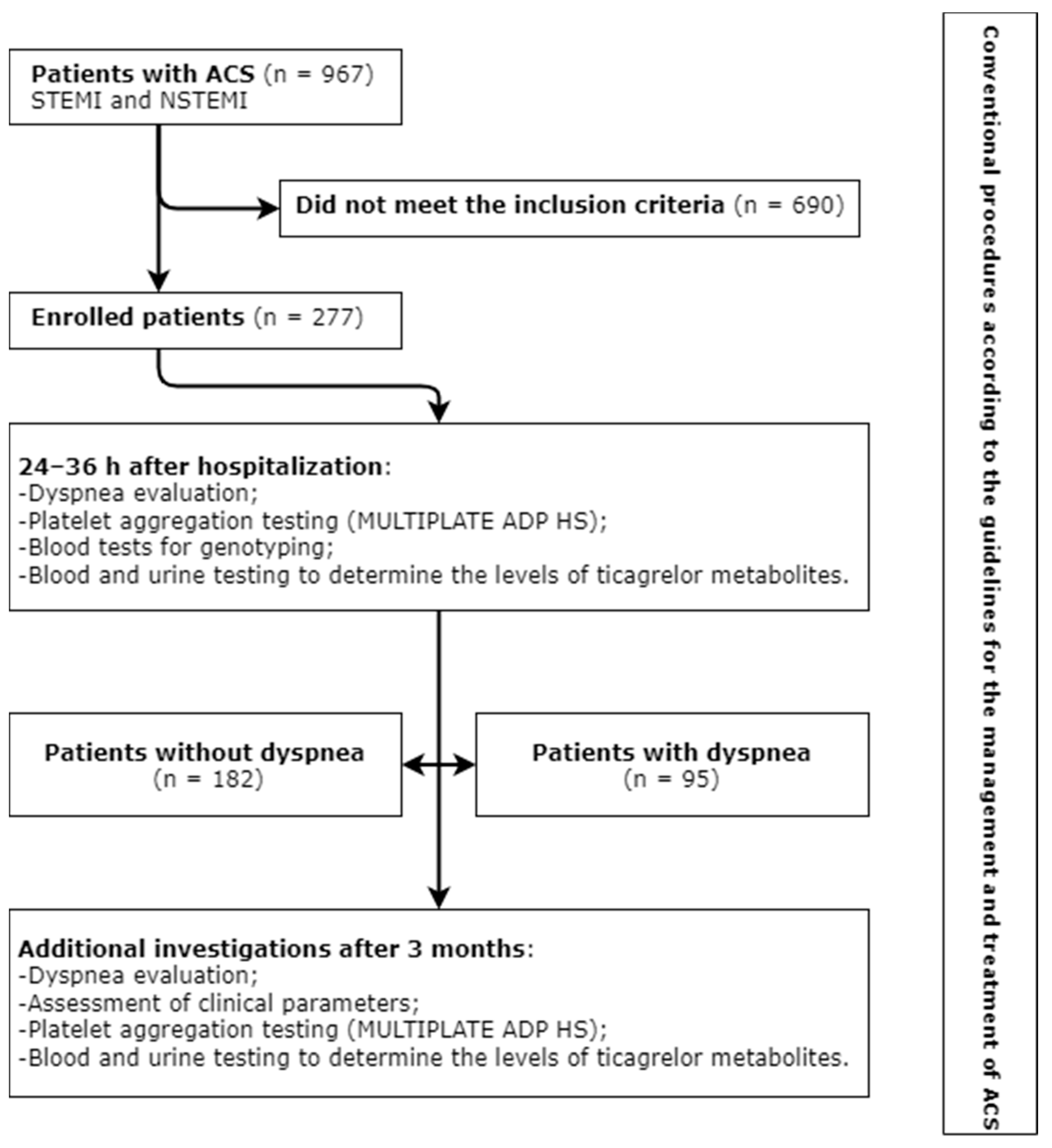
A total of 967 patients with acute myocardial infarction (with and without ST-segment elevation) were treated in the Cardiovascular Centre, Republican Šiauliai Hospital, from January 2020 to September 2021. During this period, 277 patients were eligible to be enrolled in this study based on the inclusion criteria. The study flow chart is depicted in Figure 2.
The inclusion criteria were as follows:
  • Patients treated in the Cardiovascular Centre, Republican Šiauliai Hospital, for ACS (myocardial infarction with and without ST-segment elevation), who underwent coronary angiography and PTCA with stent implantation;
  • Treatment with antiplatelet drugs (ticagrelor and aspirin) or a combination of antiplatelet drugs and anticoagulant (ticagrelor, aspirin, and dabigatran etexilate);
  • Planned 12-month treatment with ticagrelor.
  • The exclusion criteria were as follows:
  • Previous dyspnea experienced by a patient;
  • Severe comorbid disease (stage IV cancer; significant disease of another organ system, etc.) that could have an influence on the results of the performed investigations;
  • Respiratory diseases (bronchial asthma; chronic obstructive pulmonary disease; COVID-19 during ACS);
  • Previous or new-onset New York Heart Association (NYHA) class III and IV heart failure;
  • Warfarin usage (e.g., due to mechanical heart valve);
  • Patients at high risk of bleeding, who were treated with clopidogrel;
  • Social indications (e.g., a patient being cared for, no possibility to arrive for follow-up visits);
  • Patient’s refusal to take part in this study.

2.1. Investigations and Patients’ Clinical Data

During the treatment period, patients underwent conventional procedures according to the European Society of Cardiology (ESC) guidelines for the management and treatment of ACS (Table 1) [2,3]. All enrolled patients, 24–36 h after hospitalization, were divided into 2 groups (Figure 2) taking into consideration the presence or absence of dyspnea. The severity of dyspnea was rated from 0 to 4 using the modified Borg acute dyspnea scale (Table 2). New-onset dyspnea in the enrolled patients was linked to ticagrelor use after repeat evaluation and exclusion of other dyspnea-related causes (heart failure, anemia, oxygen saturation, and airway or pulmonary pathology).
Patients underwent the following additional investigations upon enrollment into this study and after 3 months:
  • Venous blood samples were collected for genotyping. Genotyping procedures were carried out at the Laboratory of Molecular Cardiology, Institute of Cardiology, Medical Academy, Lithuanian University of Health Sciences. DNA was extracted from blood using a salting-out method. The determination of gene variants was carried out using TaqMan probes (Thermo Fisher Scientific, Waltham, MA, USA), TaqMan Universal Master Mix (Thermo Fisher Scientific, Waltham, MA, USA), and PCR-grade water. The QuantStudio 5 and 3 Real-Time PCR systems (Thermo Fisher Scientific, Waltham, MA, USA) were employed. A total of 12 gene variants—C148T (rs1800787), CYP4F2 (rs3093135, rs1558139, rs2108622, and rs2074902), CYP2C19 *2 and *17 (rs4244285 and rs12248560), CYP2C9 *15 (rs72558190), ABCB1 (rs1045642), COX-2 (rs689465), PAI-1 (rs5918), CYP1A2*1C (rs2069514)—were investigated;
  • Platelet aggregation (induction with high-sensitivity ADP, ADP HS) testing was performed using a MULTIPLATE analyzer and reagents for the determination of P2Y12 receptor activity. Testing was carried out at the Laboratory Diagnostic Unit, Republican Šiauliai Hospital. Platelet aggregation testing with ADP HS was carried out 24–36 h and 3 months after the administration of a loading ticagrelor dose.
All the patients had hypertension and dyslipidemia. Standard treatment with beta blockers, angiotensin-converting enzyme inhibitors, statins, and antiplatelet drugs was prescribed.
After 3 months, patients underwent a repeat evaluation of dyspnea, bleeding data, blood testing, and other standard investigations based on the ESC recommendations (Table 1).

2.2. Statistical Analysis

The frequencies of genetic variants of fibrinogen beta chains (FBG)-C148T (rs1800787), CYP4F2 (rs3093135, rs1558139, rs2108622, and rs2074902) and CYP2C19 *2 and *17 (rs4244285 and rs12248560), ABCB1 (rs1045642), COX-2 (rs689465), and PAI-1 (rs5918) were expressed as percentages. The data of the platelet aggregation (ADP HS) testing and other laboratory tests were expressed as medians with ranges. The Mann–Whitney test was used to compare continuous data. Categorical data were compared with the chi-square test (Pearson formulae). Bonferroni correction for multiple comparisons was used to determine the incidence of dyspnea in patients according to the FBG-C148T (rs1800787), CYP4F2 (rs3093135, rs1558139, rs2108622, and rs2074902) and CYP2C19 *2 and *17 (rs4244285 and rs12248560), ABCB1 (rs1045642), COX-2 (rs689465), and PAI-1 (rs5918) genotypes; when the frequency in at least one cell of a contingency table was small (<5), the Fisher’s exact test was used. Proportions were compared with the z test. Binary logistic regression (Backward Stepwise (Wald) and ENTER methods) analysis was carried out to determine independent clinical and genetic factors associated with the development of dyspnea. The diagnostic performance of ADP HS was evaluated using receiver operating characteristic (ROC) curve analysis. To determine an optimal cut-off value, the Youden J index was used. The level of significance was set at p < 0.05.

3. Results

3.1. Characteristics of the Study Population

The majority of the study population were male (n = 208; 75.1%). STEMI was most common among the enrolled patients (53.1%). On coronary angiography, 67.8% of the patients were found to have advanced coronary lesions (two- or three-vessel coronary artery disease, CAD). Diabetes mellitus was present in 12.3% of the patients who were treated most frequently with a combination of metformin and gliclazide. Hypothyroidism was documented in 2.5% of the patients. Patients with atrial fibrillation (14.4%) received dual therapy with ticagrelor plus low-dose (110 mg) dabigatran etexilate for the prevention of thromboembolic complications, and aspirin was administered only during hospitalization (6 days on average) (Table 3) [2].
After initiation of ticagrelor use, 95 patients experienced dyspnea; 182 patients had no dyspnea.
Within the 3-month period after ACS, 15 patients experienced bleeding from the digestive tract that was confirmed by esophagogastroduodenoscopy; however, treatment with antiplatelet drugs was not discontinued. A bleeding event was considered as insignificant, and an additional therapy with drugs of the proton pump inhibitor class was administered.

3.2. Associations between Clinical and Genetic Characteristics and the Development of Dyspnea

Dyspnea was more common among patients having two-vessel CAD (p = 0.034), hypothyroidism (p = 0.049), lower ADP HS both during the first investigation (p < 0.001) and after 3 months (p = 0.009), higher creatinine concentration (p = 0.019), and those who used atorvastatin less frequently (p = 0.027) (Table 3). There was a trend for a greater incidence of dyspnea in older patients (p = 0.066).
The FBG-C148T (rs1800787) gene polymorphism was found to be associated with dyspnea (p = 0.009) (Table 4). Patients homozygous for the C allele (CC) experienced dyspnea more frequently (p = 0.014) and heterozygous (CT) less frequently (p = 0.002) (Table 4).
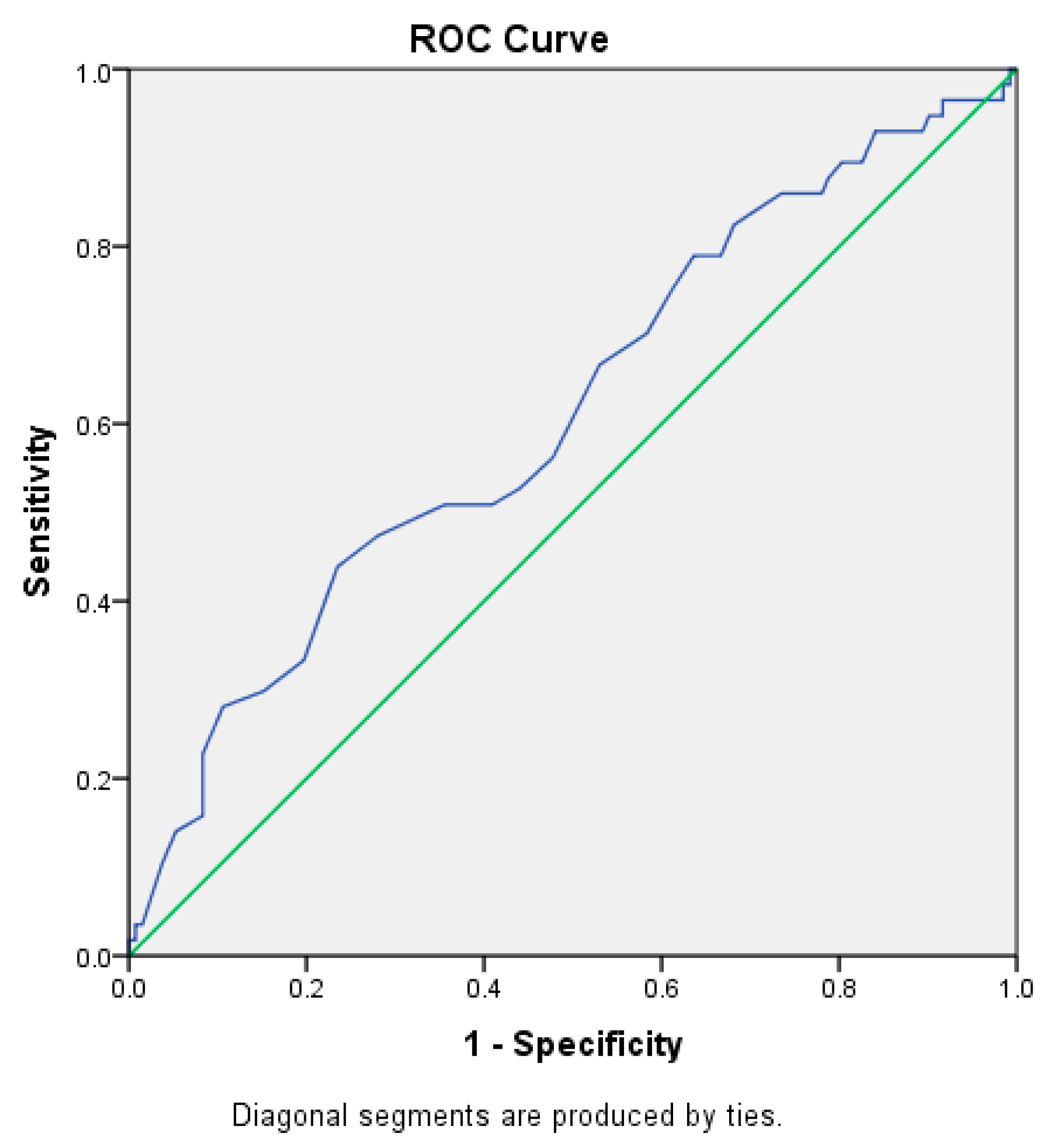
Patients experiencing ticagrelor-related dyspnea had lower ADP HS values (Table 5). By application of ROC curve analysis, an ADP HS value predicting dyspnea was determined (Figure 3). An ADP HS cut-off value of ≤19.5 U was found to be associated with the development of dyspnea (AUC = 0.612; 95% CI = 0.52–0.70; p = 0.015; sensitivity of 43.9%; specificity of 76.8%; Youden J index = 0.317). In addition, there was a trend that in the presence of an ADP HS value of ≤19.5 U, dyspnea of varying severity persisted during the entire follow-up period.

3.3. Logistic Regression Analysis for the Identification of Significant Risk Factors for Dyspnea

Logistic regression analysis showed that the ADP HS value of ≤19.5 U was associated with more than a 4-fold greater risk of developing dyspnea (OR = 4.07, p ≤ 0.001). Any dose of atorvastatin lower than 80 mg (or no atorvastatin) increased the risk of dyspnea by more than 2 times (OR = 2.18; p = 0.012) (Table 6).

4. Discussion

The aim of this study was to determine clinical and genetic factors that would allow prediction of the development of dyspnea in patients receiving ticagrelor.
Based on the results of the large-scale clinical ONSET/OFFSET trial, dyspnea was documented in 38.6, 9.3, and 8.3% of patients receiving ticagrelor, clopidogrel, and prasugrel, respectively [32,34]. In our study, dyspnea of varying severity was reported by 34.3% of patients. This study enrolled patients in whom the occurrence of dyspnea due to other causes was unlikely. The severity of dyspnea was evaluated with the modified Borg acute dyspnea scale, which was used in early clinical trials investigating the effect of ticagrelor [29,35]. As patients with diabetes mellitus often have other comorbidities that were included in the exclusion criteria, patients with diabetes mellitus in our study accounted for only 12.3%. In agreement with other studies, dyspnea experienced by our patients was not associated with the indicators of heart failure or pulmonary function impairment (confirmed by spirometry, auscultation, or chest X-ray) [31]. There is evidence that severe renal or hepatic impairment enhances the effect of ticagrelor [10]. This is in line with the results of our study. Our patients with poorer renal function experienced dyspnea more frequently. Different studies have reported that the incidence of dyspnea is greater in patients with advanced coronary atherosclerosis, thyroid pathology, and older patients, receiving ticagrelor [10]. Our data also support these statements as, based on epidemiological data, older patients in the entire population had advanced atherosclerotic lesions (two- and three-vessel CAD) and underlying diseases (i.e., hypothyroidism) more frequently.
The findings of our study confirm the results of other authors that treatment of ACS (with beta blockers and angiotensin-converting enzyme inhibitors) defined by the ESC guidelines has no impact on the incidence of ticagrelor-related dyspnea [31]. Additional analysis of the PLATO trial aimed to investigate possible interactions between ticagrelor and statins (atorvastatin and simvastatin). In the presence of ticagrelor, the simvastatin concentration increased 3–4 times in some patients while the atorvastatin concentration increased marginally (up to 23%) and was considered as clinically irrelevant [10,36]. There is evidence that ticagrelor conjugated to glucuronic acid during metabolism in the body can inhibit cytochrome P450 enzymes. The metabolism of ticagrelor is slowed down and its removal from the body is affected [37], resulting in increased levels of ticagrelor in blood. Even in 2010, the study on human hepatocytes carried out by Feidt et al. demonstrated that statins induced the activities of enzymes belonging to the cytochrome P450 family in the following order: atorvastatin > simvastatin > lovastatin > rosuvastatin. Statins showed the strongest effect on the activity of CYP2C8 followed by CYP3A4, CYP2C9, CYP2B6, and CYP2C19 [38]. In our study, all the patients received treatment with atorvastatin or rosuvastatin. Any-intensity rosuvastatin therapy had no impact on the occurrence of dyspnea while the administration of any dose of atorvastatin lower than 80 mg (or no atorvastatin) increased the likelihood of developing ticagrelor-related dyspnea by more than 2 times. We make an assumption that atorvastatin can reduce the incidence of ticagrelor-related dyspnea by having an effect on ticagrelor metabolism through the dose-dependent activation of P450 enzymes.
Variability in the therapeutic effect of clopidogrel depending on various gene polymorphisms has been documented [16]. Up to date, there are no significant results of clinical trials that could prove the impact of CYP2C19, ABCB1, or other gene polymorphisms on the pharmacodynamic parameters of ticagrelor [10]. The results of this study show that the FBG-C148T gene polymorphism was associated with dyspnea. However, multiple logistic regression analysis did not confirm the impact of this variant on the development of dyspnea in the total sample of patients. The synthesis of fibrinogen is regulated by the β-fibrinogen promoter. The -C148T allele is associated with higher levels of inflammatory markers. It is located close to the interleukin-6-response element controlling fibrinogen gene expression during the acute phase of inflammation. Thus, according to Wypasek et al., the FBG-C148T gene polymorphism is associated with the inflammatory response to advanced atherosclerotic lesion (two- and three-vessel disease) [39]. Inflammation is one of the most important components of atherosclerotic artery lesion. The more advanced the atherosclerotic lesion, the greater the inflammatory response observed [40]. In the presence of multiple atherosclerotic coronary lesions, patients usually experience the symptoms of angina pectoris, directly related to the extent of atherosclerotic disease, with dyspnea being one of the typical clinical symptoms [41]. In our study, we showed that lower platelet activity might be related to the sensation of dyspnea. Platelet activity may also show the activity of the inflammatory system [42]. Thus, it is probable that the lower rate of sensation of dyspnea is related to the lower concentration and clinical effect of ticagrelor in patients having FBG variants.
Many clinical trials have proved the impact of the CYP2C19 polymorphism on predicting the clinical outcomes of patients with ACS who underwent PTCA and stenting and received treatment with a combination of clopidogrel and aspirin. It is known that ticagrelor does not require the activation of the enzyme coded by the CYP2C19 gene [10]. Some studies have searched for possible associations between the effect of ticagrelor and the CYP2C19 polymorphism, but the data reported are controversial. The study by Xie et al. suggested that the CYP2C19 polymorphism could be related to an increased risk of bleeding in Asian patients who received treatment with ticagrelor [43]. Teng reported that CYP2C19 appeared to have no impact on the pharmacodynamic characteristics of ticagrelor [10]. The pooled analysis of the RESPOND and ONSET/OFFSET clinical trials also showed that the antiplatelet activity of ticagrelor was not related to the CYP2C19 genotype [10,44]. However, there are contradictory data indicating that the CYP2C19 genetic polymorphism is responsible for the pharmacokinetics of ticagrelor. The CYP2C19 *1*1 variant was found to be associated with lower platelet aggregation in ticagrelor users [19,45]. The results of our study did not confirm an association between the occurrence of dyspnea and the CYP2C19 variants.
Previous research has found that the CYP4F2 gene polymorphism has an impact on the effectiveness of treatment with ticagrelor. The CYP4F2 (rs3093135) T allele was associated with a greater incidence of bleeding events during therapy with ticagrelor [19]. The TT genetic variant was more common among those patients who reported bleeding events within a 3-month period after hospitalization [19]. Nie et al. observed that the CYP4F2 gene polymorphism had a remarkable effect on the pharmacokinetic parameters of ticagrelor [18]. The findings of our study did not show any association between the occurrence of dyspnea and CYP4F2 variants.
Based on the accumulated body of knowledge, we made an assumption that the occurrence of dyspnea might be related to high ticagrelor plasma levels, tightly related to platelet aggregation and atorvastatin use [26,27]. Large randomized well-controlled studies are required to identify the impact of clinical and genetic factors involved in dyspnea occurrence. Ortega-Paz et al. reported that the occurrence of dyspnea was associated with a higher plasma concentration of ticagrelor [27]. In our study, we determined the exact range of platelet aggregation (ADP HS), increasing the likelihood of dyspnea occurrence. An ADP HS value of ≤19.5 U increased the odds of developing dyspnea by more than 4 times. To our knowledge, up to date, there are no such similar studies that would confirm the diagnostic value of platelet aggregation testing in the prediction of dyspnea in patients receiving ticagrelor. This hypothesis is indirectly supported by the results of the PEGASUS-TIMI 54 clinical trial. In this clinical trial, the administration of a lower ticagrelor dose (60 mg twice a day) led to a considerably lower rate of dyspnea, but the effect on P2Y12 receptors remained sufficient [46].
Some limitations of our study have to be acknowledged. First, it was a single-center study with a small sample size that was not sufficient for the identification of all possible associations. Second, we could not report the results of the evaluation of ticagrelor metabolite levels in urine and plasma as research is still ongoing.

5. Conclusions

A lower ADP HS was related to the occurrence of dyspnea.
Atorvastatin might have an impact on the occurrence of ticagrelor-related dyspnea by affecting ticagrelor metabolism. The administration of any dose of atorvastatin lower than 80 mg (or no atorvastatin at all) increased the likelihood of developing ticagrelor-related dyspnea by more than 2 times. No impact of any genetic variant on the development of dyspnea was determined.

Author Contributions

V.T. (Vytenis Tamakauskas), V.T. (Vacis Tatarūnas) conceived the ideas and experimental design of the study, collected and analyzed data, wrote and revised the manuscript; R.Ž., N.K.-K., G.Š., and V.L. conceived and designed the study and revised the manuscript; I.Č. and J.J. conducted the experiments and analyzed the data. All authors have read and agreed to the published version of the manuscript.

Funding

This research received no external funding.

Institutional Review Board Statement

This study was conducted in accordance with the Declaration of Helsinki, and approved by Kaunas Regional Biomedical Research Ethics Committee (permission No. BE-2-42, dated 10 June 2019).

Informed Consent Statement

Informed consent was obtained from all subjects involved in this study.

Data Availability Statement

The data presented in this study are available on request from the corresponding author. The data are not publicly available due to ethical restrictions and data protection policies.

Conflicts of Interest

The authors declare no conflict of interest.

References

  1. European Health for All Database (HFA-DB). Available online: https://cisid.euro.who.int/cisid/ (accessed on 15 May 2022).
  2. Collet, J.-P.; Thiele, H.; Barbato, E.; Barthélémy, O.; Bauersachs, J.; Bhatt, D.L.; Dendale, P.; Dorobantu, M.; Edvardsen, T.; Folliguet, T.; et al. 2020 ESC Guidelines for the management of acute coronary syndromes in patients presenting without persistent ST-segment elevation. Eur. Heart J. 2021, 42, 1289–1367. [Google Scholar] [CrossRef]
  3. Ibanez, B.; James, S.; Agewall, S.; Antunes, M.J.; Bucciarelli-Ducci, C.; Bueno, H.; Caforio, A.L.P.; Crea, F.; Goudevenos, J.A.; Halvorsen, S.; et al. 2017 ESC Guidelines for the management of acute myocardial infarction in patients presenting with ST-segment elevation. Eur. Heart J. 2018, 39, 119–177. [Google Scholar] [CrossRef] [Green Version]
  4. Adamski, P.; Buszko, K.; Sikora, J.; Niezgoda, P.; Fabiszak, T.; Ostrowska, M.; Barańska, M.; Karczmarska-Wódzka, A.; Navarese, E.P.; Kubica, J. Determinants of high platelet reactivity in patients with acute coronary syndromes treated with ticagrelor. Sci. Rep. 2019, 9, 3924. [Google Scholar] [CrossRef] [PubMed] [Green Version]
  5. Mehran, R.; Kalkman, D.N.; Angiolillo, D.J. Atrial fibrillation, with ACS and PCI: Walking a tightrope. Eur. Heart J. 2019, 40, 1563–1566. [Google Scholar] [CrossRef] [PubMed]
  6. Spertus, J.A.; Kettelkamp, R.; Vance, C.; Decker, C.; Jones, P.G.; Rumsfeld, J.S.; Messenger, J.C.; Khanal, S.; Peterson, E.D.; Bach, R.G.; et al. Prevalence, predictors, and outcomes of premature discontinuation of thienopyridine therapy after drug-eluting stent placement: Results from the PREMIER registry. Circulation 2006, 113, 2803–2809. [Google Scholar] [CrossRef] [Green Version]
  7. Arora, S.; Shemisa, K.; Vaduganathan, M.; Qamar, A.; Gupta, A.; Garg, S.K.; Kumbhani, D.J.; Mayo, H.; Khalili, H.; Pandey, A.; et al. Premature Ticagrelor Discontinuation in Secondary Prevention of Atherosclerotic CVD: JACC Review Topic of the Week. J. Am. Coll. Cardiol. 2019, 73, 2454–2464. [Google Scholar] [CrossRef]
  8. Nawarskas, J.J.; Clark, S.M. Ticagrelor: A novel reversible oral antiplatelet agent. Cardiol. Rev. 2011, 19, 95–100. [Google Scholar] [CrossRef]
  9. Husted, S.; van Giezen, J.J.J. Ticagrelor: The first reversibly binding oral p2y12 receptor antagonist. Cardiovasc. Ther. 2009, 27, 259–274. [Google Scholar] [CrossRef] [Green Version]
  10. Teng, R. Ticagrelor: Pharmacokinetic, Pharmacodynamic and Pharmacogenetic Profile: An Update. Clin. Pharmacokinet. 2015, 54, 1125–1138. [Google Scholar] [CrossRef] [Green Version]
  11. Kalantzi, K.I.; Tsoumani, M.E.; Goudevenos, I.A.; Tselepis, A.D. Pharmacodynamic properties of antiplatelet agents: Current knowledge and future perspectives. Expert Rev. Clin. Pharmacol. 2012, 5, 319–336. [Google Scholar] [CrossRef]
  12. Rosa, G.M.; Bianco, D.; Valbusa, A.; Massobrio, L.; Chiarella, F.; Brunelli, C. Pharmacokinetics and pharmacodynamics of ticagrelor in the treatment of cardiac ischemia. Expert Opin. Drug Metab. Toxicol. 2016, 12, 1491–1502. [Google Scholar] [CrossRef] [PubMed]
  13. Schühlen, H. Pre-specified vs. post-hoc subgroup analyses: Are we wiser before or after a trial has been performed? Eur. Heart J. 2014, 35, 2055–2057. [Google Scholar] [CrossRef] [Green Version]
  14. Husted, S.E.; Stoery, R.F.; Bliden, K.P.; Tantry, U.S. Pharmacokinetics and Pharmacodynamics of Ticagrelor in Patients with Stable Coronary Artery Disease Results from the ONSET-OFFSET and RESPOND Studies. Clin. Pharmacokinet. 2012, 51, 397–409. [Google Scholar] [CrossRef] [PubMed]
  15. Cannon, C.P.; Husted, S.; Harrington, R.A.; Scirica, B.M.; Emanuelsson, H.; Peters, G.; Storey, R.F.; DISPERSE-2 Investigators. Safety, Tolerability, and Initial Efficacy of AZD6140, the First Reversible Oral Adenosine Diphosphate Receptor Antagonist, Compared With Clopidogrel, in Patients With Non-ST-Segment Elevation Acute Coronary Syndrome. Primary Results of the DISPERSE-2 Trial. J. Am. Coll. Cardiol. 2007, 50, 1844–1851. [Google Scholar] [CrossRef] [PubMed] [Green Version]
  16. Tatarunas, V.; Kupstyte-Kristapone, N.; Norvilaite, R.; Tamakauskas, V.; Skipskis, V.; Audrone, V.; Jurgaityte, J.; Stuoka, M.; Lesauskaite, V. The impact of CYP2C19 and CYP4F2 variants and clinical factors on treatment outcomes during antiplatelet therapy. Pharmacogenomics 2019, 20, 483–492. [Google Scholar] [CrossRef]
  17. Wallentin, L.; James, S.; Storey, R.F.; Armstrong, M.; Barratt, B.J.; Horrow, J.; Husted, S.; Katus, H.; Steg, P.G.; Shah, S.H.; et al. Effect of CYP2C19 and ABCB1 single nucleotide polymorphisms on outcomes of treatment with ticagrelor versus clopidogrel for acute coronary syndromes: A genetic substudy of the PLATO trial. Lancet 2010, 376, 1320–1348. [Google Scholar] [CrossRef]
  18. Nie, S.; Chen, K.; Guo, C.; Pei, Q.; Zou, C.; Yao, L.; Yuan, H.; Zhao, X.; Xie, R.; He, X.; et al. Effect of CYP4F2 Polymorphisms on Ticagrelor Pharmacokinetics in Healthy Chinese Volunteers. Front. Pharmacol. 2022, 12, 797278. [Google Scholar] [CrossRef]
  19. Tatarunas, V.; Aldujeli, A.; Kurnickaite, Z.; Maciulevicius, L.; Burkanas, M.; Venius, J.; Ciapiene, I.; Skipskis, V.; Norvilaite, R.; Giedraitiene, A.; et al. Blood direct PCR: Impact of CYP2C19 and CYP4F2 variants for bleeding prediction in ST-elevation myocardial infarction patients with ticagrelor. Pers. Med. 2022, 19, 207–217. [Google Scholar] [CrossRef]
  20. Tatarunas, V.; Kupstyte-Kristapone, N.; Zvikas, V.; Jakstas, V.; Zaliunas, R.; Lesauskaite, V. Factors associated with platelet reactivity during dual antiplatelet therapy in patients with diabetes after acute coronary syndrome. Sci. Rep. 2020, 10, 3175. [Google Scholar] [CrossRef] [Green Version]
  21. Nanhwan, M.K.; Ling, S.; Kodakandla, M.; Nylander, S.; Ye, Y.; Birnbaum, Y. Chronic treatment with ticagrelor limits myocardial infarct size: An adenosine and cyclooxygenase-2-dependent effect. Arterioscler. Thromb. Vasc. Biol. 2014, 34, 2078–2085. [Google Scholar] [CrossRef]
  22. Reiner, M.F.; Breitenstein, A.; Holy, E.W.; Glanzmann, M.; Amstalden, H.; Stämpfli, S.F.; Bonetti, N.R.; Falk, V.; Keller, S.; Savarese, G.; et al. Ticagrelor, but not clopidogrel active metabolite, displays antithrombotic properties in the left atrial endocardium. Eur. Heart J. 2017, 38, 916–919. [Google Scholar] [CrossRef] [PubMed] [Green Version]
  23. Gibson, J.S. Mechanism of O2-sensitive red cell properties. Blood 2016, 128, 2593–2595. [Google Scholar] [CrossRef] [PubMed] [Green Version]
  24. Gasecka, A.; Nieuwland, R.; Budnik, M.; Dignat-George, F.; Eyileten, C.; Harrison, P.; Lacroix, R.; Leroyer, A.; Opolski, G.; Pluta, K.; et al. Ticagrelor attenuates the increase of extracellular vesicle concentrations in plasma after acute myocardial infarction compared to clopidogrel. J. Thromb. Haemost. 2020, 18, 609–623. [Google Scholar] [CrossRef] [PubMed] [Green Version]
  25. Sun, C.; Zhao, W.B.; Chen, Y.; Hu, H.Y. Higher Plasma Concentrations of Platelet Microparticles in Patients With Acute Coronary Syndrome: A Systematic Review and Meta-analysis. Can. J. Cardiol. 2016, 32, 1325.e1. [Google Scholar] [CrossRef]
  26. Zhang, N.; Xu, W.; Li, O.; Zhang, B. The risk of dyspnea in patients treated with third-generation P2Y12 inhibitors compared with clopidogrel: A meta-analysis of randomized controlled trials. BMC Cardiovasc. Disord. 2020, 20, 140. [Google Scholar] [CrossRef] [PubMed]
  27. Ortega-Paz, L.; Brugaletta, S.; Ariotti, S.; Akkerhuis, K.M.; Karagiannis, A.; Windecker, S.; Valgimigli, M. Adenosine and ticagrelor plasma levels in patients with and without ticagrelor- related dyspnea. Circulation 2018, 138, 646–648. [Google Scholar] [CrossRef]
  28. Hai-Ling, L.I. Risk factors of ticagrelor-associated dyspnea in patients with acute coronary syndrome. Acad. J. Second. Mil. Med. Univ. 2020, 12, 11–17. [Google Scholar]
  29. Parshall, M.B.; Schwartzstein, R.M.; Adams, L.; Banzett, R.B.; Manning, H.L.; Bourbeau, J.; Calverley, P.M.; Gift, A.G.; Harver, A.; Lareau, S.C.; et al. An official American thoracic society statement: Update on the mechanisms, assessment, and management of dyspnea. Am. J. Respir. Crit. Care Med. 2012, 185, 435–452. [Google Scholar] [CrossRef] [Green Version]
  30. Butler, K.; Maya, J.; Teng, R. Effect of ticagrelor on pulmonary function in healthy elderly volunteers and asthma or chronic obstructive pulmonary disease patients. Curr. Med. Res. Opin. 2013, 29, 569–577. [Google Scholar] [CrossRef]
  31. Storey, R.F.; Bliden, K.P.; Patil, S.B.; Karunakaran, A.; Ecob, R.; Butler, K.; Teng, R.; Wei, C.; Tantry, U.S.; Gurbel, P.A. Incidence of dyspnea and assessment of cardiac and pulmonary function in patients with stable coronary artery disease receiving ticagrelor, clopidogrel, or placebo in the ONSET/OFFSET study. J. Am. Coll. Cardiol. 2010, 56, 185–193. [Google Scholar] [CrossRef] [Green Version]
  32. Krakowiak, A.; Kuleta, J.; Plech, I.; Zarębiński, M.; Wojciechowska, M.; Wretowski, D.; Cudnoch-Jędrzejewska, A. Ticagrelor-Related Severe Dyspnoea: Mechanisms, Characteristic Features, Differential Diagnosis and Treatment. Clin. Med. Insights Case Rep. 2020, 13, 1179547620956634. [Google Scholar] [CrossRef] [PubMed]
  33. Arif, A.S. Salicylic Acid (Aspirin); Updated 15 July 2021; StatPearls Publishing: Treasure Island, FL, USA, 2022. Available online: https://www.ncbi.nlm.nih.gov/books/NBK519032/ (accessed on 3 May 2022).
  34. Giannoni, A.; Borrelli, C.; Gentile, F.; Mirizzi, G.; Coceani, M.; Paradossi, U.; Vergaro, G.; Bramanti, F.; Iudice, G.; Emdin, M.; et al. Central Apnoeas and Ticagrelor Related Dyspnoea in Patients with Acute Coronary Syndrome Running Title: Ticagrelor and Central Apnoeas. Eur. Heart J.-Cardiovasc. Pharmacother. 2020, 7, 180–188. [Google Scholar] [CrossRef] [PubMed]
  35. Wittfeldt, A.; Emanuelsson, H.; Brandrup-Wognsen, G.; van Giezen, J.; Jonasson, J.; Nylander, S.; Gan, L.-M. Ticagrelor enhances adenosine-induced coronary vasodilatory responses in humans. J. Am. Coll. Cardiol. 2013, 61, 723–727. [Google Scholar] [CrossRef] [PubMed] [Green Version]
  36. Teng, R.; Mitchell, P.D.; Butler, K.A. Pharmacokinetic interaction studies of co-administration of ticagrelor and atorvastatin or simvastatin in healthy volunteers. Eur. J. Clin. Pharmacol. 2013, 69, 477–487. [Google Scholar] [CrossRef] [PubMed]
  37. Liu, S.; Hou, L.; Li, C.; Zhao, Y.; Yao, X.; Zhang, X.; Tian, X. Contributions of UDP-Glucuronosyltransferases to Human Hepatic and Intestinal Metabolism of Ticagrelor and Inhibition of UGTs and Cytochrome P450 Enzymes by Ticagrelor and its Glucuronidated Metabolite. Front. Pharmacol. 2021, 12, 761814. [Google Scholar] [CrossRef]
  38. Feidt, D.M.; Klein, K.; Hofmann, U.; Riedmaier, S.; Knobeloch, D.; Thasler, W.E.; Weiss, T.S.; Schwab, M.; Zanger, U.M. Profiling induction of cytochrome P450 enzyme activity by statins using a new liquid chromatography-tandem mass spectrometry cocktail assay in human hepatocytes. Drug Metab. Dispos. 2010, 38, 1589–1597. [Google Scholar] [CrossRef] [PubMed] [Green Version]
  39. Wypasek, E.; Stepien, E.; Kot, M.; Plicner, D.; Kapelak, B.; Sadowski, J.; Undas, A. Fibrinogen beta-chain -C148T polymorphism is associated with increased fibrinogen, C-reactive protein, and interleukin-6 in patients undergoing coronary artery bypass grafting. Inflammation 2012, 35, 429–435. [Google Scholar] [CrossRef] [Green Version]
  40. de Maatab, M.P.; Pietersma, A.; Kofflardd, M.; Sluiter, W.; Kluftb, C. Association of plasma fibrinogen levels with coronary artery disease, smoking and inflammatory markers. Atherosclerosis 1996, 121, 185–191. [Google Scholar] [CrossRef] [Green Version]
  41. Knuuti, J.; Wijns, W.; Saraste, A.; Capodanno, D.; Barbato, E.; Funck-Brentano, C.; Prescott, E.; Storey, R.F.; Deaton, C.; Cuisset, T.; et al. 2019 ESC Guidelines for the diagnosis and management of chronic coronary syndromes. Eur. Heart J. 2020, 41, 407–477. [Google Scholar] [CrossRef] [Green Version]
  42. Arman, M.; Payne, H.; Ponomaryov, T.; Brill, A. Role of Platelets in Inflammation. In The Non-Thrombotic Role of Platelets in Health and Disease; InTech: Vienna, Austria, 2015. [Google Scholar] [CrossRef]
  43. Xie, Q.; Xiang, Q.; Liu, Z.; Mu, G.; Zhou, S.; Zhang, Z.; Ma, L.; Gong, Y.; Jiang, J.; Cui, Y. Effect of CYP2C19 genetic polymorphism on the pharmacodynamics and clinical outcomes for patients treated with ticagrelor: A systematic review with qualitative and quantitative meta-analysis. BMC Cardiovasc. Disord. 2022, 22, 1–11. [Google Scholar] [CrossRef]
  44. Tantry, U.S.; Bliden, K.P.; Wei, C.; Storey, R.F.; Armstrong, M.; Butler, K.; Gurbel, P.A. First analysis of the relation between CYP2C19 genotype and pharmacodynamics in patients treated with ticagrelor versus clopidogrel: The ONSET/OFFSET and RESPOND genotype studies. Circ. Cardiovasc. Genet. 2010, 3, 556–566. [Google Scholar] [CrossRef] [PubMed] [Green Version]
  45. Tatarunas, V.; Kupstyte, N.; Zaliunas, R.; Giedraitiene, A.; Lesauskaite, V. The impact of clinical and genetic factors on ticαelor and clopidogrel antiplatelet therapy. Pharmacogenomics 2017, 18, 969–979. [Google Scholar] [CrossRef] [PubMed] [Green Version]
  46. Bonaca, M.P.; Bhatt, D.L.; Cohen, M.; Steg, P.G.; Storey, R.F.; Jensen, E.C.; Magnani, G.; Bansilal, S.; Fish, M.P.; Im, K.; et al. Long-Term Use of Ticagrelor in Patients with Prior Myocardial Infarction. N. Eng. J. Med. 2015, 372, 1791–1800. [Google Scholar] [CrossRef] [PubMed]
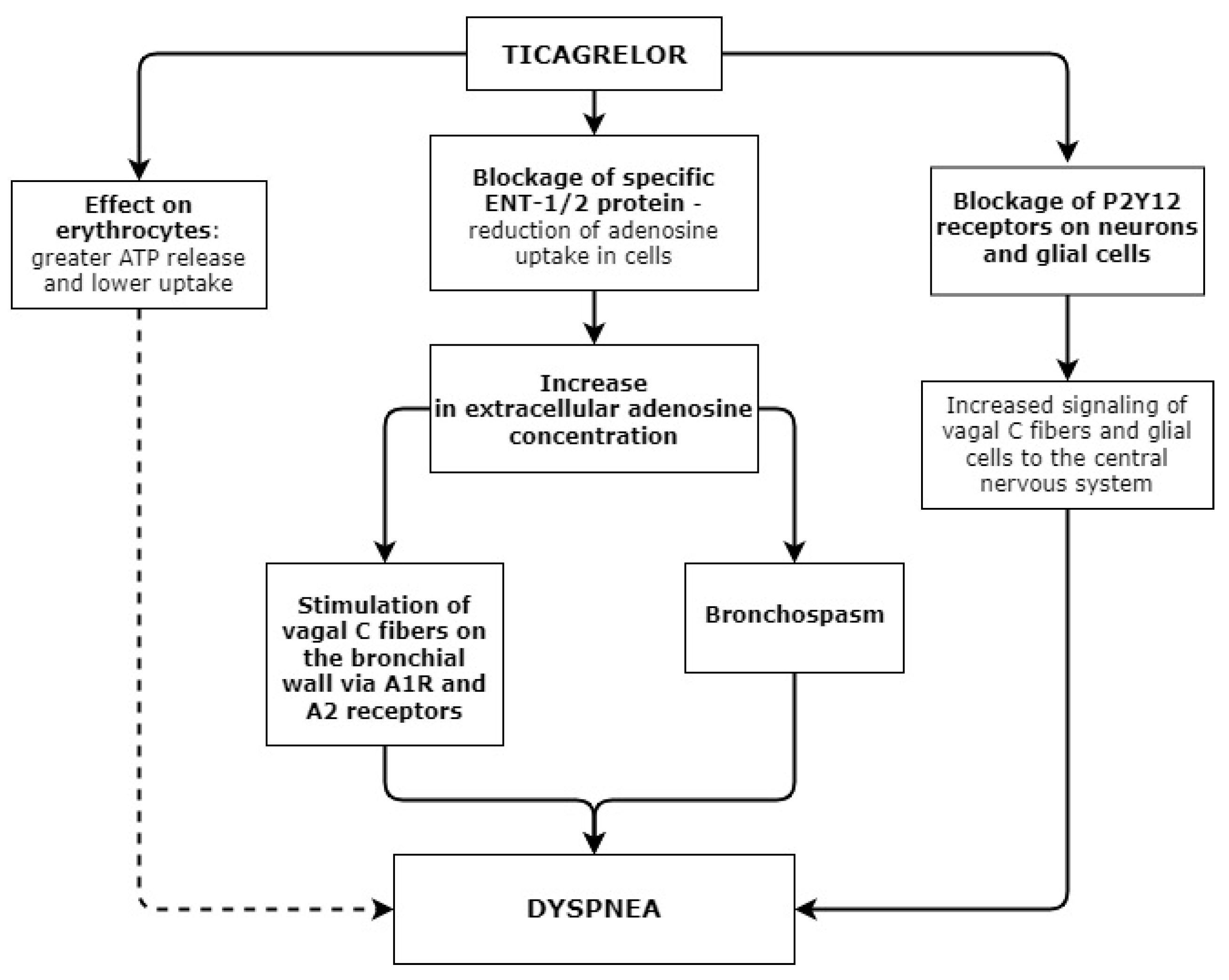
Figure 1. Possible mechanisms of ticagrelor-related dyspnea.
Applsci 12 10021 g001
Figure 2. Study flow chart.
Applsci 12 10021 g002
Figure 3. ROC curve analysis of ADP HS for the prediction of dyspnea. AUC = 0.612, 95% CI = 0.523–0.701, p = 0.015. ADP HS cut-off value = 19.5; sensitivity = 43.9%, specificity = 76.5%. Youden index J = 0.317.
Applsci 12 10021 g003
Table 1. Investigations and their timing.
InvestigationTiming
Complete blood count and blood biochemistry (e.g., potassium and creatinine concentration, lipid panel, troponin I, CRB, glycemia)On hospitalization, during treatment based on the need, and after 3 months
Troponin I dynamics6 h after hospitalization
Coronary angiography and PTCA with stent implantationUp to 60 min after hospitalization
Additional blood and urine testing following the study protocol24–36 h after hospitalization and 3 months
Dyspnea evaluation24–36 h from hospitalization and after 3 months
2D echocardiography and BNP measurement36–48 h from hospitalization and after 3 months
Other instrumental investigations (e.g., ECG)During treatment based on the need and after 3 months
Table 2. Modified Borg acute dyspnea scale.
DegreeSymptoms
0No symptoms of dyspnea
1Mild dyspnea
2Moderate dyspnea
3Severe dyspnea
4Very severe dyspnea followed by forced sitting position and panic attack
Table 3. Characteristics of the study population.
CharacteristicDyspneap
No (n = 182)Yes (n = 95)
Gender, n (%) 0.696
Male138 (75.8)70 (73.7)
Female44 (24.2)25 (26.3)
Age, years64 (42–88)66 (32–90)0.066
BMI, kg/m229 (21–50)28 (21–45)0.859
MI, n (%) 0.720
NSTEMI84 (46.2)46 (48.4)
STEMI98 (53.8)49 (51.6)
CAD, n (%)
One-vessel62 (31.1)27 (28.4)0.162
Two-vessel57 (31.3)42 (44.2)
Three-vessel63 (34.6)26 (27.4)0.034
DM, n (%)23 (12.6)11 (11.6)0.799
Smoking, n (%)50 (27.5)29 (30.5)0.068
Hypothyroidism, n (%)2 (1.1)5 (5.3)0.049
AF, n (%)24 (13.2)16 (16.8)0.411
Troponin I, ng/mL495 (20–89977)496 (20–85725)0.697
Troponin I after 6 h, ng/mL7816 (20–635352)4324 (20–147527)0.146
BNP, pmol/L131 (11–1858)140 (6–2000)0.89
BNP after 3 months, pmol/l70 (9–368)51 (5–271)0.708
ADP HS, U25 (9–147)19 (2–133)<0.001
ADP HS after 3 months, U23 (9–58)20 (1–32)0.009
Creatinine, µmol/L80 (42–480)85 (41–182)0.019
CRP, mg/L8.25 (0.3–145)5.8 (0.3–152.5)0.615
LDL-C, mmol/L3.5 (1.4–6.7)3.3 (1.4–7.1)0.348
HGB, g/L144 (93–387)142 (85–345)0.726
LVEF, %50 (20–55)45 (20–55)0.429
Medications, n (%)
Beta blockers177 (97.8)92 (97.9)0.894
ACE inhibitors177 (97.8)93 (98.9)0.974
Statins (irrespective of dose), n (%)
Atorvastatin95 (52.5)40 (42.6)0.027
Rosuvastatin86 (47.5)54 (57.4)0.255
Anticoagulants21 (11.5)15 (15.8)0.318
Values are the median (range) unless indicated otherwise. BMI—body mass index; NSTEMI—non-ST-segment elevation myocardial infarction; STEMI—ST-segment elevation myocardial infarction; CAD—coronary artery disease; DM—diabetes mellitus; AF—atrial fibrillation; BNP—B-type natriuretic peptide; LDL-C—low-density lipoprotein cholesterol; CRP—C-reactive protein; HGB—hemoglobin; ACE—angiotensin-converting enzyme; LVEF—left ventricular ejection fraction. ADP HS—platelet aggregation with high-sensitivity ADP.
Table 4. Incidence of dyspnea by different genotypes in the entire study population.
GenesDyspneaχ2p
NoYes
CYP4F2 (rs2108622)
CC83 (62.9)31 (54.4)1.3070.520
CT41 (31.1)21 (36.8)
TT8 (6.1)5 (8.8)
C alleleT allele207 (78.4)57 (21.6)83 (72.8)31 (27.2)1.3990.236
CYP4F2 (rs1558139)
AA23 (17.4)14 (24.6)1.6060.448
AG76 (57.6)32 (56.1)
GG33 (25.0)11 (19.3)
A allele122 (46.2)60 (52.6)1.3140.251
G allele142 (53.8)54 (47.4)
CYP4F2 (rs3093135)
AA18 (13.6)10 (17.5)1.8670.393
AT79 (59.8)28 (49.1)
TT35 (26.5)19 (33.3)
A allele115 (43.6)48 (42.1)0.0680.793
T allele149 (56.4)66 (57.9)
CYP4F2 (rs2074902)
CC3 (2.3)3 (5.3)1.5660.474
CT36 (27.3)17 (29.8)
TT93 (70.5)37 (64.9)
C allele42 (15.9)23 (20.1)1.0170.313
T allele222 (84.1)91 (79.8)
CYP2C19 (rs4244285) (*2)
AA1 (0.8)1 (1.8)0.7770.851
AG27 (20.5)11 (19.3)
GG104 (78.8)45 (78.9)
A allele29 (11.0)13 (11.4)0.0140.905
G allele235 (89.0)101 (88.6)
ABCB1 (rs1045642)
CC37 (28.0)14 (24.6)0.2520.882
CT70 (53.0)32 (56.1)
TT25 (18.9)11 (19.3)
C allele144 (54.6)60 (52.6)0.1170.731
T allele120 (45.4)54 (47.4)
FBG-C148T (rs1800787)
CC60 (45.5) 137 (64.9) 19.4890.009
CT59 (44.7) 212 (21.1) 2
TT13 (9.8)8 (14.0)
C allele179 (67.8)86 (75.4)2.2140.136
T allele85 (32.2)28 (24.6)
COX-2 (rs689465)
CC2 (1.5)0 (0.0)0.5321.000
CT22 (16.8)9 (15.8)
TT107 (81.7)48 (84.2)
C allele26 (9.9)9 (7.9)0.3870.533
T allele236 (90.1)105 (92.1)
PAI-1 (rs5918)
CC5 (3.8)2 (3.5)0.6190.840
CT49 (37.1)18 (31.6)
TT78 (59.1)37 (64.9)
C allele59 (22.4)22 (19.3)0.440.507
T allele205 (77.6)92 (80.7)
CYP2C19 (rs12248560) (*17)
CC63 (47.7)36 (63.2)3.8710.138
CT61 (46.2)18 (31.6)
TT8 (6.1)3 (5.3)
C allele187 (70.8)90 (79.0)2.6770.101
T allele77 (29.2)24 (21.0)
Values are the number (percentage). 1 p = 0.014; 2 p = 0.002.
Table 5. Associations between the results of ADP HS testing and dyspnea.
TimingADP HS, Up
Without DyspneaWith Dyspnea
Patients with diabetes mellitus (n = 34)
On enrolment25 (16–71)15 (10–41)0.002
After 3 months23 (12–51)22 (20–32)0.836
Patients without diabetes mellitus (n = 243)
On enrolment25 (9–147)19 (2–133)<0.001
After 3 months23 (9–58) 20 (1–79)0.004
One-vessel CAD (n = 89)
On enrolment25 (12–71)22 (12–47)0.048
After 3 months21 (9–43)19 (12–28)0.018
Two- and three-vessel CAD (n = 188)
On enrolment25 (9–147)18 (2–133)<0.001
After 3 months24 (11–58)21 (1–79)0.022
Values are median (range). CAD—coronary artery disease, ADP HS—platelet aggregation with high-sensitivity ADP.
Table 6. Factors increasing the risk of dyspnea.
VariableOR (95% CI)p
ADP HS > 19.51
ADP HS ≤ 19.54.07 (2.37–6.99)≤0.001
Maximal atorvastatin dose (80 mg)1
Any atorvastatin dose (or no atorvastatin)2.18 (1.18–4.01)0.012
Publisher’s Note: MDPI stays neutral with regard to jurisdictional claims in published maps and institutional affiliations.