Abstract
Early detection of caries is an urgent problem in the dental clinic. Current caries detection methods do not detect early enamel caries accurately, and do not show microstructural changes in the teeth. Optical coherence tomography (OCT) can provide imaging of tiny, demineralized regions of teeth in real time and noninvasively detect dynamic changes in lesions with high resolution and high sensitivity. Over the last 20 years, researchers have investigated different methods for quantitative assessment of early caries using OCT. This review provides an overview of the principles of enamel caries detection with OCT, the methods of characterizing caries lesion severity, and correlations between OCT results and measurements from multiple histological detection techniques. Studies have shown the feasibility of OCT in quantitative assessment of early enamel lesions but they vary widely in approaches. Only integrated reflectivity and refractive index measured by OCT have proven to have strong correlations with mineral loss calculated by digital microradiography or transverse microradiography. OCT has great potential to be a standard inspection method for enamel lesions, but a consensus on quantitative methods and indicators is an important prerequisite. Our review provides a basis for future discussions.
1. Introduction
Dental caries is one of the most common chronic diseases in people worldwide because of its high incidence and the wide range of the affected population [1]. Caries is a disease of dental hard tissue caused by oral microorganisms. Normally the teeth are in a dynamic balance of alternating demineralization and remineralization, in which demineralization is the dissolution of dental minerals in the presence of acid, from which inorganic ions such as calcium and phosphate are removed, and remineralization refers to the reprecipitation and crystallization of minerals in tissues that have been demineralized [2]. If demineralization continues, caries will occur. Early enamel caries involves the demineralization of the tooth surface, and does not show substantial defects. There is no significant difference between sound enamel and early enamel caries by visual observation. At this time, it is possible to promote the repair of carious tissues through non-destructive treatment of remineralization and interrupt caries progression. However, further irreversible dental hard tissue loss will occur without interventional treatment, and then only traumatic treatment can be performed [3]. Therefore, the detection of early caries is of great importance.
Common clinical diagnostic methods mainly include visual inspection and X-ray radiography. Visual inspection lacks objectivity and has limited accuracy, while X-ray radiography can only detect severe caries lesions [4,5]. These conventional methods cannot detect early caries, which makes the early diagnosis of caries difficult and delays the best time for treatment [6]. In recent years, optical-based caries detection methods, such as Raman spectroscopy [7,8], quantitative light-induced fluorescence (QLF) [9,10] and fiber-optic transillumination [11,12], have overcome some disadvantages of traditional methods, but they do not show the internal microstructural features of teeth and cannot quantify dynamic changes in early stages of enamel lesions [13].
Optical Coherence Tomography (OCT) is a noninvasive imaging method with high resolution and high sensitivity, regarded as an “optical biopsy” [14,15]. It has a wide range of medical applications, including ophthalmology [16,17], dentistry [18,19], dermatology [20,21], and gastroenterology [22,23]. For dental caries, OCT can detect tiny, demineralized areas on the tooth surface and inside, overcoming the disadvantages of other optical detection methods [24]. In dentistry, much research has demonstrated that OCT can image dental tissue clearly and be applied to the quantitative assessment of early enamel caries [25,26,27,28]. Mineral loss is the gold standard in cariology research that determines the degree of demineralization of the lesion. Only when the quantitative index of enamel caries obtained by OCT has a definite correlation with the enamel mineral loss, the severity of early enamel caries can be quantified more objectively and accurately.
To advance the application of OCT for early caries detection, a standardized and validated method to achieve quantitative detection of early caries is required. The purpose of this paper is to present an overview of the principles, methods and applications for the quantitative evaluation of early enamel caries by OCT and to provide a basis for the establishment of a uniform clinical standard in the future. This paper first discusses the optical properties of teeth and their changes with tooth demineralization, secondly introduces methods of artificial caries preparation, and then summarizes current research advances in evaluation of early enamel lesions using different quantitative indicators and discusses the limitations of the methods.
2. Optical Properties of Teeth
Biological tissues’ optical properties have important theoretical significance and application value for the diagnosis of diseases. The variation in optical properties of tooth tissue during demineralization are the basis of OCT caries detection [29].
- (1)
- Refractive Index
Refractive index describes the reflection and refraction of light as well as the temporal properties of light transmission within a tissue. The refractive index of tooth enamel is 1.62 to 1.65 [30,31]. Demineralization in caries lesions alters the refractive index of enamel [32], and can be considered as an index of its scattering properties [33].
Since hydroxyapatite in tooth enamel is an anisotropic crystal, tooth enamel has birefringent properties [18]. When polarized light propagates through biological tissues, its polarization state is altered by the scattering properties of the tissue. By detecting changes in the polarization state of backscattered light from dental tissue, microstructural information of the tissue can be obtained, and caries lesions can be detected.
- (2)
- Scattering Properties
Dependent on the structure of hard dental tissue, two types of scattering occur when light is transmitted inside the tooth. Dental enamel is an ordered array of hydroxyapatite crystals surrounded by a protein/lipid/water matrix. The diameter of hydroxyapatite crystals is about , and the crystals are clustered together into enamel rods with a diameter of 4 [34]. In the near-IR (NIR), the crystal diameter is much smaller than the source wavelength, so it is in accordance with the Rayleigh scattering law. The diameter of the enamel rods is comparable to the source wavelength, so it obeys the Mie scattering law.
Enamel has the greatest transparency in the NIR close to 1310 nm. The attenuation coefficients of sound enamel at 1310 nm and 1550 nm are 3.1 and 3.8 [35], respectively, which are much lower than in the visible region. Thus, the NIR spectrum is suited for tooth imaging to identify lesions [36]. Micropores form in the lesion as the mineral crystals are partially dissolved during the demineralization process, and behave as scattering centers. During initial lesion development, the scattering coefficient has an exponential increase with increasing mineral loss. As the severity of the lesion increases, the scattering gradually increases [29].
- (3)
- Absorption Properties
The absorption of enamel is quite weak in NIR. The absorption coefficient of enamel at 1310 nm is around 0.12 [36], which is much smaller than the scattering coefficient. Therefore, scattering plays a major role in light transmission through the tooth.
3. Preparation of Artificial Carious Lesions
The study of early caries using OCT requires tracking of the caries process, but natural early caries takes 6–18 months, and there are many influencing factors. Therefore, the preparation of artificial caries using isolated teeth is the main way to study early caries. The key to preparing artificial caries is to simulate the chemical and microbial environment of natural caries formation. There are two main methods to prepare artificial caries: chemical and biofilm methods. Chemical methods can be used for enamel surface demineralization by adjusting the chemical state and pH of a using a partially saturated acidic buffer [37,38] or an acid gel [39]. This method is easy and the most widely used, while a biofilm method is performed by placing a cariogenic suspension on the surface of the tooth, altering the metabolic environment of the bacteria and creating a carious sample that more closely resembles natural caries [40,41]. However, the biofilm method is generally used for studies related to the pathogenesis of dental caries because it is difficult to control the experimental conditions.
4. Quantitative Assessment Methods of Early Enamel Caries with OCT
Most of the current caries detection studies based on OCT use frequency domain OCT, which eliminates axial scanning of the reference arm and overcomes the disadvantage of slow scanning speed of time-domain OCT (TD-OCT) [42]. Moreover, some functional OCT systems are employed for caries detection due to the birefringent properties of dental hard tissues. Polarization-sensitive OCT (PS-OCT) as a type of functional OCT system can provides additional polarization-sensitive information about the sample compared to conventional OCT systems [43,44,45,46]. In addition, as a new type of PS-OCT, Cross Polarization OCT (CP-OCT) not only reflects the polarization-sensitive structural information of the sample, but also better reveals the superficial microstructure of the sample by attenuating the effect of strong reflected light on the sample surface [47].
4.1. Quantitative Assessment Based on Lesion Depths
During the demineralization process, the OCT image contrast of caries is formed as the mineral content (MC) of the enamel decreases and the optical properties change [48]. Therefore, lesion depths determined by OCT images can be used to quantify early enamel caries. First, the filters are applied to remove speckle noise and enhance contrast for the OCT B-scan image, then the A-scan in the region of interest (ROI) is searched for the first pixel that exceeds the intensity threshold point, and the distance from the enamel surface to this pixel is taken as the lesion depth. The mean lesion depth is obtained by calculating the lesion depth for each A-scan in the ROI. It has been suggested to select times the peak intensity as the signal intensity threshold. The lesion depth measured by OCT is proportional to the demineralization time [49,50,51,52,53]. Le et al. [49] investigated bovine enamel caries lesions with 1310-nm PS-OCT. Figure 1 shows the PS-OCT B-scan of one of the bovine enamel blocks in the perpendicular axis. It can be seen that the lesion depth increases with the enhancement of the back-scattered intensity around the enamel surface, and the lesion severity increases. The results show the mean lesion depths varies significantly from 10 to 75 for demineralization over 0–4 days. Moreover, there is strong correlation (r = 0.85) between the mean depth of lesion measured by PS-OCT and polarized light microscopy (PLM). Jones et al. [50] prepared artificial caries on the occlusal surfaces by using a 14-day pH cycle model and detected them with PS-OCT and digital microradiography (DM). It was found that the image contrast between the sound and lesion area in the perpendicular axis was stronger than that in the parallel axis. The mean lesion depth of caries lesions calculated from the perpendicular axis images of the ten teeth was 150 ± 30 , which was highly correlated with the lesion depths obtained from DM (r = 0.811). Meng [51] and Yao et al. [52] observed an approximately linear relationship between lesion depth and demineralization time using TD-OCT. Park et al. [53] established assessment criteria for OCT using lesion depths, and conducted a concordance study between OCT and light microscopy using ex vivo teeth, which showed moderate concordance (k = 0.54, p ≤ 0.001) with no significant difference (p = 0.25). Then, smooth surface in vitro and in vivo evaluations were performed using OCT and the International Caries Detection and Assessment System (ICDAS). The extent of caries was seen to vary considerably within each ICDAS category using OCT, which could effectively complement the visual assessment with ICDAS. Yavuz et al. [54] utilized 840-nm SD-OCT to assess the remineralization of artificial enamel caries. The results showed there was a significant reduction in the lesion depth after remineralization, 311.80 (344.38), 320.10 (244.36) and 312.70 (203.80) for the three remineralization agents, respectively. The measured lesion depth was also compared with a surface microhardness analysis but there was no correlation between the two.

Figure 1.
PS-OCT B-scan of bovine enamel in the perpendicular axis. D0 represents the sound area and D1–D4 represent areas demineralized for 1–4 days. A red-white-blue color chart was used, with red indicating strong reflectivity and blue indicating low reflectivity. This figure was adapted from [49].
Lesion depth is a common quantitative index used in early caries studies. The main challenge for its calculation is the difficulty in selecting an intensity threshold as the end point of a lesion, as the range of OCT images is relatively high. The method of selecting times the peak intensity as the signal intensity threshold does not always work effectively [55]. Le et al. [49] used edge-finding algorithms based on this method to measure lesion depth. Other studies designed algorithms to determine the lesion boundary in the image and obtained lesion depth [51,56,57]. A summary of the above research results is shown in Table 1.

Table 1.
Results of quantifying enamel caries with lesion depths.
Lesion depths of caries can visually indicate the severity of caries to some extent. However, they can only reflect part of the characteristics of the initial stage of enamel lesions, as the amount of mineral loss may be different in a certain depth range [58]. Furthermore, there is a lack of solid criteria for determining the cut-off point to define lesion depth.
4.2. Quantitative Assessment Based on Reflectivity
Many studies have used reflectivity for the quantitative assessment of early caries, since the reflectivity of caries lesions can be obtained directly from OCT signals. A commonly used quantitative index related to reflectivity is the integrated reflectivity for caries detection.
A line profile for each lesion depth is taken from B-scans, and the integrated reflectivity can be calculated by integrating the reflectivity from the enamel surface to various depths [50]. The observed optical depth should be divided by the enamel refraction index (n = 1.63) to determine the real lesion depth when using the line profile.
Most studies have confirmed that integrated reflectivity increases after demineralization and decreases after remineralization [49,50,57,59,60]. Le et al. [49] utilized a fixed depth algorithm and an edge detection algorithm to calculate integrated reflectivity. In the first algorithm the integration was performed to a fixed depth that needed to be greater than the maximum lesion depth, while the second algorithm could obtain the depth of the lesion. The results showed both algorithms were able to detect the difference in demineralization from 0 to 4 days, except that the fixed depth algorithm yielded a higher integrated reflectivity. Jones et al. [50] calculated the mean integrated reflectivity of artificial caries prepared by applying a 14-day pH cycle model based on the perpendicular axis PS-OCT images, and the result was 450 ± 110 arbitrary units. Meanwhile, it was demonstrated that the integrated reflectivity calculated by PS-OCT was linearly correlated with the relative mineral loss determined by DM (r = 0.755). Nee et al. [59] detected demineralization around adhesive-bound orthodontic brackets in vivo using CP-OCT for a period of 1 year and acquired 2D projection images of with automated algorithms. The results indicated for both adhesives increased remarkably with time, varying in the range from 10.2 (10.5) to 29.7 (9.4) . PS-OCT was applied to monitor the process of remineralization of caries using an acid remineralization model by Kang et al. [60]. There were significant alterations in the integrated reflectivity of the lesion region after remineralization, from 257 ± 60.2 to 168 ± 58.5 .
Amaechi et al. [61] scanned demineralized bovine teeth using 850-nm OCT and demonstrated that of the enamel reduced with the time of demineralization. They proposed the percentage reflectivity loss () as a quantitative index as follows:
where is the reflectivity of sound enamel and is the reflectivity of enamel lesion.
The results showed that increased from 54.0 ± 11.27 to 86.64 ± 7.57 with demineralization time. In a follow-on study it was demonstrated that was linearly correlated with both the mineral loss determined by transverse microradiography (TMR) () [62] and the percentage of fluorescence loss calculated () by QLF () [63]. However, is rarely applied. A summary of the above research results is shown in Table 2.

Table 2.
Results of quantifyin0p-[g enamel caries with reflectivity.
In addition, other researchers have used the mean relative reflectivity (mRR) proposed in retinal OCT imaging to assess fissure caries by using 1325-nm SS-OCT [64]. The mRR is calculated from the difference between the fissure area signal and sound enamel signal. Although the mRR of demineralized fissures were at least 6 times higher than those of sound fissures at 250, 500 and 1000 depths beneath the surface, the mRR was unable to accurately describe lesion mineral density ().
In summary, the integrated reflectivity of enamel lesion calculated by OCT is linearly correlated with mineral loss. Most results confirm that the integrated reflectivity of enamel increases after demineralization, but individual studies show the opposite. Researchers often use the integrated reflectivity in combination with lesion depth to evaluate the lesion severity. However, the calculation of the integrated reflectivity suffers from similar problems to the lesion depth calculation.
4.3. Quantitative Assessment Based on Attenuation Coefficient
The attenuation coefficient is the sum of the absorption coefficient and scattering coefficient [35]. The attenuation characteristics of sound and carious enamel are different because of the alteration in optical properties of the teeth after demineralization. Hence, the attenuation coefficient can be used to quantify early enamel caries. Attenuation coefficients can be obtained by fitting the normalized A-scan signal with the Beer-Lambert law equation [65] as follows:
where is the OCT signal intensity, is the attenuation coefficient, and is the depth beneath the tooth surface.
Mandurah et al. [66] reported that attenuation coefficients for sound areas of the samples were the smallest, ranging from 0.08 to 0.29 , increasing to a range of 1.34 to 3.4 after demineralization, and decreasing after remineralization with mean values of 0.81 and 0.85 . Moreover, there was a strong linear regression (r = −0.97) between the measured by OCT and integrated nanohardness (INH) measured by a nanoindentation device. Hardness has been recognized as a measure of hard tissue’s mineral density for a long time [67]. Maia et al. [68] studied morphological alterations between sound enamel and artificial white spot lesions in human teeth using OCT and QLF. The attenuation coefficient increases of enamel lesions ranged between 27.8% and 62.5%, while fluorescence intensity reduction ranged between 11.9% and 34.2%. Therefore, it was demonstrated that determined by OCT was more sensitive to alterations than fluorescence measured by QLF. Cara et al. [69] verified that the attenuation coefficient could be employed for the initial lesion to effectively discriminate between sound and demineralized enamel with 0.93 sensitivity and 0.96 specificity.
A weaker attenuation of the OCT signal in enamel lesions was observed in Popescu et al.’s work [70,71]. The mean attenuation coefficient was 1.35 for sound enamel and 0.77 for caries lesions [70]. They attributed the results to the high porosity of demineralized enamel. One possible reason for the contradictory results of the studies mentioned above is the use of different wavelengths. Mandurah et al.’s study used the 1310-nm SS-OCT system, while Popescu et al.’s study used the 850-nm OCT system. The optical properties of enamel at the two wavelength ranges are different. A summary of the above research results is shown in Table 3.

Table 3.
Results of quantifying enamel caries with the attenuation coefficient.
The Beer-Lambert equation used in the methods is based on a single scattering model. The single scattering model only considers single scattering, while the demineralization of caries enhances the effect of multiple scattering. This leads to bias of the obtained attenuation coefficients. In addition, reliably extracting attenuation coefficients from OCT signals can be affected by noise. These features diminish the utility of employing the attenuation coefficient as a marker for early enamel caries detection.
4.4. Quantitative Assessment Based on Degree of Polarization
Demineralized enamel results in rapid depolarization of polarized light in the NIR due to increased scattering [72], which has been confirmed by PS-OCT measurements [73]. Polarization imaging can provide higher contrast images of early enamel caries. Since OCT is an interferometric imaging method, only the contribution of fully polarized light can be measured. Thus, the degree of polarization within a single speckle is always equal to 1. However, when depolarized, the polarization state of the adjacent speckles is uncorrelated. Therefore, the degree of polarization uniformity (DOPU) has been proposed to assess carious lesions [74]. DOPU can be derived by an averaging of Stokes vectors over adjacent speckles, as follows:
where , and are the mean values of the Stokes vector elements within a certain evaluation kernel. It can be seen that the value of DOPU depends on the number of speckles in the chosen kernel.
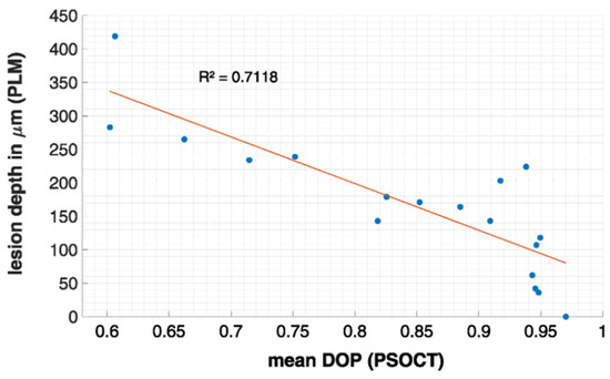
The combination of the DOPU algorithm and PS-OCT was first applied to detect carious lesions by Golde et al. [74], and has been used for ophthalmologic research [75]. They measured three tooth samples with different proximal lesions, and the significant DOPU contrast provided better identification of lesions in comparison with reflectivity images. Furthermore, the effect of different DOPU evaluation kernel sizes on the resulting contrast was investigated. In a following study, they improved the DOPU algorithm by noise-immune processing, and adopted this approach to examine two tooth samples with stains and occlusal lesions [76]. Then, the research group measured the DOPU of bovine enamel at different stages of demineralization by using PS-OCT, and compared it with lesion depth obtained from PLM measurements [77]. The results showed that there was no depolarization in sound enamel, but an increased depolarization after 15 days of demineralization, corresponding to a decrease in DOPU. There was a high linear correlation () between the DOPU and measured lesion depth with PLM, as shown in Figure 2. The summary of the above research results is shown in Table 4.

Figure 2.
Correlation between the calculated mean DOP by PS-OCT and determined lesion depths by PLM. This figure was adapted from [77].

Table 4.
Results of quantifying enamel caries with degree of polarization.
While the above results indicate the feasibility of assessing the demineralization stage by DOPU, there is a need to investigate the validity of DOPU at various polarization changes and the correlation between DOPU and mineral loss measured by TMR for further studies.
4.5. Quantitative Assessment Based on Refractive Index
Demineralization causes a change in the refractive index of enamel, and accurate measurement of this change can assist in the identification of early caries [32]. The refractive index of teeth can be determined with the optical path-length matching method using OCT [78]. Samples are put onto a metal plate to acquire OCT images. The depth position of the reflection surface of the metal plate before the sample is placed is . After adding samples, the depth positions of the sample surface and the metal plate surface are and , respectively. The thickness of the sample is . Then, the refractive index of teeth is determined by [79]:
Hariri et al. [80] measured refractive index of sound bovine enamel, demineralized for 2 months, and remineralized for 2 months, by 1310-nm SS-OCT with an axial/lateral resolution 11/17 , and analyzed mineral content by TMR. The results showed that at an n range between 1.52 and 1.63, the mineral content ranged between 50 and 87 (vol.%). This indicates there were strong positive linear correlations between and mineral content in both demineralized enamel () and remineralized enamel (). However, this method required sectioning of the sample to measure the refractive index, which is destructive and cannot be applied in clinical practice.
4.6. Quantitative Assessment Based on Scattering Coefficient
Since the scattering properties of the enamel changes significantly after demineralization, the scattering coefficient can serve as an indicator of enamel lesion severity. A single scattering model combined with dynamic focusing can be used to determine the scattering coefficients of sound and carious enamel [81]. The OCT signal intensity is:
where is the depth, is the focal plane position, is Rayleigh length, and is the OCT depth profile.
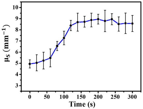
Tsai et al. [81] applied acid gel to demineralize enamel and scanned the sample in vitro before and after demineralization using 850-nm SD-OCT with an axial/lateral resolution 3/4 . The estimated scattering coefficient is shown in Figure 3. The scattering coefficient increased with the demineralization time and leveled out at times greater than 120 s. Moreover, the average scattering coefficients were 4.60 and 8.46 for sound and carious enamel, respectively.

Figure 3.
Variation of scattering coefficient with demineralization time. This figure was adapted from [81] and was created with Microsoft Word (Microsoft Corp., Redmond, WA, USA).
However, as mentioned above, the single scattering model used above is inaccurate for enamel caries. According to the optical properties of the caries lesion, it shows a significant growth in the scattering coefficient of enamel during the production of the initial lesion. Hence, if an accurate scattering coefficient is used for the quantitative assessment of early caries, early demineralization can be detected more sensitively. However, there are few studies using scattering coefficient to quantitatively evaluate early caries.
4.7. Quantitative Assessment Based on the Surface Roughness of Enamel
Acid or bacterial erosion alters the surface roughness of enamel. The root mean square of the surface roughness can be expressed as [81]:
where is the total number of A-scans, and are the surface and underlying depth positions of lesion area in A-scan, respectively.
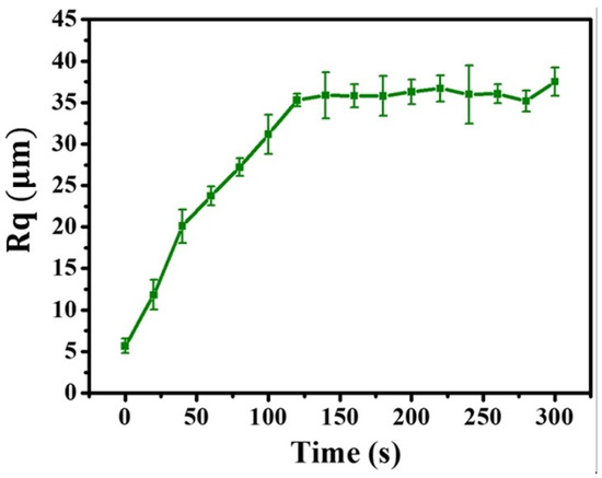
Tsai et al. [81] estimated the surface roughness of the demineralized enamel, as shown in Figure 4. The surface roughness of the enamel increased gradually with demineralization time, and tended to level out, varying from 5.11 to 31.7 . Although the results demonstrated that the surface roughness could be applied for the detection of early caries, there were estimation errors compared with the results of scanning electron microscopy (SEM), and the effect of artificial caries and natural caries on enamel surface roughness may be different. In addition, there are few relevant studies.

Figure 4.
Variation of the surface roughness with demineralization time. This figure was adapted from [81] and was created with Microsoft Word (Microsoft Corp., Redmond, WA, USA).
4.8. Quantitative Assessment Based on the Volume of Residual Enamel
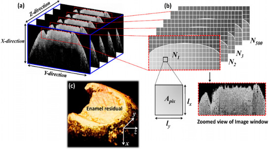
Demineralization caused by caries lesions changes the volume of residual enamel. Wijesinghe et al. [82] obtained cross-sectional images of sound, partially demineralized and completely demineralized teeth in vitro by using 1310-nm SD-OCT with an axial/lateral resolution 6/25 , and measured the volume of residual enamel with an automated calculation method based on pixel intensity. The volumetric evaluation algorithm is shown in Figure 5. For the precise selection of residual enamel, an image window is applied to the 2D OCT images, as shown in Figure 5a. Then, the pixels that satisfy the pre-determined intensity threshold range are selected as shown in Figure 5b. Finally, a 3D OCT volumetric image is obtained as shown in Figure 5c. The volume of residual enamel is determined by:
where is the number of pixels in each window that satisfy the predetermined intensity cut-off points, n is the number of 2D images contained in the 3D image. , and are the pixel sizes in the x, y and z directions, respectively.

Figure 5.
Evaluation algorithm for the volume of residual enamel. (a) 2D images with the applied image window; (b) Pixels that meet the predetermined intensity cut-off points; (c) 3D volumetric image. This figure was adapted from [82].
The volume of residual enamel for carious samples, partially demineralized samples and sound samples ranged from 12.26 to 28.72 . The progression of dental caries is determined by detecting changes in the volume of residual enamel. When reduction in tooth volume is identified, medication can be taken immediately to inhibit the development of caries. The key in this method is the determination of threshold parameters, which requires the evaluation and standardization of volumetric information for multiple in vivo teeth to enhance accuracy.
4.9. Quantitative Assessment Based on the Dehydration Parameter
Recent research has revealed the impact of hydration on OCT images by conventional polarization-insensitive OCT [83,84]. A method for assessing early enamel lesions with the dehydration parameter (DH) based on the integrated reflectivity has been presented [85]. The dehydration parameter refers to the positive difference between the two OCT signals of tooth enamel under dry and hydrated conditions, i.e., the integrated area between the two signals.
Nazari et al. [85] detected sound and demineralized bovine enamel blocks after 3, 9, and 15 days of demineralization using 1310-nm SS-OCT with axial/lateral resolution 11/17 , and calculated the dehydration parameters. The experimental results showed DH for sound and demineralized bovine enamel ranged from 272(204) to 3304(751), and a strong linear correlation () between the dehydration parameter and the square root of demineralization day. The benefit of this method is that the DH calculation does not involve determination of the cut-off depth, and the evaluation results are not influenced by surface reflections. Although this method has the potential to quantitatively assess early enamel caries, there are few relevant studies, and optimization of the methodology requires evaluation of large amounts of demineralized and remineralized enamel.
5. Conclusions
In conclusion, the common aim of the discussed studies was to investigate and improve the capabilities of OCT in monitoring the pathophysiological process of early enamel caries and remineralization. Researchers have proposed multiple quantitative indicators for the assessment of early enamel caries and have studied correlations with the results of multiple histological detection techniques. By far the most widely used quantitative indicators include lesion depth, integrated reflectivity and attenuation coefficients. The differences in quantitative results are mainly attributed to the use of different systems, different methods and different sample preparation. Among them, there is a high linear correlation between depths of enamel lesions determined by OCT and DM, as well as between integrated reflectivity calculated by OCT and mineral loss determined by DM. However, the assessment methods using these three quantitative indicators still have certain limitations and lack objectivity and accuracy. It is a remarkable fact that the scattering coefficient of tooth enamel is very sensitive to changes in mineral content and increases significantly with increasing mineral loss at the initial stages of the enamel caries process. Therefore, the early detection of enamel lesions using the scattering coefficient based on OCT is a potential research direction. Meanwhile, most studies on quantitative assessment of enamel caries have been performed in vitro due to the limitations of the probe, and in vivo studies are mainly focused on the buccal and incisal/occlusal surfaces of premolars and anterior teeth. With the development of intraoral probes [4,86], the problem of device availability is being solved slowly. However, a very important issue is the lack of consensus on the method, and OCT has not been applied for the clinical diagnosis of early caries. Although OCT has made great progress as a noninvasive imaging method for quantitatively assessing early enamel caries, efforts to standardize rigorous methodology in future research are crucial for detection, diagnosis, and treatment guidance of early enamel lesions using OCT.
Author Contributions
Conceptualization, all authors; methodology, B.S., J.N., X.Z.; formal analysis, B.S., J.N.; investigation, B.S., J.N.; resources, all authors; data curation, B.S., J.N.; writing—original draft preparation, B.S., J.N.; writing—review and editing, all authors; visualization, B.S., J.N.; supervision, X.Z., X.D.; project administration, B.S., J.N.; funding acquisition, B.S., J.N. All authors have read and agreed to the published version of the manuscript.
Funding
This research was funded by the Natural Science Foundation of Tianjin (grant number 19JCYBJC16200) and the Science and Technology Program of Tianjin (grant number 20YDTPJC01530).
Institutional Review Board Statement
Not applicable.
Informed Consent Statement
Not applicable.
Data Availability Statement
All the abovementioned data can be found in the references.
Conflicts of Interest
The authors declare no conflict of interest.
References
- Baelum, V.; Fejerskov, O. Caries diagnosis: A mental resting place on the way to intervention. In Dental Caries–The Disease and Its Clinical Management; Blackwell Munksgaard: London, UK, 2003; pp. 101–110. [Google Scholar]
- Dirks, O.B. Posteruptive Changes in Dental Enamel. J. Dent. Res. 1966, 45, 503–511. [Google Scholar] [CrossRef]
- Tenbosch, J.J.; Verdonschot, E.H.; Vaarkamp. Light propagation through teeth containing simulated caries lesions. Phys. Med. Biol. 1995, 40, 1375–1387. [Google Scholar]
- Schneider, H.; Ahrens, M.; Strumpski, M.; Rüger, C.; Häfer, M.; Hüttmann, G.; Theisen-Kunde, D.; Schulz-Hildebrandt, H.; Haak, R. An Intraoral OCT Probe to Enhanced Detection of Approximal Carious Lesions and Assessment of Restorations. J. Clin. Med. 2020, 9, 3257. [Google Scholar] [CrossRef] [PubMed]
- Schneider, H.; Park, K.J.; Häfer, M.; Rüger, C.; Schmalz, G.; Krause, F.; Schmidt, J.; Ziebolz, D.; Haak, R. Dental Applications of Optical Coherence Tomography (OCT) in Cariology. Appl. Sci. 2017, 7, 472. [Google Scholar] [CrossRef]
- Brouwer, F.; Askar, H.; Paris, S.; Schwendicke, F. Detecting Secondary Caries Lesions: A Systematic Review and Meta-analysis. J. Dent. Res. 2016, 95, 143–151. [Google Scholar] [CrossRef] [PubMed]
- Jones, R.S.; Huynh, G.D.; Jones, G.C.; Fried, D. Near-infrared transillumination at 1310-nm for the imaging of early dental decay. Opt. Exp. 2003, 11, 2259–2265. [Google Scholar] [CrossRef]
- Schaefer, G.; Pitchika, V.; Litzenburger, L.; Hickel, R.; Kühnisch, J. Evaluation of occlusal caries detection and assessment by visual inspection, digital bitewing radiography and near-infrared light transillumination. Clin. Oral Investig. 2018, 22, 2431–2438. [Google Scholar] [CrossRef]
- Alammari, M.R.; Smith, P.W.; De Josselin De Jong, E.; Higham, S.M. Quantitative light-induced fluorescence (QLF): A tool for early occlusal dental caries detection and supporting decision making in vivo. J. Dent. 2013, 41, 127–132. [Google Scholar] [CrossRef]
- Gomez, J. Detection and diagnosis of the early caries lesion. BMC Oral Health 2015, 15, 3. [Google Scholar] [CrossRef]
- Abogazalah, N.; Ando, M. Alternative methods to visual and radiographic examinations for approximal caries detection. J. Oral Sci. 2017, 59, 315–322. [Google Scholar] [CrossRef]
- Pretty, I.A. Caries detection and diagnosis: Novel technologies. J. Dent. 2006, 34, 727–739. [Google Scholar] [CrossRef]
- Patil, C.A.; Bosschaart, N.; Keller, M.D.; Leeuwen, T.G.; Mahadevan-Jansen, A. Combined Raman spectroscopy and optical coherence tomography device for tissue characterization. Opt. Lett. 2008, 33, 1135–1137. [Google Scholar] [CrossRef] [PubMed]
- Qiao, W.; Chen, Z. All-optically integrated photoacoustic and optical coherence tomography: A review. J. Innov. Opt. Health Sci. 2017, 10, 1730006. [Google Scholar] [CrossRef]
- Tearney, G.J.; Brezinski, M.E.; Bouma, B.E.; Boppart, S.A.; Pitvis, C.; Southern, J.F.; Fujimoto, J.G. In vivo endoscopic optical biopsy with optical coherence tomography. Science 1997, 276, 2037–2039. [Google Scholar] [CrossRef]
- Hangai, M.; Ojima, Y.; Gotoh, N.; Inoue, R.; Yasuno, Y.; Makita, S.; Yamanari, M.; Yatagai, T.; Kita, M.; Yoshimura, N. Three-dimensional Imaging of Macular Holes with High-speed Optical Coherence Tomography. Ophthalmology 2007, 114, 763–773. [Google Scholar] [CrossRef] [PubMed]
- Yasuno, Y.; Hong, Y.; Makita, S. In vivo high-contrast imaging of deep posterior eye by 1-μm swept source optical coherence tomography and scattering optical coherence angiography. Opt. Exp. 2007, 15, 6121–6139. [Google Scholar] [CrossRef]
- Baumgartner, A.; Dichtl, S.; Hitzenberger, C.K.; Sattmann, H.; Robl, B.; Moritz, A.; Fercher, A.F.; Sperr, W. Polarization–Sensitive Optical Coherence Tomography of Dental Structures. Caries Res. 2000, 14, 59–69. [Google Scholar] [CrossRef] [PubMed]
- Colston, B.W.; Sathyam, U.S.; DaSilva, L.B.; Everett, M.J.; Stroeve, P.; Otis, L.L. Dental OCT. Opt. Exp. 1998, 3, 230–238. [Google Scholar] [CrossRef]
- Pagnoni, A.; Knuettel, A.; Welker, P.; Rist, M.; Stoudemayer, T.; Kolbe, L.; Sadiq, I.; Kligman, A.M. Optical coherence tomography in dermatology. Skin Res. Technol. 1999, 5, 83–87. [Google Scholar] [CrossRef]
- Pierce, M.C.; Strasswimmer, J.; Park, H.; Cense, B.; Boer, J.F. Birefringence measurements in human skin using polarization-sensitive optical coherence tomography. J. Biomed. Opt. 2004, 9, 287–291. [Google Scholar] [CrossRef] [PubMed]
- Evans, J.A.; Poneros, J.M.; Bouma, B.E.; Bressner, J.; Halpern, E.F.; Shishkov, M.; Lauwers, G.Y.; Kenudson, M.M.; Nishioka, N.S.; Tearney, G.J. Optical coherence tomography to identify intramucosal carcinoma and high-grade dysplasia in Barrett’s esophagus. Clin. Gastroenterol. Hepatol. 2006, 4, 38–43. [Google Scholar] [CrossRef]
- Poneros, J.M.; Brand, S.; Bouma, B.E.; Tearney, G.J.; Compton, C.C.; Nishioka, N.S. Diagnosis of Specialized Intestinal Metaplasia by Optical Coherence Tomography. Gastroenterology 2001, 51, 7–12. [Google Scholar] [CrossRef] [PubMed]
- Yang, Z.; Shang, J.; Liu, C.; Zhang, J.; Hou, F.; Liang, Y. Intraoperative imaging of oral-maxillofacial lesions using optical coherence tomography. J. Innov. Opt. Health Sci. 2020, 13, 2050011. [Google Scholar] [CrossRef]
- Na, J.; Baek, J.H.; Choi, E.S.; Ryu, S.Y.; Chang, J.; Lee, C.S.; Lee, B.H. Assessment of dental-caries using optical coherence tomography. Int. Soc. Opt. Eng. SPIE 2006, 6137, 18–27. [Google Scholar]
- Na, J.; Baek, J.H.; Ryu, S.Y.; Lee, C.; Lee, B.H. Tomographic imaging of incipient dental-caries using optical coherence tomography and comparison with various modalities. Opt. Rev. 2009, 16, 426–431. [Google Scholar] [CrossRef]
- Holtzman, J.S.; Osann, K.; Pharar, J. Ability of Optical Coherence Tomography to Detect Caries Beneath Commonly Used Dental Sealants. Lasers Surg. Med. 2010, 42, 752–759. [Google Scholar] [CrossRef]
- Shimada, Y.; Sadr, A.; Burrow, M.F.; Tagami, J.; Ozawa, N.; Sumi, Y. Validation of swept-source optical coherence tomography (SS-OCT) for the diagnosis of occlusal caries. J. Dent. 2010, 38, 655–665. [Google Scholar] [CrossRef]
- Darling, C.L.; Huynh, G.; Fried, D. Light scattering properties of natural and artificially demineralized dental enamel at 1310 nm. J. Biomed. Opt. 2006, 11, 034023. [Google Scholar] [CrossRef]
- Ohmi, M.; Ohnishi, Y.; Yoden, K.; Haruna, M. In vitro simultaneous measurement of refractive index and thickness of biological tissue by the low coherence interferometry. IEEE Trans. Biomed. Eng. 2000, 47, 1266–1270. [Google Scholar] [CrossRef]
- Wang, X.J.; Milner, T.E.; Boer, J.F.d.; Zhang, Y.; Pashley, D.H.; Nelson, J.S. Characterization of dentin and enamel by use of optical coherence tomography. Appl. Opt. 1999, 38, 2092–2096. [Google Scholar] [CrossRef]
- Besic, F.C.; Wiemann, M.R. Dispersion Staining, Dispersion, and Refractive Indices in Early Enamel Caries. J. Dent. Res. 1972, 51, 973–985. [Google Scholar] [CrossRef]
- Knuettel, A.R.; Bonev, S.M.; Knaak, W. New method for evaluation of in vivo scattering and refractive index properties obtained with optical coherence tomography. J. Biomed. Opt. 2004, 9, 265–273. [Google Scholar] [CrossRef] [PubMed]
- Fried, D.; Glena, R.E.; Featherstone, J.D.B.; Seka, W. Nature of light scattering in dental enamel and dentin at visible and near-infrared wavelengths. Appl. Opt. 1995, 34, 1278–1285. [Google Scholar] [CrossRef] [PubMed]
- Jones, R.; Fried, D. Attenuation of 1310-nm and 1550-nm laser light through sound dental enamel. Lasers Dent. VIII 2002, 4610, 187–190. [Google Scholar]
- Spitzer, D.; Bosch, J.T. The absorption and scattering of light in bovine and human dental enamel. Calcif. Tiss. Res. 1975, 17, 129–137. [Google Scholar] [CrossRef] [PubMed]
- Chan, K.H.; Chan, A.C.; Darling, C.L.; Fried, D. Methods for Monitoring Erosion Using Optical Coherence Tomography. Lasers Dent. XIX 2013, 8566, 35–40. [Google Scholar]
- Lee, C.; Darling, C.L.; Fried, D. Polarization-sensitive optical coherence tomographic imaging of artificial demineralization on exposed surfaces of tooth roots. Dent. Mater. 2009, 25, 721–728. [Google Scholar] [CrossRef]
- Şen, S.; Erber, R.; Deurer, N.; Orhan, G.; Lux, C.J.; Zingler, S. Demineralization Detection in Orthodontics Using an Ophthalmic Optical Coherence Tomography Device Equipped with a Multicolor Fluorescence Module. Clin. Oral Investig. 2020, 24, 2579–2590. [Google Scholar] [CrossRef]
- Azevedo, C.S.; Trung, L.C.E.; Simionato, M.R.L.; Freitas, A.Z.; Matos, A.B. Evaluation of caries-affected dentin with optical coherence tomography. Braz. Oral Res. 2011, 25, 407–413. [Google Scholar] [CrossRef] [PubMed]
- Freitas, A.Z.; Zezell, D.M.; Mayer, M.P.A.; Ribeiro, A.C.; Gomes, A.S.L.; Jr., N.D.V. Determination of dental decay rates with optical coherence tomography. Laser Phys. Lett. 2009, 6, 896–900. [Google Scholar] [CrossRef]
- Espigares, J.; Sadr, A.; Hamba, H.; Shimada, Y.; Otsuki, M.; Tagami, J.; Sumi, Y. Assessment of natural enamel lesions with optical coherence tomography in comparison with microfocus X-ray computed tomography. J. Med. Imag. 2015, 2, 014001. [Google Scholar] [CrossRef] [PubMed]
- Hee, M.R. Optical Coherence Tomography of the Human Retina. Arch. Ophthalmol. 1995, 113, 325–332. [Google Scholar] [CrossRef] [PubMed]
- Kurokawa, K.; Sasaki, K.; Makita, S.; Yamanari, M.; Cense, B.; Yasuno, Y. Simultaneous high-resolution retinal imaging and high-penetration choroidal imaging by one-micrometer adaptive optics optical coherence tomography. Opt. Exp. 2010, 18, 8515–8527. [Google Scholar] [CrossRef]
- Boer, J.F.; Milner, T.E.; Gemert, M.J.; Nelson, J.S. Two-dimensional birefringence imaging in biological tissue by polarization-sensitive optical coherence tomography. Opt. Lett. 1997, 22, 934–936. [Google Scholar] [CrossRef]
- Saxer, C.E.; Boer, J.F.; Hyle Park, B.H.; Zhao, Y.; Chen, Z.; Nelson, J.S. High-speed fiber-based polarization-sensitive optical coherence tomography of in vivo human skin. Opt. Lett. 2000, 25, 1355–1357. [Google Scholar] [CrossRef] [PubMed]
- Chan, K.H.; Tom, H.; Lee, R.C.; Kang, H.; Simon, J.C.; Staninec, M.; Darling, C.L.; Pelzner, R.B.; Fried, D. Clinical monitoring of smooth surface enamel lesions using CP-OCT during nonsurgical intervention. Lasers Surg. Med. 2016, 48, 915–923. [Google Scholar] [CrossRef] [PubMed]
- Chen, Y.; Otis, L.; Piao, D.; Zhu, Q. Characterization of dentin, enamel, and carious lesions by a polarization-sensitive optical coherence tomography system. Appl. Opt. 2005, 44, 2041–2048. [Google Scholar] [CrossRef]
- Le, M.H.; Darling, C.L.; Fried, D. Automated analysis of lesion depth and integrated reflectivity in PS-OCT scans of tooth demineralization. Lasers Surg. Med. 2010, 42, 62–68. [Google Scholar] [CrossRef] [PubMed]
- Jones, R.S.; Darling, C.L.; Featherstone, J.D.B.; Fried, D. Imaging Artificial Caries on the Occlusal Surfaces with Polarization-Sensitive Optical Coherence Tomography. Caries Res. 2006, 40, 81–89. [Google Scholar] [CrossRef]
- Meng, Z.; Yao, X.; Yao, H.; Liu, T.; Li, Y.; Wang, G. Detecting early artificial caries by using optical coherence tomography. Chin. J. Lasers 2010, 37, 2709–2713. [Google Scholar] [CrossRef]
- Yao, H.; Li, Y.; Wang, G.; Yao, X.; Meng, Z.; Jin, S.; Liang, Y.; Zhang, L.; Liu, T. Quantification detecting artificial early caries with an All-fiber-OCT system. Int. J. Biomed. Eng. 2009, 37, 65–69+66. [Google Scholar]
- Park, K.J.; Schneider, H.; Ziebolz, D.; Krause, F.; Haak, R. Optical coherence tomography to evaluate variance in the extent of carious lesions in depth. Lasers Med. Sci. 2018, 33, 1573–1579. [Google Scholar] [CrossRef]
- Yavuz, B.S.; Kargul, B. Comparative evaluation of the spectral-domain optical coherence tomography and microhardness for remineralization of enamel caries lesions. Dent. Mater. J. 2021, 40, 1115–1121. [Google Scholar] [CrossRef] [PubMed]
- Can, A.M.; Darling, C.L.; Ho, C.; Fried, D. Non-destructive assessment of inhibition of demineralization in dental enamel irradiated by a λ = 9.3-μm CO2 laser at ablative irradiation intensities with PS-OCT. Lasers Surg. Med. 2008, 40, 342–349. [Google Scholar] [CrossRef]
- Staninec, M.; Douglas, S.M.; Darling, C.L.; Chan, K.; Kang, H.; Lee, R.C.; Fried, D. Non-destructive clinical assessment of occlusal caries lesions using near-IR imaging methods. Lasers Surg. Med. 2011, 43, 951–959. [Google Scholar] [CrossRef] [PubMed]
- Jones, R.S.; Fried, D. Remineralization of Enamel Caries Can Decrease Optical Reflectivity. J. Dent. Res. 2006, 85, 804–808. [Google Scholar] [CrossRef] [PubMed]
- Jones, R.S.; Darling, C.L.; Featherstone, J.D.B.; Fried, D. Remineralization of in vitro dental caries assessed with polarization-sensitive optical coherence tomography. J. Biomed. Opt. 2006, 11, 014016. [Google Scholar] [CrossRef] [PubMed]
- Nee, A.; Chan, K.; Kang, H.; Staninec, M.; Darling, C.L.; Fried, D. Longitudinal monitoring of demineralization peripheral to orthodontic brackets using cross polarization optical coherence tomography. J. Dent. 2014, 42, 547–555. [Google Scholar] [CrossRef][Green Version]
- Kang, H.; Darling, C.L.; Fried, D. Nondestructive monitoring of the repair of enamel artificial lesions by an acidic remineralization model using polarization-sensitive optical coherence tomography. Dent. Mater. 2012, 28, 488–494. [Google Scholar] [CrossRef]
- Amaechi, B.T.; Higham, S.M.; Podoleanu, A.G.; Rogers, J.A.; Jackson, D.A. Use of optical coherence tomography for assessment of dental caries: Quantitative procedure. J. Oral Rehabil. 2001, 28, 1092–1093. [Google Scholar] [CrossRef]
- Amaechi, B.T.; Podoleanu, A.; Komarov, G.; Higham, S.M.; Jackson, D.A. Optical coherence tomography for dental caries detection and analysis. Lasers Dent. VIII 2002, 4610, 100–108. [Google Scholar]
- Amaechi, B.T.; Podoleanu, A.G.; Higham, S.M.; Jackson, D.A. Correlation of quantitative light-induced fluorescence and optical coherence tomography applied for detection and quantification of early dental caries. J. Biomed. Opt. 2003, 8, 642–647. [Google Scholar] [CrossRef] [PubMed]
- Liu, X.; Jones, R.S. Evaluating a novel fissure caries model using swept source optical coherence tomography. Dent. Mater. J. 2013, 32, 906–912. [Google Scholar] [CrossRef][Green Version]
- Sowa, M.G.; Popescu, D.P.; Werner, J.; Hewko, M.; Ko, A.C.T.; Payette, J.; Dong, C.C.S.; Cleghorn, B.; Choo-Smith, L.P. Precision of Raman depolarization and optical attenuation measurements of sound tooth enamel. Anal. Bioanal. Chem. 2007, 387, 1613–1619. [Google Scholar] [CrossRef] [PubMed]
- Mandurah, M.; Sadr, A.; Shimada, Y.; Kitasako, Y.; Nakashima, S.; Bakhsh, T.A.; Tagami, J.; Sumi, Y. Monitoring remineralization of enamel subsurface lesions by optical coherence tomography. J. Biomed. Opt. 2013, 18, 046006. [Google Scholar] [CrossRef]
- Featherstone, J.D.B.; Cate, J.M.; Shariati, M.; Arends, J. Comparison of artificial caries-like lesions by quantitative microradiography and microhardness profiles. Caries Res. 1983, 17, 385–391. [Google Scholar] [CrossRef] [PubMed]
- Maia, A.M.A.; Freitas, A.Z.; Campello, S.; Gomes, A.S.L.; Karlsson, L. Evaluation of dental enamel caries assessment using quantitative light induced fluorescence and optical coherence tomography. J. Biophotonics 2016, 9, 596–602. [Google Scholar] [CrossRef]
- Cara, A.C.B.; Zezell, D.M.; Ana, P.A.; Maldonado, E.P.; Freitas, A.Z. Evaluation of two quantitative analysis methods of optical coherence tomography for detection of enamel demineralization and comparison with microhardness. Lasers Surg. Med. 2014, 46, 666–671. [Google Scholar] [CrossRef]
- Popescu, D.P.; Sowa, M.G.; Hewko, M.D.; Choo-Smith, L.P. Assessment of early demineralization in teeth using the signal attenuation in optical coherence tomography images. J. Biomed. Opt. 2008, 13, 054053. [Google Scholar] [CrossRef]
- Sowa, M.G.; Popescu, D.P.; Friesen, J.R.; Hewko, M.D.; Choo-Smith, L.-P. A comparison of methods using optical coherence tomography to detect demineralized regions in teeth. J. Biophotonics 2011, 4, 814–823. [Google Scholar] [CrossRef]
- Darling, C.L.; Fried, D. Polarized light propagation through sound and carious enamel at 1310-nm. Proc. SPIE–Int. Soc. Opt. Eng. 2006, 6137, 151–158. [Google Scholar]
- Everett, M.J.; Colston, B.W.; Sathyam, U.S.; Silva, L.B.; Fried, D.; Featherstone, J.D. Noninvasive diagnosis of early caries with polarization-sensitive optical coherence tomography (PS-OCT). Lasers Dent. V SPIE 2022, 3593, 177–182. [Google Scholar]
- Golde, J.; Tetschke, F.; Walther, J.; Rosenauer, T.; Hempel, F.; Hannig, C.; Koch, E.; Kirsten, L. Detection of carious lesions utilizing depolarization imaging by polarization sensitive optical coherence tomography. J. Biomed. Opt. 2018, 23, 071201–071208. [Google Scholar] [CrossRef] [PubMed]
- Götzinger, E.; Pircher, M.; Geitzenauer, W.; Ahlers, C.; Baumann, B.; Michels, S.; Schmidt-Erfurth, U.; Hitzenberger, C.K. Retinal pigment epithelium segmentation by polarization sensitive optical coherence tomography. Opt. Exp. 2008, 16, 16410–16422. [Google Scholar] [CrossRef]
- Golde, J.; Tetschke, F.; Vosahlo, R.; Walther, J.; Hannig, C.; Koch, E.; Kirsten, L. Assessment of occlusal enamel alterations utilizing depolarization imaging based on PS-OCT. Biomed. Opt. Imag. SPIE 2019, 11078, 11078_24. [Google Scholar]
- Tetschke, F.; Golde, J.; Rosenauer, T.; Basche, S.; Walther, J.; Kirsten, L.; Koch, E.; Hannig, C. Correlation between Lesion Progression and Depolarization Assessed by Polarization-Sensitive Optical Coherence Tomography. Appl. Sci. 2020, 10, 2971. [Google Scholar] [CrossRef]
- Meng, Z.; Yao, X.; Yao, H.; Liang, Y.; Liu, T.; Li, Y.; Wang, G. Measurement of the refractive index of human teeth by optical coherence tomography. J. Biomed. Opt. 2009, 14, 034010. [Google Scholar] [CrossRef]
- Hariri, I.; Sadr, A.; Shimada, Y.; Nakashima, S.; Sumi, Y.; Tagami, J. Relationship between Refractive Index and Mineral Content of Enamel and Dentin Using SS-OCT and TMR. Lasers Dent. XVIII 2012, 8208, 117–122. [Google Scholar]
- Hariri, I.; Sadr, A.; Nakashima, S.; Shimada, Y.; Tagami, J.; Sumi, Y. Estimation of the Enamel and Dentin Mineral Content from the Refractive Index. Caries Res. 2013, 47, 18–26. [Google Scholar] [CrossRef]
- Tsai, M.T.; Wang, Y.L.; Yeh, T.W.; Lee, H.C.; Chen, W.J.; Ke, J.L.; Lee, Y.J. Early detection of enamel demineralization by optical coherence tomography. Sci. Rep. 2019, 9, 17154. [Google Scholar] [CrossRef]
- Wijesinghe, R.E.; Cho, N.H.; Park, K.; Jeon, M.; Kim, J. Bio-Photonic Detection and Quantitative Evaluation Method for the Progression of Dental Caries Using Optical Frequency-Domain Imaging Method. Sensors 2016, 16, 2076. [Google Scholar] [CrossRef] [PubMed]
- Natsume, Y.; Nakashima, S.; Sadr, A. Estimation of lesion progress in artificial root caries by swept source optical coherence tomography in comparison to transverse microradiography. J. Biomed. Opt. 2011, 16, 071408. [Google Scholar] [CrossRef] [PubMed]
- Shimamura, Y.; Murayama, R.; Kurokawa, H.; Miyazaki, M.; Mihata, Y.; Kmaguchi, S. Influence of tooth-surface hydration conditions on optical coherence-tomography imaging. J. Dent. 2011, 39, 572–577. [Google Scholar] [CrossRef] [PubMed]
- Nazari, A.; Sadr, A.; Funollet, M.C.; Nakashima, S.; Shimada, Y.; Tagami, J.; Sumi, Y. Effect of hydration on assessment of early enamel lesion using swept-source optical coherence tomography. J. Biophotonics 2012, 6, 171–177. [Google Scholar] [CrossRef] [PubMed]
- Won, J.; Huang, P.C.; Spillman, D.R.; Chaney, E.J.; Adam, R.; Klukowska, M.; Barkalifa, R.; Boppart, S.A. Handheld optical coherence tomography for clinical assessment of dental plaque and gingiva. J. Biomed. Opt. 2020, 25, 116011. [Google Scholar] [CrossRef] [PubMed]
Publisher’s Note: MDPI stays neutral with regard to jurisdictional claims in published maps and institutional affiliations. |
© 2022 by the authors. Licensee MDPI, Basel, Switzerland. This article is an open access article distributed under the terms and conditions of the Creative Commons Attribution (CC BY) license (https://creativecommons.org/licenses/by/4.0/).