Article

Biomechanical Comparison of Salvage Pedicle Screw Augmentations Using Different Biomaterials

1
Department of Biomedical Engineering, Chang Gung University, Taoyuan 333, Taiwan
2
Department of Orthopedic Surgery, Spine Section, Bone and Joint Research Center, Chang Gung Memorial Hospital, Taoyuan 333, Taiwan
3
Department of Orthopedic Surgery, New Taipei Municipal TuCheng Hospital (Built and Operated by Chang Gung Medical Foundation), New Taipei City 236, Taiwan
4
Department of Mechanical Engineering, Chang Gung University, Taoyuan 333, Taiwan
*
Authors to whom correspondence should be addressed.
Appl. Sci. 2022, 12(15), 7792; https://doi.org/10.3390/app12157792
Submission received: 8 June 2022 / Revised: 11 July 2022 / Accepted: 12 July 2022 / Published: 3 August 2022
(This article belongs to the Section Biomedical Engineering)

Abstract

:
Allograft bone particles, hydroxyapatite/β-hydroxyapatite-tricalcium phosphate (HA/β-TCP), calcium sulfate (CS), and polymethylmethacrylate (PMMA) bone cement are biomaterials clinically used to fill defective pedicles for pedicle screw augmentation. Few studies have systematically investigated the effects of various biomaterials utilized for salvage screw stabilization. The aim of this study was to evaluate the biomechanical properties of screws augmented with these four different materials and the effect of different pilot hole sizes and bone densities on screw fixation strength. Commercially available synthetic bones with three different densities (7.5 pcf, 15pcf, 30 pcf) simulating different degrees of bone density were utilized as substitutes for human bone. Two different pilot hole sizes (3.2 mm and 7.0 mm in diameter) were prepared on test blocks to simulate primary and revision pedicle screw fixation, respectively. Following separate specimen preparation with these four different filling biomaterials, a screw pullout test was conducted using a material test machine, and the average maximal screw pullout strength was compared among groups. The average maximal pullout strength of the materials, presented in descending order, was as follows: bone cement, calcium sulfate, HA/β-TCP, allograft bone chips and the control. In samples in both the 3.2 mm pilot-hole and 7.0 mm pilot-hole groups, the average maximal pullout strength of these four materials increased with increasing bone density. The average maximal pullout strength of the bone cement augmented salvage screw (7.0 mm) was apparently elevated in the 7.5 pcf test block. Salvage pedicle screw augmentation with allograft bone chips, HA/β-TCP, calcium sulfate, and bone cement are all feasible methods and can offer better pullout strength than materials in the non-augmentation group. Bone cement provides the most significantly augmented effect in each pilot hole size and bone density setting and could be considered preferentially to achieve larger initial stability during revision surgery, especially for bones with osteoporotic quality.

1. Introduction

Spinal pedicle screws were first used by Boucher [1] in the 1950s and have been commonly used in surgeries for various spinal disorders to acquire adequate spinal stability [2,3,4]. Because of rising life expectancy, the demand for spinal surgery among elderly individuals has increased, and lumbar spinal fusion with instrumentation has become one of the most commonly performed spinal surgery procedures [5,6]. Spinal instrumentation in the elderly population has obviously increased in recent years (doubled in the 1980s and tripled in the 1990s) and is expected to continue to increase [7]. A literature review reported a screw loosening rate of 0.6–11% and a breakage rate of 0.6–25% [8]. Moreover, the reported incidence of deep infection after lumbar instrumented fusion was 1.3–7.2% [9,10], and the rate of concurrent screw loosening was up to 84% when a migrated interbody cage was noted [11]. Therefore, revision surgeries to correct failed instrumentation are progressively more common, and the surgical strategies for salvaging need to be handled more carefully.
When revision surgery is performed under the circumstance of pedicle screw loosening, screw exchange with a larger length or greater diameter is often used initially [12,13]. However, the increase in length and diameter has limitations because of the probability of vertebral anterior cortex penetration and pedicle fracture. There are some other strategies, including different directions of screw trajectory and pedicle screw augmentation using various biomaterials, to obtain adequate spinal stability and avoid extension of a fused segment [14,15,16,17,18].
Several biomaterials, such as allograft bone particles (ABP), hydroxyapatite/β-hydroxyapatite-tricalcium phosphate (HA/β-TCP), calcium sulfate (CS), and polymethylmethacrylate (PMMA) bone cement, were clinically used to fill the defective pedicle for pedicle screw augmentation. Allograft bone particles represent a suitable alternative to autogenous bone and are derived from humans as well. Allografts are osteoconductive and weakly osteoinductive (growth factors may still be present, depending on the processing). HA [Ca10(PO4)6(OH)2] is the crystalline form of TCP. HA is a relatively inert substance that was retained in vivo for prolonged periods of time, whereas the more porous β-TCP typically underwent biodegradation within 6 weeks of its introduction into the area of bone formation. HA offered very high mechanical strength, while β-TCP had poor mechanical qualities. Therefore, generally, the base was biphasic calcium phosphate (BCP), which combined 40–60% β-TCP with 60–40% HA, which may yield a more physiological balance between mechanical support and bone resorption [19,20]. Calcium sulfate CS [CaSO4], also known as gypsum, was first implanted in humans as a void filler of osteomyelitis by 1892 [21]. CS was usually resorbed within 6–8 weeks. Supporters of CS claimed that the CS particles provided an effective gap filler with osteoconductive properties, allowed for vascular ingrowth, and resorbed rapidly and completely, allowing for physiologic bone healing [22]. PMMA bone cement is widely used in pedicle screw augmentation and has been shown to significantly improve fixation strength [23,24]. However, it has several disadvantages, including excessive Young’s modulus, short handling time, the risk of thermal injury and intraspinal occupancy [25,26,27,28].
As mentioned, these biomaterials have their own advantages and disadvantages as filling materials for pedicle screw augmentation. However, few studies have systematically explored the effects of various biomaterials used for salvage screw stabilization. The aim of this study was to evaluate the biomechanical properties of screws augmented with these four different materials, and the effect of different pilot hole sizes and bone densities on screw fixation strength.

2. Materials and Methods

2.1. Test Blocks and Pilot Hole Sizes

Commercially available synthetic bones (test blocks) with three different densities (Pacific Research Laboratory Inc., Vashon Island, WA, USA) simulating different degrees of bone density were utilized as substitutes for human bone. This eliminated the effects of the individual differences in bone properties and morphometry [29]. Synthetic bones had a rectangular shape (test block) with dimensions of 13 cm × 18 cm × 4 cm. Three types of test blocks made of open cell foams with a density of 7.5 pcf (pounds per cubic foot; 0.12 g/cm3; Model: #1522- 507), 15 pcf (0.24 g/cm3; Model: #1522- 524), and 30 pcf (0.48 g/cm3; Model: #1522- 525), which simulated cadaveric spinal bones with bone quality of osteoporosis, normal and higher than normal density, respectively (Figure 1). Two different pilot-hole sizes (3.2 mm and 7.0 mm in diameter) were prepared on test blocks to simulate primary and revision pedicle screw fixation.

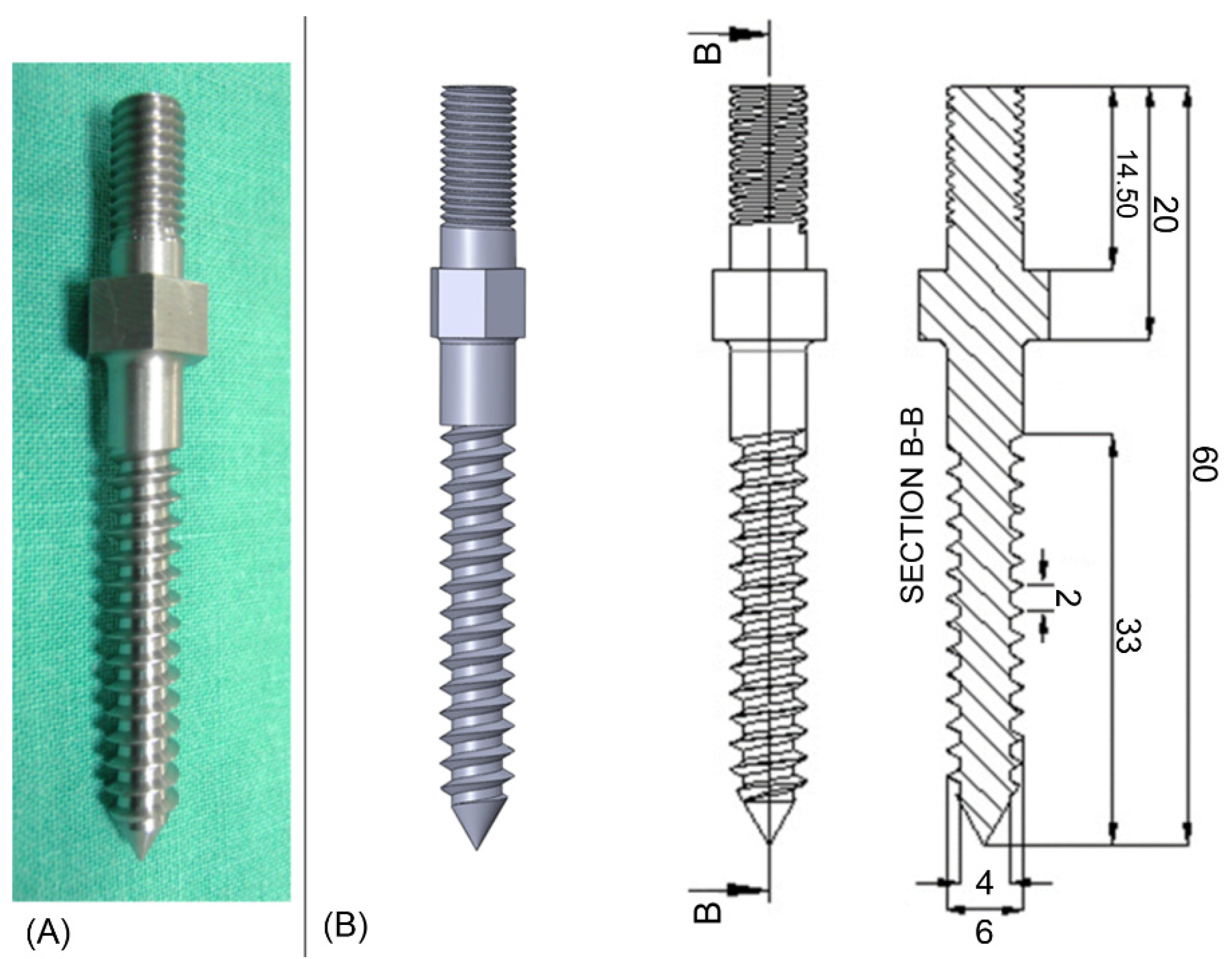
2.2. Pedicle Screw Geometries

The shape of the screws maintained a constant outer/inner diameter (6.0 mm/4.0 mm) from hub to tip. For all screws, the length of the thread coverage was controlled at 33 mm. The thread pitch was 2 mm, and the type of threads was V-shaped. The schematic drawings and photographs of the pedicle screws are illustrated in Figure 2.

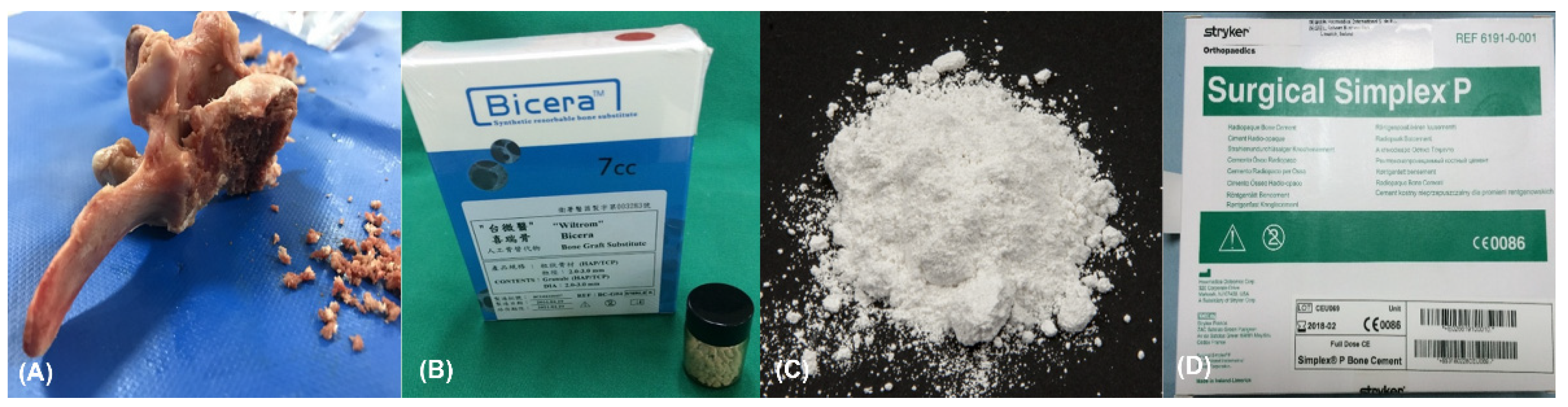
2.3. Filling Biomaterials and Preparation

Four kinds of biomaterials were filled into predrilled holes (pilot holes). These included (1) allograft bone particles (ABP) obtained from porcine vertebral bodies; (2) hydroxyapatite-tricalcium phosphate (HA/β-TCP) (Wiltrom Bicera Bone Graft Substitute, HA/β-TCP, 60 weight percent/40 weight percent, Taiwan); (3) calcium sulfate (CS) (Terra alba, Perma-Cemet, Taiwan); and (4) polymethylmethacrylate (PMMA) bone cement (Stryker Howmedica Simplex P Bone Cement, Kalamazoo, MI, USA). These four kinds of filling biomaterials are shown in Figure 3.
The CS powder was mixed with water at a ratio of 1.15 (CaSO4/H2O: 90 g/80 g) to obtain maximal Young’s modulus of compression, which was analyzed in our prior experiment. After the mixing, CS was poured into the syringe and injected into the pilot hole. The screw was then inserted, and the specimen was kept for one day to allow the material to solidify.
The PMMA powder and the monomer were mixed in a ratio of 2:1 as the original composition and stirred for 2–4 min. The injection timing was chosen between the liquid phase and the paste phase of the bone cement. Following screw insertion, the specimen was held for one day to ensure completion of the curing process.

2.4. Experimental Grouping: Primary and Revision

The experiment was divided into two parts according to the pilot-hole diameter. The pilot-hole diameter in the first part was 3.2 mm, simulating primary surgery. Three groups of test blocks (7.5, 15, and 30 pcf) were used (30 in each group), and each group of test block was further divided into five subgroups (control, PMMA, CS, HA/β-TCP, and ABP), based on different test materials added in each test block group. After the pilot hole was created, the screw was directly inserted into the test block as the control group. Pilot holes in other test pieces were stuffed with one of four test materials, followed by screw insertion in the experimental group. Six specimens were repeated for each group.
The pilot-hole diameter in the second part was 7.0 mm, simulating revision surgery. The experimental grouping for the second part was identical to those in the first part. Because the outer diameter of the screw was 6 mm, which is smaller than the diameter of the pilot hole, the data of the control group in the second part (7.0 mm) adopted the data from the first part (3.2 mm). The experimental flowchart is shown in Figure 4.

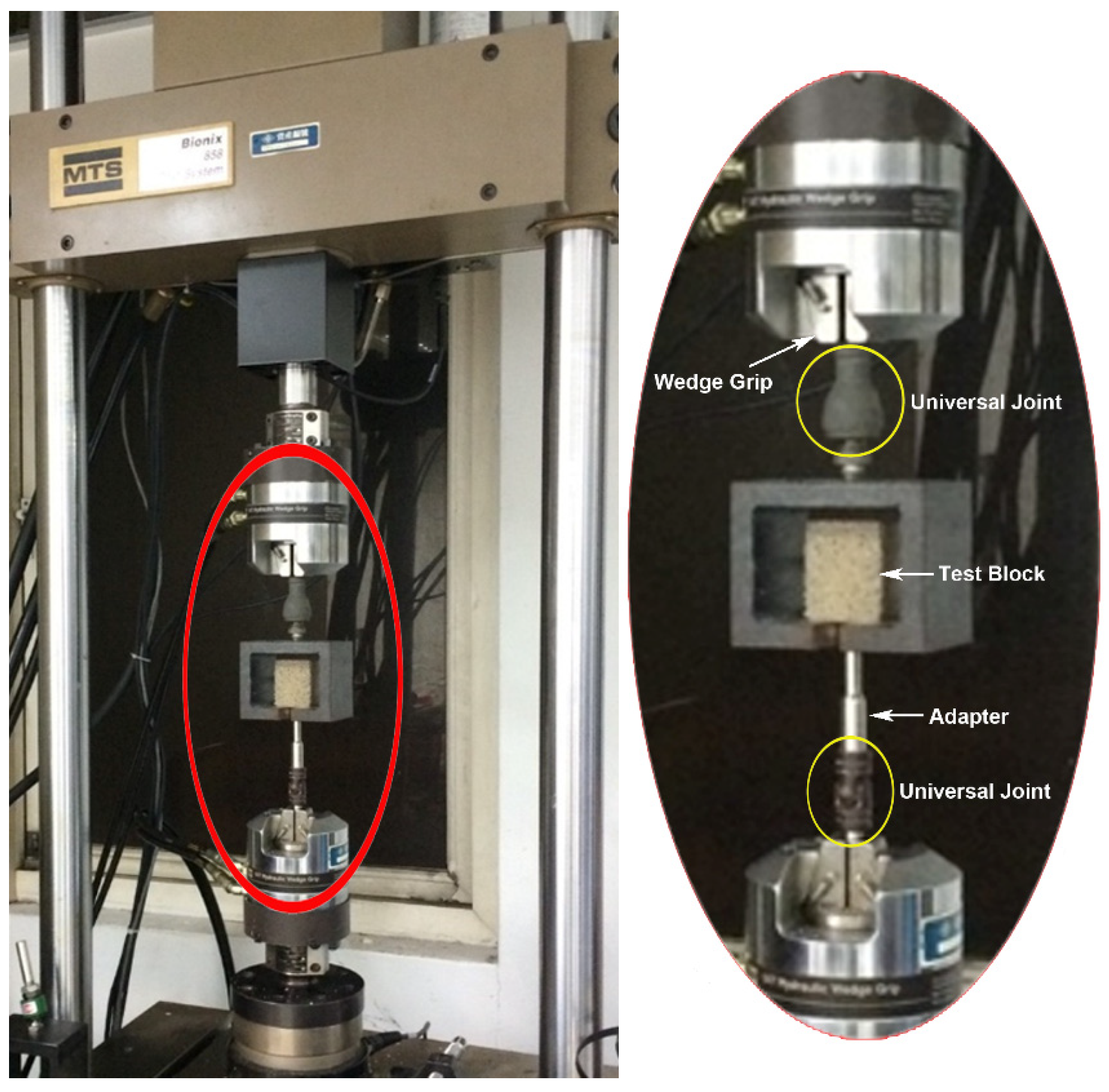
2.5. Biomechanical Pullout Testing

Both the trajectory axis perpendicular to the insertional plane of the test block and consistent insertional depth were confirmed using X-ray imaging prior to pullout testing (Figure 5). After screw insertion, the specimen was placed on a custom-made universal fixture that was capable of self-alignment to ensure the long axis of the screw was coaxial with the testing machine (Bionix 858, MTS Corp., Eden Prairie, MN, USA) pullout ram. The pedicle screw head was fixed into a cylindrical adapter with an inner thread matching the outer thread of the screw head. The adapter was then clamped to the lower wedge grip of testing machine. The experimental setup of the screw pullout test is shown in Figure 6. After the specimen was mounted, a pullout force was applied at a constant rate of 5 mm/min. The force acting on the screw during the testing was continuously recorded in 0.05 mm increments until failure. The peak force recorded during the pullout test was defined as the maximum pullout strength for comparison.

2.6. Statistical Analysis

To evaluate the effects of filling biomaterials for salvage pedicle screw augmentations, the ultimate pullout forces were compared by statistical analysis. All the measurements are expressed as the mean ± standard deviation (SD). Statistical analysis was performed using SPSS software (SPSS for Windows version 12.0, SPSS, Inc., Chicago, IL, USA). The Mann–Whitney U test was used to evaluate the differences between groups. A p value < 0.05 was considered significant.

3. Results

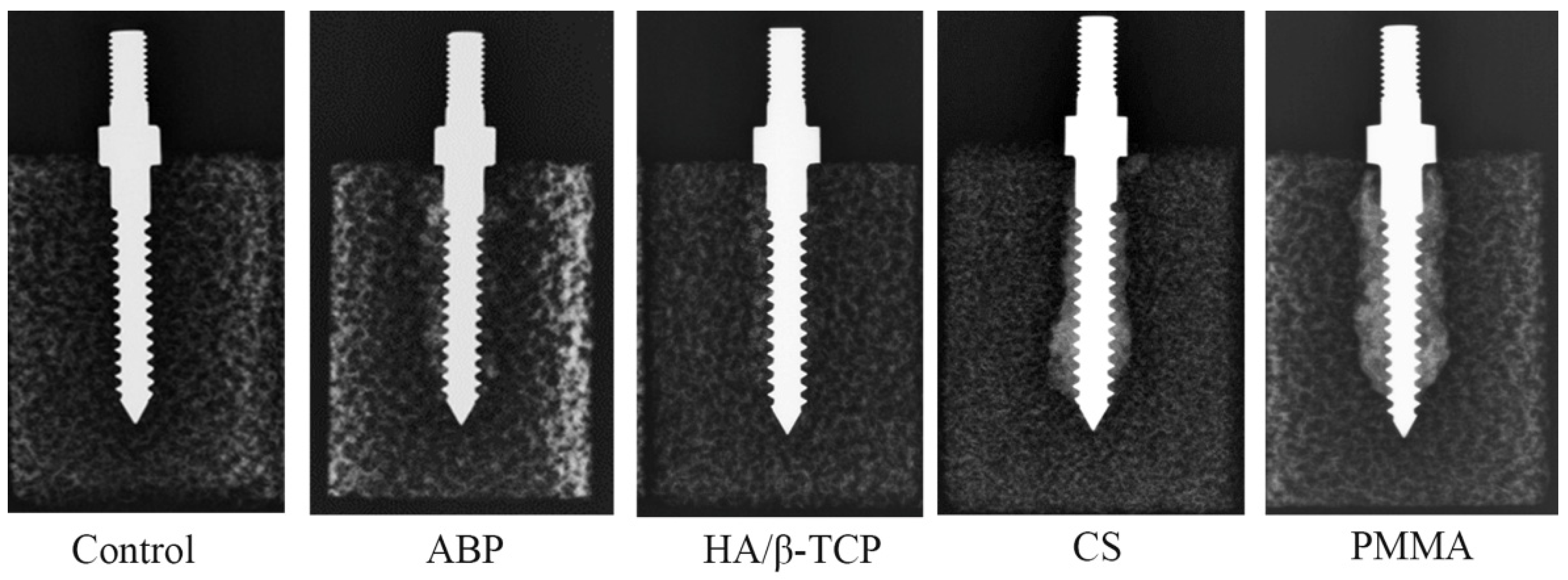
3.1. Radiological Images after the Insertion of Screws with Various Materials

Prior to biomechanical pullout testing, X-ray images were obtained after pedicle screw insertion in the test blocks with various filling biomaterials. The radiopacities of these materials were different. CS and PMMA demonstrated greater radiopacity in comparison to ABP and HA/β-TCP (Figure 5). This finding could be interpreted as suggesting that paste-like materials, such as CS and PMMA, are distributed more homogeneously around the screw than particle-like materials, such as ABP and HA/β-TCP.

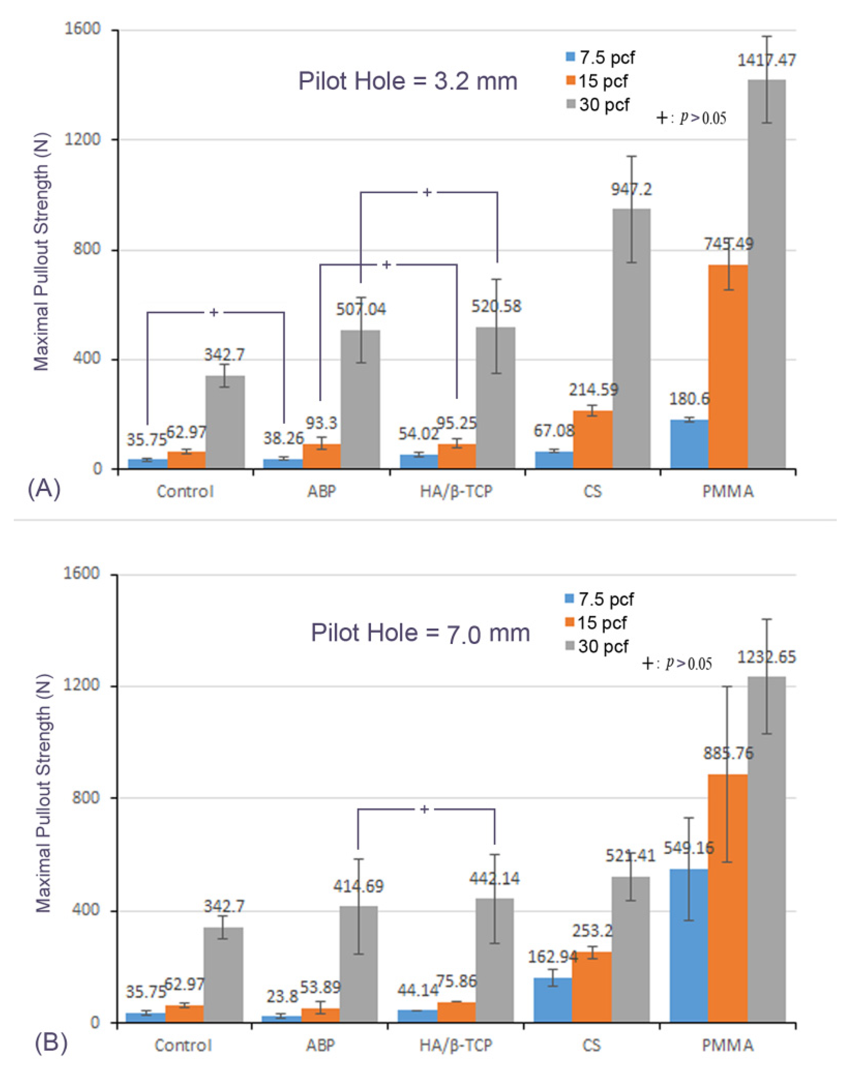
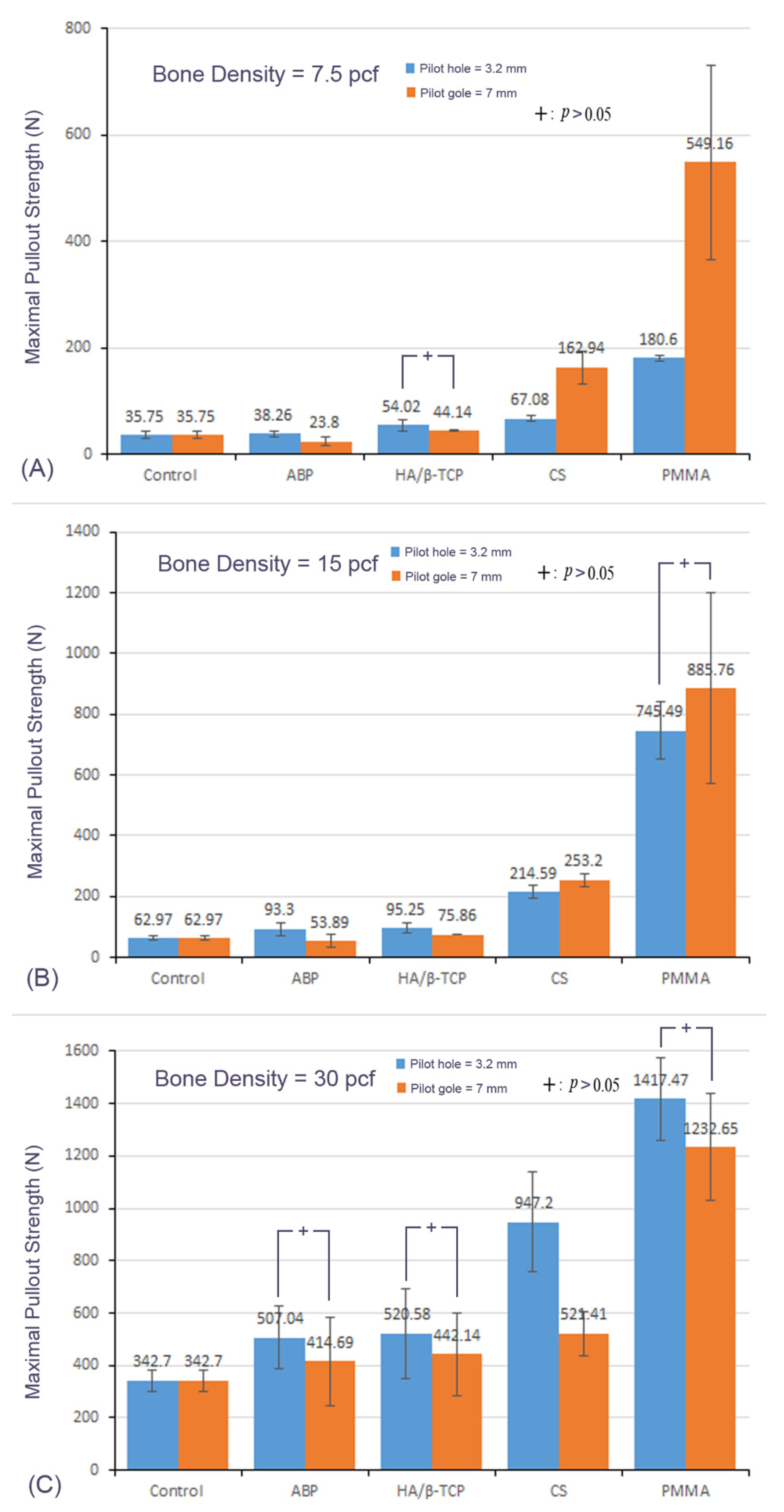
3.2. Mean Maximal Pullout Strength of Different Materials with the Different Pilot-Hole Sizes and Bone Densities

In samples in the 3.2 mm pilot-hole group, in which primary surgery was simulated, the control group, ABP, HA/β-TCP, CS, and PMMA in samples in the 7.5 pcf group showed a mean maximal pullout strength of 35.75 ± 6.35, 38.26 ± 4.99, 54.02 ± 10.14, 67.08 ± 5.91, and 180.60 ± 6.09 N, respectively, compared with 62.97 ± 8.26, 93.30 ± 22.31, 95.25 ± 16.55, 214.59 ± 19.92, and 745.49 ± 94.20 N, respectively, in samples in the 15 pcf group, and 342.70 ± 40.94, 507.04 ± 117.92, 520.58 ± 171.90, 947.20 ± 191.97, 1417.47 ± 158.22 N, respectively, in samples in the 30 pcf group.
In samples in the 7.0 mm pilot-hole group, in which revision surgery was simulated, the mean maximal pullout strength in samples in the 7.5 pcf group for the control group, ABP, HA/β-TCP, CS, and PMMA was 35.75 ± 6.35, 23.80 ± 7.97, 44.14 ± 1.74, 162.94 ± 29.90, and 549.16 ± 183.42 N, respectively, compared with 62.97 ± 8.26, 53.89 ± 20.68, 75.86 ± 0.33, 253.20 ± 21.30, and 885.76 ± 315.39 N, respectively, in samples in the 15 pcf group, and 342.70 ± 40.94, 414.69 ± 168.87, 442.14 ± 156.84, 521.41 ± 85.54, and 1232.65 ± 204.27 N, respectively, in samples in the 30 pcf group.
From the above results, the maximal pullout strength of these five conditions in descending order was PMMA, CS, HA/β-TCP, ABP and the control (Figure 7).

3.3. Effect of Bone Density

In samples in both the 3.2 mm pilot-hole and 7.0 mm pilot-hole groups, the mean maximal pullout strength of these four materials increased with increasing bone density (Figure 7).

3.4. Effect of the Pilot-Hole Size

In samples in both the 7.5 pcf and 15 pcf groups, the mean maximal pullout strength of CS and PMMA increased from the 3.2 mm pilot hole to the 7.0 mm pilot hole. In contrast, the mean maximal pullout strength of the ABP and HA/β-TCP decreased from the 3.2 mm pilot hole to the 7.0 mm pilot hole. In samples in the 30 pcf group, the maximal pullout strength of these four materials decreased as the hole increased from a 3.2 mm pilot hole to a 7.0 mm pilot hole (Figure 8).

4. Discussion

When revision spine surgery was performed under various conditions, coexisting pedicle screw loosening was often encountered intraoperatively. Screw augmentation using different biomaterials is an important salvaging strategy to obtain adequate spinal stability and avoid extension of a fused segment. This study performed a biomechanical evaluation using four common biomaterials for screw augmentation to simulate primary and revision surgery with loosened pedicle screws. The results demonstrated that the mean maximal pullout strength after biomaterial filling was indeed increased compared with the control group, and bone cement offered the most significantly augmented effect in each pilot-hole size and bone-density condition. Moreover, the maximal pullout strength of different materials increased with increasing bone density. The strength of our study was that we simulated screw loosening in revision surgery using a 7.0 mm pilot hole and systematically investigated the effects of various biomaterials utilized for salvage screw stabilization, which has rarely been reported in the previous literature to our knowledge.
From a clinical point of view, PMMA, HA/β-TCP and bone chips (from either autogenous or allogenous bone) are materials that are available in a timely manner during surgery. Pedicle screw augmentation with PMMA can quickly provide good initial stability because the whole curing time is relatively low (of the order of 10 min or less). However, the risks of PMMA used for screw augmentation are cement leakage, which increases the difficulty and complexity of the next potential surgery. Commercialized HA/β-TCP and ABP were also easily used during the surgery. We could use the prepared screw path with HA/β-TCP or ABP and then insert the screw. Although the initial augmentation via HA/β-TCP and ABP may not be as good as that of PMMA, HA/β-TCP and CS had osteoconductive properties that served scaffolds onto which bone cells can attach, migrate, and grow. CS has osteoconductive ability as well and can serve as a bone graft for bone defects, but the preparation time needed is approximately 30–45 min [22], which leads to limitations in spine surgery.
In the present study, the mean maximal pullout strength in descending order was as follows: PMMA, CS, HA/β-TCP, ABP and control. PMMA and CS are paste-like materials that are distributed more homogeneously around the screw than particle-like materials and can offer increased stability. Among these materials, PMMA had the highest Young’s modulus and mechanical strength, so the pullout strength was obviously higher than that of the other materials.
Our findings were in line with other studies with similar procedures, e.g., there was a close correlation between bone mineral density and screw pullout strength [30,31]. With the increase in the bone density, the mean maximal pullout strength of different materials increased in each group of different filling materials. This finding may be caused by the following: the area of the augmented screw−bone interface in the high mineral density bone was larger than that in the low mineral density bone.
When comparing the difference in the pullout strength between the 3.2 mm and 7.0 mm pilot holes, we found that the mean maximal pullout strength in samples in the 7.0 mm group was similar (slightly larger or smaller) to that in the 3.2 mm group, which illustrated that pedicle screw augmentation with these materials was certainly helpful. Moreover, the mean maximal pullout strength of the PMMA-augmented salvage screw (7.0 mm) was apparently elevated in the 7.5 pcf test block. That is, PMMA bone cement augmentation should be considered to have more initial stability during revision surgery, especially in osteoporotic bone quality.
Seong Yi et al. published a study [17] in which the authors compared pullout strength after pedicle screw augmentation with hydroxyapatite, calcium phosphate, and PMMA bone cement. The mean pullout strength, compared with that of controls, increased by 12.5% in HA-augmented screws (p = 0.600) and by 14.9% in calcium phosphate-augmented hemivertebrae (p = 0.234). The pullout strength of polymethylmethacrylate- versus hydroxyapatite-augmented pedicle screws was 60.8% higher (p = 0.028). Even though the pullout strength of HA was not significant, they concluded that HA was likely a better clinical alternative to PMMA, as HA augmentation, unlike PMMA augmentation, stimulates bone growth and can be revised. In our opinion, both HA (or bone chips) and PMMA augmentation are feasible methods that depended on intraoperative screw purchase, the etiology of screw loosening, and surgical planning. For example, patients who are young or concurrent with spine infection should not undergo bone cement augmentation due to a long life expectancy and foreign body consideration. For patients who simultaneously underwent anterior surgery with additional anterior support, the requirement for the strength of the pedicle screw fixation was not as strict as those who underwent posterior revision surgery alone.
Chongyu Jia et al. [32] investigated pedicle screw fixation augmented with allograft bone particles (ABPs) in osteoporotic vertebrae. Their results showed that trajectory augmentation with allograft bone particles can significantly increase the strength of the augmented screws. Full-trajectory augmentation was suggested compared with half-trajectory augmentation. The authors also analyzed proper ABP size and recommend using 1 mm ABP (0.3 g ABP for a 5.5 mm × 40 mm screw and 0.5 g ABP for a 6.5 mm × 45 mm screw) for augmentation. In addition, the optimal filling volume of the bone cement was 75% of the trajectory volume (approximately 1.03 mL) for osteoporotic vertebrae in the primary surgery, which was suggested by Fan et al. [33]. Rescue augmentation with cement was studied by Lukas Weiser et al. [34], who reported that the fatigue load reached 207 ± 75 N for the non-augmented screws, 301 ± 96 N for the initial cement augmentation, and 370 ± 87 N for the rescue augmentation. The volume of cement used for the rescue augmentation ranged between 2 and 5 mL, depending on the radiological assessment of the distribution. They concluded that cement augmentation of initially loosened pedicle screws was a promising option to restore adequate screw stability.
Inevitably, this study had some limitations. The first limitation was that the homogenous bone density of the test blocks cannot represent the real structure of the vertebrae in the clinic. However, the test blocks were made of uniform polyurethane foam, which reduced the influence of the variability of properties and the morphometry of cadaveric bones and provided an effective platform to compare the mechanical characteristics of various filling materials in different degrees of bone mineral density. The second limitation was that ABP, HA/β-TCP, and CS were absorbable and osteoconductive biomaterials. In the process of bone formation and remodeling, the effects of these osteoconductive biomaterials on the mechanical properties of screws may change. Thus, this issue should be explored through further animal experiments. Third, only one brand of bone cement and one design of pedicle screws were used in this study. This was because the purpose of this experiment focused on comparing the characteristics of filling biomaterials, so the influence of different bone cement and screw designs was not investigated in this study. Finally, only a static loading (screw pullout test in synthetic bone) was conducted without considering other physiological loading modes. In actual physiological conditions, the screw/biomaterials/bone interfaces are subjected to complex dynamic loadings, which may have an impact on long-term screw-fixation strength due to the effects of fretting and stress shielding. Although the pullout test is typically used for evaluating screw fixation ability, further investigation on the effects of other biomechanical effects such as fretting and stress shielding, would be beneficial. Nevertheless, in the present study, all experimental steps were conducted to preserve uniformity and reproducibility. We believe that our results offer valuable information for spine surgeons who need to deal with pedicle screw loosening during revision spine surgery.

5. Conclusions

Salvage pedicle screw augmentation with ABP, HA/β-TCP, CS, and PMMA bone cement are all feasible methods and can offer better pullout strength than non-augmentation. PMMA bone cement provides the most significant augmentation in each pilot hole size and bone density condition and could be considered preferentially to obtain larger initial stability during revision surgery, especially in osteoporotic bone quality. However, ABP and HA/β-TCP have osteoconductive properties and can avoid possible complications caused by PMMA bone cement. Therefore, the most suitable filling materials depend on intraoperative screw purchase, etiology of screw loosening, and surgical planning.

Author Contributions

Conceptualization, P.-L.L., Y.-D.L. and C.-L.T.; methodology, M.-K.H. and Y.-J.L.; software, D.-M.L. and Y.-J.L.; validation, P.-L.L., Y.-D.L., T.-T.T. and P.-L.L.; formal analysis, M.-K.H., Y.-J.L., D.-M.L. and C.-L.T.; investigation, Y.-D.L., D.-M.L. and C.-L.T.; data curation: P.-L.L., T.-T.T. and P.-L.L.; supervision: C.-L.T.; writing—original draft preparation, Y.-D.L.; writing—review and editing, P.-L.L., M.-K.H. and C.-L.T.; funding acquisition, C.-L.T. All authors have read and agreed to the published version of the manuscript.

Funding

This work was supported by the Ministry of Science and Technology in Taiwan (Grant No. MOST 107-2221-E-182-013; MOST 109-2221-E-182-006-MY2).

Institutional Review Board Statement

Not applicable.

Informed Consent Statement

Not applicable.

Data Availability Statement

The data used to support the finding of this study are available from the corresponding author upon request.

Conflicts of Interest

The authors declare no conflict of interest.

References

  1. Boucher, H.H. A method of spinal fusion. J. Bone Joint Surg. Br. 1959, 41, 248–259. [Google Scholar] [CrossRef] [PubMed]
  2. Elder, B.D.; Lo, S.-F.L.; Holmes, C.; Goodwin, C.R.; Kosztowski, T.A.; Lina, I.A.; Locke, J.E.; Witham, T.F. The biomechanics of pedicle screw augmentation with cement. Spine J. 2015, 15, 1432–1445. [Google Scholar] [CrossRef] [PubMed]
  3. Gaines, R.W., Jr. The use of pedicle screw internal fixation for the operative treatment of spinal disorders. J. Bone Joint Surg. Am. 2000, 82, 1458–1476. [Google Scholar] [CrossRef] [PubMed]
  4. Nakashima, H.; Yukawa, Y.; Imagama, S.; Kanemura, T.; Kamiya, M.; Yanase, M.; Ito, K.; Machino, M.; Yoshida, G.; Ishikawa, Y.; et al. Complications of cervical pedicle screw fixation for nontraumatic lesions: A multicenter study of 84 patients. J. Neurosurg. Spine 2012, 16, 238–247. [Google Scholar] [CrossRef] [PubMed] [Green Version]
  5. Martin, B.I.; Deyo, R.A.; Mirza, S.K.; Turner, J.A.; Comstock, B.A.; Hollingworth, W.; Sullivan, S.D. Expenditures and health status among adults with back and neck problems. JAMA 2008, 299, 656–664. [Google Scholar] [CrossRef] [Green Version]
  6. Deyo, R.A.; Gray, D.T.; Kreuter, W.; Mirza, S.; Martin, B.I. United States Trends in Lumbar Fusion Surgery for Degenerative Conditions. Spine 2005, 30, 1441–1445. [Google Scholar] [CrossRef]
  7. Szpalski, M.; Gunzburg, R.; Aebi, M. The aging of the population: A growing concern for spine care in the twenty-first century. Eur. Spine J. 2003, 12, S81–S83. [Google Scholar] [CrossRef] [Green Version]
  8. Esses, S.I.; Sachs, B.L.; Dreyzin, V. Complications Associated with the Technique of Pedicle Screw Fixation A Selected Survey of ABS Members. Spine 1993, 18, 2231–2239. [Google Scholar] [CrossRef]
  9. Mirovsky, Y.; Floman, Y.; Smorgick, Y.; Ashkenazi, E.; Anekstein, Y.; Millgram, M.A.; Giladi, M. Management of Deep Wound Infection After Posterior Lumbar Interbody Fusion with Cages. J. Spinal Disord. Tech. 2007, 20, 127–131. [Google Scholar] [CrossRef]
  10. Kobayashi, K.; Ando, K.; Kato, F.; Kanemura, T.; Sato, K.; Hachiya, Y.; Matsubara, Y.; Kamiya, M.; Sakai, Y.; Yagi, H.; et al. Reoperation within 2 years after lumbar interbody fusion: A multicenter study. Eur. Spine J. 2018, 27, 1972–1980. [Google Scholar] [CrossRef]
  11. Li, Y.-D.; Chi, J.-E.; Chiu, P.-Y.; Kao, F.-C.; Lai, P.-L.; Tsai, T.-T. The comparison between anterior and posterior approaches for removal of infected lumbar interbody cages and a proposal regarding the use of endoscope-assisted technique. J. Orthop. Surg. Res. 2021, 16, 1–11. [Google Scholar] [CrossRef] [PubMed]
  12. Talu, U.; Kaya, I.; Dikici, F.; Sar, C. Pedicle screw salvage: The effect of depth and diameter on pullout strength: A biomechanical study. Acta Orthop Traumatol Turc. 2004, 34, 300–307. [Google Scholar]
  13. Hsieh, M.-K.; Liu, M.-Y.; Chen, J.-K.; Tsai, T.-T.; Lai, P.-L.; Niu, C.-C.; Tai, C.-L. Use of longer sized screws is a salvage method for broken pedicles in osteoporotic vertebrae. Sci. Rep. 2020, 10, 1–12. [Google Scholar] [CrossRef] [PubMed]
  14. Kaye, I.D.; Prasad, S.K.; Vaccaro, A.R.; Hilibrand, A.S. The Cortical Bone Trajectory for Pedicle Screw Insertion. JBJS Rev. 2017, 5, e13. [Google Scholar] [CrossRef] [PubMed]
  15. Delgado-Fernandez, J.; García-Pallero, M.; Blasco, G.; Pulido-Rivas, P.; Rafael, G.S. Review of Cortical Bone Trajectory: Evidence of a New Technique. Asian Spine J. 2017, 11, 817–831. [Google Scholar] [CrossRef] [Green Version]
  16. Hashemi, A.; Bednar, D.; Ziada, S. Pullout strength of pedicle screws augmented with particulate calcium phosphate: An experimental study. Spine J. 2009, 9, 404–410. [Google Scholar] [CrossRef]
  17. Yi, S.; Rim, D.-C.; Park, S.W.; Murovic, J.A.; Lim, J.; Park, J. Biomechanical Comparisons of Pull Out Strengths After Pedicle Screw Augmentation with Hydroxyapatite, Calcium Phosphate, or Polymethylmethacrylate in the Cadaveric Spine. World Neurosurg. 2015, 83, 976–981. [Google Scholar] [CrossRef]
  18. Liu, M.-Y.; Tsai, T.-T.; Lai, P.-L.; Hsieh, M.-K.; Chen, L.-H.; Tai, C.-L. Biomechanical comparison of pedicle screw fixation strength in synthetic bones: Effects of screw shape, core/thread profile and cement augmentation. PLoS ONE 2020, 15, e0229328. [Google Scholar] [CrossRef]
  19. Agrawal, M.; Katti, D.; Boyan, B.; McMillan, J.; Lohmann, C.; Ranly, D.; Schwartz, Z. Chapter 13—Bone Graft Substitutes: Basic Information for Successful Clinical Use with Special Focus on Synthetic Graft Substitutes; ASTM International: West Conshohocken, PA, USA, 2009; p. 231. [Google Scholar] [CrossRef]
  20. Campana, V.; Milano, G.; Pagano, E.; Barba, M.; Cicione, C.; Salonna, G.; Lattanzi, W.; Logroscino, G. Bone substitutes in orthopaedic surgery: From basic science to clinical practice. J. Mater. Sci. Mater. Med. 2014, 25, 2445–2461. [Google Scholar] [CrossRef]
  21. Peltier, L.F.; Bickel, E.Y.; Lillo, R.; Thein, M.S. The Use of Plaster of Paris to Fill Defects in Bone. Ann. Surg. 1957, 146, 61–69. [Google Scholar] [CrossRef]
  22. Kumar, Y.; Nalini, K.B.; Menon, J.; Patro, D.K.; Banerji, B.H. Calcium sulfate as bone graft substitute in the treatment of osseous bone defects, a prospective study. J. Clin. Diagn. Res. 2013, 7, 2926–2928. [Google Scholar] [CrossRef] [PubMed]
  23. Chen, L.-H.; Tai, C.-L.; Lee, D.-M.; Lai, P.-L.; Lee, Y.-C.; Niu, C.-C.; Chen, W.-J. Pullout strength of pedicle screws with cement augmentation in severe osteoporosis: A comparative study between cannulated screws with cement injection and solid screws with cement pre-filling. BMC Musculoskelet Disord. 2011, 12, 33. [Google Scholar] [CrossRef] [PubMed] [Green Version]
  24. Liu, D.; Wu, Z.-X.; Pan, X.-M.; Fu, S.-C.; Gao, M.-X.; Shi, L.; Lei, W. Biomechanical comparison of different techniques in primary spinal surgery in osteoporotic cadaveric lumbar vertebrae: Expansive pedicle screw versus polymethylmethacrylate-augmented pedicle screw. Arch. Orthop. Trauma. Surg. 2011, 131, 1227–1232. [Google Scholar] [CrossRef] [PubMed]
  25. Stürup, J.; Nimb, L.; Kramhøft, M.; Jensen, J.S. Effects of polymerization heat and monomers from acrylic cement on canine bone. Acta Orthop. Scand. 1994, 65, 20–23. [Google Scholar] [CrossRef]
  26. Lai, P.-L.; Tai, C.-L.; Chen, L.-H.; Nien, N.-Y. Cement leakage causes potential thermal injury in vertebroplasty. BMC Musculoskelet. Disord. 2011, 12, 116. [Google Scholar] [CrossRef] [Green Version]
  27. Lai, P.-L.; Tai, C.-L.; Chu, I.-M.; Fu, T.-S.; Chen, L.-H.; Chen, W.-J. Hypothermic manipulation of bone cement can extend the handling time during vertebroplasty. BMC Musculoskelet. Disord. 2012, 13, 198. [Google Scholar] [CrossRef] [Green Version]
  28. Tai, C.-L.; Lai, P.-L.; Lin, W.-D.; Tsai, T.-T.; Lee, Y.-C.; Liu, M.-Y.; Chen, L.-H. Modification of Mechanical Properties, Polymerization Temperature, and Handling Time of Polymethylmethacrylate Cement for Enhancing Applicability in Vertebroplasty. BioMed Res. Int. 2016, 2016, 1–8. [Google Scholar] [CrossRef] [Green Version]
  29. Gibson, L.; Ashby, M. Cancellous bone. Cellular Solids: Structure & Properties; Pergamon Press: New York, NY, USA, 1988; pp. 316–331. [Google Scholar]
  30. Amirouche, F.; Solitro, G.F.; Magnan, B.P. Stability and Spine Pedicle Screws Fixation Strength—A Comparative Study of Bone Density and Insertion Angle. Spine Deform. 2016, 4, 261–267. [Google Scholar] [CrossRef]
  31. Weiser, L.; Huber, G.; Sellenschloh, K.; Viezens, L.; Püschel, K.; Morlock, M.M.; Lehmann, W. Insufficient stability of pedicle screws in osteoporotic vertebrae: Biomechanical correlation of bone mineral density and pedicle screw fixation strength. Eur. Spine J. 2017, 26, 2891–2897. [Google Scholar] [CrossRef]
  32. Jia, C.; Zhang, R.; Xing, T.; Gao, H.; Li, H.; Dong, F.; Zhang, J.; Ge, P.; Song, P.; Xu, P.; et al. Biomechanical properties of pedicle screw fixation augmented with allograft bone particles in osteoporotic vertebrae: Different sizes and amounts. Spine J. 2019, 19, 1443–1452. [Google Scholar] [CrossRef]
  33. Fan, H.T.; Zhang, R.J.; Shen, C.L.; Dong, F.L.; Li, Y.; Song, P.W.; Gong, C.; Wang, Y.J. The Biomechanical Properties of Pedicle Screw Fixation Combined with Trajectory Bone Cement Augmentation in Osteoporotic Vertebrae. J. Spinal Disord. Tech. 2016, 29, 78–85. [Google Scholar] [CrossRef] [PubMed]
  34. Weiser, L.; Huber, G.; Sellenschloh, K.; Püschel, K.; Morlock, M.M.; Viezens, L.; Lehmann, W. Rescue Augmentation: Increased Stability in Augmentation After Initial Loosening of Pedicle Screws. Glob. Spine J. 2021, 11, 679–685. [Google Scholar] [CrossRef] [PubMed] [Green Version]
Figure 1. Three types of test blocks made of open cell foams with a density of (A) 7.5, (B) 15, and (C) 30 pcf.
Applsci 12 07792 g001
Figure 2. (A) Photograph and (B) schematic drawings of the pedicle screw. Section B-B in the right of drawing of (B) indicates the vertical cross section along line B-B.
Applsci 12 07792 g002
Figure 3. Four kinds of filling biomaterials: (A) allograft bone chips; (B) hydroxyapatite-tricalcium phosphate (HA/β-TCP); (C) calcium sulfate; (D) polymethylmethacrylate (PMMA) bone cement.
Applsci 12 07792 g003
Figure 4. The experimental flowchart.
Applsci 12 07792 g004
Figure 5. Radiological images of the test blocks after the insertion of screws with various filling biomaterials.
Applsci 12 07792 g005
Figure 6. Photograph showing experimental testing configuration using a custom-made universal fixture that was capable of self-alignment to ensure the long axis of the screw was coaxial with the testing machine.
Applsci 12 07792 g006
Figure 7. Comparisons of mean maximal pullout strengths for screws augmented with different filling biomaterials using 7.5 pcf, 15 pcf, and 30 pcf test blocks in (A) 3.2 mm and (B) 7.0 mm pilot holes. Groups without significant differences are indicated with the “+” symbol.
Applsci 12 07792 g007
Figure 8. Comparisons of mean maximal pullout strengths for screws augmented with different filling biomaterials between 3.2 mm and 7.0 mm pilot holes using (A) 7.5 pcf, (B) 15 pcf, and (C) 30 pcf test blocks. Groups without significant differences are indicated with the “+” symbol.
Applsci 12 07792 g008
Publisher’s Note: MDPI stays neutral with regard to jurisdictional claims in published maps and institutional affiliations.

