Article

Effects of Caffeine and 5-Caffeoylquinic Acid on Blood Cell In Vitro Cytokine Production in Response to Lipopolysaccharide Stimulation

1
Research Group on Evidence, Lifestyles and Health, Department of Fundamental Biology and Health Sciences, Research Institute of Health Sciences (IUNICS), University of the Balearic Islands, 07122 Palma, Spain
2
Research Group on Evidence, Lifestyles and Health, Department of Nursing and Physiotherapy, Research Institute of Health Sciences (IUNICS), University of the Balearic Islands, 07122 Palma, Spain
3
Health Research Institute of the Balearic Islands (IdISBa), 07120 Palma, Spain
4
School of Life Sciences, Pharmacy and Chemistry, Faculty of Science Engineering and Computing, Kingston University London, Penryhn Road, Surrey KT1 2EE, UK
*
Authors to whom correspondence should be addressed.
Appl. Sci. 2022, 12(14), 7322; https://doi.org/10.3390/app12147322
Submission received: 14 June 2022 / Revised: 6 July 2022 / Accepted: 18 July 2022 / Published: 21 July 2022
(This article belongs to the Special Issue Effects of Caffeine on Human Health)

Abstract

:
Previous in vitro studies the have suggested anti-inflammatory properties of caffeine. Coffee is also rich in phenolic acids with several potential health benefits (chlorogenic acids), mainly 5-caffeoylquinic acid (5-CQA). The aim of the present study was to determine the effect of caffeine and 5-CQA, using concentrations comparable to typical human consumption, on LPS-induced cytokine production in whole blood in vitro cultures. Whole blood samples (n = 10) were stimulated for 24 h with LPS in the presence of caffeine or 5-CQA. The concentrations of various cytokines, malondialdehyde (MDA) and adenosine-3′,5′-cyclic monophosphate (cAMP) were determined in the culture media. Significant effects of caffeine were found only for the stimulated production of TNF-α (p = 0.014) and cAMP (p = 0.001). However, only the highest caffeine concentration (50 μM) induced lower TNF-α (p = 0.016) and higher cAMP (p = 0.006) levels in comparison to the control. The 5-CQA only influenced the stimulated production of IL-6 (p = 0.002), with lower values for the highest 5-CQA concentrations (2 μM: p = 0.012; 20 μM: p < 0.001). Neither caffeine (p = 0.227) nor 5-CQA (p = 0.259) influenced MDA concentrations. In conclusion, both caffeine and 5-CQA exerted limited in vitro anti-inflammatory effects. For caffeine, these effects were attained with high physiological concentrations. However, for 5-CQA, supraphysiological concentrations were needed.

1. Introduction

Coffee is one of the most commonly consumed beverages in the world [1]. For many populations, coffee is the major source of caffeine [1], the most widely consumed psychostimulant [2]. However, coffee is also a very rich source of chlorogenic acids (CGAs), naturally-produced phenolic acids that are derived from the esterification of cinnamic acids and quinic acid [3]. The most abundant CGA in coffee is 5-caffeoylquinic acid (5-CQA), which accounts for approximately 80% of the total CGA content [4]. Furthermore, coffee is the main source of CGAs in the human diet, with less contribution provided from other products such as tea and some fruits and vegetables [5].
It has been reported that a regular intake of coffee is associated with a reduced risk of chronic diseases, such as type 2 diabetes [6,7], metabolic syndrome [8], cardiovascular diseases [9,10], and cancer [10]. Furthermore, consumption of coffee has been recently associated with lower all-cause cancer mortality [11]. Interestingly, some studies have shown an anti-inflammatory effect of regular coffee consumption [12,13,14,15].
Due to having antioxidant and anti-inflammatory properties, the health benefits of coffee have been attributed, at least in part, to CGAs [3,14,16]. Studies testing the anti-inflammatory activity of CGAs are scarce, but some have confirmed this anti-inflammatory effect [3]. Two studies carried out with mouse macrophages demonstrated how the presence of 5-CQA (the CGA most commonly tested), in cell culture suppressed the lipopolysaccharide (LPS)-stimulated production of pro-inflammatory compounds [17,18]. On the other hand, studies in animal models have shown that the administration of 5-CQA causes suppresses in the circulating levels of pro-inflammatory cytokines [19,20,21]. However, no studies using human cells, particularly blood cells, focused on to analyzing the possible anti-inflammatory effect of chlorogenic acids currently exist in the literature. Furthermore, in some studies, the 5-CQA concentrations used were in the μM range [17,18], whereas concentrations observed in human plasma after coffee ingestion are typically in the nM range [22]. Therefore, it would be of interest to analyze the anti-inflammatory effect of 5-CQA, as it is the most abundant CGA in coffee, using human blood cells stimulated with LPS, a commonly used model.
However, the potential effects of caffeine should also be considered. Caffeine can interact with almost all tissues in the organism, including the immune system [23]. In this regard, a number of animal and in vitro studies have been focused on the effects of caffeine on immune related parameters, such as tumor necrosis factor (TNF)-α and antibody production, hypersensitivity and natural killer cell cytotoxicity, indicating an anti-inflammatory role for caffeine [23]. As such, some studies have reported that caffeine suppresses TNF-α production in vitro [24,25], but only when caffeine concentrations are higher than the physiological ones.
Taking into account these considerations, the aim of the present study was to determine the effect of caffeine and 5-CQA, using physiological concentrations comparable to typical human consumption, on the LPS-induced cytokine production in whole blood in vitro cultures. Due to the low concentrations tested, limited effects of caffeine and 5-CQA on cytokine production are expected.

2. Materials and Methods

2.1. Study Participants

All of the participants were classified as healthy individuals recruited from the University of the Balearic Islands, Spain, and were aged 30–45 years. The exclusion criteria included: common colds, flus (or similar diseases), infectious diseases, allergic episodes and/or consumption of anti-inflammatory medication within the two weeks preceding the study, and regular drugs or alcohol (more than one daily alcoholic drink in women and two daily alcohol drinks in men) consumption. Informed written consent was obtained from ten volunteers (five men and five women). All of the measurements and experiments were completed for all of the participants, leading to the final number of ten participants. This number of participants is similar and even higher than previously considered [24,25]. The protocol was performed in accordance with the Declaration of Helsinki for research on human participants and was approved by the Balearic Islands Clinical Investigation Ethics Committee (IB 3493/17 PI).

2.2. Laboratory Visit

After an overnight fast of approximately 12 h, the participants arrived at the laboratory between 08:00 a.m. and 10:00 a.m. The volunteers were requested to abstain from any caffeine-containing food and beverages for at least 24 h prior at arriving to the laboratory. Each participant was required to void their bladder before body mass and stature were recorded. Body mass was measured to the nearest 0.1 kg using an electronic scale (Seca 700, Seca GmbH, Hamburg, Germany) and stature was measured to the nearest 0.5 cm using a stadiometer (Seca 220 (CM) Telescopic Height Rod for Column Scales, Seca GmbH, Hamburg, Germany). Body mass index (BMI) was calculated as body mass (kg) divided by stature (m) squared (kg·m−2). The participants then sat quietly for 10 min before a venous blood sample was taken. Whole blood venous samples were collected into suitable Vacutainer® blood collection tubes with ethylenediaminetetraacetic acid (EDTA) or heparin as an anticoagulant (BD, Madrid, Spain). The total blood volume was approximately 8 mL. Leukocyte numbers were quantified using the EDTA blood sample with an automatic flow cytometer analyzer (ABX Pentra 60, Horiba Medical, Montpellier, France). Blood EDTA samples were then centrifuged at 1000× g for 10 min to extract the plasma. The plasma was subsequently stored at −70 °C until subsequent analysis. Whole blood collected with heparin was used for the subsequent blood culture in vitro experiments.

2.3. Plasma Caffeine Measurement

To ensure that no residual caffeine was present in the participants’ plasma samples, caffeine levels were measured in the EDTA-plasma by a high-performance liquid chromatography (HPLC) method previously described [26]. Caffeine was undetectable in all of the participants’ whole blood samples (n = 10).

2.4. Blood Culture, In Vitro Blood LPS Stimulation and Effects of Caffeine and 5-CQA

Whole heparin blood samples (n = 10) obtained by venepuncture were subsequently diluted with a 1:3 ratio with culture media (RPMI-1640 Medium, Sigma, St. Louis, MO, USA). The diluted whole blood was then pipetted into the wells of a sterile 12-well plate (Sarstedt, Nümbrecht, Germany). To each well, 50 μL of either medium (control sample), pharmaceutical grade caffeine powder (1,3,7-trimethylxanthine, dissolved in medium to yield final concentrations in a culture of 5, 10, 15, 30, and 50 μM) or 5-CQA (dissolved in medium to yield final concentrations in a culture of 5, 20, 200 nM, and 2 and 20 μM) were added. For each experiment, caffeine and 5-CQA, a different set of controls was applied. Following a 1 h incubation at 37 °C in a 5% CO2 atmosphere, a total of 50 μL of LPS (Escherichia coli serotype 055:B5; Sigma, St. Louis, MO, USA; final concentration 10 ng/mL dissolved in culture media) or the same volume of culture media (spontaneous production) were added to each well. Culture plates were then incubated for a further 24 h at 37 °C in a 5% CO2 atmosphere. Immediately following incubation, the samples were then centrifuged at 1000× g for 10 min to obtain the supernatants. The supernatant aliquots were then stored at −70 °C until measurements were performed.

2.5. Cytokine Measurements in Cell Culture Supernatants

The concentrations of interleukin (IL)-6, IL-10, IL-12, IL-1β and TNF-α were determined in culture supernatants from both caffeine and 5-CQA experiments using commercially available enzyme-linked immunosorbent assay (ELISA) kits (Invitrogen, Carlsbad, CA, USA), with a spectrophotometric microplate reader (PowerWavei; BioTek, Winooski, VT, USA). Blood monocyte numbers were used to normalize cytokine production (difference between cytokine concentration in stimulated and unstimulated cultures) on a per cell basis [27].

2.6. Adenosine-3′,5′-Cyclic Monophosphate (cAMP) Measurement

In this case, cAMP concentrations were measured in culture supernatants from the caffeine treated samples using a commercially available ELISA kit (Arbor Assays, Eisenhower Place Ann Arbor, MI, USA) with a spectrophotometric microplate reader (PowerWavei; BioTek, Winooski, VT, USA). Here, cAMP concentrations in cultures were expressed as the difference between concentrations in stimulated and unstimulated samples.

2.7. Malondialdehyde (MDA) Measurement

MDA was determined in culture supernatants following an adaptation of a previously described HPLC method [28]. In this method, MDA was derivatized using 2-thiobarbituric acid (TBA), leading to the formation of the fluorescent MDA–TBA complex. MDA culture concentrations were expressed as the difference between the supernatant concentrations in stimulated and unstimulated samples.

2.8. Statistical Analysis

Statistical analysis was performed using IBM SPSS Statistics 27.0 software (SPSS/IBM, Chicago, IL, USA). All of the data were normally distributed (Kolmogorov–Smirnov test) and results were reported as arithmetic mean ± standard deviation (SD). General characteristics of the participants were compared between sexes using a t-test for unpaired data. The data were analyzed using the one-way analysis of variance (ANOVA). For any statistically significant effects, post hoc comparisons were analyzed using the Dunnett’s two-tailed test, with the control sample as the reference. Eta squared (η2) was determined as a measure of effect size for the ANOVA. The statistical significance was set at p < 0.05.

3. Results

3.1. Characteristics of Participants in the Study

The general characteristics of the participants in the study, as a whole and stratified by sex, are shown in Table 1. No differences were found between men and women in any of the measured parameters (p > 0.05).

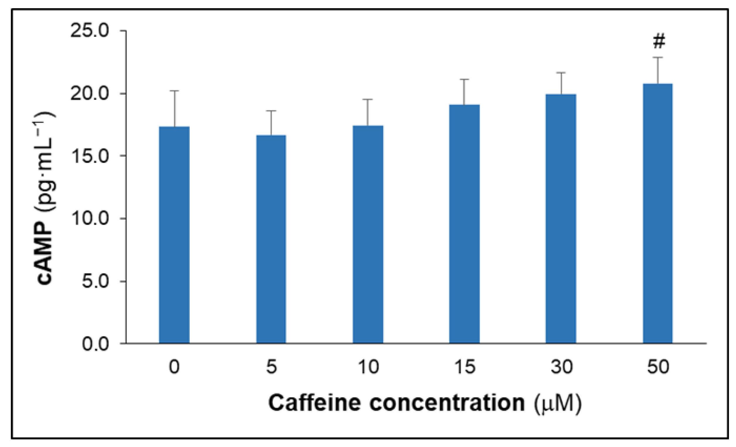
3.2. Effects of Caffeine on Culture Cytokine, MDA and cAMP Concentrations

Significant effects of caffeine were found for TNF-α stimulated production (p = 0.014) (Table 2). Caffeine induced lower TNF-α concentrations, with a significant decrease in comparison to the control only for the highest (50 μM) caffeine concentration (p = 0.016). Caffeine did not influence IL-10 (p = 0.208), IL-6 (p = 0.543), IL-12 (p = 0.700) nor IL-1β (p = 0.501)—stimulated production. No effects of caffeine were observed for unstimulated cytokine production (Table S1). Furthermore, caffeine did not influence MDA culture concentrations (p = 0.227).
When the effect of caffeine on the stimulated cytokine production was analyzed in samples stratified by sex, no significant effect was observed (results not shown).
Figure 1 shows cAMP culture concentrations expressed as the difference between the concentrations in the stimulated and unstimulated samples. A significant effect of caffeine was found for supernatant cultured cAMP concentrations (p = 0.001, η2 = 0.354). However, only the highest caffeine concentration had a significantly higher cAMP concentration in comparison with the control (p = 0.006). For the remaining concentrations, no significant differences were observed, with p-values ranging from 0.052 to 1.000.

3.3. Effects of 5-CQA on Culture Cytokine and MDA Concentrations

Table 3 shows the effect of the different concentrations of 5-CQA on cytokine-stimulated production. 5-CQA significantly influenced the stimulated production of IL-6 (p = 0.002). The 2 μM (p = 0.012) and 20 μM (p < 0.001) concentrations of 5-CQA induced significantly lower IL-6 concentrations than in the control. However, no effects of 5-CQA were observed on the stimulated production of IL-10 (p = 0.438), TNF-α (p = 0.068), IL-12 (p = 0.650) or IL-1β (p = 0.571). No effects of 5-CQA were observed for unstimulated cytokine production (Table S2). 5-CQA did not influence MDA culture concentrations (p = 0.259).
When the influence of 5-CQA on the stimulated-cytokine production was analyzed in samples stratified per sex, no significant effect was observed (results not shown).

4. Discussion

The current study determined the effect of physiological concentrations of caffeine and 5-CQA on the LPS-induced cytokine production in whole blood cultures. The main findings were that caffeine induces lower TNF-α in vitro concentrations. These effects were observed for caffeine concentrations that can be attained after in ingestion of two to three cups of coffee and suggest a limited anti-inflammatory effect of caffeine. On the other hand, 5-CQA at physiological concentrations did not influence in vitro cytokine-stimulated production. This is the first known study using human blood-derived cells, to analyze the anti-inflammatory effect of chlorogenic acid, 5-CQA.
A diluted whole-blood method was applied for the determination of cytokine-stimulated production. When whole blood, rather than isolated cells, is used, the natural cell–cell interactions are preserved avoiding artifacts from preparation, whereas the methods used to isolate peripheral blood mononuclear cells modify the lymphocyte to monocyte ratio and remove endogenous immunomodulators. Therefore, in vivo conditions are more accurately represented using whole-blood culture methods [29]. In addition, physiological concentrations of both caffeine and 5-CQA were used. In this regard, two studies performed by our research group have shown plasma concentrations around 40–50 μM after ingesting 6 mg caffeine/kg body weight [26,30], the caffeine content of three to four cups of coffee. Furthermore, we found that plasma caffeine concentrations after ingesting one cup of coffee are around 10 μM (unpublished results). These observations fit with results reported by others, with peak concentrations of 1–10 μM after ingesting a cup of coffee, and 20–50 μM following the ingestion of two to three cups of coffee or an equivalent caffeine amount [2,31,32]. These observations lead us to propose that caffeine concentrations higher than 50 μM are not relevant and, therefore, we focused the present study on a range of concentrations relevant to habitual levels of caffeine consumption (0–50 μM). Considering the caffeine ingestion needed to achieve values close to 50 μM, these figures would not be commonly attained. Regarding 5-CQA, concentrations around the ones observed in plasma after coffee drinking, 5–12 nM [22], were tested. Furthermore, to allow the comparison with similar studies using much higher concentrations, values in the μM range were also considered.
The results from the present study indicate an anti-inflammatory effect of caffeine, characterized by a less pronounced production of in vitro pro-inflammatory TNF-α. Two previous studies reported inhibitory effects of caffeine on stimulated in vitro TNF-α production in whole blood cultures [24,25]. In the present study, the inhibitory effects of caffeine were observed for the 50 μM concentration. Therefore, as has been indicated above, it is unlikely that this effect could be observed commonly in vivo due to the high caffeine intake required to achieve this concentration. Actually, in previous studies the inhibitory effect of caffeine was observed for higher concentrations, 200 μg/mL (around 1 mM), which supposes a concentration 20-fold higher than the highest concentration tested in the present study, and also using S. pneumoniae rather than LPS as a stimulant [24], and 100 μM [25]. It is difficult to ascertain the reasons for the different caffeine concentrations required to produce significant decreases in TNF-α production. In fact, the only differences that can be surmised are methodological, such as different blood dilution, different LPS concentrations, or blood donors as in the study from Horrigan, a low number (eight), only female participants were considered. However, as observed in the present study, Horrigan et al. [25] reported, albeit non-significant, a downward trend from the lower concentration tested (10 μM). The results could not be considered essentially different because in the present study, a non-significant downward trend from the lower concentration tested (10 μM) was also observed. It is also noteworthy that a wide range of caffeine concentrations have been suggested to inhibit TNF-α production when different cell cultures have been used. In this sense, a study using human adipose tissue primary cultures, found an inhibitory effect of caffeine on TNF-α spontaneous production with a caffeine concentration as low as 0.5 μM [33]. Less pronounced levels in TNF-α production have been attributed to increased levels of the immunomodulator cAMP [34] and mediated by the cAMP/ protein kinase A (PKA) pathway [25]. In this regard, a significant effect of caffeine increasing cAMP concentration was observed, with significantly higher values for the highest caffeine concentration. It should be considered that in the present study cAMP levels were measured only in supernatants; however, because of their intracellular role, the concentrations in supernatants may not properly reflect intracellular cAMP concentrations.
Changes in cAMP concentrations in the presence of caffeine could be expected because caffeine, acting as an adenosine antagonist, influences cAMP concentrations. Furthermore, caffeine, as an inhibitor of cAMP-phosphodiesterase, induces higher cAMP concentrations [23]. Actually, the anti-inflammatory effects of caffeine, at least in in vitro studies, have been mainly attributed to the inhibition of cAMP-phosphodiesterase (PDE) [23]. It is worth noting that a previous study reported a significant inhibition of PDE following a coffee intervention, highlighting that this inhibition was not directly dependent on caffeine consumption [35]. Therefore, while these authors recognized a role for caffeine, they concluded that other components of coffee could play an important role in the modulation of cAMP contributing to the potential health benefits of coffee [35]. In this regard, cAMP has been reported to stimulate the synthesis of the anti-inflammatory IL-10 [34]. However, in agreement with previous studies [24,25], no significant effects of caffeine were observed on IL-10 concentrations. On the other hand, cAMP has been suggested to inhibit IL-12. However, and in the same line as IL-10, no effect of caffeine was observed on IL-12 production [24,25]. The mechanisms leading to the differential effect of caffeine on cytokine production remains to be elucidated. However, similar effects have been reported for other methylxanthines such as theophylline or pentoxifylline [24,36]. On the other hand, it was shown in an animal model that adenosine receptor agonists increased LPS-induced plasma IL-10 levels [37]. Therefore, it was suggested that although caffeine did not modify IL-10 and IL-12 production in vitro, these interleukins may be modulated in vivo by caffeine and other adenosine receptor agonists [25].
Since oxidative stress is involved in pro-inflammatory cytokine production, mainly through the activation of the redox-sensitive Nuclear Factor κB (NF-κB) [38,39], MDA was measured in cell culture supernatants. However, no effect of caffeine or 5-CQA was observed on MDA levels. Despite antioxidant effects having been attributed to caffeine [40], this result is mainly relevant for the 5-CQA experiments. In this regard, chlorogenic acids have been characterized as important antioxidants [3,41], exerting, among other effects, an attenuation of NF-κB [17,18,42]. In agreement with this observation, two studies carried out with mouse macrophages demonstrated how the presence of 5-CQA (the most used CGA in studies), in cell culture with LPS-stimulation suppressed the expression of pro-inflammatory compounds such as cyclooxygenase [18] as well as IL-1β, TNF-α and IL-6 [17]. In these studies, the lower pro-inflammatory expression was associated with attenuated NF-κB [17,18]. However, it should be noted that in both studies 5-CQA concentrations in the range of μM, much higher than physiological ones, were used. In the present study, 5-CQA influenced only IL-6 production, with suppressed levels, but only for the highest 5-CQA concentrations tested, 2 μM and 20 μM. This result is in agreement with the effect observed in cultures of mouse macrophages using similar high 5-CQA concentrations and with the role of 5-CQA in attenuating NF-κB activation [17]. On the other hand, when physiological concentrations were considered, no effect of 5-CQA was observed. Due to the relationship between oxidative stress and activation of the NF-κB [38], the limited effects of 5-CQA on cytokine production in the present study may be related to the lack of effects on culture MDA concentrations. However, it cannot be discarded that chlorogenic acids play an important role in vivo as has been suggested from studies focused on the health effects of coffee consumption [3,14,16]. In this regard, studies in animal models have shown that the administration of 5-CQA lowers the circulating concentrations of pro-inflammatory cytokines such as TNF-α or IL-1β [19,20,21]. On the contrary, supplementations in humans using coffee preparation have shown little (mainly increasing adiponectin concentrations) or no effect on inflammatory markers (reviewed by Paiva et al. [43]). Moreover, only a higher increase in total plasma antioxidant capacity, without affecting the inflammatory response, was observed in response to exercise after supplementation with high-chlorogenic-acid coffee [44].
The authors acknowledge some of the limitations that are present in this study. No intracellular measurements, mainly cAMP but also MDA, were performed. The concentrations of cAMP and MDA were measured only in culture supernatants. Furthermore, NF-κB was not measured. The effects of caffeine and 5-CQA were not tested together. Since in coffee, as well as in other products such as tea, they are found in combination, it would be of interest to test the combined effects of both substances.

5. Conclusions

The results from the present study indicate an anti-inflammatory effect of caffeine, at least under in vitro conditions, characterized by an inhibition of the pro-inflammatory TNF-α production. However, caffeine concentrations higher than usual physiological plasma values are needed to produce this effect. 5-CQA concentrations relevant to human coffee consumption did not influence in vitro cytokine-stimulated production, as only a suppression in IL-6 with the highest, non-physiological, concentrations were observed. More studies are therefore required to clarify whether these concentrations could influence low grade systemic inflammation.

Supplementary Materials

The following supporting information can be downloaded at: https://www.mdpi.com/article/10.3390/app12147322/s1, Table S1. Effects of caffeine on spontaneous cytokine production. Table S2. Effects of 5-CQA on spontaneous cytokine production.

Author Contributions

Conceptualization, P.T.; methodology, P.T., S.M., A.R.-S. and L.R.; formal analysis, P.T. and H.J.M.; investigation, L.R., S.M., A.R.-S. and P.T.; resources, P.T. and S.M., data curation, A.R.-S. and P.T.; writing-original draft preparation, L.R.; writing-review and editing, P.T. and H.J.M.; visualization, A.R.-S.; supervision, P.T. and S.M.; project administration, P.T. All authors have read and agreed to the published version of the manuscript.

Funding

This research was funded in part by the Government of the Balearic Islands and the European Regional Development Funds (FEDER) “Una manera de hacer Europa”, Acció Especial AAEE041/2017.

Institutional Review Board Statement

The study was conducted in accordance with the Declaration of Helsinki, and approved by the Balearic Islands Clinical Investigation Ethics Committee (protocol code IB 3493/17 PI, date of approval 8 September 2017) for studies involving humans.

Informed Consent Statement

Informed consent was obtained from all participants involved in the study.

Data Availability Statement

The data presented in this study are available on request from the corresponding author.

Acknowledgments

The authors want to thank all participants in the study.

Conflicts of Interest

The authors declare no conflict of interest.

References

  1. Reyes, C.M.; Cornelis, M.C. Caffeine in the diet: Country-level consumption and guidelines. Nutrients 2018, 10, 1772. [Google Scholar] [CrossRef] [PubMed] [Green Version]
  2. Faudone, G.; Arifi, S.; Merk, D. The medicinal chemistry of caffeine. J. Med. Chem. 2021, 64, 7156–7178. [Google Scholar] [CrossRef] [PubMed]
  3. Liang, N.; Kitts, D.D. Role of Chlorogenic Acids in Controlling Oxidative and Inflammatory Stress Conditions. Nutrients 2015, 8, 16. [Google Scholar] [CrossRef] [PubMed] [Green Version]
  4. Neveu, V.; Perez-Jiménez, J.; Vos, F.; Crespy, V.; du Chaffaut, L.; Mennen, L.; Knox, C.; Eisner, R.; Cruz, J.; Wishart, D.; et al. Phenol-Explorer: An online comprehensive database on polyphenol contents in foods. Database 2010, 2010, bap024. [Google Scholar] [CrossRef]
  5. Farah, A.; de Paula Lima, J. Consumption of chlorogenic acids through coffee and health implications. Beverages 2019, 5, 11. [Google Scholar] [CrossRef] [Green Version]
  6. Akash, M.S.H.; Rehman, K.; Chen, S. Effects of coffee on type 2 diabetes mellitus. Nutrition 2014, 30, 755–763. [Google Scholar] [CrossRef]
  7. Ding, M.; Bhupathiraju, S.N.; Chen, M.; van Dam, R.M.; Hu, F.B. Caffeinated and decaffeinated coffee consumption and risk of type 2 diabetes: A systematic review and a dose-response meta-analysis. Diabetes Care 2014, 37, 569–586. [Google Scholar] [CrossRef] [Green Version]
  8. Shang, F.; Li, X.; Jiang, X. Coffee consumption and risk of the metabolic syndrome: A meta-analysis. Diabetes Metab. 2016, 42, 80–87. [Google Scholar] [CrossRef]
  9. Ding, M.; Satija, A.; Bhupathiraju, S.N.; Hu, Y.; Sun, Q.; Han, J.; Lopez-Garcia, E.; Willett, W.; van Dam, R.M.; Hu, F.B. Association of coffee consumption with total and cause-specific mortality in 3 large prospective cohorts. Circulation 2015, 132, 2305–2315. [Google Scholar] [CrossRef] [Green Version]
  10. Grosso, G.; Godos, J.; Galvano, F.; Giovannucci, E.L. Coffee, Caffeine, and Health Outcomes: An Umbrella Review. Annu. Rev. Nutr. 2017, 37, 131–156. [Google Scholar] [CrossRef] [Green Version]
  11. Torres-Collado, L.; Compañ-Gabucio, L.M.; González-Palacios, S.; Leyre, N.-B.; Oncina-Cánovas, A.; Vioque, J.; García-de la Hera, M. Coffee Consumption and All-Cause, Cardiovascular, and Cancer Mortality in an Adult Mediterranean Population. Nutrients 2021, 13, 1241. [Google Scholar] [CrossRef] [PubMed]
  12. Williams, C.J.; Fargnoli, J.L.; Hwang, J.J.; van Dam, R.M.; Blackburn, G.L.; Hu, F.B.; Mantzoros, C.S. Coffee Consumption Is Associated with Higher Plasma Adiponectin Concentrations in Women with or Without Type 2 Diabetes A prospective cohort study. Diabetes Care 2008, 31, 504–507. [Google Scholar] [CrossRef] [PubMed] [Green Version]
  13. Kotani, K.; Tsuzaki, K.; Sano, Y.; Maekawa, M.; Fujiwara, S.; Hamada, T.; Sakane, N. The relationship between usual coffee consumption and serum C-reactive protein level in a Japanese female population. Clin. Chem. Lab. Med. 2008, 46, 1434–1437. [Google Scholar] [CrossRef] [PubMed]
  14. Lopez-Garcia, E.; van Dam, R.M.; Qi, L.; Hu, F.B. Coffee consumption and markers of inflammation and endothelial dysfunction in healthy and diabetic women. Am. J. Clin. Nutr. 2006, 84, 888–893. [Google Scholar] [CrossRef] [PubMed] [Green Version]
  15. Maki, T.; Pham, N.M.; Yoshida, D.; Yin, G.; Ohnaka, K.; Takayanagi, R.; Kono, S. The relationship of coffee and green tea consumption with high-sensitivity C-reactive protein in Japanese men and women. Clin. Chem. Lab. Med. 2010, 48, 849–854. [Google Scholar] [CrossRef]
  16. Tajik, N.; Tajik, M.; Mack, I.; Enck, P. The potential effects of chlorogenic acid, the main phenolic components in coffee, on health: A comprehensive review of the literature. Eur. J. Nutr. 2017, 56, 2215–2244. [Google Scholar] [CrossRef]
  17. Hwang, S.J.; Kim, Y.-W.W.; Park, Y.; Lee, H.-J.J.; Kim, K.-W.W. Anti-inflammatory effects of chlorogenic acid in lipopolysaccharide-stimulated RAW 264.7 cells. Inflamm. Res. 2013, 63, 81–90. [Google Scholar] [CrossRef]
  18. Shan, J.; Fu, J.; Zhao, Z.; Kong, X.; Huang, H.; Luo, L.; Yin, Z. Chlorogenic acid inhibits lipopolysaccharide-induced cyclooxygenase-2 expression in RAW264.7 cells through suppressing NF-KB and JNK/AP-1 activation. Int. Immunopharmacol. 2009, 9, 1042–1048. [Google Scholar] [CrossRef]
  19. Chauhan, P.S.; Satti, N.K.; Sharma, P.; Sharma, V.K.; Suri, K.A.; Bani, S. Differential effects of chlorogenic acid on various immunological parameters relevant to rheumatoid arthritis. Phytother. Res. 2012, 26, 1156–1165. [Google Scholar] [CrossRef]
  20. Yun, N.; Kang, J.-W.; Lee, S.-M. Protective effects of chlorogenic acid against ischemia/reperfusion injury in rat liver: Molecular evidence of its antioxidant and anti-inflammatory properties. J. Nutr. Biochem. 2012, 23, 1249–1255. [Google Scholar] [CrossRef]
  21. Shin, H.S.; Satsu, H.; Bae, M.J.; Zhao, Z.; Ogiwara, H.; Totsuka, M.; Shimizu, M. Anti-inflammatory effect of chlorogenic acid on the IL-8 production in Caco-2 cells and the dextran sulphate sodium-induced colitis symptoms in C57BL/6 mice. Food Chem. 2015, 168, 167–175. [Google Scholar] [CrossRef] [PubMed]
  22. Monteiro, M.; Farah, A.; Perrone, D.; Trugo, L.C.; Donangelo, C. Chlorogenic acid compounds from coffee are differentially absorbed and metabolized in humans. J. Nutr. 2007, 137, 2196–2201. [Google Scholar] [CrossRef] [PubMed] [Green Version]
  23. Horrigan, L.A.; Kelly, J.P.; Connor, T.J. Immunomodulatory effects of caffeine: Friend or foe? Pharmacol. Ther. 2006, 111, 877–892. [Google Scholar] [CrossRef] [PubMed]
  24. Van Furth, A.M.; Seijmonsbergen, E.M.; Langermans, J.A.; van der Meide, P.H.; van Furth, R. Effect of xanthine derivates and dexamethasone on Streptococcus pneumoniae-stimulated production of tumor necrosis factor alpha, interleukin-1 beta (IL-1 beta), and IL-10 by human leukocytes. Clin. Diagn. Lab. Immunol. 1995, 2, 689–692. [Google Scholar] [CrossRef] [PubMed] [Green Version]
  25. Horrigan, L.A.; Kelly, J.P.; Connor, T.J. Caffeine suppresses TNF-alpha production via activation of the cyclic AMP/protein kinase A pathway. Int. Immunopharmacol. 2004, 4, 1409–1417. [Google Scholar] [CrossRef]
  26. Tauler, P.; Martinez, S.; Moreno, C.; Monjo, M.; Martinez, P.; Aguilo, A. Effects of caffeine on the inflammatory response induced by a 15-km run competition. Med. Sci. Sport. Exerc. 2013, 45, 1269–1276. [Google Scholar] [CrossRef]
  27. Segre, E.; Fullerton, J.N. Stimulated whole blood cytokine release as a biomarker of immunosuppression in the critically ill: The need for a standardized methodology. Shock 2016, 45, 490–494. [Google Scholar] [CrossRef] [Green Version]
  28. Agarwal, R.; Chase, S.D. Rapid, fluorimetric-liquid chromatographic determination of malondialdehyde in biological samples. J. Chromatogr. B. Anal. Technol. Biomed. Life Sci. 2002, 775, 121–126. [Google Scholar] [CrossRef]
  29. Damsgaard, C.T.; Lauritzen, L.; Calder, P.C.; Kjær, T.M.R.; Frøkiær, H. Whole-blood culture is a valid low-cost method to measure monocytic cytokines—A comparison of cytokine production in cultures of human whole-blood, mononuclear cells and monocytes. J. Immunol. Methods 2009, 340, 95–101. [Google Scholar] [CrossRef]
  30. Tauler, P.; Martinez, S.; Martinez, P.; Lozano, L.; Moreno, C.; Aguiló, A. Effects of Caffeine Supplementation on Plasma and Blood Mononuclear Cell IL-10 Levels After Exercise. Int. J. Sport Nutr. Exerc. Metab. 2016, 26, 8–16. [Google Scholar] [CrossRef]
  31. De Leon, J.; Diaz, F.J.; Rogers, T.; Browne, D.; Dinsmore, L.; Ghosheh, O.H.; Dwoskin, L.P.; Crooks, P.A. A pilot study of plasma caffeine concentrations in a US sample of smoker and nonsmoker volunteers. Prog. Neuro-Psychopharmacol. Biol. Psychiatry 2003, 27, 165–171. [Google Scholar] [CrossRef]
  32. Horrigan, L.A.; Diamond, M.; Connor, T.J.; Kelly, J.P. Caffeine inhibits monocyte and neutrophil chemotaxis at concentrations relevant to normal human consumption. In Proceedings of the International Cytokine Society Annual Meeting, Dublin, Ireland, 20–24 September 2003; pp. 49–54. [Google Scholar]
  33. Dray, C.; Daviaud, D.; Guigné, C.; Valet, P.; Castan-Laurell, I. Caffeine reduces TNFα up-regulation in human adipose tissue primary culture. J. Physiol. Biochem. 2007, 63, 329–336. [Google Scholar] [CrossRef] [PubMed]
  34. Eigler, A.; Siegmund, B.; Emmerich, U.; Baumann, K.H.; Hartmann, G.; Endres, S. Anti-inflammatory activities of cAMP-elevating agents: Enhancement of IL-10 synthesis and concurrent suppression of TNF production. J. Leukoc. Biol. 1998, 63, 101–107. [Google Scholar] [CrossRef] [PubMed]
  35. Montoya, G.A.; Bakuradze, T.; Eirich, M.; Erk, T.; Baum, M.; Habermeyer, M.; Eisenbrand, G.; Richling, E. Modulation of 3′,5′-cyclic AMP homeostasis in human platelets by coffee and individual coffee constituents. Br. J. Nutr. 2014, 112, 1427–1437. [Google Scholar] [CrossRef] [PubMed] [Green Version]
  36. Marcinkiewicz, J.; Grabowska, A.; Lauterbach, R.; Bobek, M. Differential effects of pentoxifylline, a non-specific phosphodiesterase inhibitor, on the production of IL-10, IL-12 p40 and p35 subunits by murine peritoneal macrophages. Immunopharmacology 2000, 49, 335–343. [Google Scholar] [CrossRef]
  37. Haskó, G.; Szabó, C.; Németh, Z.H.; Kvetan, V.; Pastores, S.M.; Vizi, E.S. Adenosine receptor agonists differentially regulate IL-10, TNF-alpha, and nitric oxide production in RAW 264.7 macrophages and in endotoxemic mice. J. Immunol. 1996, 157, 4634–4640. [Google Scholar]
  38. Socała, K.; Szopa, A.; Serefko, A.; Poleszak, E.; Wlaź, P. Neuroprotective effects of coffee bioactive compounds: A review. Int. L. Mol. Sci. 2020, 22, 107. [Google Scholar] [CrossRef]
  39. Lawrence, T. The nuclear factor NF-kappaB pathway in inflammation. Cold Spring Harb. Perspect. Biol. 2009, 1, a001651. [Google Scholar] [CrossRef] [Green Version]
  40. Barcelos, R.P.; Lima, F.D.; Carvalho, N.R.; Bresciani, G.; Royes, L.F. Caffeine effects on systemic metabolism, oxidative-inflammatory pathways, and exercise performance. Nutr. Res. 2020, 80, 1–17. [Google Scholar] [CrossRef]
  41. Gómez-Ruiz, J.Á.; Leake, D.S.; Ames, J.M. In vitro antioxidant activity of coffee compounds and their metabolites. J. Agric. Food Chem. 2007, 55, 6962–6969. [Google Scholar] [CrossRef]
  42. Zatorski, H.; Sałaga, M.; Zielińska, M.; Piechota-Polańczyk, A.; Owczarek, K.; Kordek, R.; Lewandowska, U.; Chen, C.; Fichna, J. Experimental colitis in mice is attenuated by topical administration of chlorogenic acid. Naunyn-Schmiedebergs Arch. Pharmacol. 2015, 388, 643–651. [Google Scholar] [CrossRef] [PubMed] [Green Version]
  43. Paiva, C.L.R.S.; Beserra, B.T.S.; Reis, C.E.G.; Dorea, J.G.; Da Costa, T.H.M.; Amato, A.A. Consumption of coffee or caffeine and serum concentration of inflammatory markers: A systematic review. Crit. Rev. Food Sci. Nutr. 2019, 59, 652–663. [Google Scholar] [CrossRef] [PubMed]
  44. Nieman, D.C.; Goodman, C.L.; Capps, C.R.; Shue, Z.L.; Arnot, R. Influence of 2-weeks ingestion of high chlorogenic acid coffee on mood state, performance, and postexercise inflammation and oxidative stress: A randomized, placebo-controlled trial. Int. J. Sport Nutr. Exerc. Metab. 2018, 28, 55–65. [Google Scholar] [CrossRef] [PubMed] [Green Version]
Figure 1. Effects of caffeine and LPS-stimulation on culture cAMP concentrations. Values are expressed as means ± SD. # indicates significant differences vs. control. cAMP: Adenosine-3′,5′-cyclic monophosphate.
Applsci 12 07322 g001
Table 1. General characteristics of participants in the study.
ParameterAll
(n = 10)
Men
(n = 5)
Women
(n = 5)
p Value
Age (years)36.5 ± 6.535.8 ± 8.137.2 ± 5.30.754
Body mass (kg)68.5 ± 11.670.6 ± 14.666.4 ± 9.00.599
Stature (cm)169.7 ± 7.0173.6 ± 5.7165.8 ± 6.30.073
BMI (kg·m−2)23.8 ± 3.423.3 ± 3.924.2 ± 3.30.722
Leukocytes (103·μL−1)5.28 ± 1.075.34 ± 1.405.22 ± 0.780.871
Lymphocytes (103·μL−1)1.53 ± 0.371.65 ± 0.391.41 ± 0.350.334
Monocytes (103·μL−1)0.35 ± 0.130.38 ± 0.170.32 ± 0.070.495
Values are expressed as means ± SD.
Table 2. Effects of caffeine on LPS-stimulated cytokine production and MDA concentration.
Caffeine Concentrations (μM)ANOVA
Control510153050p2)
IL-10 (pg·10−3 cells)21.9 ± 3.422.1 ± 6.520.6 ± 3.922.7 ± 4.524.0 ± 4.924.6 ± 3.90.208 (0.137)
IL-6 (pg·10−3 cells)385.2 ± 88.6448.2 ± 96.3451.5 ± 99.8412.4 ± 82.5440.6 ± 107396.1 ± 93.60.543 (0.070)
TNF-α (pg·10−3 cells)58.6 ± 10.457.5 ± 9.855.2 ± 9.449.2 ± 9.147.9 ± 8.345.1 ± 7.7 #0.014 * (0.250)
IL-12 (pg·10−3 cells)4.13 ± 0.844.50 ± 0.785.01 ± 0.114.91 ± 0.664.36 ± 0.84.50 ± 0.950.700 (0.053)
IL-1β (pg·10−3 cells)17.2 ± 5.019.3 ± 6.018.2 ± 5.319.4 ± 4.816.8 ± 6.320.1 ± 5.10.501 (0.073)
MDA (nM)0.62 ± 0.100.63 ± 0.120.72 ± 0.130.72 ± 0.120.62 ± 0.110.58 ± 0.110.227 (0.147)
Values are expressed as means ± SD and represents the percentage with respect to the control. * p < 0.05 indicates significant differences (ANOVA one-way test). # Indicates significant differences vs. control. η2 value is provided as a measure of effect size.
Table 3. Effects of 5-CQA on LPS-stimulated cytokine production and MDA concentration.
5-CQA ConcentrationsANOVA
Control5 nM20 nM200 nM2 μM20 μMp2)
IL-10 (pg·10−3 cells)21.2 ± 4.520.8 ± 6.620.2 ± 5.719.2 ± 6.817.4 ± 6.217.8 ± 6.50.438 (0.083)
IL-6 (pg·10−3 cells)385.2 ± 92.4351.2 ± 83.1326.5 ± 93.9291.9 ± 86.1252.0 ± 85.8 #202.2 ± 82.5 #0.002 * (0.286)
TNF-α (pg·10−3 cells)58.2 ± 9.855.1 ± 10.155.4 ± 8.049.2 ± 9.946.9 ± 7.344.7 ± 8.50.068 (0.169)
IL-12 (pg·10−3 cells)4.48 ± 0.104.22 ± 0.974.53 ± 0.124.24 ± 0.144.05 ± 0.983.75 ± 0.100.650 (0.058)
IL-1β (pg·10−3 cells)16.2 ± 4.018.5 ± 5.017.8 ± 5.318.4 ± 5.815.8 ± 7.714.8 ± 5.70.571 (0.067)
MDA (nM)0.61 ± 0.110.65 ± 0.140.70 ± 0.140.68 ± 0.140.70 ± 0.110.65 ± 0.100.259 (0.111)
Values are expressed as means ± SD. * p < 0.05 indicates significant differences (ANOVA one-way test). # Indicates significant differences vs. control. η2 value is provided as a measure of effect size.
Publisher’s Note: MDPI stays neutral with regard to jurisdictional claims in published maps and institutional affiliations.

