Article

Therapeutic Potential of Ajwa Dates (Phoenix dactylifera) Extract in Prevention of Benzo(a)pyrene-Induced Lung Injury through the Modulation of Oxidative Stress, Inflammation, and Cell Signalling Molecules

by
Saleh A. Almatroodi
1,
Amjad Ali Khan
2,
Abdulaziz A. Aloliqi
3,
Mansoor Ali Syed
4 and
Arshad Husain Rahmani
1,*
1
Department of Medical Laboratories, College of Applied Medical Sciences, Qassim University, Buraydah 52571, Saudi Arabia
2
Department of Basic Health Science, College of Applied Medical Sciences, Qassim University, Buraydah 52571, Saudi Arabia
3
Department of Medical Biotechnology, College of Applied Medical Sciences, Qassim University, Buraydah 52571, Saudi Arabia
4
Translational Research Lab, Department of Biotechnology, Faculty of Natural Sciences, Jamia Millia Islamia, New Delhi 110025, India
*
Author to whom correspondence should be addressed.
Appl. Sci. 2022, 12(13), 6784; https://doi.org/10.3390/app12136784
Submission received: 16 May 2022 / Revised: 29 June 2022 / Accepted: 2 July 2022 / Published: 4 July 2022
(This article belongs to the Special Issue Application of Natural Products with Anti-inflammatory Potentials)

Abstract

:
Chronic respiratory diseases are a leading cause of lung-related death worldwide. The vital factors causing lung pathogenesis include consistent exposure to tobacco smoke, air pollution, and occupational risks. Regarding the significant morbidity and mortality linked to lung pathogenesis, there are neither conclusive treatments nor wholly preventive strategies. In the present study, the protective mechanism of Ajwa date extract (ADE), on Benzopyrene [B(a)P]-induced lung injury in animal models was investigated using antioxidant, lipid peroxidation, anti-inflammatory activities, angiogenesis, histopathological studies, and apoptosis assays. B(a)P treatment significantly decreased the level of antioxidant enzymes such as catalase (Cat) (13.4 vs. 24.7 U/mg protein), Superoxide dismutase (SOD) (38.5 vs. 65.7 U/mg protein), Glutathione peroxidase (GPx) (42.4 vs. 57.3 U/mg protein) and total antioxidant capacity (TAC) (49.8 vs. 98.7 nM) as compared to the treatment group (p < 0.05). B(a)P treatment led to increased expression of pro-inflammatory markers such as TNF-α (88.5 vs. 72.6 pg/mL), IFN-γ (4.86 vs. 3.56 pg/mL), interleukin-6 (IL-6) (109.6 vs. 85.4 pg/mL) and CRP (1.84 vs. 0.94 ng/mL) as compared to the treatment group (p < 0.05). The data shows a significant increase in lipid peroxidation and angiogenesis factors such as vascular endothelial growth factor (VEGF) by B(a)P treatment (p < 0.05). However, ADE treatment showed an improvement of these factors. In addition, ADE treatment significantly ameliorated histopathological changes, collagen fiber deposition, and expression pattern of VEGF and Bax proteins. Furthermore, the flow cytometry data demonstrated that B(a)P intoxication enhanced the apoptosis ratio, which was significantly improved with ADE treatment. Finally, we may infer that Phyto-constituents of ADE have the potential to protect against B(a)P-induced lung pathogenesis. Therefore, Ajwa dates might be used to develop a possible potent alternative therapy for lung pathogenesis.

1. Introduction

Chronic respiratory diseases, including chronic obstructive pulmonary disease, pulmonary fibrosis, and lung cancer, enormously affect health and these diseases are linked to millions of deaths worldwide [1]. The respiratory diseases are strongly related to smoke inhalation, and benzopyrene [B(a)P] is the main component of tobacco smoke. This compound belongs to the family of polycyclic aromatic hydrocarbons and is known as the most harmful compound in lung pathogenesis [2,3,4,5]. It is recognized that B(a)P undergoes successive metabolic activation stages, mainly through cytochrome P450, that shows a vital role in the drug’s metabolism and produces 7,8-diol-9,10-epoxide-benzo(a)pyrene. This product is supposed to be the definitive carcinogenic metabolite of benzopyrene that causes formation of DNA adducts [6]. For the activation of B(a)P, the enzymes involved and the subsequent binding to DNA have been well-proven in lung-based studies [7], and cause numerous toxicities such as airway inflammation [8]. Acute exposure to B(a)P alters the cellular antioxidant levels [9] and causes numerous changes in the lungs, including edema, inflammation, and epithelial damage [10]. Moreover, numerous studies have proven that B(a)P induces inflammatory responses, and inflammatory mediators are increased during B(a)P-induced genotoxicity [11,12].
The current mode of treatment in lung pathogenesis with anti-inflammatory drugs is not very effective and causes various side effects. As a result, in order to explore a therapeutic approach, it is becoming more and more important to identify the pathobiological processes associated with lung pathogenesis and associated complications. The use of herbal-based natural compounds affects cellular mechanisms, and the role of natural products in inflammatory pulmonary diseases has been growing [13]. It has been discovered that using medicinal plants to treat and manage diseases in various traditional medical systems is a secure and cost-effective substitute for allopathic medications. Herbal-based natural compounds show an effect in disease management through enhancement of antioxidant enzyme activities, immunomodulation, anti-inflammatory activities, reduction of reactive oxygen species (ROS), and modulation of other biological activities. Finding a potent natural treatment for lung pathogenesis requires investigating medicinal plants and using a drug-repurposing strategy to identify potential therapeutic drugs against lung pathogenesis.
Ajwa dates (Phoenix dactylifera) are a member of the Arecaceae family, specifically grown in the region of Medina, Saudi Arabia [14]. This fruit contains various ingredients including glycosides, flavonoids, polyphenols, and phytosterols [15]. These ingredients play a significant role in disease management through antioxidant and anti-inflammatory activities, liver protection [16,17], nephroprotective activity [18], and antihyperlipidemic activity [19]. In addition, organic solvent extracts of Ajwa date flesh and pits have been found to contain high radical-scavenging and antioxidant activities [20]. Although Ajwa has been found to have positive effects on a number of chronic diseases, the precise mechanism is still unknown. In this study, Ajwa date extract (ADE) was assessed for in vivo protective roles against benzopyrene-induced lung injury in rats as well as revealed its mechanisms associated with oxidative stress, inflammatory process, angiogenesis, and apoptosis.

2. Materials and Methods

2.1. Reagents and Chemicals

Ajwa dates used in the current study were purchased from Medina city (Al Madinah Al Munawwarah) of Medina province, Saudi Arabia. Benzopyrene [B(a)P] was purchased from Sigma-Aldrich Chemical Co. (St. Louis, MO, USA). The antioxidant enzyme kits were obtained from Abcam Company (Abcam, Cambridge, UK). TNF-α, IFN-γ, interleukin-6, and CRP were also purchased from Abcam Company (Abcam, Cambridge, UK). Picro Sirius red and Masson’s trichrome staining kits were obtained from Abcam Company (Abcam, Cambridge, UK). In addition, antibodies (Bax, and VEGF) used in the current study were purchased from the same company. High-analytical-grade reagents and supporting chemicals were used in this study. The rat chow was purchased from a local market in Buraydah (Qassim) of Saudi Arabia.

2.2. Preparation of Ethanolic Extract of Ajwa Pulp (ADE)

One hundred grams of dried fruit pulp was separated and cleaned with distilled water. The dried pulp was finely ground to prepare powder. A total 50 g of dry powder was soaked in 500 mL of 95% ethanol on a magnetic shaker continuously for three days at 25 °C. Filtration was used to purify the extract and condensed by a rotary evaporator at decreased pressure at 40 °C. Prepared extract was kept for storage at 4 °C until further use. The extract % yield was measured with below mentioned the equation.
(%) Yield = [Weight of extract sample/sample Initial weight] × 100

2.3. Phytochemical Screening

Screening of phytochemicals such as carbohydrates, phenolics, anthraquinone, alkaloids, flavonoids, saponins, and tannins, as well as phenolic compounds were estimated as previously explained [21]. Carbohydrates were detected by adding 1 mL iodine solution with 3 mL ethanol ADE separately. Carbohydrates may be detected if the hue changes to purple. Saponins were found by combining 7 mL extract with 7 mL distilled water and forcefully stirring the mixture before heating the test tubes. The formation of stable foam is the indication of the presence of saponins. In the meantime, 1.5 mL of extract and 1.5 mL of distilled water were put in a test tube. After adding a few drops of ferric chloride (FeCl3), a green precipitate formation indicates the presence of condensed tannins. When 20% NaOH solution was mixed with extracts (2.0 mL), a rich yellow color was developed, and it is a confirmation of presence of flavonoids in the extract. However, dilute HCl addition causes a yellowish hue that then becomes colorless. Presence of phenolic compounds was confirmed as emergence of blue-black color after the addition of a few drops of FeCl3. To Ajwa date extract.

2.4. Total Phenolic Content

Folin–Ciocalteu reagent was used to measure the total phenolic content. In a vial, 500 µL of extract (1 mg/mL) was added, along with 10% Folin–Ciocalteu reagent (2.5 mL). Finally, 2000 µL of 7.5% Na2CO3 was added in each tube. At room temperature, test tubes were incubated in dark conditions for approximately half an hour, and at 760 nm absorbance was measured. A standard calibration plot was made using different concentrations of gallic acid (50–250 g/mL). The amount of total phenolic content in the extract was calculated using this standard. Gallic acid equivalents (GAE) were used to express the total phenolic content. All tests were completed in triplicate, and the results were presented as milli gram GAE/gram extract. Total phenolic content was measured through the following formula:
Total phenolic content = M × N/P
where, M refers to gallic acid (mg/mL) concentration N denotes plant extract volume (mL), and P refers to weight of pure plant extract (g).

2.5. Evaluation of the Amount of Total Flavonoids

Total flavonoid content of ADE was evaluated by using the protocol from previously published articles [22,23,24]. Total flavonoid concentration was measured using quercetin calibration curve, and results presented in mg of quercetin equivalent per gm of extract (mg QUE/g).

2.6. Animals

Male albino wistar rats weighing 200 ± 25 g were obtained from King Saud University, Riyadh, Saudi Arabia. After the acclimatization period of one week, the animals were randomly separated into four experimental groups of eight rats per group. The rats were housed in plastic cages and maintained at a temperature of 23 ± 2 °C with humidity maintained at 45–55% in a 12-h light and dark condition environment. The animals were given access to normal rat pellets as well as water ad libitum.

2.7. Animal Ethics

All the animals were treated in accordance with proper guidelines from Ethics Committee for the Care and Use of Laboratory Animals of the Qassim University, Saudi Arabia (10241-cams1-2020-1-3-I). This research project was approved by animal ethics committee of Qassim University, and all efforts were made to minimize the animals’ suffering.

2.8. Study Design and Treatment Plan

To evaluate the potential protective effects of ADE on lungs, experiments were conducted in four different animal groups as described in Table 1.
At the end of the treatment, all rats were killed by cervical decapitation, and lungs were removed quickly and washed properly with phosphate buffer saline (PBS). One portion of the lung was homogenized with a tissue homogenizer in 50 mM potassium phosphate buffer, pH 7.0, and the homogenate was centrifuged at 4 °C for 15 min at 10,000 rpm. All the supernatant samples were preserved at −20 °C for biochemical analysis. Moreover, for biochemical analysis, blood was also obtained and the serum was separated. The other portion of the lung was fixed in a formaldehyde neutral buffer solution for histopathological analysis such as hematoxylin-eosin (HE), Masson’s trichrome, and Sirius red, for immunohistochemical and Tunel assay analysis.

2.9. Measurement of Antioxidant Enzymes and Oxidative Stress Markers

Catalase, Superoxide dismutase (SOD) and Glutathione peroxidase (GPx) were measured in lung tissue as per the manufacturer’s instructions. The absorbance was taken at a specific wavelength by using a micro-plate reader, and results were interpreted accordingly. The total antioxidant capacity (TAC) in lung tissue was evaluated using the kit from Abcam, UK and the protocol was followed as per the manufacturer’s instructions.

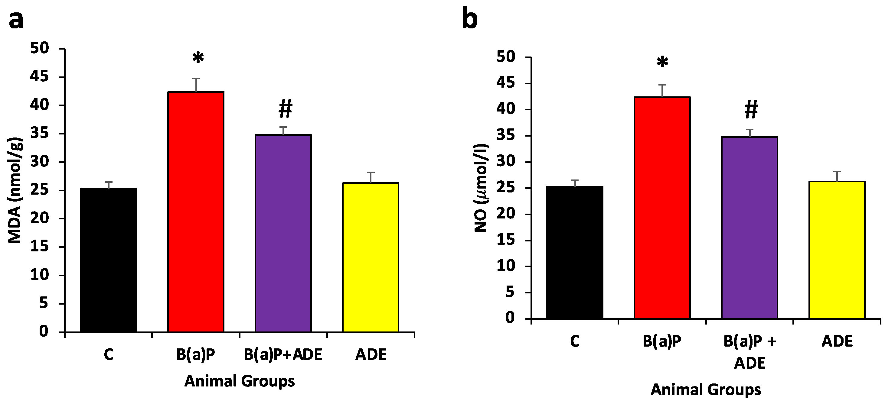
2.10. Measurement of Malondialdehyde and Nitric Oxide Content

Malondialdehyde (MDA), the main indicator of lipid peroxidation, was measured in the lung tissues using thiobarbituric acid reactive substances (TBARS). A product formation appearing after the reaction of TBARS with MDA was measured at 532 nm.
Nitric Oxide levels in lung tissues were measured by the kits, and protocol was followed as per manufacturer’s instructions. The absorbance was taken at a 549 nm wavelength by using a micro-plate reader and result was interpreted accordingly.

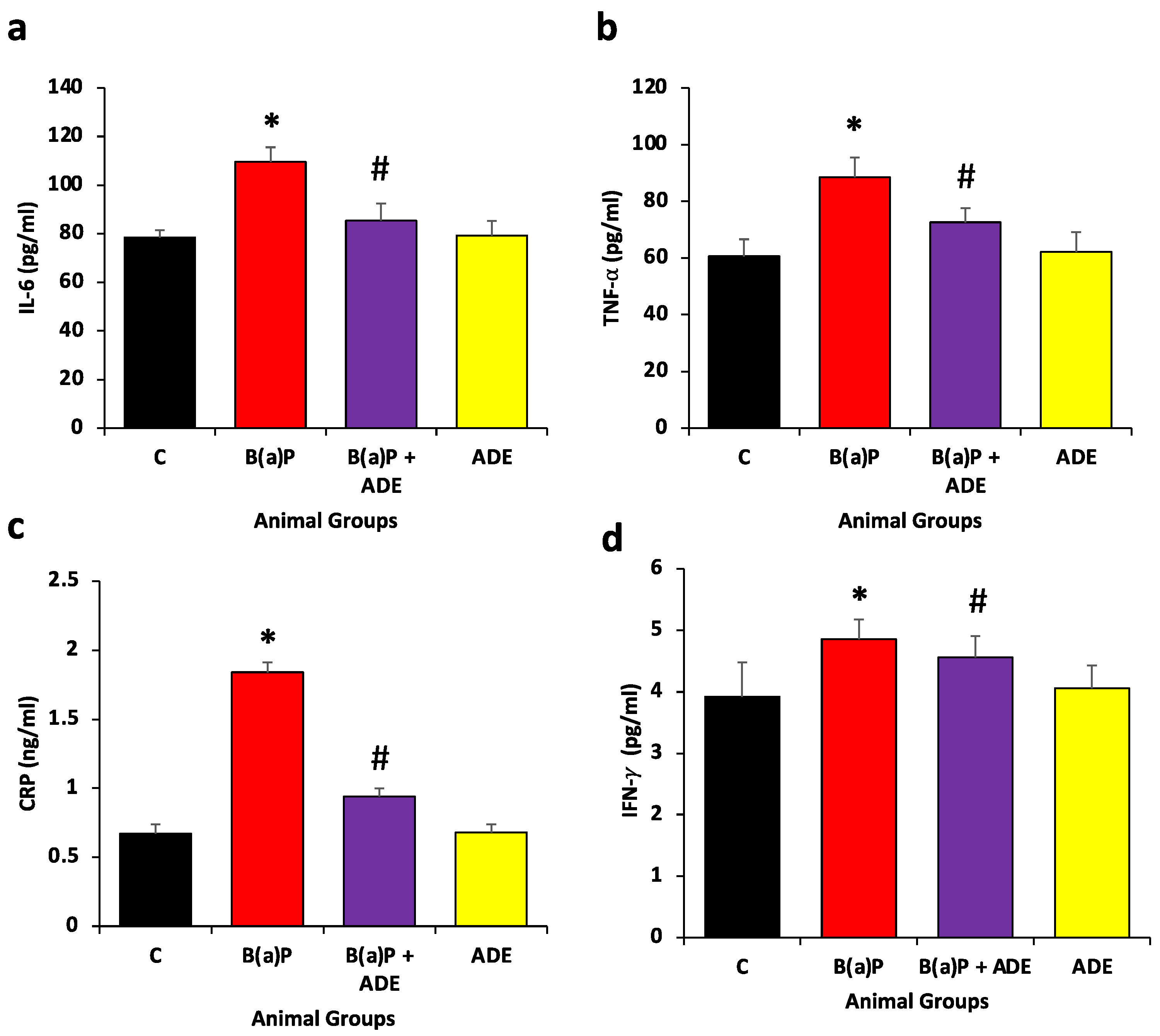
2.11. Quantification of Inflammatory Markers

The estimation of different markers of inflammation such as TNF-α, IFN-γ, IL-6, and CRP were performed by using the lung tissue homogenate by using the colorimetric assay kits from Abcam, UK. The procedure was followed as per the instructions from the manufacturer. All the samples were run in duplicate, and the mean absorbance was used for the calculation.

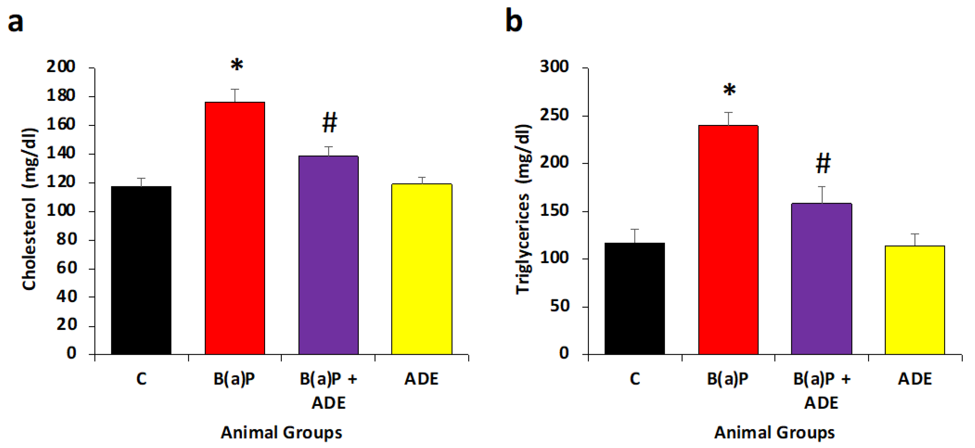
2.12. Measurement of Lipid Profile

Triglycerides (TG) and total cholesterol (TC) were measured, and the results were analyzed consequently.

2.13. Measurement of VEGF Level

VEGF is a potent angiogenic factor and plays a significant role in diseases’ development and progression. The level of VEGF was evaluated in lung tissue using the kit provided by Abcam, Cambridge, UK. All the samples were run in duplicate, and the average absorbance was measured at 450 nm.

2.14. Assessment of Lung Histopathology

The lung tissues from all the animals were suspended in 10% neutral buffered formalin and dehydrated through graded ethanol, cleared with xylene, and embedded in paraffin wax. The tissues were sectioned at 5 μm thickness (Leica, Wetzlar, Germany) and stained with hematoxylin and eosin to report the histological damage in these tissues. The histological changes were observed with an Olympus microscope and the results were interpreted accordingly. Sirius red and Masson’s trichrome staining were used for determining the collagen/fiber deposition.

2.15. Immunohistochemical Analysis of Bax and VEGF Protein Expressions

In order to determine the expression of Bax and VEGF protein in the lung tissue, the 5 µm thick sections were processed for immunohistochemical staining by following the protocols from previously-published studies [26,27]. The scoring for protein expressions were calculated by two independent pathologists in a blinded manner. The expression, or positivity score, was calculated in a semi-quantitative manner, in which the staining intensity was multiplied by the percentage of positive cells. Percentage of positive e cells was categorized as follows: 0 (negative), 1 (15%), 2 (16–50%), 3 (51–75%) and 4 (more than 75%).

2.16. Terminal Deoxynucleotidyl Transferase-Mediated dUTP-Biotin Nick-End Labelling (TUNEL) Assay

TUNEL assay was performed on all lung tissue sections to evaluate the apoptosis level. Deparaffinization and rehydration of tissue sections were performed, and PBS was used to wash it. The TUNEL staining was performed according to the manufacturer’s instructions including quenching, equilibration, labeling reaction, termination of labeling reaction, blocking, and development by adding DAB and counterstaining. The nuclei were stained and examined with a light microscope to detect DNA fragmentation. The TUNEL-positive nuclei were calculated, photographs were captured, and the results were analyzed.

2.17. Transmission Electron Microscopy

Phosphate-buffered glutaraldehyde (2.5%) was used to fix the lung tissues and the specimens were dehydrated and epoxy resin was used for embedding. The ultra-microtome was used to obtain thin sections. Uranyl acetate and lead citrate were used to stain the ultrathin sections, and the sections were photographed using transmission electron microscopy.

2.18. Apoptosis Analysis by Flow Cytometry

Flow cytometry assay was performed on all experimental group samples to measure the apoptosis through FITC/Annexin V Apoptosis detection kit, as per the manufacturer’s instructions. The cells were re-suspended in sample buffer followed by ribonuclease addition, and a temperature of 37 °C was used to incubate it with FITC/Annexin V and PI in the dark for staining of cells. Furthermore, the cells were centrifuged, resuspended in binding buffer, and examined using a flow cytometer (Miltenyi Biotec, Bergisch Gladbach, Germany).

2.19. Statistical Analysis

The in vivo data were obtained from different treatment groups and expressed as mean ± SEM. Tukey’s test for multiple comparisons using one-way analysis of variance (ANOVA) by SPSS software was used to evaluate the significance of the differences among various groups. All the results were analysed and considered statistically significant at p < 0.05.

3. Results

Increased oxidative stress, inflammation, angiogenesis, and alterations of lung architecture are commonly associated with lung pathogenesis induced by B(a)P. Current investigation indicates that Ajwa dates show potential therapeutic action against B(a)P induced lung injury through its antioxidant and anti-inflammatory activity, as well as by modulation of various biological effects including lipid peroxidation, angiogenesis and apoptotic pathways.

3.1. Preliminary Screening for the Presence of Flavonoid and Phenolic Compounds

Ajwa dates are most typically grown in Medina, Saudi Arabia, and are generally black in color. Table 2 shows the color, odor, texture, and yield (%) of ethanolic extract of Ajwa. Table 3 shows the results of phytochemical screening of ADE extract. The dry weight of Ajwa fruit used in preparation of ethanol extract was 50 g, the yield was 2.89%, and the color was reddish brown, with a sticky texture (Table 2). Phytochemical screening confirmed the presence of saponins, alkaloids, glycosides, flavonoids, phenolic compounds, and terpenoids in Ajwa extract (Table 3). The total phenolic content (TPC) level in fruit pulp extracts was 534.30 ± 0.026 milli gram gallic acid equivalent/gram dry weight of extract and sugar reduction, particularly fructose, ascorbic acid, and protein, may have an impact on total polyphenol content. The TPC in ethanol extract of fruit pulp was 234.59 ± 0.072 mg quercetin equivalents/g dry weight of extract, respectively.

3.2. Measurement of ADE Activity on Antioxidant Enzymes

To evaluate the protective effects of ADE on lung oxidative stress status, the levels of different antioxidant enzymes were measured. The activities of antioxidant enzymes including catalase, SOD, GPx, and total antioxidant capacity (TAC) were meaningfully decreased in the B(a)P-treated group when compared with the normal/control group (p < 0.05) (Figure 1). Treatment with ADE efficiently enhanced the decreased catalase, GPx, SOD, and TAC when compared to the benzopyrene-only-treated group (p < 0.05).

3.3. Measurement of ADE Effect on MDA and NO Contents

One significant biomarker of oxidative stress is malondialdehyde (MDA). Malondialdehyde (MDA) is a lipid peroxidation byproduct found in the lung and serum and is used to estimate lipid peroxidation. Our data showed that the oxidative stress markers, including MDA (43.8 nmol/g) and NO levels (42.3 µmol/L), were considerably elevated in the B(a)P-treated group when compared to the normal control group (MDA 24.9 nmol/g, and NO 24.2 µmol/L) (p < 0.05). However, ADE showed in ADE-treated groups, MDA (35.6 nmol/g), and NO level (36.1 µmol/L) was meaningfully decreased when compared to the B(a)P-treated group only (p < 0.05) (Figure 2). The results confirm the inhibition potential of ADE against membrane damage.

3.4. Effects of ADE on Inflammatory Markers

The lung-protective effect of ADE was evaluated via the measurement of pro-inflammatory mediators. In the B(a)P-treated group only, the level of pro-inflammatory cytokines such as TNF-α, IFN-γ, interleukin-6 and CRP was significantly increased, whereas these mediators were decreased significantly in the ADE + B(a)P-treated group as compared with the B(a)P-only-treated group (Figure 3).

3.5. Measurement of Antihyperlipidemic Effects of ADE

In all experimental animal groups, the lipid profile was estimated to evaluate the antihyperlipidemic activity of ADE. In the disease control group, a significant (p < 0.05) increase in triglyceride and total cholesterol level compared to the normal control group was observed. After 6 weeks of the treatment (ADE 1 g/kg b.w.), there were significant decreases in TG and TC levels when compared to the B(a)P-treated group (p < 0.05) (Figure 4). This result evidently supports the hypothesis that Ajwa dates have anti-hyperlipidemic effect.

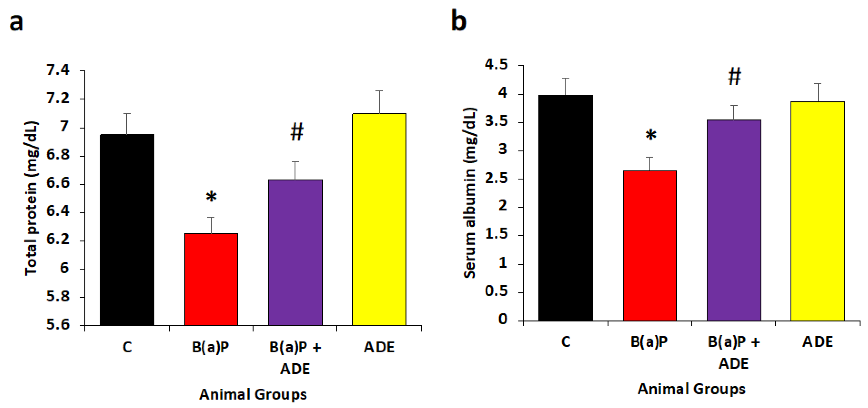
3.6. Measurement of ADE Effect on Total Protein and Albumin Levels

The total protein and albumin status measurements play a significant role in evaluation of disease development and progression [28]. Moreover, albumin is a significant constituent of plasma antioxidant activity that mainly binds divalent cations, free fatty acids, as well as hydrogen oxychloride (HOCI) [29]. In this study, it was found that total protein and albumin concentrations significantly decreased in the B(a)P-treated group as compared to the control group (p < 0.05). Total proteins in the B(a)P-treated group were found to be 6.25 mg/dL as compared to control 6.88 mg/dL. However, serum albumin was 2.61 mg/dL in comparison with the control (3.92 mg/dL). Moreover, total protein and albumin levels increased significantly in ADE-treated group rats as compared to B(a)P-treated animals and were 6.62 mg/dL and 3.48 mg/dL, respectively (p < 0.05) (Figure 5). In addition, animal groups treated with ADE only displayed 7.3 mg/dL, and 3.7 mg/dL of total protein and serum albumin, respectively.

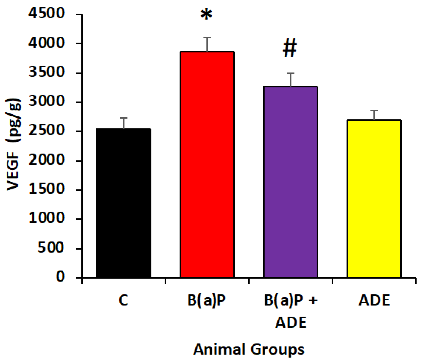
3.7. Effect of ADE on Angiogenic Marker Level

In the B(a)P group, level of VEGF was significantly high as compared to the control groups. The level of VEGF in the B(a)P group (3756 ± 243.07 pg/g tissue) exhibited significant elevation as compared to the control rats (2447 ± 190.06 pg/g tissue). The ADE-treated groups showed a significant decrease in VEGF level (3405.38 pg/g tissue) compared to diseases control [B(a)P-treated group] (Figure 6). However, the VEGF level in Ajwa-treated rats was only 2970 mg/dL. These finding suggest that Ajwa date reduces the angiogenic factor (VEGF) induced by B(a)P in rats and prevents lung pathogenesis.

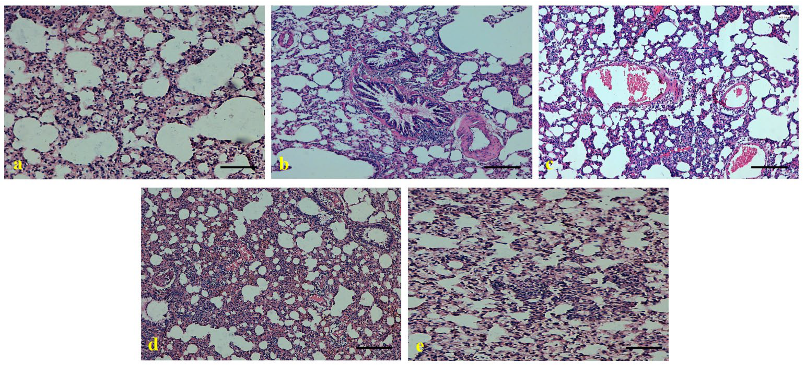
3.8. Role of Ajwa Date Extract on the Maintenance of Lung Architecture

Lung tissues of the control group animals show normal architecture, alveolar tissues, and normal bronchial structure. The B(a)P-treated group experienced lung damage including interstitial inflammatory cell infiltration, hemorrhage, congestion, edema, and fibrosis. Conversely, animals treated with ADE showed remarkable improvement in lung architecture as compared to the B(a)P-treated group. Lung damage was found to be significantly less in the group co-administered with ADE + B(a)P, as ADE decreases the injury of lung epithelium, infiltration of inflammatory cells, congestion, and edema, and maintains the lung architecture (Figure 7).

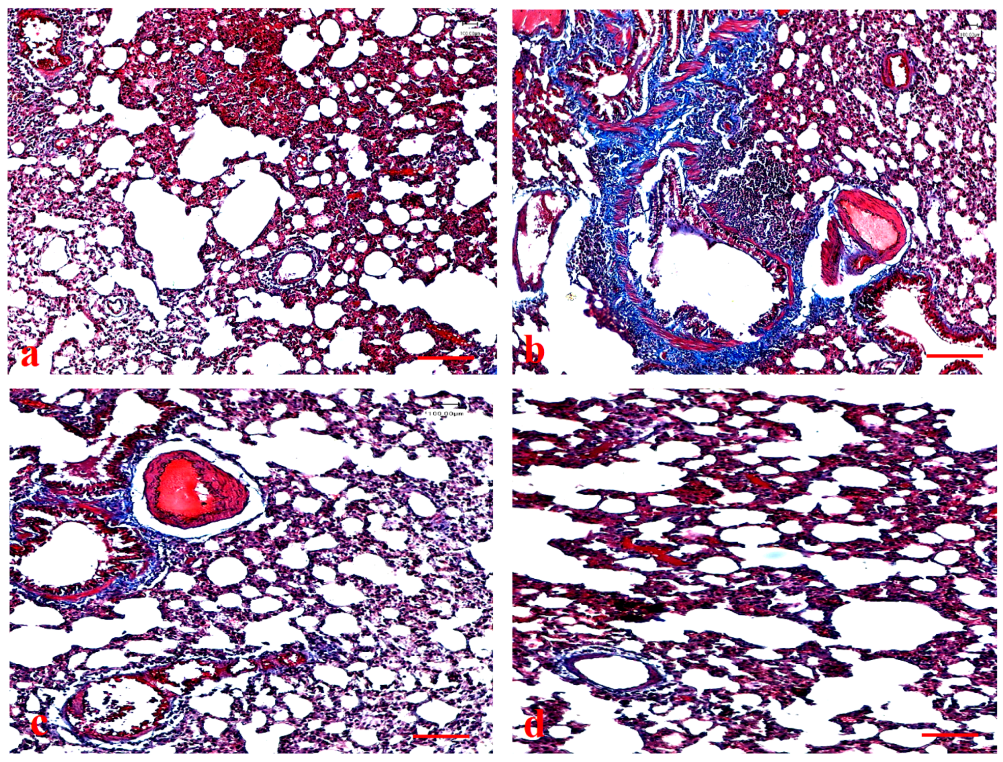
3.9. Effects of Ajwa Date Extract on Lung Fibrosis Caused by B(a)P in Rats

To evaluate fibrosis, staining with Masson’s trichrome and Sirius red was performed, and the results were interpreted. The B(a)P-treated group indicated noticeable accumulation of collagen in the lung as compared to the control group. Treatment with ADE (1 mg/kg b.w.) caused significant decrease in the collagen as compared with the B(a)P-treated group (Figure 8).
Moreover, in the B(a)P-treated group, fibrosis levels were significant in lung tissue rats. Treatment with ADE (1 mg/kg b.w.) caused significant decrease in the fiber as compared with B(a)P-treated group, as confirmed by Sirius red staining (Figure 9).

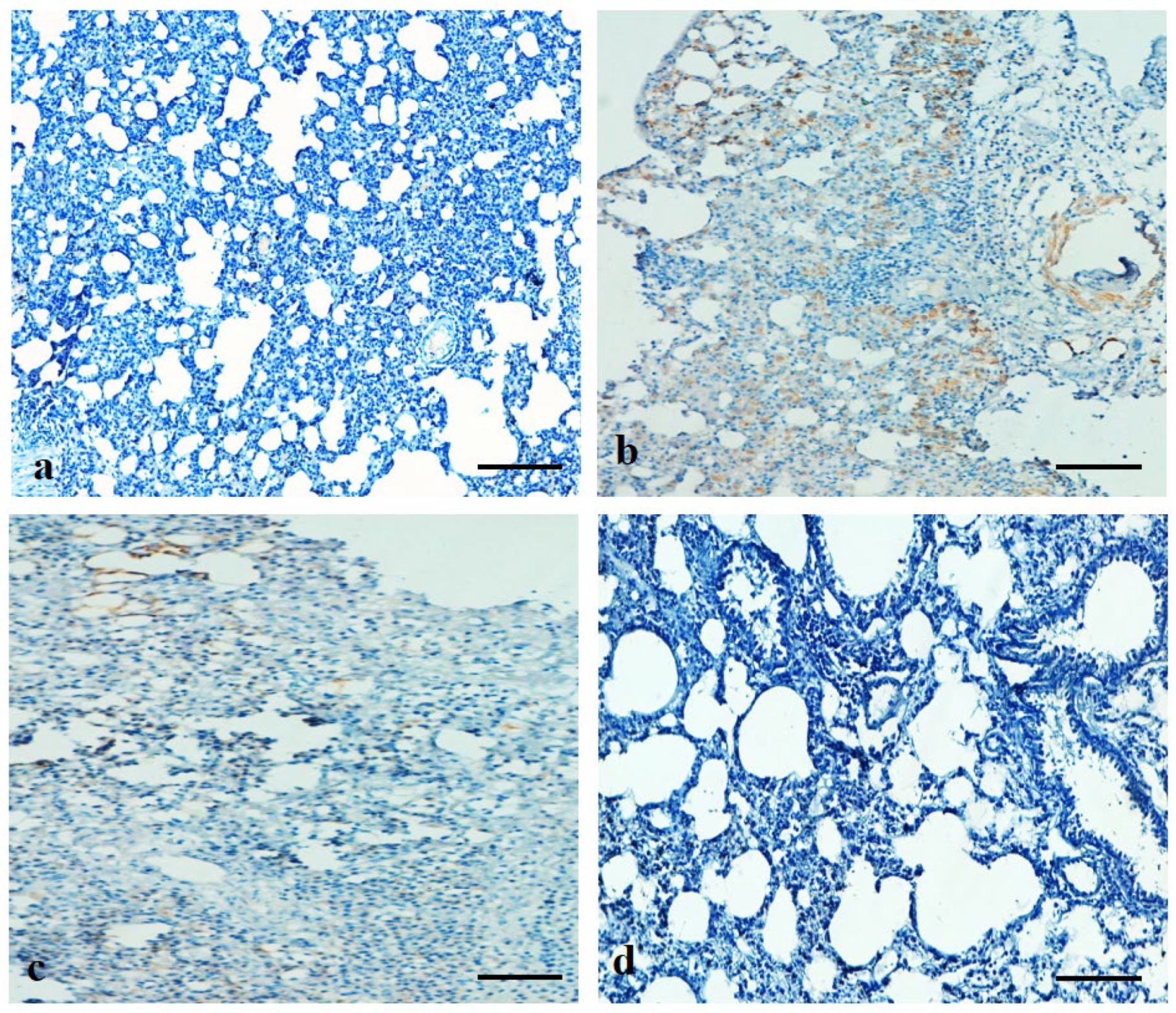
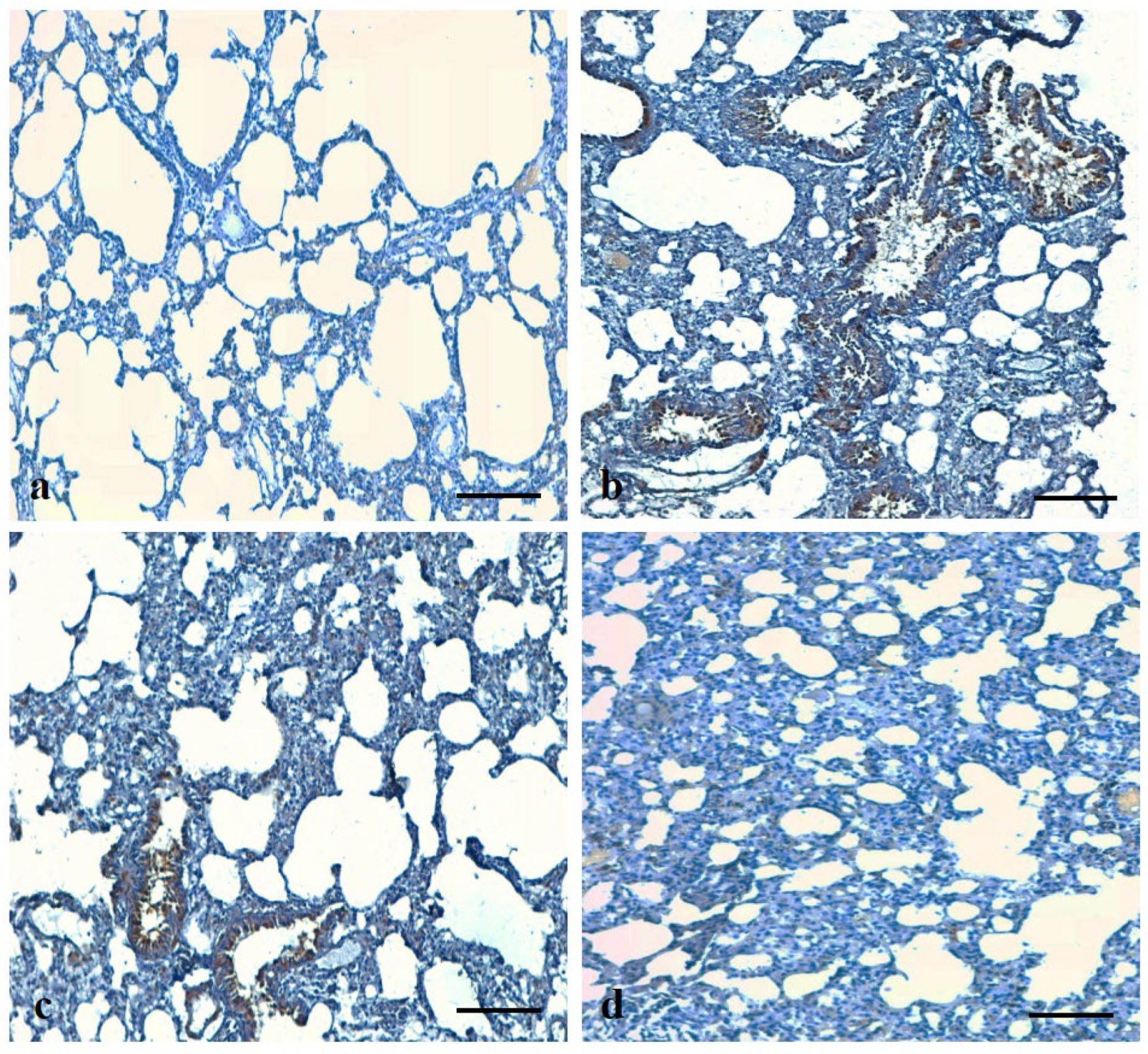
3.10. Effects of ADE on Expression Pattern of Bax and VEGF Protein in Lung Tissue

Immunohistochemistry (IHC) staining revealed that Bax level was significantly increased in lung tissues of B(a)P-induced group as compared to control group. Conversely, Bax expression decreased in rats treated with B(a)P + ADE (Figure 10).
The expression pattern of VEGF was evaluated in all experimental groups. It was observed that VEGF expression is significantly high in the B(a)P-treated group as compared to control. Conversely, VEGF expression decreased in rats treated with B(a)P + ADE (Figure 11).

3.11. ADE attenuates B(a)P-Induced Apoptosis in Lung Tissue

The TUNEL assay was performed on all experimental groups to measure DNA fragmentation, and nuclei stained dark brown were considered positive. In control sections, TUNEL positive cells were not noticed. When compared with the control group, TUNEL positivity increased significantly in the B(a)P-treated group. However, pre-treatment with ADE showed a low number of TUNEL positive cells as compared with the B(a)P group. This finding revealed that ADE plays a significant role in the attenuation of apoptotic cell death (Figure 12).

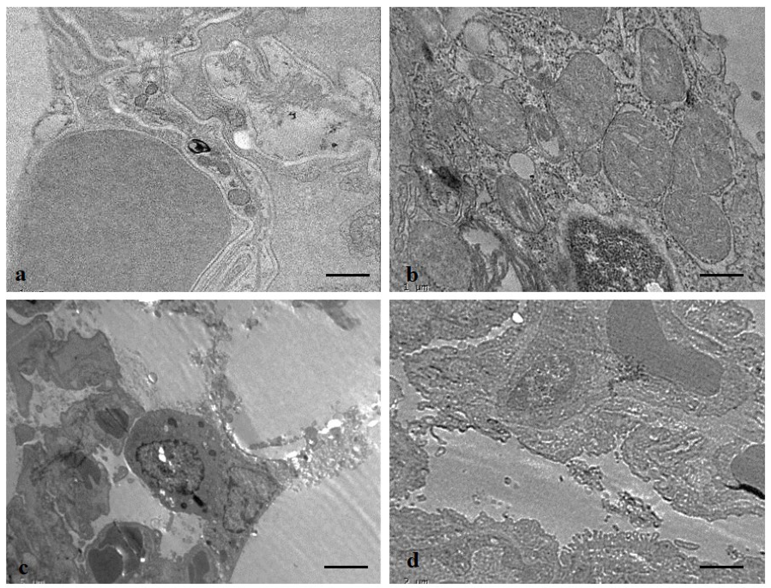
3.12. Protective Role of ADE on Lung Tissue Ultrastructure

The ultrastructural changes in the lung tissues were examined using transmission electron microscopy (TEM). The control group samples demonstrated normal lung architecture. In the B(a)P-treated group, various types of alterations were seen as numerous swollen mitochondria, alveolar space, alveolar constriction, granular cytoplasm, vacuoles, and degenerative necrotic nucleated cells. The B(a)P-intoxicated rats treated with ADE showed fewer alterations in the organelle structures (Figure 13).

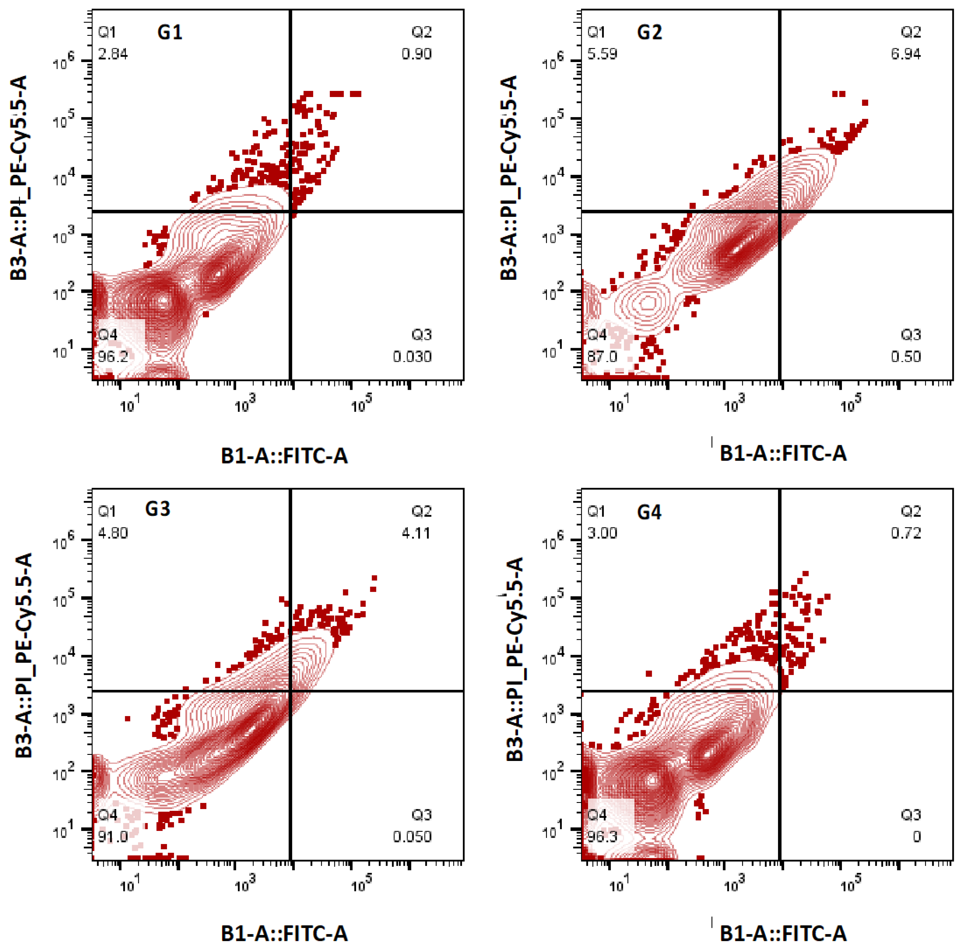
3.13. Effect of ADE on Apoptotic Cell Death

Apoptotic cell death level was measured using flow cytometry, and data exhibited that cell death was increased in the B(a)P-treated group (87.0%) as compared to the normal control with viable cell count (96.2%). It was noticed that the viable cell count was restored to some extent (91.0%) in animals treated with B(a)P+ADE. The animals treated with ADE only showed almost the same viable cell count as normal control (96.3%) (Figure 14).

4. Discussion

Medicinal plants and their bioactive compounds are rich sources of polyphenols, and these compounds inhibit various types of pathogenesis [30]. Date fruits shows role in disease management through their free radical scavenging properties.
B(a)P is a commonly used carcinogen to cause lung pathogenesis, including cancer in experimental animal models. This toxicant causes changes in antioxidant enzyme levels, causes inflammation, and damages lung tissue architecture [31]. Excess reactive oxygen species (ROS) play a significant role in oxidative damages, which is chiefly characterized by lipid peroxidation and DNA damage [32]. Malondialdehyde (MDA) is a significant product in the process of lipid peroxidation, and the content of MDA is measured to judge the degree of lipid peroxidation [32]. In addition, excess ROS produced by B(a)P alters the redox cycle that alters the redox status in the system [33].
In the current study, it was observed that MDA levels were meaningfully increased in the benzopyrene-treated animals as compared to the control group, which might be due to the excessive amounts of ROS produced in response to the administration of B(a)P [34]. In ADE-treated groups, the MDA level was significantly decreased as compared to the B(a)P-treated group. These results confirmed that B(a)P causes oxidative damage with MDA concentration increment. In agreement with these results, the previous finding reported that ADE produced a significant decrease in lipid peroxides measured as MDA content and a significant increase in antioxidant enzymes level [35].
Nitric oxide and associated compounds are formed by an extensive variety of residential as well as inflammatory cells in the airways [36]. A study based on animal model reported that nitric oxide level was substantially increased in the B(a)P-treated group as compared to the control group [37,38]. In the present study, it was noticed that NO levels were increased in the B(a)P-treated group animals whereas ADE treatment significantly decreases the NO levels. The previous finding is in accordance with the current finding as NO level was high in or after B(a)P treatment group as compared to the control group. Moreover, pretreatment with natural compounds significantly reduced NO content in the BALF and lung tissue of the treatment group [10,37,38].
Angiogenesis, the formation of new blood vessels, plays a significant role in disease development and progression. VEGF induced by allergic reaction, facilitates vascular remodeling as well as angiogenesis that is linked with airway remodeling [39]. The levels of VEGF have been shown to be increased in chronic lung diseases [40]. In accordance with previous findings [41], in the present study, an increase in levels of VEGF in B(a)P-induced lung injury rat models was noted, whereas VEGF levels were significantly decreased in the B(a)P + ADE-treated group. In the current study, it was also noticed that the antioxidant enzymes level decreased in B(a)P-treated rats. ADE treatment groups exhibited increased levels of these enzymes as compared to B(a)P-treated rats. This finding is in accordance with other previous findings which reported that B(a)P treatment decreased the enzyme levels and natural compounds enhanced the antioxidant enzyme levels [41,42]. Moreover, prior studies have also verified that antioxidant enzyme levels are lower in the B(a)P-treated group as compared to the normal control groups. Conversely, extract treatment noticeably restored the activities of such antioxidant enzymes [43]. Furthermore, another study based on liver cancer reported that ADE-treated group showed increase in the activity of antioxidant enzymes as compared to the DEN-treated group [44].
B(a)P treatment administered to the animals causes lung injury and other types of changes, confirmed by pathological changes: intra-alveolar hemorrhage, collagen fiber deposition, inflammatory cell infiltration, and edema, which were measured to show an important role in the lung injury [10,45,46]. In the current study, it was observed that B(a)P treatment causes lung injury, and various other pathological alterations, although co-administered ADE and B(a)P treatment showed a role in the maintenance of lung architecture. This result was in agreement with previous reports, as B(a)P caused lung injury, necrosis, alteration in epithelium lung architecture [47], and lung epithelium thickening as well as a mass infiltration of inflammatory cells [48]. The current finding is consistent with the earlier findings that reported that ADE showed a role in the histoarchitecture maintenance [49] and ADE treatment reduced edema, necrosis, and inflammatory cells infiltration and reestablished the cardiomyocytes architecture [50].
The tumor suppressor protein p53 causes cell cycle arrest and induces apoptosis in lung injury. Though p53 modulates apoptosis through complex and partly understood mechanisms, one recognized pathway includes activating the mitochondria-regulated death pathway via increasing gene expression of proapoptotic stimuli while decreasing the expression of antiapoptosis [51,52,53,54,55]. Immunohistochemistry findings established that Bax and VEGF levels and TUNEL staining were upregulated in the B(a)P-caused toxicity in rats, which decreased after treatment with ADE. An earlier study reported that the level of activated p53 was significantly influenced by B(a)P treatment, while this effect was noticeably decreased after curcumin and VE co-treatment. Moreover, the ratio of Bax/Bcl-2 was also considerably increased in cells exposed to B(a)P and this increase was reversed by VE co-treatment [56]. Activated Bax was significantly expressed in the lung tissues in the bleomycin-treated group and was attenuated by BIP-V5 treatment [57].
Moreover, TEM results also showed various changes as numerous swollen mitochondria, alveolar space, alveolar constriction, granular cytoplasm, and degenerative necrotic nucleated cells in the B(a)P-induced lung-injury group, which was improved in the ADE treatment group.
There are several anti-inflammatory allopathic drugs that are being currently used to treat lung pathogenesis. These drugs are effective but also cause negative side effects on health. A safe and affordable alternative to allopathic medications has been found to be the use of medicinal plants to treat and manage diseases in various traditional medical systems. Ajwa dates play a vital role in health management by boosting the functions of the immune system, scavenging of reactive oxygen species, and anti-microbial activity. The overall outcome of this study demonstrated the role of Ajwa dates in the management of lung pathogenesis through antioxidant and anti-inflammatory activity. It also maintains lung tissue architecture as is evident by the decrease in the inflammatory cells, hemorrhages, congestion, and fibrosis. In addition, Ajwa date treatment significantly ameliorates collagen fiber deposition and expression pattern of VEGF and Bax proteins. In addition, it also modulates various biological activities including lipid peroxidation, angiogenesis, and apoptotic pathways. Our study, for the first time, investigated the health benefit effects of Ajwa date against lung pathogenesis and associated complications under in vivo conditions. Therefore, current investigations on Ajwa date might act as a path towards developing a potent alternative therapy against respiratory diseases, especially chronic lung diseases, in the future.

5. Conclusions

This is the first study investigating the therapeutic implications of Ajwa date and its possible mechanisms of action in lung pathogenesis. The lung protective effect, as noticed in the current study, could be due to the action of various ingredients, possibly through the inhibition of oxidative stress, inflammation, and apoptosis in lung epithelial cells. B(a)P treatment significantly decreased the level of antioxidant enzymes and total antioxidant capacity as compared to the treatment group. B(a)P treatment also led to increased levels of pro-inflammatory markers. Moreover, Ajwa date shows a role in the enhancement of antioxidant enzymes and reduction in inflammatory markers. It also maintains lung tissue architecture, as is evident by decrease in the inflammatory cells, hemorrhages, congestion, edema, and fibrosis. In addition, ADE treatment significantly ameliorates collagen fiber deposition and the expression pattern of VEGF and Bax proteins. Henceforth, Ajwa date can be used as a potential therapeutic strategy to treat benzopyrene-induced lung pathogenesis. The different formulation-based strategy of Ajwa date extract can be used for the treatment of lung pathogenesis induced by different toxic agents.

Author Contributions

Conceptualization, S.A.A., A.H.R., M.A.S. and A.A.K.; methodology, S.A.A., M.A.S., A.A.K., A.A.A. and A.H.R.; investigation, S.A.A., A.A.K. and A.H.R.; writing—original draft preparation, S.A.A., A.A.K. and A.H.R.; writing—review and editing, S.A.A., A.A.K., A.A.A. and A.H.R.; supervision, S.A.A., A.A.K. and A.H.R. All authors have read and agreed to the published version of the manuscript.

Funding

This work was funded as well as supported by the Qassim University, Deanship of Scientific Research (Grant No. 10241-cams1-2020-1-3-I).

Institutional Review Board Statement

The animals were maintained at animal facility of the College of Applied Medical Sciences (CAMS) accordance with the guidelines of the Qassim University on Animal Care. The animal experiments were carried out as per the guidelines of CAMS, Qassim University and approved by the Institutional Animal Ethics Committee (10241-cams1-2020-1-3-I), Qassim University.

Informed Consent Statement

Not applicable.

Data Availability Statement

The data used to support the findings of this study are included within the article.

Conflicts of Interest

The authors declare no conflict of interest.

References

  1. GBD 2017 Disease and Injury Incidence and Prevalence Collaborators. Global, Regional, and National Incidence, Prevalence, and Years Lived with Disability for 354 Diseases and Injuries for 195 Countries and Territories, 1990–2017: A Systematic Analysis for the Global Burden of Disease Study 2017. Lancet 2018, 392, 1789–1858. [Google Scholar] [CrossRef] [Green Version]
  2. Piccardo, M.T.; Stella, A.; Valerio, F. Is the smokers exposure to environmental tobacco smoke negligible? Environ. Health 2010, 9, 5. [Google Scholar] [CrossRef] [PubMed] [Green Version]
  3. Yeo, C.D.; Kim, J.W.; Ha, J.H.; Kim, S.J.; Lee, S.H.; Kim, I.K. Chemopreventive effect of phosphodieasterase-4 inhibition in ben-zo(a)pyrene-induced murine lung cancer model. Exp. Lung Res. 2014, 40, 500–506. [Google Scholar] [CrossRef] [PubMed]
  4. Jahangir, T.; Sultana, S. Benzo(a)pyrene-induced genotoxicity: Attenuation by farnesol in a mouse model. J. Enzym. Inhib. Med. Chem. 2008, 23, 888–894. [Google Scholar] [CrossRef] [Green Version]
  5. Kometani, T.; Yoshino, I.; Miura, N.; Okazaki, H.; Ohba, T.; Takenaka, T.; Shoji, F.; Yano, T.; Maehara, Y. Benzo[a]pyrene promotes proliferation of human lung cancer cells by accelerating the epidermal growth factor receptor signaling pathway. Cancer Lett. 2009, 278, 27–33. [Google Scholar] [CrossRef]
  6. Gelboin, H.V. Benzo[alpha]pyrene metabolism, activation and carcinogenesis: Role and regulation of mixed-function oxidases and related enzymes. Physiol. Rev. 1980, 60, 1107–1166. [Google Scholar] [CrossRef] [Green Version]
  7. Baer-Dubowska, W.; Kulinska, A.; Jarmuz, M.; Brauze, D.; Gnojkowski, J. DNA adducts detected by synchronous fluorescence spectrophotometry in rat liver, kidney, lung and spleen after intraperitoneal administration of benzo[a]pyrene. Acta Biochim. Pol. 1993, 40, 63–65. [Google Scholar] [CrossRef]
  8. Podechard, N.; Lecureur, V.; Le Ferrec, E.; Guenon, I.; Sparfel, L.; Gilot, D.; Gordon, J.R.; Lagente, V.; Fardel, O. Interleukin-8 induction by the environmental contaminant benzo(a)pyrene is aryl hydrocarbon receptor-dependent and leads to lung inflammation. Toxicol. Lett. 2008, 177, 130–137. [Google Scholar] [CrossRef]
  9. Lina, T.; Yang, M.S. Benzo[a]pyrene-induced elevation of GSH level protects against oxidative stress andenhances xenobioticdetoxificationinhumanHepG2 cells. Toxicology 2007, 235, 1–10. [Google Scholar] [CrossRef]
  10. Qamar, W.; Khan, A.Q.; Khan, R.; Lateef, A.; Tahir, M.; Rehman, M.U.; Ali, F.; Sultana, S. Benzo(a)pyrene-induced pulmonary inflammation, edema, surfactant dysfunction, and injuries in rats: Alleviation by farnesol. Exp. Lung Res. 2012, 38, 19–27. [Google Scholar] [CrossRef]
  11. Smerdova, L.; Neca, J.; Svobodova, J.; Topinka, J.; Schmuczerova, J.; Kozubik, A.; Machala, M.; Vondracek, J. Inflammatory mediators accelerate metabolism of benzo[a]pyrene in rat alveolar type II cells: The role of enhanced cytochrome P450 1B1 expression. Toxicology 2013, 314, 30–38. [Google Scholar] [CrossRef]
  12. Uno, S.; Makishima, M. Benzo [a] pyrene toxicity and inflammatory disease. Curr. Rheumatol. Rev. 2009, 5, 266–271. [Google Scholar] [CrossRef]
  13. Slader, C.A.; Reddel, H.K.; Jenkins, C.R.; Armour, C.L.; Bosnic-Anticevich, S.Z. Complementary and alternative medicine use in asthma: Who is using what? Respirology 2006, 11, 373–387. [Google Scholar] [CrossRef]
  14. Ragab, A.R.; Elkablawy, M.A.; Sheik, B.Y.; Baraka, H.N. Antioxidant and tissueprotective studies on Ajwa extract: Dates from al-Madinah al-Monwarah, Saudia Arabia. J. Environ. Anal. Toxicol. 2013, 3, 163. [Google Scholar]
  15. Zhang, C.R.; Aldosari, S.A.; Vidyasagar, P.S.; Nair, K.M.; Nair, M.G. Antioxidant and anti-inflammatory assays confirm bioactive compounds in Ajwa Date fruit. J. Agric. Food Chem. 2013, 61, 5834–5840. [Google Scholar] [CrossRef]
  16. Sheikh, B.Y.; Elsaed, W.M.; Samman, A.H.; Sheikh, B.Y.; Ladin, A.-M.M.B. Ajwa dates as a protective agent against liver toxicity in rat. Eur. Sci. J. 2014, 10, 358–368. [Google Scholar]
  17. Rahmani, A.H.; Khan, A.A.; Babiker, A.Y.; Khan, M.A.; Alzohairy, M.A. Therapeutic implications of ajwa dates (Phoenix dactylifera) in the inhibition of liver tissue alterations through the modulation of vascular endothelial growth factor and phosphatase, and tensin homolog gene. Pharmacogn. Res. 2018, 10, 151–155. [Google Scholar] [CrossRef]
  18. Ali, A.; Abdu, S. Antioxidant protection against pathological mycotoxins alterations on proximal tubules inrat kidney. Funct. Foods Health Dis. 2011, 1, 118–134. [Google Scholar] [CrossRef]
  19. Alqarni, M.M.; Osman, M.A.; Al-Tamimi, D.S.; Gassem, M.A.; Al-Khalifa, A.S.; Al-Juhaimi, F.; Mohamed Ahmed, I.A. Antioxidant and antihyperlipidemic effects of Ajwa date (Phoenix dactylifera L.) extracts in rats fed a cholesterol-rich diet. J. Food Biochem. 2019, 43, e12933. [Google Scholar] [CrossRef]
  20. Shahbaz, K.; Asif, J.A.; Liszen, T.; Nurul, A.A.; Alam, M.K. Cytotoxic and Antioxidant Effects of Phoenix dactylifera L. (Ajwa Date Extract) on Oral Squamous Cell Carcinoma Cell Line. BioMed Res. Int. 2022, 2022, 5792830. [Google Scholar] [CrossRef]
  21. Alsahli, M.A.; Anwar, S.; Alzahrani, F.M.; Almatroudi, A.; Alfheeaid, H.; Khan, A.A.; Allemailem, K.S.; Almatroodi, S.A.; Rahmani, A.H. Health Promoting Effect of Phyllanthus emblica and Azadiractha indica against Advanced Glycation End Products Formation. Appl. Sci. 2021, 11, 8819. [Google Scholar] [CrossRef]
  22. Anwar, S.; Almatroudi, A.; Allemailem, K.S.; Jacob Joseph, R.; Khan, A.A.; Rahmani, A.H. Protective Effects of Ginger Extract against Glycation and Oxidative Stress-Induced Health Complications: An In Vitro Study. Processes 2020, 8, 468. [Google Scholar] [CrossRef]
  23. Anwar, S.; Almatroodi, S.A.; Almatroudi, A.; Allemailem, K.S.; Joseph, R.J.; Khan, A.A.; Alrumaihi, F.; Alsahli, M.A.; Husain Rahmani, A. Biosynthesis of silver nanoparticles using Tamarix articulata leaf extract: An effective approach for attenuation of oxidative stress mediated diseases. Int. J. Food Prop. 2021, 24, 677–701. [Google Scholar] [CrossRef]
  24. Almatroodi, S.A.; Anwar, S.; Almatroudi, A.; Khan, A.A.; Alrumaihi, F.; Alsahli, M.A.; Rahmani, A.H. Hepatoprotective Effects of Garlic Extract against Carbon Tetrachloride (CCl4)-Induced Liver Injury via Modulation of Antioxidant, Anti-Inflammatory Activities and Hepatocyte Architecture. Appl. Sci. 2020, 10, 6200. [Google Scholar] [CrossRef]
  25. Hassan, S.K.; Mousa, A.M.; El-Sammad, N.M.; Abdel-Halim, A.H.; Khalil, W.K.; Elsayed, E.A.; Anwar, N.; Linscheid, M.W.; Moustafa, E.S.; Hashim, A.N.; et al. Antitumor activity of Cuphea ignea extract against benzo(a)pyrene-induced lung tumorigenesis in Swiss Albino mice. Toxicol. Rep. 2019, 6, 1071–1085. [Google Scholar] [CrossRef]
  26. Rahmani, A.; Alzohairy, M.; Khadri, H.; Mandal, A.K.; Rizvi, M.A. Expressional evaluation of vascular endothelial growth factor (VEGF) protein in urinary bladder carcinoma patients exposed to cigarette smoke. Int. J. Clin. Exp. Pathol. 2012, 5, 195–202. [Google Scholar]
  27. Babiker, A.Y.; Almatroudi, A.; Allemailem, K.S.; Husain, N.E.O.S.; Alsammani, M.A.; Alsahli, M.A.; Rahmani, A.H. Clinicopathologic Aspects of Squamous Cell Carcinoma of the Uterine Cervix: Role of PTEN, BCL2 and P53. Appl. Sci. 2018, 8, 2124. [Google Scholar] [CrossRef] [Green Version]
  28. Saad, B.; Azaizeh, H.; Abu-Hijleh, G.; Said, S. Safety of traditional Arab herbal medicine. Evid. Based Complement. Alternat. Med. 2006, 3, 433–439. [Google Scholar] [CrossRef]
  29. Akiibinu, O.M.; Ogunyemi, O.E.; Arinola, O.G.; Adenaike, A.F.; Adegoke, O.D. Assessment of antioxidants and nutritional status of pulmonary tuberculosis patients in Nigeria. Eur. J. Gen. Med. 2008, 5, 208–211. [Google Scholar]
  30. Almatroodi, S.A.; AlRumaihi, F.; Alsahli, M.A.; Alhommrani, M.F.; Khan, A.; Rahmani, A.H. Curcumin, an Active Constituent of Turmeric Spice: Implication in the Prevention of Lung Injury Induced by Benzo(a) Pyrene (BaP) in Rats. Molecules 2020, 25, 724. [Google Scholar] [CrossRef] [Green Version]
  31. Schieber, M.; Chandel, N.S. ROS function in redox signaling and oxidative stress. Curr. Biol. 2014, 24, R453–R462. [Google Scholar] [CrossRef] [PubMed] [Green Version]
  32. Pan, L.Q.; Ren, J.; Liu, J. Responses of antioxidant systems and LPO level to benzo(a)pyrene and benzo(k)fluoranthene in the haemolymph of the scallop Chlamys ferrari. Environ. Pollut. 2006, 141, 443–451. [Google Scholar] [CrossRef] [PubMed]
  33. Kiruthiga, P.V.; Pandian, S.K.; Devi, K.P. Silymarin protects PBMC against B(a)P induced toxicity by replenishing redox status and modulating glutathione metabolizing enzymes—An in vitro study. Toxicol. Appl. Pharmacol. 2010, 247, 116–128. [Google Scholar] [CrossRef] [PubMed]
  34. El-Kott, A.F. Anti-angiogenic effectiveness of the pomegranate against benzo (a)pyrene induced lung carcinoma in mice. Cancer Res. 2015, 11, 164–174. [Google Scholar] [CrossRef] [Green Version]
  35. Elsadek, B.; El-Sayed, E.S.; Mansour, A.; Elazab, A. Abrogation of carbon tetrachloride-induced hepatotoxicity in Sprague-Dawley rats by Ajwa date fruit extract through ameliorating oxidative stress and apoptosis. Pak. J. Pharm. Sci. 2017, 30, 2183–2191. [Google Scholar]
  36. Gaston, B.; Kobzik, L.; Stamler, J.S. Distribution of nitric oxide synthase in the lung. In Nitric Oxide and the Lung; Zapol, W.M., Bloch, K.D., Eds.; Dekker: New York, NY, USA, 1997; pp. 75–86. [Google Scholar]
  37. Alzohairy, M.A.; Khan, A.A.; Alsahli, M.A.; Almatroodi, S.A.; Rahmani, A.H. Protective Effects of Thymoquinone, an Active Compound of Nigella sativa, on Rats with Benzo(a)pyrene-Induced Lung Injury through Regulation of Oxidative Stress and Inflammation. Molecules 2021, 26, 3218. [Google Scholar] [CrossRef]
  38. Baradaran Rahimi, V.; Rakhshandeh, H.; Raucci, F.; Buono, B.; Shirazinia, R.; Samzadeh Kermani, A.; Maione, F.; Mascolo, N.; Askari, V.R. Anti-Inflammatory and Anti-Oxidant Activity of Portulaca oleracea Extract on LPS-Induced Rat Lung Injury. Molecules 2019, 24, 139. [Google Scholar] [CrossRef] [Green Version]
  39. Wang, K.; Liu, C.T.; Wu, Y.H.; Feng, Y.L.; Bai, H.L. Budesonide/formoterol decreases expression of vascular endothelial growth factor (VEGF) and VEGF receptor 1 within airway remodelling in asthma. Adv. Ther. 2008, 25, 342–354. [Google Scholar] [CrossRef]
  40. Gomulka, K.; Liebhart, J.; Gladysz, U.; Medrala, W. VEGF serum concentration and irreversible bronchoconstriction in adult asthmatics. Adv. Clin. Exp. Med. 2019, 28, 759–763. [Google Scholar] [CrossRef]
  41. Alzohairy, M.A.; Khan, A.A.; Ansari, M.A.; Babiker, A.Y.; Alsahli, M.A.; Almatroodi, S.A.; Rahmani, A.H. Protective Effect of Quercetin, a Flavonol against Benzo(a)pyrene-Induced Lung Injury via Inflammation, Oxidative Stress, Angiogenesis and Cyclooxygenase-2 Signalling Molecule. Appl. Sci. 2021, 11, 8675. [Google Scholar] [CrossRef]
  42. Ali, R.; Shahid, A.; Ali, N.; Hasan, S.K.; Majed, F.; Sultana, S. Amelioration of benzo [a] pyrene-induced oxidative stress and pulmonary toxicity by naringenin in Wistar rats: A plausible role of COX-2 and NF-κB. Hum. Exp. Toxicol. 2017, 36, 349–364. [Google Scholar] [CrossRef]
  43. Shahid, A.; Ali, R.; Ali, N.; Kazim Hasan, S.; Barnwal, P.; Mohammad Afzal, S.; Vafa, A.; Sultana, S. Methanolic bark extract of Acacia catechu ameliorates benzo(a)pyrene induced lung toxicity by abrogation of oxidative stress, inflammation, and apop-tosis in mice. Environ. Toxicol. 2017, 32, 1566–1577. [Google Scholar] [CrossRef]
  44. Khan, F.; Khan, T.J.; Kalamegam, G.; Pushparaj, P.N.; Chaudhary, A.; Abuzenadah, A.; Kumosani, T.; Barbour, E.; Al-Qahtani, M. Anti-cancer effects of Ajwa dates (Phoenix dactylifera L.) in diethylnitrosamine induced hepatocellular carcinoma in Wistar rats. BMC Complement. Altern Med. 2017, 17, 418. [Google Scholar] [CrossRef]
  45. Barnwal, P.; Vafa, A.; Afzal, S.M.; Shahid, A.; Hasan, S.K.; Alpashree; Sultana, S. Benzo(a)pyrene induces lung toxicity and inflammation in mice: Prevention by carvacrol. Hum. Exp. Toxicol. 2018, 37, 752–761. [Google Scholar] [CrossRef]
  46. Khan, S.; Naseem, I. A comparative insight into the oxidative damage and cell death potential of photoilluminated aminophylline—Riboflavin system in normal and cancer lung cells of swiss albino mice. Toxicol. In Vitro 2019, 61, 104651. [Google Scholar] [CrossRef]
  47. Shahid, A.; Ali, R.; Ali, N.; Kazim Hasan, S.; Rashid, S.; Majed, F.; Sultana, S. Attenuation of genotoxicity, oxidative stress, apoptosis and inflammation by rutin in benzo(a)pyrene exposed lungs of mice: Plausible role of NF-kappaB, TNF-alpha and Bcl-2. J. Complement. Integr. Med. 2016, 13, 17–29. [Google Scholar] [CrossRef]
  48. Islam, J.; Shree, A.; Afzal, S.M.; Vafa, A.; Sultana, S. Protective effect of Diosmin against benzo(a)pyrene-induced lung injury in Swiss Albino Mice. Environ. Toxicol. 2020, 35, 747–757. [Google Scholar] [CrossRef]
  49. Sabbah, M.F.; Alshubali, F.; Baothman, O.A.; Zamzami, M.A.; Shash, L.; Hassan, I.A. Cardioprotective Effect of Ajwa Date Aqueous Extract on Doxorubicin-Induced Toxicity in Rats. Biomed. Pharmacol. J. 2018, 11, 1521–1536. [Google Scholar] [CrossRef]
  50. Al-Yahya, M.; Raish, M.; AlSaid, M.S.; Ahmad, A.; Mothana, R.A.; Al-Sohaibani, M.; Al-Dosari, M.S.; Parvez, M.K.; Rafatullah, S. ‘Ajwa’ dates (Phoenix dactylifera L.) extract ameliorates isoproterenol-induced cardiomyopathy through downregulation of oxidative, inflammatory and apoptotic molecules in rodent model. Phytomedicine 2016, 23, 1240. [Google Scholar] [CrossRef]
  51. Oren, M. Decision making by p53: Life, death and cancer. Cell Death Differ. 2003, 10, 431–442. [Google Scholar] [CrossRef]
  52. Schuler, M.; Green, D.R. Transcription, apoptosis and p53: Catch-22. Trends Genet. 2005, 21, 182–187. [Google Scholar] [CrossRef]
  53. Miyashita, T.; Reed, J.C. Tumor suppressor p53 is a direct transcriptional activator of the human bax gene. Cell 1995, 80, 293–299. [Google Scholar]
  54. Nakano, K.; Vousden, K.H. PUMA, a novel proapoptotic gene, is induced by p53. Mol. Cell 2001, 7, 683–694. [Google Scholar] [CrossRef]
  55. Yu, J.; Zhang, L. The transcriptional targets of p53 in apoptosis control. Biochem. Biophys. Res. Commun. 2005, 331, 851–858. [Google Scholar] [CrossRef]
  56. Zhu, W.; Cromie, M.M.; Cai, Q.; Lv, T.; Singh, K.; Gao, W. Curcumin and vitamin E protect against adverse effects of benzo[a]pyrene in lung epithelial cells. PLoS ONE 2014, 9, e92992. [Google Scholar] [CrossRef] [Green Version]
  57. Suzuki, K.; Yanagihara, T.; Yokoyama, T.; Maeyama, T.; Ogata-Suetsugu, S.; Arimura-Omori, M.; Mikumo, H.; Hamada, N.; Harada, E.; Kuwano, K.; et al. Bax-inhibiting peptide attenuates bleomycin-induced lung injury in mice. Biol. Open 2017, 6, 1869–1875. [Google Scholar] [CrossRef] [Green Version]
Figure 1. The antioxidant enzymes including (a) catalase, (b) SOD, (c) GPx, and (d) total antioxidant capacity were significantly decreased in B(a)P-treated group. The levels of enzymes were significantly enhanced in the ADE treatment group. The number signifies the mean ± SEM with 8 animals per group. The statistical differences are represented with (*) asterisk representing significance at (p < 0.05) as compared to the control group and (#) hash represents (p < 0.05) when compared with B(a)P-treated group.
Applsci 12 06784 g001
Figure 2. Effect of ADE on (a) MDA and (b) NO level in different treatment animal groups. MDA and NO were meaningfully increased in the B(a)P-treated group. The MDA and NO levels significantly decreased in the ADE treatment group. The number signifies the mean ± SEM with 8 animals per group. The statistical differences are represented with an asterisk (*) representing significance at (p < 0.05) as compared to the normal control group and hash (#) represents (p < 0.05) when compared with B(a)P-treated group only.
Applsci 12 06784 g002
Figure 3. Effect of ADE on proinflammatory markers. Pro-inflammatory cytokines such as (a) IL-6, (b) TNF-α, (c) CRP, and (d) IFN-γ, were significantly increased in B(a)P-treated groups, whereas this level significantly decreased in the ADE treatment group. The number indicates the mean ± SEM with 8 animals per group. The statistical differences are represented with an asterisk (*) representing significance at (p < 0.05) as compared to the normal control group and hash (#) represents when compared with B(a)P-treated group only (p < 0.05).
Applsci 12 06784 g003
Figure 4. The level of (a) total cholesterol and (b) triglyceride level was significantly increased in B(a)P-treated group. This level of these parameters significantly decreased in the ADE treatment group. The numbers signify the mean ± SEM with 8 animals per group. The statistical differences are represented with asterisk (*) representing significance at (p < 0.05) as compared to the normal control group and hash (#) represents (p < 0.05) as compared with B(a)P-treated group only.
Applsci 12 06784 g004
Figure 5. The level of (a) total proteins and (b) serum albumin levels decreased in B(a)P-treated group. The total protein and albumin levels significantly increased in the ADE treatment group. The numbers signify the mean ± SEM with 8 animals per group. The statistical differences are represented with asterisk (*) representing significance at (p < 0.05) as compared to the normal control group and hash (#) represents (p < 0.05) as compared with B(a)P-treated group only.
Applsci 12 06784 g005
Figure 6. The angiogenic marker (VEGF) level in the different treatment groups. The values indicate the mean ± SEM, with 8 animals/group. In the B(a)P group, level of VEGF was significantly higher as compared to the control groups. The treatment with ADE significantly decreased the VEGF level (p < 0.05). The significant differences (p < 0.05) are represented by an asterisk (*) between the control group and B(a)P groups and (#) between the control group and B(a)P+ Ajwa date extract (ADE) groups.
Applsci 12 06784 g006
Figure 7. Pathological changes in the lung tissues of different treatment groups of rats. (a) Lung architecture in control group rats showed normal architecture, alveolar space, alveolar tissues and normal bronchial (b,c) B(a)P-treated group suffered lung damage including interstitial inflammatory cell infiltrate, hemorrhage, congestion, edema, and fibrosis. (d) Animals treated with ADE showed remarkable improvement of lung architecture. (e) In ADE-treated group only, lung tissue shows typical architecture (Scale bar = 100 μm).
Applsci 12 06784 g007
Figure 8. Effect of ADE on lung fibrosis. (a) Normal fiber was seen in control group; (b) severe collagen deposition in disease control group (c) ADE + B(a)P treatment group shows low collagen fibers (d) normal collagen in ADE-only-treated group (Scale bar = 100 μm).
Applsci 12 06784 g008
Figure 9. Effect of ADE on lung fibrosis (a) in control group, normal fiber was seen; (b) B(a)P-treated group showed thick fiber deposition (c) ADE + B(a)P treatment group show low fiber deposition (d) in ADE-only-treated group, normal fiber was seen (Scale bar = 100 µm).
Applsci 12 06784 g009
Figure 10. The expression pattern of Bax was evaluated. (a) in the control group, there was no expression of Bax protein; (b) in the disease control, Bax protein expression was high (c) in the ADE plus B(a)P treatment group, the expression of Bax protein was decreased (d) In ADE-treated group only, there was no expression of Bax protein. (Scale bar = 100 µm).
Applsci 12 06784 g010
Figure 11. Expression pattern of VEGF was evaluated. (a) In the control group, there was no expression of VEGF protein; (b) in the B(a)P-only-treated group, VEGF protein expression was high (c) in the ADE + B(a)P treatment group, the expression of VEGF protein was decreased (d) in ADE-treated group only, VEGF protein did not show any expression (Scale bar = 100 µm).
Applsci 12 06784 g011
Figure 12. The effect of ADE was analyzed on apoptosis using TUNEL assay. (a) in the control group, brown-stained nuclei were not seen; (b) significant number was seen in the B(a)P-treated group only (c) the group treated with ADE plus B(a)P displayed a significant decrease in the apoptosis (d): the group treated with ADE only did not show any brown-stained nuclei (Scale bar = 100 μm).
Applsci 12 06784 g012
Figure 13. Transmission electron microscopy images in the different experimental groups (a) the control group lung samples show normal lung architecture; (b) In the B(a)P treatment group, various types of alterations were seen as numerous swollen mitochondria, alveolar space, alveolar constriction, granular cytoplasm, and degenerative necrotic nucleated cells; (c) B(a)P+ADE-treated group showed fewer alterations in the organelle structures; (d) ADE-only-treated group shows normal lung architecture (Scale bar = 2–5 μm).
Applsci 12 06784 g013
Figure 14. Effect of ADE on apoptosis using flow cytometry. (G1) 96.2% cells were viable in the control group; (G2) B(a)P-only group, total viable cells decreased to 87.0; (G3) ADE plus B(a)P group reduced the apoptosis and the viable cells were 91.0%; (G4) ADE-nly group displayed no noteworthy difference as compared with the control group.
Applsci 12 06784 g014
Table 1. Animal groups and treatment plan.
Group NumberShort NameTreatment Plan
Group ICThe normal saline solution was given orally to rats
Group IIB(a)PBenzopyrene (B(a)P) in corn oil (50 mg/kg b.w.) [25] was given orally twice a week for 6 successive weeks
Group IIIB(a)P +ADE1 g/kg b.w. of ADE was given orally daily for 6 weeks and given B(a)P in olive oil (50 mg/kg b.w.) 2 times/week for 6 continuous weeks
Group IVADE1 g/kg b.w. ADE was given orally daily for 6 weeks.
Abbreviation: C = control; B(a)P = benzopyrene; ADE = Ajwa date extract.
Table 2. Preliminary screening of Ajwa extract.
Preliminary ScreeningAjwa Date Extract
Dry weight of Ajwa fruit50 g
Yield2.89%
ExtractEthanol
ColorReddish brown
OdourNo specific
TextureSticky
Table 3. Phytochemical screening of Ajwa date ethanolic extract.
Phytochemical ConstituentsFruit Pulp
Saponins+
Alkaloids +
TanninsND
Glycosides +
Flavonoids+
Phenolic compounds +
Terpenoids+
+ denotes present; ND denotes Not Determined.
Publisher’s Note: MDPI stays neutral with regard to jurisdictional claims in published maps and institutional affiliations.

