Article

Evaluating Effective Dose: A Comparison of Methods Based on Organ Dose Calculations versus Dose-Length Product and Monte Carlo Simulation

1
Department of Radiological Sciences, College of Applied Medical Science, Imam Abdulrahman Bin Faisal University, Dammam 34212, Saudi Arabia
2
Department of Scientific Research, Deanship of Scientific Research, Imam Abdulrahman Bin Faisal University, Dammam 34212, Saudi Arabia
3
Department of Computer Sciences and Networks, College of Sciences, University of Northern Borders, Arar 91431, Saudi Arabia
4
Radiology Department, King Fahad University Hospital, Alkhobar 34445, Saudi Arabia
5
Department of Applied Radiologic Technology, College of Applied Medical Sciences, University of Jeddah, Jeddah 21959, Saudi Arabia
*
Author to whom correspondence should be addressed.
Appl. Sci. 2022, 12(13), 6691; https://doi.org/10.3390/app12136691
Submission received: 1 June 2022 / Revised: 20 June 2022 / Accepted: 27 June 2022 / Published: 1 July 2022

Abstract

:
Computed tomography (CT) has had a massive impact on diagnostic radiology over the past few decades. Serious concerns exist because of the increase in the effective radiation dose associated with CT scans, which could pose significant health risks. In CT, the effective dose can be estimated by Monte Carlo simulations. The aim of the study was to estimate and compare the effective doses for CT from organ dose-based calculations using the tissue weighting factors of the International Commission on Radiological Protection publications (ICRP 60, 103), Monte Carlo CT-Expo v2.6, and dose-length product (DLP)-based estimates. For 165 CT scans, the effective doses (Ed) of the most common routine radiological investigations were assessed. There were 112 male patients (68%) and 53 female patients (32%). When compared to organ dose-based estimates, the DLP-based estimates of the effective dose produced by applying ICRP 60 coefficients were less than 55–57% (head) and more than 18.1% (chest) and 20% (abdomen). The ICRP 103 values of the Ed were less than 79% (head) and more than 17% (chest), and they changed randomly with the tissue weighting factors for the abdomen. For Monte Carlo CT-Expo, the Ed values were lower by 54% (head), 6% (abdomen), and more than 2% (chest) compared to organ dose-based estimates. Effective doses calculated using the tissue-weighting factors of ICRP 103 values comparable to ICRP 60 differ greatly by an average of 2.3, 2.9, and 4.5 mSv for the head, chest, and abdomen, respectively. In conclusion, all estimates of Ed are subject to the biases inflicted by the assumptions in the methods used; therefore, no significant agreement should be expected. The reason for evaluating ICRP 60 is to make a point that ICRP’s update is indeed more accurate. The variability associated with the use of various methodologies to estimate and compare the effective dose Ed in CT scans was shown to be significant in this study.

1. Introduction

Computed tomography has had a massive influence on diagnostic radiology over the past few decades. Serious concerns exist about the increased radiation associated with CT scans, which may pose significant health risks. Although the benefits of computed tomography in medicine are well known, the radiation doses associated with CT have drawn the attention of imaging experts and professionals to discuss how patient doses can be reduced. Ionizing radiation is produced by computed tomography, which carries a modest risk [1,2]. Radiation workers are subject to ionizing radiation exposure during the procedures of common radiological investigations. Therefore, the awareness and knowledge of radiation dose exposure are important [3]. In Saudi Arabia, medical imaging applications provided by computed tomography have significant benefits, leading to an increase in the use of CT scanners and healthcare technology. The increase of ionizing radiation from CT in diagnostic and interventional radiology increases the radiation dose exposure to individuals or populations. The development of new CT technologies would require improvements to protect populations from high doses of radiation [4]. CT technologies have recently evolved and improved in terms of parameters, effectiveness, and efficiency, and they have been widely used to reduce CT dose and improve patient protection [5,6,7,8]. The effective dose Ed is one measure that could trigger the ionizing radiation exposure risk from CT. The Ed underestimates the risk of detrimental biologic effects from partial-body exposure instead of whole-body exposure [9]. Different methods of estimating and comparing effective doses have been established and reported by many researchers [10,11,12]. The continuing and rapid development of new CT modalities necessitates the adoption of such measures to ensure the containment of any excessive patient doses relative to clinical needs. This study was intended to determine, estimate, and compare the Ed using different methods: ICRP 60, 103 organ dose-based calculations (Table 1), Monte Carlo CT-Expo v2.6, and dose-length product (DLP)-based estimates.

2. Materials and Methods

Calculation of Effective Dose (Ed)

This study, conducted at King Fahad University Hospital, aimed to estimate and compare different methods used to calculate the effective doses: the computational method based on CT dose index (CTDIvol), the dose–length product (DLP) using Monte Carlo simulation CT-Expo 2.6v, and the well-known standard method based on organ dose estimates which uses tissue weighting factors specified by ICRP [13,14,15]. The effective doses (Ed) of the most common routine radiological investigations were evaluated for 165 CT studies. A total of 112 patients were male (68%), and 53 were female (32%).
In this work, the effective dose (Ed) was calculated for CT scan studies as follows:
Effective dose (Ed) = DLP × k
where DLP is the parameter displayed on the screen after the scan and k is the coefficient factor reported by European Commission NRPB-W67 (2005) with reference to regional anatomy (Table 2). This is referenced as EDLP, which refers to the effective dose calculated with reference to the dose length product values displayed on the screen. For example, in Table 3, the first row is as follows:
EDLP = DLP × K = 634 × 0.0021 = 1.3314
The effective doses represented by E60 and E103 were calculated using organ dose-based estimates (tissues weighting factor) from ICRP 60, 103.
Monte Carlo simulation is considered to be the best complete computational method for the estimation of organ and tissue radiation. Additionally, it accounts for many scanner specifications such as beam collimation, filtration, scanner geometry, and tube potential. The effective doses were estimated by Monte Carlo simulation and referenced as Es60 and Es103 for comparison. In this work, the CT-Expose light model simulation was used, in which the calculation can be performed independently of the type of scanner in use and only requires the corresponding CTDIvol and DLP values to be set as inputs for the simulation (Table 4). The values of CTDIvol and DLP were used as inputs for the simulation. Currently, all newly developed CT scanners have protocol information and parameters (CTDIvol, DLP) that provide an effective dose calculation displayed on the screen [16]. In computed tomography, CTDI is the measure of the radiation output obtained from the absorbed dose at a line parallel to the rotation of the z-axis, when the X-ray tube has a single rotation. In ICRU report no. 87, CTDI is defined by [17]:
CTDI = 1 n T   + A z d z
A(z) is air kerma, z and n are tomographic sections, and T is the width. The absorbed dose accumulated in a range of minus (±50 mm) of the beam center, giving the scale of CTDI100:
CTDI 100 = 1 n T   50 + 50 A z d z
CTDI100 is measured by an ionizing chamber, and the weighted CTDI (CTDIw) is given as:
CTDIW = 1 3 . CTDIC + 2 3 · CTDIP
The change in the radiation dose due to the pitch factor (P) is known as volume (CTDIvol):
CTDIvol = CTDIW P
The dose-length product is introduced to provide the scanning range and is directly estimated as:
Dose length product = CTDIvol × scan length
DLP accounts for all the energy absorbed in mGy.cm [18]. Table 1 shows the tissue weighting factors for ICRP 60, published in 1991, and different tissue weighting factors for primary organs updated by ICRP 103, published in 2007. Because of the changes in ICRP 103, CT examinations can provide estimates of the effective dose, differing substantially with regard to the ICRP reports.
According to the European Guidelines: the effective dose Ed can be estimated from DLP values by:
Effective dose (Ed) = DLP × k
Ed is measured in mSv, while k (in units of mSv/(mGycm)) is the coefficient factor taken from ICRP103 and is considered only for the regional anatomy scanned. This method of calculation underestimates the Ed when the dose length product is estimated using the CTDIvol and scan range, because the length that is irradiated is usually more than the prescribed scan length [19,20]. As a result, the newly developed CT scanners display DLP and contain the entire length that was irradiated, not only the length prescribed for the scan. In this study, we used the published coefficient factors in Table 2 to estimate the effective dose.
The effective dose can be calculated using different Monte Carlo simulation software packages [23]. Two valuable data resources are the National Radiological Protection Board in the UK and the Institute of Radiation Protection in Germany [24,25]. Monte Carlo simulation is considered to be the best complete computational method for the estimation of organ and tissue radiation doses in computed tomography. The simulations account for many scanner specifications such as beam collimation, filtration, scanner geometry, tube potential, and the CT dose parameters (CTDIvol, DLP, scan length L) for a given CT exam. In this work, Monte Carlo simulation CT-Expo version 2.6, which is Microsoft Excel Spreadsheet-written in Visual Basic for the calculation of the patient dose in CT examinations, was used (Figure 1) [26]. CT-Expo v2.6 facilitates the calculation of the dose quantities such as weighted (CTDIw), volume (CTDIvol), size-specific dose estimate (SSDE), dose-length product (DLP), organ doses, and effective dose according to ICRP 60 and 103. Using CT-Expo v2.6, the dose calculations for all age groups and genders can be estimated in all existing scanner models. In CT-Expo v2.6, the ‘Light’ module, which offers a simplified calculation method with predefined standard CT examinations of standard-sized adults, was used. The calculation can be performed independently of the type of scanner in use and only requires the corresponding CTDIvol and DLP values. These are provided by most scanners. Hence, only the effective and organ doses remain to be assessed. Microsoft Excel 2010 was used to perform the effective dose calculations. The statistical analysis was performed using SPSS (Version 25, SPSS Inc., Chicago, IL, USA) and Microsoft Excel 2010. The data are presented as the mean ± standard deviations. All the displayed protocol information regarding the radiation dose to the patient or dosimetry metrics (CTDIvol and DLP) is recorded.

3. Results and Discussion

The effective doses (Ed) were calculated from CT retrospective data examinations uing tissue weighting factors and organ dose estimates from ICRP (60, 103) and CT-Expo v2.6 compared with DLP-based effective dose estimates (Table 3).
The effective dose (EDLP) underestimated the Ed for tube voltage (120 kVp) in CT examinations in a manner comparable to the organ-based estimation of E60, and the dissimilarities in terms of percentages were −59% (head), 18.1% (chest), and −20% (abdomen). The calculated organ-based effective dose E103 underestimates EDLP only for the head exams, and the largest difference (EDLPE103) was −5.17 mSv, −79%. The EDLP underestimated all the examinations of organ-based calculations relative to the CT-Expo Monte Carlo Simulations of Es60, except for the chest exam, and the percentages differences E DLP E s 60 E s 60 % were −54% and −6% for the head and abdomen, respectively. The chest exam was approximately the same, with an increase of 0.1 mSv, a comparative increase of 2%. All other calculations of EDLP using the coefficient K values for ICRP103 (Table 3) show the energy independently, and the largest relative differences ( E DLP E 103 E 103   % ) for the abdomen (3.76 mSv, 49%). Table 4 shows the results of the estimated effective dose based on the organ dose of ICRP 60, 103 compared with Monte Carlo simulation CT-Expo-v2.6.
The results in Table 4 show the differences between the maximum and minimum (range, scope Eb) estimated effective doses for Es60, Es103, E60, and E103, the average was 0.56 mSv (head), −1.88 mSv (chest), and −2.26 mSv (abdomen) of the effective dose Es60 values. Comparing the four effective doses, Es60 was the largest (12.1 mSv (abdomen)) and the smallest (1.1 mSv (head)). Considering the recommended ICRP 103 rather than ICRP 60 and using tissue weighting factors to calculate the effective dose, the abdomen and chest effective doses increased by 1.4 mSv because of the increase in the tissue weighting factors. Comparing the calculated and simulated effective dose (EDLP−Es60), for the CT head, the effective dose decreased by 1.5 mSv, 54%. These changes prove that the effective dose is dependent on the parameters used to calculate—whether in the simulation or the derived coefficient values. As a result, the value of the effective dose Ed varies depending on the tissue weighting factors used. The coefficient k values (Table 2) are based on the average of the data collected from different CT scanners. Therefore, the EDLP based on ICRP 60 underestimates the Es60 based on CT-Expo2.6 simulation by 1.5 mSv and the E60 based on organ dose by 1.87 mSv for the retrospective data studied in the head region. This underestimation was noticed as a result of the uncertainties in estimating the Ed based on organ doses. Generally speaking, the uncertainty of the Ed values modeled by Monte Carlo calculations can be improved only if the statistical fluctuation of the data or phantom simulated is highly significant; however, uncertainties or errors will exist due to simulated data variations.
The relative uncertainty of using organ doses to estimate the effective dose was reported by Martin for a reference patient as ±40% [27]. The International Cooperation of Radiological Protection has defined the effective dose as a single parameter to throw back an all-inclusive risk averaged for the overall age and gender for a reference patient [28]. Hence, the CT-Expo2.6 or the DLP-based coefficients are not suitable to estimate the effective dose of a single patient [28]. The results of this study reveal that determining an effective dose using DLP underestimated the effective dose calculated based on organ doses. Different tube voltages were chosen because the effective doses based on DLP were independent of the tube potential and would be the same for all of them. However, EDLP underestimated the effective dose relative to the Es60 calculated using CT-Expo, E60, and E103 by 45–54%, 55–59%, and 77–79%, respectively (Table 3). In the future study, the limitations presented in this work will need to be considered. The retrospective data were collected from different scanners, which showed variations in estimating the effective doses for the most common radiological investigations using coefficient k values and DLP compared to the estimated effective dose based on organ doses. Although the Monte Carlo simulation CT-Expo v2.6 was updated for all new scanners, using the organ dose coefficient, there are still variations in the scanner matching. In conclusion, all estimates of individual organ doses and calculations of the effective dose are subject to the biases inflicted by the assumptions of the phantom model, scanner geometry, beam quality, and exam input variables; therefore, no significant agreement should be expected.

4. Conclusions

The significant variability of the different methods used to estimate and compare the effective dose in CT exams was identified in this study. The effective dose calculated using the tissue-weighting factors of ICRP 103 values comparable to ICRP 60 differed significantly by an average of 2.3, 2.9, and 4.5 mSv for the head, chest, and abdomen, respectively. The use of coefficient factors to convert DLP to Ed resulted in an underestimation of the effective dose relative to the Es60 calculated using CT-Expo, E60, and E103 by 45–54%, 55–59%, and 77–79%, respectively. When the recommended ICRP 103 was used instead of ICRP 60 and tissue weighting factors were used to calculate the effective dose, the abdomen and the chest effective doses increased by 1.39 mSv due to the increase in the tissue weighting factors. Monte Carlo simulation is regarded as the most comprehensive computational method for estimating organ and tissue radiation doses. When compared to organ dose-based estimations, the calculated effective dose values calculated using Monte Carlo were lower in this study for all of the organs under investigation. Because all of the methods used to estimate the effective dose are energy-independent and subject to the biases imposed by the assumptions of the phantom model, scanner geometry, beam quality, and exam input variables, no significant agreement was found. The study’s novelty shows that the variability associated with the employment of different methodologies to estimate and compare the effective dose Ed in CT exams is significant.

Author Contributions

Conceptualization, E.A., A.A. and A.T.; Methodology, A.T., O.M., E.A. and A.A.; Validation, O.M. and S.A.; Software, E.A., E.O. and M.E.; Analysis, A.M., E.A., W.A. and A.A.; Investigation, S.A., R.A., A.T., E.A., M.A. and R.A.; Writing—original draft and preparation, E.A., A.T., A.A. and O.M.; Writing—review and editing, R.A., S.A., E.A., A.M., A.A.-O. and A.T. All authors have read and agreed to the published version of the manuscript.

Funding

This research received no external funding.

Institutional Review Board Statement

This study was approved by the IRB Committee at Imam Abdulrahman bin Faisal University, reference number: IRB-2022-03-122.

Informed Consent Statement

Informed consent was obtained from all subjects involved in the study.

Data Availability Statement

The data will be available on request.

Acknowledgments

The authors would like to thank Ali Alkhamis, Mortadh Alsalem, and Yahya Bumijdad for their help in this work.

Conflicts of Interest

The authors declare no conflict of interest.

References

  1. Huang, R.; Liu, X.; He, L.; Zhou, P.-K. Radiation Exposure Associated With Computed Tomography in Childhood and the Subsequent Risk of Cancer: A Meta-Analysis of Cohort Studies. Dose-Response 2020, 18, 1559325820923828. [Google Scholar] [CrossRef] [PubMed]
  2. Albert, J.M. Radiation Risk From CT: Implications for Cancer Screening. Am. J. Roentgenol. 2013, 201, W81–W87. [Google Scholar] [CrossRef] [PubMed]
  3. Abuelhia, E. Awareness of ionizing radiation exposure among junior doctors and senior medical students in radiological investigations. J. Radiol. Prot. 2016, 37, 59–67. [Google Scholar] [CrossRef] [PubMed]
  4. Abuelhia, E.; Alghamdi, A. Evaluation of arising exposure of ionizing radiation from computed tomography and the associated health concerns. J. Radiat. Res. Appl. Sci. 2020, 13, 295–300. [Google Scholar] [CrossRef] [Green Version]
  5. International Commission on Radiological Protection. Managing Patients’ Dose in Computer Tomography—International Commission on Radiological Protection Publication 87. In Annals of the ICRP 30; Pergamon: Oxford, UK, 2000; No. 4. [Google Scholar]
  6. Golding, S.J.; Shrimpton, P.C. Radiation dose in CT: Are we meeting the challenge? Br. J. Radiol. 2002, 75, 1–4. [Google Scholar] [CrossRef] [PubMed]
  7. Kalender, W. Dose management in multi–slice spiral computed tomography. Eur. Radiol. Suppl. 2004, 14, 40–49. [Google Scholar] [CrossRef]
  8. Huda, W. Medical radiation dosimetry. In Syllabus: Categorical Course in Diagnostic Radiology: From Invisible to Visible—The Science and Practice of X-ray Imaging and Radiation Dose Optimizatio; Radiological Society of North America: Oak Brook, IL, USA, 2006; pp. 29–40. [Google Scholar]
  9. Martin, C. Effective dose in medicine. Ann. ICRP 2020, 49 (Suppl. 1), 126–140. [Google Scholar] [CrossRef] [PubMed]
  10. Newman, B.; Ganguly, A.; Kim, J.-E.; Robinson, T. Comparison of Different Methods of Calculating CT Radiation Effective Dose in Children. Am. J. Roentgenol. 2012, 199, W232–W239. [Google Scholar] [CrossRef] [PubMed]
  11. Inoue, Y.; Yonekura, Y.; Nagahara, K.; Uehara, A.; Ikuma, H. Conversion from dose-length product to effective dose in computed tomography venography of the lower extremities. J. Radiol. Prot. 2022, 42, 011521. [Google Scholar] [CrossRef] [PubMed]
  12. De Mattia, C.; Campanaro, F.; Rottoli, F.; Colombo, P.E.; Pola, A.; Vanzulli, A.; Torresin, A. Patient organ and effective dose estimation in CT: Comparison of four software applications. Eur. Radiol. Exp. 2020, 4, 1–16. [Google Scholar] [CrossRef] [PubMed] [Green Version]
  13. Nenot, J.-C.; Brenot, J.; Laurier, D.; Rannou, A.; Thierry, D. ICRP Publication 103 The 2007 Recommendations of the International Commission on Radiological Protection (INIS-FR--18-0662); Institut de Radioprotection et de Surete Nucleaire: Fontenay-aux-Roses, France, 2009. [Google Scholar]
  14. International Commission on Radiological Protection. Recommendations of the international commission on radiological protection. Ann. ICRP 1991, 21, 60. [Google Scholar]
  15. International Commission on Radiological Protection. Recommendations of the international commission on radiological protection. Ann. ICRP 2007, 37, 103. [Google Scholar]
  16. Image Gently. Image Gently Web Site. 2016. Available online: http://www.pedrad.org/associations/5364/ig/ (accessed on 5 March 2022).
  17. International Commission on Radiation Measurements and Units. Radiation Dose and Image Quality Assessment: In Computer Tomography. J. ICRU 12. ICRU Report No. 87. 2012. Available online: https://cds.cern.ch/record/2307593 (accessed on 8 January 2022).
  18. McNitt-Gray, M.F. AAPM/RSNA physics tutorial for residents: Topics in CT-radiation dose in CT. Radio Graph. 2002, 22, 1541–1553. [Google Scholar] [CrossRef] [PubMed]
  19. Crawford, C.R.; King, K.F. Computed tomography scanning with simultaneous patient translation. Med. Phys. 1990, 17, 967–982. [Google Scholar] [CrossRef] [PubMed]
  20. Nicholson, R.; Fetherston, S. Primary radiation outside the imaged volume of a multislice helical CT scan. Br. J. Radiol. 2002, 75, 518–522. [Google Scholar] [CrossRef] [PubMed]
  21. Parakh, A.; Kortesniemi, M.; Schindera, S.T. CT Radiation Dose Management: A Comprehensive Optimization Process for Improving Patient Safety. Radiology 2016, 280, 663–673. [Google Scholar] [CrossRef] [PubMed]
  22. European Guidelines for Quality Criteria for Computed Tomography; European Commission: Luxembourg, 2000.
  23. McCollough, C.H.; Schueler, B.A. Calculation of effective dose. Med. Phys. 2000, 27, 828–837. [Google Scholar] [CrossRef] [PubMed]
  24. Jones, D.G.; Shrimpton, P.C. Survey of CT Practice in the UK. Part 3: Normalized Organ Doses Calculated Using Monte Carlo Techniques, NRPB-250; National Radiological Protection Board: Oxon, UK, 1991. [Google Scholar]
  25. Tang, X.; Geng, C.; Chen, F.; Liu, Y.; Chen, D.; Xie, Q. The Simulation of Effective Dose of Human Body from External Exposure by Monte Carlo Methods. In Proceedings of the 2011 First International Workshop on Complexity and Data Mining, Nanjing, China, 24–28 September 2011; pp. 152–155. [Google Scholar] [CrossRef]
  26. Stmm, G.; Nagel, H.D. CT-Expo V 2.6. A Tool for Dose Evaluation in Computed Tomography User’s Guide; Buchholz: Hannover, Germany, 2019. [Google Scholar]
  27. Lewis, M.; Edyvean, S.; Sassi, S.; Kiremidjian, H.; Keat, N.; Britten, A. Estimating patient dose on current CT scanners: Results of the ImPACT CT dose survey. RAD Mag. 2000, 26, 1. [Google Scholar]
  28. Christner, J.A.; Kofler, J.M.; McCollough, C.H. Estimating Effective Dose for CT Using Dose–Length Product Compared with Using Organ Doses: Consequences of Adopting International Commission On Radiological Protection Publication 103 Or Dual-Energy Scanning. Am. J. Roentgenol. 2010, 194, 881–889. [Google Scholar] [CrossRef] [PubMed]
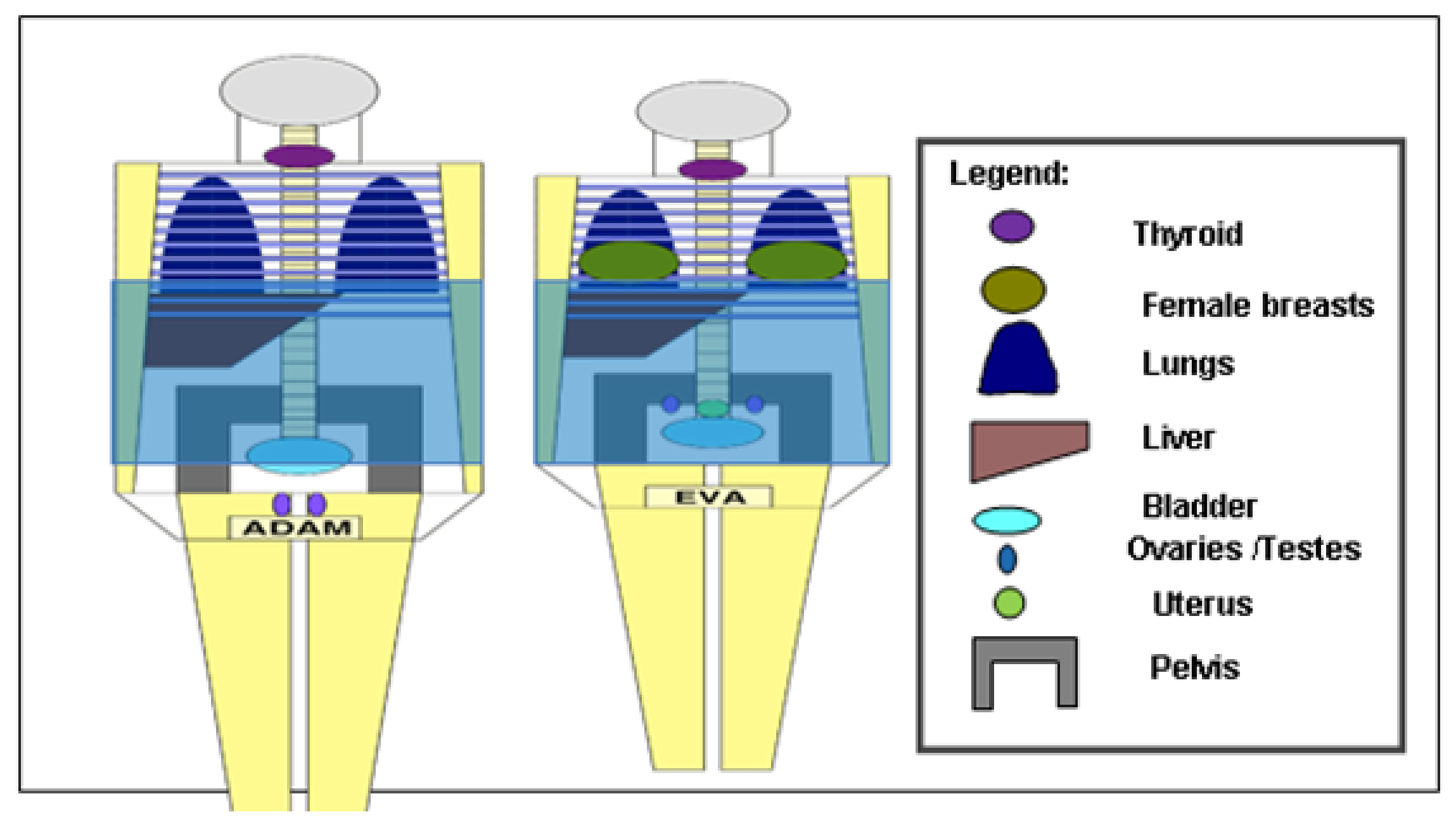
Figure 1. CT-Expo scan range for adults; all organs doses HT are based on conversion coefficients for adult standard patients (AdAM, EVA).
Applsci 12 06691 g001
Table 1. ICRP 60 and 103 tissue weighting factors.
Body OrgansGonadsLungStomachBreastLiverThyroidBladderBrainReminder
PublicationsICRP
60
0.200.120.120.050.050.050.05-0.05
ICRP
103
0.080.120.120.120.040.040.040.010.12
Table 2. Reported DLP to Ed conversion factors.
Regional AnatomyDose Length Product to Ed Conversion Factors (k)
[mSv/(mGy × cm)]
Phantom
Module
(cm)
Jess. et al. [21]European Commission Guidelines [22]European
Commission
NRPB-W67 (2005)
Head0.00210.00230.002116
Chest0.0140.0170.01432
Abdomen0.0120.0150.01532
Pelvis0.0190.0190.01532
National Radiological Protection Board (NRPB). Dose-length product (DLP), Ed = k × DLP.
Table 3. DLP-based effective dose estimates and comparison for ICRP 60, 103 and CT-Expo.
Regional
Anatomy
Tube Voltages (kVp)DLP
(mGycm)
K Values
(mSv)/
(mGycm)
EDLP
(mSv)
EDLP−Es60EDLP−E60EDLP−E103 E D L P E s 60 E s 60 % E D L P E 60 E 60 % E D L P E 103 E 103 %
Head120634 ± 1.30.00211.3−1.5−1.87−5.04−54−59−79
120334.8 ± 0.40.00210.7−0.8−0.97−2.65−53−58−79
120265.6 ± 0.10.00210.6−0.5−0.73−2.06−45−55−77
120657.2 ± 1.20.00211.4−1.5−1.89−5.17−52−57−79
140428.5 ± 0.30.00210.9−1.0−1.24−3.38−5358−79
Chest120539.7 ± 2.10.0147.60.24.91.120318117
120459.9 ± 0.20.0146.40.14.10.880217816
100333.8 ± 0.40.0144.70.13.030.690218117
100380.9 ± 0.20.0145.30.13.40.730217916
100412.3 ± 1.10.0145.80.13.740.850218217
Abdomen120685.5 ± 0.40.01510.3−0.66.872.07−0620025
120430.2 ± 3.10.0156.5−0.44.351.34−0620226
120758.3 ± 0.20.01511.41.38.223.76−1325849
140636.4 ± 0.10.0159.6−2.55.810.5−211535.0
140725.8 ± 0.40.01510.9−0.77.271.63−0620018
Es The effective dose was calculated using CT-Expo Monte Carlo simulation.
Table 4. Effective dose (Ed) using organ dose-based estimates and comparison for ICRP 60, 103 and Monte Carlo.
Regional
Anatomy
Tube
Voltages
(kVp)
CTDIvol (mGy)DLP
(mGycm)
Es60
(mSv)
Es103
(mSv)
E60
(mSv)
E103
(mSv)
E103−E60Scope of
Eb (mSv)
Scope of
Eb/Es60% (mSv)
Scope of
Eb/E60% (mSv)
Head12033.43 ± 0.2634 ± 1.32.83.23.176.343.173.54126.43111.67
12016.28 ± 0.1334.8 ± 0.41.51.71.673.351.671.85123.33110.51
12016.08 ± 0.1265.6 ± 0.11.11.31.332.661.331.56141.82117.47
12028.85 ± 0.7657.2 ± 1.22.93.33.296.573.293.67126.55111.69
14047.3 ± 1.1428.5 ± 0.31.92.12.144.282.142.38125.26111.09
Chest12024.47 ± 0.6539.7 ±2.17.47.72.706.483.785.067.57185.28
10011.54 ± 1.2459.9 ± 0.26.36.62.305.523.224.368.25186.97
1205.47 ± 0.03333.8 ± 0.44.64.81.674.012.343.1368.04187.54
14013.02 ± 0.07380.9 ± 0.25.25.41.904.572.673.567.31183.78
12012.88 ± 1.0412.3 ± 1.15.75.92.064.952.893.8467.37186.27
Abdomen12013.53 ± 0.09685.5 ± 0.410.912.23.438.234.807.4768.53217.94
12010.22 ± 1.1430.2 ± 3.16.97.72.155.163.015.5580.43258.02
12013.41 ± 0.05758.3 ± 0.210.111.43.187.644.458.2281.39258.35
12015.89 ± 0.4636.4 ± 0.112.113.53.799.105.319.7180.25256.10
14013.41 ± 1.3725.8 ± 0.411.612.93.639.275.089.2779.91255.44
(Es60, Es103) Monte Carlo simulated. Eb Range of E = (Max−Min) effective dose for the set of Es60, Es103, E60, E103.
Publisher’s Note: MDPI stays neutral with regard to jurisdictional claims in published maps and institutional affiliations.