Abstract
There is a bidirectional interaction between the gut microbiota and human health status. Disturbance of the microbiota increases the risk of pathogen infections and other diseases. The use of bacteriophages as antibacterial therapy or prophylaxis is intended to counteract intestinal disorders. To deliver bacteriophages unharmed into the gut, they must be protected from acidic conditions in the stomach. Therefore, an encapsulation method based on in situ complexation of alginate (2%), calcium ions (0.5%), and milk proteins (1%) by spray drying was investigated. Powdered capsules with particle sizes of ~10 µm and bacteriophage K5 titers of ~107 plaque-forming units (pfu) g−1 were obtained. They protected the bacteriophages from acid (pH 2.5) in the stomach for 2 h and released them within 30 min under intestinal conditions (in vitro). There was no loss of viability during storage over two months (4 °C). Instead of consuming bacteriophage capsules in pure form (i.e., as powder/tablets), they could be inserted into food matrices, as exemplary shown in this study using cereal cookies as a semi-solid food matrix. By consuming bacteriophages in combination with probiotic organisms (e.g., via yoghurt with cereal cookies), probiotics could directly repopulate the niches generated by bacteriophages and, thus, contribute to a healthier life.
1. Introduction
Human gut microbiome and health are strongly correlated to each other. Diseases such as gastrointestinal, liver, inflammatory bowel, Alzheimer’s diseases, and colorectal cancer are associated with changes or an imbalance in the microbiome [1,2,3]. Numerous studies have already highlighted the role of the microbiome, for instance, in regulating epithelial development, providing nourishment, and instructing innate immunity [4,5,6,7]. The composition of the intestinal microbiota is relatively constant throughout life; however, some changes such as taking antibiotics, bacterial infections, or a long-term change in diet may influence it. Short-term and long-term effects of antibiotics on the human gut microbiota were reported by several authors, some of which can lead to acute or chronic diseases [8,9,10].
In recent years, a variety of studies haves been conducted to define the composition of the human intestinal microbiota and to identify influencing factors [11,12,13,14]. According to these studies, diet, probiotics, prebiotics, and microbiota transplantation are emphasized as potential factors that can modulate the composition and diversity of the gut microbiota. Moreover, because of their ability to infect specific bacteria, bacteriophages (viruses specifically infecting bacterial cells) have been suggested for the treatment of gastrointestinal diseases and for use as therapeutic agents [1,15]. Bacteriophages are natural and essential components of the human gastrointestinal tract. Their numbers are reported to range between 108 and 1010 plaque forming units (pfu) per gram of feces (strictly speaking, these are referred to as virus-like particles) [16,17,18]. Therefore, the recognition of bacteriophages in applications to shape and modulate the human gut microbiota has been increasing.
The application of bacteriophages in the treatment of diseases actually dates back to the 1920s [19]. Studies conducted on 153 patients in the first bacteriophage therapy center in the European Union (in Poland) provided good clinical results regarding the therapeutic use of bacteriophages [20], suggesting that further investigations in this area were warranted. In vivo bacteriophage therapy studies of the last decade with humans and animals were summarized in review articles by Malik et al., and Melo et al. [21,22]. Furthermore, bacteriophages are also used in food companies as biosanitizers, biocontrol for pathogenic bacteria, and biopreservatives in the production of meat, vegetables, and fruits [23,24,25,26]. Formulations containing bacteriophages have a ‘generally recognized as safe’ status (GRAS status) from the American Food and Drug Administration (FDA) or the European Food Safety Authority (EFSA) and are used legally in the USA, Canada, Australia, and in some parts of Europe [25,27,28,29].
Delivery of bacteriophages to their target place (i.e., to the human gut) via food matrices has two main challenges. The first challenge is preserving enough active bacteriophages to maintain target values after production, storage (e.g., storage in a food product), and administration stages [30]. The second challenge is preserving the activity of bacterio-phages through the gastrointestinal tract, since their activities are affected by physicochemical factors (e.g., pH value, enzymes, temperature, salt concentrations, and ions) and biological factors (e.g., lytic cycles, burst size, and host physiological conditions) [31]. In order to protect bacteriophages from the mentioned factors, microencapsulation methods (i.e., entrapment of bacteriophages in an encapsulation system) using freeze drying, spray drying, ultrasonic vacuum spray drying, electrospraying, extrusion, emulsion, and supercritical techniques have been studied and reviewed intensively [21,32,33,34,35]. As encapsulation materials, aginate solutions together with milk proteins such as alginate-caseinate [36,37] and alginate-whey protein [33,38] formulations were employed succesfully. In food processing, microencapsulation techniques are commonly utilized for sensitive food ingredients (e.g., proteins, oils, aroma, and probiotics) [34,39,40]. However, to the best of our knowledge, the application of encapsulated bacteriophages in a food system has not yet been studied. Though bacteriophage therapeutic products are available in some Eastern European countries (e.g., the Republic of Georgia), the use of bacteriophages in food products as functional ingredients still requires the approval of the European Union [28,41,42]. More research is needed in this field, since the functional application of bacteriophages via food products could demonstrate promising opportunities and lower the hurdles for their possible approval [29,42].
In this study, bacteriophages were encapsulated using in situ complexation of alginate, calcium ions, and milk proteins during spray drying. The resulting capsules were incorporated into a food product (e.g., milk or cereal product) for comfortable reception. The capsules should have a particle diameter below 100 µm in order not to influence the sensory properties. Furthermore, the capsules should be able to be stored for a period of time without loss of functionality, have adequate protection against the acidic conditions in the stomach, and should release the bacteriophages in intestinal conditions (in vitro) completely. Therefore, it was hypothesized that encapsulated bacteriophages can be transferred into the human gut via food to eliminate undesired bacteria and, thus, create a niche at their target. The newly generated niche could be directly repopulated, for example, by probiotic organisms. Hence, it would be advisable to consume bacteriophages in combination with probiotic organisms, exemplary via yoghurt (with probiotics) and a cereal cookie (with bacteriophages). In analogy to probiotics, we would like to introduce the neologism “bacteriophage-biotics” for therapeutically active bacteriophages. The present work exclusively focuses on in vitro studies using a harmless model system with a probiotic host (Escherichia coli Nissle 1917) and bacteriophage K5 and serves as proof-of-concept in general. After successful demonstration of the functional principle, the model system could be used to perform in vivo human feasibility studies. In future work, the model bacteriophages could then be replaced by other bacteriophages that have a health-related or medically relevant purpose for humans.
2. Materials and Methods
2.1. Host Strain and Bacteriophage
2.1.1. Selection and Morphology
All experiments were conducted with Escherichia coli Nissle 1917 (DSM 6601, abbreviated as EcN), which was generously provided by Ardeypharm GmbH (Herdecke, Germany). The corresponding bacteriophage is called K5 and was purchased from SSI Diagnostica A/S (Hillerød, Denmark).
The morphology was investigated via atomic force microscopy using the Nanoscope IIIa Multimode with the E scanner, which has a maximum scan size of 10 × 10 µm, and the Nanoscope V5.31r1 software (Veeco Instruments Ltd., Plainview, NY, USA) and silicon nitride micro cantilevers (Oxford Instruments GmbH, Wiesbaden Germany (formerly: Olympus, Tokyo, Japan); NanoAndMore GmbH, Wetzlar, Germany) under tapping mode (AC) in air and at room temperature (20–25 °C).
2.1.2. Cultivation, Propagation, and Enumeration
Cultivation of EcN was performed at 37 °C and 100 rpm in lysogeny broth according to Lennox (LB (Lennox)) (Th. Geyer GmbH and Co. KG, Renningen, Germany). Bacteriophage K5 lysate was propagated with the aid of a fresh EcN culture in 10 mL scale in LB broth until the optical density (OD620 nm) was < 0.1 (PF-12 Plus, Macherey-Nagel GmbH and Co. KG, Düren, Germany). Subsequently, it was purified by centrifugation (15,294× g, 7 °C, 10 min) and sterile filtration using 0.2 µm sterile filters (Sartorius AG, Göttingen, Germany). Purified bacteriophage K5 lysates were stored at 4 °C. Bacteriophage titers (in pfu mL−1) were determined using the agar overlay method (plaque assay) on LB (Lennox) agar (Th. Geyer GmbH and Co. KG, Renningen, Germany). After overnight incubation at 37 °C, plaques were counted and titers were evaluated according to the weighed arithmetic mean with the consideration of at least 10 plaques per plate [43,44].
2.1.3. Thermal Inactivation of Bacteriophage K5
Bacteriophage K5 was characterized regarding its behavior with response to heat. Pure bacteriophage K5 lysate (Section 2.1.2) with an initial bacteriophage titer of N* = 7.2 × 10⁸–7.3 × 10⁹ pfu mL−1 was filled into screw-capped stainless steel tubes (inner volume: 1.5 mL, hermetically sealed, see details in [45]) and heated at temperatures of 68–75 °C in a water bath (HAAKE S 14P, Thermo Fisher Scientific Inc., Waltham, MA, USA). To avoid uneven temperature distribution within the tubes, the experiment was started with a warm-up phase of 1 min [45,46]. Then, holding times ranging from t = 0–10 min were applied, followed by a cooling step in ice water. Bacteriophage titers were determined after the 1 min warm-up phase (N0) and after respective holding times (Nt) using the plaque assay (Section 2.1.2).
The thermal inactivation data can be used to determine the decimal reduction time Dϑ, which is a measure of the heat stability of organisms. It is defined as the time required to decrease the bacteriophage titer by 90% (1 log unit) of the initial titer N0 at a defined temperature ϑ. Mathematically, it is described by the negative inverse slope of the log-linear phase of the thermal inactivation curve (Equation (1)):
where Dϑ is the decimal reduction time at a defined temperature (min) and m is the slope of the log-linear regression line (min−1) [47].
Additionally, the data were modeled according to the Arrhenius equation as described previously [43,45] (Equation (2)):
where Nt is the bacteriophage titer (pfu mL−1) after the thermal treatment at time t (s), N0 is the bacteriophage titer after the 1 min warm-up phase (pfu mL−1), n is the order of reaction (–) and unequal to 1, kref is the death rate at the reference temperature (s−1 (pfu mL−1)(1−n)), Ea is the activation energy (J mol−1), R is the universal gas constant (8.314 J mol−1 K−1), T is the absolute temperature (K), and Tref is the reference temperature (K) [47].
2.1.4. Acidic Inactivation of Bacteriophage K5
Analogous to Section 2.1.3, bacteriophage K5 was characterized with respect to its survival behavior in an acidic environment. Therefore, 0.1 mL pure bacteriophage K5 lysate (Section 2.1.2) with an initial bacteriophage titer of N* = 2.3 × 10⁷ pfu mL−1 was incubated in 9.9 mL of an acidic solution. Acidic solutions were composed of 2 g L−1 NaCl (Carl Roth GmbH and Co. KG, Karlsruhe, Germany) adjusted with HCl (Th. Geyer GmbH and Co. KG, Renningen, Germany) to pH values of 2.0, 3.0, 3.5, 4.0, and 5.0 (inoLab® 7110, Xylem Analytics Germany Sales GmbH and Co. KG, WTW, Weilheim, Germany). A pure NaCl solution served as a reference (native pH 6.6). The first sample was taken directly after mixing (t = 20 s). After that, incubation times ranged from t = 1–120 min. Samples were neutralized immediately in a 0.1 M sodium phosphate buffer (pH 6.8) (Na2HPO4, Carl Roth GmbH and Co. KG, Karlsruhe, Germany; NaH2PO4, AppliChem GmbH, Darmstadt, Germany) using a 1:1 ratio of sample and buffer. Bacteriophage titers were determined after 20 s (N0) and after respective incubation times (Nt) using the plaque assay (Section 2.1.2).
2.2. Encapsulation of Bacteriophage K5 by Spray Drying
K5 bacteriophages were encapsulated by an in situ cross linking of alginate and bivalent calcium ions during spray drying. This process was invented and patented by Jeoh Zicari et al. [48] and slightly modified in this study. An overview of the process is given in Figure 1.
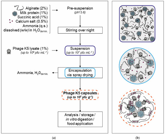
Figure 1.
In situ complexation of alginate, calcium ions, and milk proteins during spray drying (method modified according to [48]). (a) Incubation in simulated gastric fluid (SGF). Results are given as bacteriophage titer of solid capsules (NC in pfu g–1). (b) Schematic drawing (not to scale) of the encapsulation process with suspension (above, blue frame), atomized droplet (center, turquoise frame), and capsule (bottom, orange frame) (figure modified according to [49]).
In the first step, a pre-suspension of the matrix components was prepared. This contained demineralized water, 2% (w/w) sodium alginate of low viscosity (Merck KGaA, Darmstadt, Germany), 1% (w/w) milk protein (either micellar casein powder or whey protein isolate powder; both produced by the Dairy for Research and Training, University of Hohenheim, Stuttgart, Germany), 1% (w/w) succinic acid (Carl Roth GmbH and Co. KG, Karlsruhe, Germany), and 0.5% (w/w) calcium hydrogen phosphate dihydrate (Carl Roth GmbH and Co. KG, Karlsruhe, Germany). The pre-suspension had a pH value of ~3.5 but was adjusted with a 25% ammonia solution (Carl Roth GmbH and Co. KG, Karlsruhe, Germany) to a pH of 5.6. For practical reasons, the calcium salt and the alginate were added after the pH adjustment and the pH value was checked again. After continuously stirring overnight at 20 °C, 1% purified bacteriophage K5 lysate (Section 2.1.2) was added and stirred for 1 h.
Spray drying was performed using the Mini Spray Dryer B-290 (BÜCHI Labortechnik GmbH, Essen, Germany) and the settings listed in Table 1. Meanwhile, the suspension was stirred continuously. The produced bacteriophage K5 capsules (powder) were collected and stored in sealed reaction vessels (Section 2.5.1).

Table 1.
Spray drying parameters and settings used for encapsulation.
2.3. Physical Characterization of Bacteriophage K5 Capsules
2.3.1. Humidity and Water Activity
The humidity was determined gravimetrically using the halogen dryer MB35 (OHAUS Europe GmbH, Nänikon, Switzerland) at 120 °C and with an automatic end point determination and a sample amount of 0.5 g. The water activity analyzer aw sprint TH 500 (Novasina AG, Lachen, Switzerland) was used to measure the water activity at 25 °C using 1.5 g of the sample. An eVC-21 filter and a grid were used to protect the integrated electrolyte sensor.
2.3.2. Particle Size
The particle size of the dry bacteriophage K5 capsules was determined by static light scattering using the Mastersizer 2000 and the Malvern Panalytical software (Malvern Panalytical GmbH, Herrenberg, Germany). As a dispersion agent, 96% ethanol (Distillery for Research and Training, University of Hohenheim, Stuttgart, Germany) was used and pumped at 1900 rpm. Refractive indices were 1.36 for ethanol [51] and 1.51 for alginate [49] as the main component of the capsule matrix. Volume-based diameters d10,3, d50,3, and d90,3 (i.e., corresponding to the percentages 10, 50, and 90% of particles under the reported particle size) were calculated based on the measurement.
2.3.3. Scanning Electron Microscopy
To assess the topography of the bacteriophage K5 capsules, the JEOL JSM-IT500 scanning electron microscope and the InTouchScope software (JEOL Ltd., Tokyo, Japan) were used. A secondary electron detector (SED), a working distance (WD) of 11 mm, and an acceleration voltage of 3.0 kV (P.C. 41 HV) were chosen as operating conditions. A magnification of ×1.500 is shown in this study.
2.4. Biological Characterization of Bacteriophage K5 Capsules
2.4.1. Capsule Break-Up, Enumeration, and Encapsulation Efficiency
Enumeration of bacteriophage K5 in the capsules can only take place after break-up of the capsules and release of the bacteriophages into a liquid. For this purpose, microsphere breaking solution (MBS) with a pH of 7.5 was prepared, consisting of 0.2 mM sodium hydrogen carbonate (Carl Roth GmbH and Co. KG, Karlsruhe, Germany), 0.05 mM tri-sodium citrate dihydrate (VWR International GmbH, Bruchsal, Germany), and 0.05 mM Tris(hydroxymethyl)aminomethane hydrochloride (Tris HCl) (Merck KGaA, Darmstadt, Germany) [52]. A mass of 0.1 g capsules was added to 9.9 mL MBS and incubated at 37 °C and 100 rpm for 1–2 h until no solid particles were visible. Bacteriophage titers of dissolved capsules NC# (pfu mL−1 of 1% capsules in MBS) were determined using the plaque assay (Section 2.1.2) and converted to titers in dry capsules NC (pfu g−1 of 100% capsules) by multiplication by a factor of 100.
The encapsulation efficiency (Equation (3)) provided information about the success of the encapsulation process and the extent of surviving/non-lost bacteriophages. For this purpose, an optimal spray drying (i.e., complete loss of aqueous and volatile components) was assumed and the bacteriophage titers of the suspension and in the capsules were put into proportion. Therefore, NC#, which represents the titer for 1% dissolved capsules in MBS, was converted into NC## by multiplying by a factor of 4.5, representing the titer for 4.5% dissolved capsules in MBS and, thus, a “reconstituted suspension” with the same dry matter (4.5%) as the suspension used for encapsulation.
where E is the encapsulation efficiency (%), NC# is the bacteriophage titer of 1% capsules dissolved in MBS (pfu mL−1), NC## is the bacteriophage titer of 4.5% capsules dissolved in MBS (pfu mL−1), and NS is the bacteriophage titer of the suspension before encapsulation (pfu mL−1) (adapted from Soykut et al. [53]).
2.4.2. In Vitro Digestion
Static in vitro digestion was performed following a protocol described by Samtlebe et al. [33]. The stability of the encapsulated bacteriophages (Section 2.2) with an initial bacteriophage titer NC* = 1.5 × 10⁷–3.0 × 10⁷ pfu g−1 was analyzed in simulated gastric fluid (SGF). This was prepared by adding 3.2 mg L−1 pepsin from porcine gastric mucosa (≥3200 international units mg−1, Merck KGaA, Darmstadt, Germany) to 2 g L−1 NaCl adjusted with HCl to a pH value of 2.5. A mass of 0.1 g capsules was added to 9.9 mL pre-warmed SGF and incubated at 37 °C and 100 rpm for t = 1–120 min. Shortly before expiration of the respective incubation times in SGF, the samples were centrifuged at 2000× g and 20 °C for 3 min. The supernatant was discarded, and 1 mL of 0.1 M sodium phosphate buffer (pH 6.8) was pipetted to the sedimented capsules to neutralize any remaining acid. To dissolve the capsules and release the bacteriophages that remained intact, 8.9 mL of MBS were added to a total volume of 10 mL and incubated at 37 °C and 100 rpm for 1–2 h. The evaluation was carried out using the plaque assay (Section 2.1.2 and Section 2.1.4).
The release of the bacteriophages from the capsules was investigated in simulated intestinal fluid (SIF). The capsules had an initial bacteriophage titer of NC*,# = 1.5 × 10⁵–3.0 × 10⁵ pfu mL−1. This consisted of 10 mg mL−1 pancreatin from porcine pancreas (Merck, KGaA, Darmstadt, Germany) dissolved in 50 mM sodium phosphate buffer (pH 6.8). A mass of 0.1 g capsules was added to 9.9 mL pre-warmed SIF and incubated at 37 °C and 100 rpm for t = 1–120 min. Directly after the respective incubation times, plaque assays were performed (Section 2.1.2 and Section 2.1.4).
2.5. Application of Bacteriophage K5 Capsules in Food Matrices
2.5.1. Storage of Capsules
Storage stability studies were performed over a period of 60 days at 23, 0, and −20 °C. Capsules were portioned and filled into sealed reaction vessels in which they could not absorb moisture. The evaluation was conducted after dissolution in MBS using the plaque assay (Section 2.1.2 and Section 2.1.4).
2.5.2. Milk and Cereal Products
Encapsulated K5 bacteriophages should be added to a liquid food matrix, e.g., to milk products. For this purpose, a preliminary test was carried out in skim milk (0.3% fat, AF Deutschland GmbH, Düsseldorf, Germany) and full fat milk (3.5% fat, Molkerei Weihenstephan GmbH and Co. KG, Freising, Germany) before a more complex matrix such as yoghurt or quark was investigated. A mass of 0.1 g capsules was mixed with 9.9 g of the respective milk and stored for up to 60 days at 5–10 °C. Samples were drawn regularly, centrifuged (15,294× g, 20 °C, 10 min), dissolved in MBS after discarding the supernatant, and enumerated using the plaque assay (Section 2.1.2 and Section 2.1.4).
Furthermore, another product group was tested, namely, a dry and semi-solid cereal product. Therefore, cereal cookies were formulated based on conventional knowledge and by using only two basic ingredients. First, a glucose syrup was prepared by mixing 9 g glucose syrup powder (Glucidex® spray-dried glucose syrup, Roquette Freres, Lestrem, France) with 5 g demineralized water, cooking it until the mixture appeared clear and of low viscosity, and cooling it to 40–50 °C to portion it (in-house process). For the cereal cookies, 1.5 g extra fine oat flakes (Edeka Zentrale AG and Co. AG, Hamburg, Germany) were mixed with 1.0 g glucose syrup and 0.05 g capsules. The cookie mixture was brought into shape using a cylindrical metal form with a diameter of 2.6 cm and was dried for 2 h at 37 °C (in-house process). Samples were stored at 23 °C in closed vessels for up to 28 days. Water activity was measured as described in Section 2.3.1. In preparation for quantification of bacteriophage K5, 20 mL MBS were added to the sample to soften the structure and to release the bacteriophage K5 (Section 2.1.4). Afterwards, the samples were centrifuged (15,294× g, 20 °C, 10 min) and enumerated using the plaque assay (Section 2.1.2).
2.6. Data Analysis
All experiments and measurements were performed in triplicate, except for the storage trials at 23 °C (Section 2.5.1; singe determination) and the experiments with the cereal cookies (Section 2.5.2; double determination). Arithmetic means and standard errors were calculated for all analyses. Values were rounded to one or two significant digits of the standard error. Data were analyzed and diagrams were prepared using Sigma Plot 12.5 software (SPSS Inc., Chicago, IL, USA). Wet-bulb temperature was calculated using Matlab® R2021b software (The MathWorks Inc., Natick, MA, USA).
3. Results and Discussion
3.1. Morphology of Escherichia coli Nissle 1917 and Bacteriophage K5
EcN is a non-pathogenic probiotic strain with the serotype O6:K5:H1. It is flagellated and, thus, is mobile and can interact with mucus and adhere to the human intestinal tract [54]. Its K5 polysaccharide capsule, comprised of α-N-acetylglucosamines and ß-glucuronic acids, represents the specific binding site for bacteriophage K5. Bacteriophage K5 encodes for the depolymerase endo-α-N-acetylglucosaminidase, which enables the hydrolysis of polysaccharides in the EcN capsule [55,56].
Bacteriophage K5 is assigned to a group of bacteriophages that are similar to T7 bacteriophages (belonging to the so-called “T7 supergroup” or are “T7-like”). Typically, they are composed of an icosahedral capsid, a short tail, and fibers [57]. To the best of our knowledge, there is not much information about bacteriophage K5 regarding its dimensions or other characteristics available in the literature.
EcN and bacteriophage K5 were visualized by atomic force microscopy (Figure 2). Well recognizable are the EcN cells with their flagella, as well as the bacteriophage K5 capsids. The capsid diameter was determined to be 68–88 nm on average. This is slightly larger than the capsid of the bacteriophage T7 (61–63 nm) and other T7-like bacteriophages (e.g., SP6 and K1-5: 65–68 nm) [57,58]. However, the larger appearance could also be associated with the applied methodology; the measured values should be verified in the future using a scanning electron microscopy image.
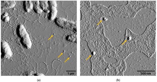
Figure 2.
Atomic force microscopy image of (a) Escherichia coli Nissle 1917 and (b) bacteriophage K5. Bacteriophages (selection) are indicated by yellow arrows in both pictures.
As mentioned before, EcN and bacteriophage K5 were particularly well suited for the following study as a test model as they enabled laboratory work without strict precautionary safety measures and work in food-grade areas. Furthermore, they would be beneficial for future feasibility studies involving animals or humans.
3.2. Heat Sensitivity of Bacteriophage K5
Pure bacteriophage K5 lysate was used for the thermal inactivation experiments. Depending on whether K5 appears to be resistant against heat or not, an appropriate encapsulation method can be chosen or process parameters can be adjusted. In addition, the data may be relevant for the later application of bacteriophage capsules in food matrices and their production (e.g., admixture before or after heating steps). Figure 3 shows the survival behavior at temperatures of 68–75 °C.
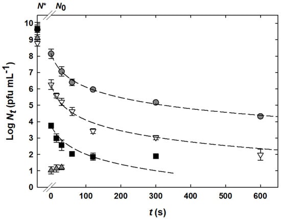
Figure 3.
Thermal inactivation of pure bacteriophage K5 lysate at temperatures of 68 °C ( ), 70 °C (
 ), 72 °C (
 ), and 75 °C (
 ). Experimental data are displayed by symbols. N* indicates the initial bacteriophage titer before any treatment. Modeled inactivation curves are represented by dashed lines and were calculated using Equation (2) and the following values for the variable parameters (± standard error s): n = 1.42 ± 0.06, kref = (2.8 ± 1.0) × 10−3 s−1 (pfu mL−1)(1−n), Ea = (9.57 ± 1.38) × 102 kJ mol−1, Tref = 345.15 K. The correlation coefficient r2 was 0.98. The limit of quantification was 101 pfu mL−1.
), 70 °C (), 72 °C (), and 75 °C (). Experimental data are displayed by symbols. N* indicates the initial bacteriophage titer before any treatment. Modeled inactivation curves are represented by dashed lines and were calculated using Equation (2) and the following values for the variable parameters (± standard error s): n = 1.42 ± 0.06, kref = (2.8 ± 1.0) × 10−3 s−1 (pfu mL−1)(1−n), Ea = (9.57 ± 1.38) × 102 kJ mol−1, Tref = 345.15 K. The correlation coefficient r2 was 0.98. The limit of quantification was 101 pfu mL−1.
The highest bacteriophage K5 reduction took place in each case within the one-minute heating phase, with reductions of 1.5, 2.5, 5.9, and 8.1 log units for 68, 70, 72, and 75 °C, respectively. Within the following 300 to 600 s, they were reduced by 3.8, 4.3, and 1.9 log units at the temperatures of 68, 70, and 72 °C, respectively. However, no further reduction was observed at 75 °C, as the titers were close to the limit of quantification (101 pfu mL−1).
Modeling of the data was performed using the Arrhenius-based model. Thereby, non-linear curve progressions with tailing were observed (except for the data obtained at 75 °C). The order of reaction n was 1.42 ± 0.06, the death rate kref was (2.8 ± 1.0) × 10−3 s−1 (pfu mL−1)(1−n), and the activation energy Ea was (9.57 ± 1.38) × 102 kJ mol−1 (all parameters given with standard error s). As a reference temperature Tref, 345.15 K was defined.
The decimal reduction times Dϑ were calculated (again, except for the data obtained at 75 °C) to be D68 °C = 0.52 min (r2 = 0.91), D70 °C = 0.74 min (r2 = 0.98), and D72 °C = 0.42 min (r2 = 0.97). The Dϑ-values were similar and decreased slightly with increasing temperature (not significant). This effect is also visible in the curve progressions themselves in Figure 3, which appear nearly parallel. Therefore, determination of another kinetic parameter, the zϑ-value (which represents the temperature increase that is necessary to achieve the same reduction effect in 1/10 of the time), was omitted.
The curve progressions, as well as the Dϑ-values, at the different temperatures were different than expected. The strong reduction within the heating phase (steep slope) in connection with a short linear phase (flat slope) and a long tailing range suggests that rather the “real” linear phase of the experimental course was not recorded. Reasons for this could be a too low initial titer (N*) and a too great inactivation within the heating phase in which no data points were recorded. However, in order to ensure even temperature distribution in the stainless steel tubes, the heating-up phase cannot or can only be slightly shortened, as previously stated by Müller-Merbach et al. [46]. To obtain better data for kinetic evaluation in the future, pure bacteriophage lysate with a higher titer should be produced and used for the inactivation experiments. For example, the CsCl ultracentrifugation method according to Sambrook and Russel results in titers of up to 1013 pfu mL−1 depending on the type of bacteriophage [59]. Nevertheless, because of these observations it can be stated that the bacteriophage K5 belongs to the rather heat sensitive bacteriophages.
To the best of our knowledge, there is no other study that ever investigated the bacteriophage K5 behavior against heat. However, there is some information available regarding other Escherichia coli bacteriophages or bacteriophages of other bacterial species. Atomic force microscopy demonstrated that heat treatment of the Escherichia coli bacteriophage T7 at 65 °C led to destabilized particles with loss of capsid tails and DNA. In contrast, heat treatment at 80 °C showed particles with higher stability, which was explained by partial protein denaturation. Although the bacteriophages were not tested for plaque-forming ability in the study, it can be assumed that the bacteriophages were no longer capable of attaching to their host cells, injecting their DNA, and lysing the host cells [60]. Pollard and Solosko treated the Escherichia coli bacteriophages T4 and λ in nutrient broth at temperatures between 50 and 75 °C. They observed linear reductions of approximately 1.5 log units at 65 °C after 70 min, whereas the same reduction was reached at 70 °C after 15 min. Below or at 60 °C, the bacteriophages appeared to be almost stable [61]. Compared with bacteriophage K5 in the present study, these two other bacteriophages seem more heat resistant. On the other hand, similar behavior to bacteriophage K5 was observed with the Bacillus amyloliquefaciens bacteriophage BA01. It was inactivated by 5 log units within 30 min at 65 °C and by 7–8 log units (estimated, since bacteriophage titers were below the limit of quantification) within 5 min at 70 °C [62]. A large amount of research has focused on the heat inactivation of bacteriophages against lactic acid bacteria, especially Lactococcus lactis, Streptococcus thermophilus, and Leuconostoc spp., as their inactivation is of great importance for the processing of fermented milk products [63]. Among them, there are heat sensitive, as well as heat resistant, bacteriophages. For instance, Lactococcus lactis bacteriophage P001 was inactivated in nutrient broth by 7 log units within 10 min at 70 °C, whereas bacteriophage P008 was inactivated by only 2.5 log units at the same temperature and time [46]. The particularly heat resistant bacteriophage P680 was inactivated at 70 °C, only by 1 log unit at 75 °C within 10 min, and still detectable after 1 min at 95 °C [45,64].
The comparison to other bacteriophages leads to the statement that bacteriophage K5 has to be considered as heat sensitive, therefore further processes, i.e., encapsulation and incorporation into food, must be designed as mild as possible. If such processes are aligned to the weakest bacteriophage, they will work with many other bacteriophages as well.
3.3. Acid Sensitivity of Bacteriophage K5
Pure bacteriophage K5 lysate was used to investigate the survival at pH values between 2.0 and 6.6 (Figure 4). In contrast to thermal inactivation experiments (Section 3.2), sensitivity in acidic environments should investigate the need for a protective measure, such as encapsulation, in order to deliver bacteriophages unscathed through the stomach into the gut. Thereby, the investigated pH range corresponds to the pH of the human stomach. It is lower when empty and increases when food is consumed [65,66]. NaCl solution at a concentration of 2 g L−1 was used as the dispersion medium, since it corresponds to the salt content in the human stomach and to the salt content of the simulated gastric fluid used for the in vitro digestion experiments (Section 2.4.2) [67].
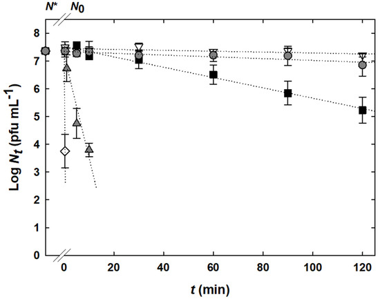
Figure 4.
Inactivation of bacteriophage K5 in 2.0 g L−1 NaCl solutions adjusted with HCl to pH values of 3.0 ( ), 3.5 (), 4.0 (), 5.0 (), and 6.6 () (reference). Experimental data are displayed by symbols. N* indicates the initial bacteriophage titer before any treatment. Linear regressions are shown by dotted lines. The limit of quantification was 2 × 10² pfu mL−1.
), 3.5 (), 4.0 (), 5.0 (), and 6.6 () (reference). Experimental data are displayed by symbols. N* indicates the initial bacteriophage titer before any treatment. Linear regressions are shown by dotted lines. The limit of quantification was 2 × 10² pfu mL−1.
Bacteriophage K5 incubated at pH 5.0 was stable for 120 min. A reduction by 2.1 log units within 120 min was observed at a pH of 4.0. In contrast, at pH 3.5, the bacteriophage titer was reduced by 3.6 log units after 10 min and then no longer detectable at all. The decrease was even more drastic at a pH value of 3.0, at which no more bacteriophages could be detected after 30 s. At the lowest pH of 2.0, bacteriophage titers were under the quantification limit; therefore, no data is shown in Figure 4. Hereby, it should be considered that the experiment had a limit of quantification of 2 × 10² pfu mL−1, because the respective acidic solutions had to be neutralized by adding a buffer and were thus diluted accordingly.
In contrast to the heat inactivation experiments, all curve progressions were linear (n = 1) and without tailing. This is to be justified with the immediate effect of the acid (no heating-up phase/no adaptation phase necessary). The solution in which the bacteriophages were suspended had no negative influence on the survival behavior, shown by the results of the reference (2 g L−1 NaCl solution with a native pH). These results demonstrate that bacteriophage K5 is unstable at pH values below 4.0 and should be protected from the acidic pH in the stomach using, e.g., encapsulation.
These results correspond to other findings in the literature. In general, bacteriophages are described to be active in solutions with pH values ranging from 5.0 to 8.0 [44]. Several studies found that bacteriophages and other viruses have a general susceptibility to acidic (and basic) solutions [64,68,69,70,71]. They may lose their viability at extreme pH values due to surface protein denaturation or deformation, as well as due to hydrolysis or other changes of the nucleic acid [72]. The T7 bacteriophage was reduced by 4 log units within 10 min when it was exposed to nitrous acid at pH 4.0. At a pH of 5.0, the reduction was below 1 log unit after the same time [68]. Feng et al., investigated the survival of the Escherichia coli bacteriophages MS2 and Qß at pH values between 3 and 11 at different temperatures. Although bacteriophage MS2 had a greater resistance, a significant reduction was seen below pH 5 and above pH 9 [69]. Capra et al., measured up to 6 log unit reductions for Lactobacillus casei bacteriophage J-1 and for Lactobacillus paracasei bacteriophage PL-1 incubated for 30 min in MRS broth with pH values below 4 [70]. Similar observations were made for Lactococcus lactis bacteriophage P680 in a nitric acid cleaning solution with pH values of 2 and 3. After 10 min incubation, P680 was inactivated by 6 log units at pH 2, but by less than 0.5 log units at pH 3 [64]. Similar effects were also found when using commercial sanitizers and disinfectants (e.g., peroxide, acetic acid, phosphoric acid, and nitric acid) against Lactococcus lactis bacteriophages, Lactiplantibacillus plantarum bacteriophages (formerly referred to as Lactobacillus plantarum bacteriophages), and Streptococcus thermophilus bacteriophages [71].
3.4. Bacteriophage K5 Capsules Powder and Its Characteristics
Bacteriophage capsules were produced according to the in situ microencapsulation method described in Section 2.2. Independent of the type of milk protein added to the matrix (either micellar casein or whey protein isolate), a white fine non-adhesive powder was obtained, representatively shown for the variant with micellar casein in Figure 5a. The smaller visible aggregates were very easy to break up with a spatula. Figure 5b shows the topography of the capsules, which consisted of a majority of smaller but also larger particles with a pulled-in surface. Roughly measured, the diameters of the capsules were between 2.5 and 10 µm.
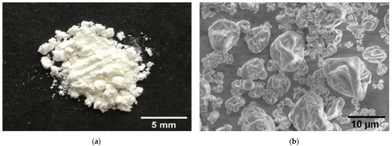
Figure 5.
Bacteriophage K5 capsules, exemplary shown for the variant with micellar casein. (a) White powdery product after spray drying. (b) Topography recorded using scanning electron microscopy and a magnification of 1.500×.
Additionally, Table 2 contains characteristic parameters of the bacteriophage K5 capsules, such as humidity, water activity, particle size, bacteriophage titer, and encapsulation efficiency. In general, both variants did not differ greatly between each other. Only the values of the particle size measurement were slightly higher in the variant with whey protein than in the variant with micellar casein.
Table 2.
Analysis of the bacteriophage K5 capsules regarding physical and biological parameters. Capsules were mainly composed of sodium alginate and milk protein (either micellar casein or whey protein isolate).
Both products had a low humidity content of around 9% and a low water activity (Table 2). Water activity was only measured for the product containing micellar casein, but it must be similar for the product with whey protein isolate. A low humidity is a quality parameter and particularly advantageous for future large-scale production, storage, and application. In this context, attention should be paid to the storage conditions and a dry environment as the particles can otherwise easily absorb water [73].
The humidity of the capsules in this study is in accordance with other literature data. According to Malik et al., a residual moisture content of 5% or more is the rule for spray drying, since the drying process does not vaporize all the liquid and moisture remains in the capsules [74,75]. Vinner et al., produced Salmonella bacteriophage Felix O1 microcapsules with Eudragit S100® and trehalose, with a final humidity of 9% [76]. Hussein et al., obtained 5.5–8.5% humidity in sodium alginate capsules when spray dried at different temperatures [77]. However, it is not the absolute humidity itself that matters, but the storage stability, which is discussed in more detail in Section 3.5.
The particle size (d50,3) was around 10 µm for both matrices and in general no particle fractions were above 50 µm. This fits to our overall aim of producing small capsules that are not sensorially recognizable in food products. According to Hansen et al., particle diameters should not exceed 100 µm in order not to influence the sensory properties of yoghurt [78]. The biggest particles with diameters of up to ~ 46 µm, included in the d90 values, could represent aggregates consisting of several smaller particles. However, these aggregates can be easily broken up, for example, by ultrasonic treatment [49].
Although the composition of the matrix, the solids concentration of the feed, the gas volume flow, the nozzle of the spray dryer, and other equipment parameters have an influence on the size of spray-dried particles, the size magnitude itself stays constant. Compared with the present data, many other studies found similar particle sizes, i.e., in the lower µm range, for spray-dried capsules. Vinner et al., measured a particle size (d50) of 6 µm for their polymer saccharide capsules [76]. Matinkhoo et al., and Leung et al., obtained diameters below 5 µm in a powdery product mainly made of trehalose and amino acids [79,80]. Sodium alginate capsules containing Bacillus subtilis or Salmonella spp. bacteriophages resulted in particle sizes (d50) between 9 and 24 µm [53].
Other encapsulation techniques, such as emulsion or extrusion, can hardly compete with the capsules obtained by spray drying. With the help of a similar capsule matrix of 1.6% alginate and 10% whey protein isolate, Samtlebe et al., were able to produce uniformly spherical but significantly larger capsules than the present study, with diameters of 1–2 mm [33]. Capsules produced by emulsion and enzymatically triggered gelation of milk protein for Lactococcus lactis P008 bacteriophages were considerably smaller at 132 μm, although not in the size range of spray-dried capsules [33]. By optimizing the polymer/protein ratio of a suspension with sodium alginate (1.53%), sodium caseinate (2.72%), and Salmonella bacteriophages Felix O1 using vibrational nozzle technique, minimum particle diameters of 0.16 mm were achieved after extrusion [36].
The most important quality parameter of the capsules is the encapsulation efficiency, which was roughly 4% in this study (Table 2) (calculated with absolute bacteriophage titers; calculation with logarithmized bacteriophage titers would correspond to an encapsulation efficiency of around 80%). In addition to forcing low humidity and low particle diameter, it should be considered that bacteriophages can be affected by heat, drying, and mechanical stress. A compromise should, therefore, be made between these conflicting concerns. As already shown and discussed in Section 3.2, bacteriophage K5 is heat sensitive and easily inactivated. However, in spray drying, the critical factors are the wet-bulb temperature and the outlet temperature (not the inlet temperature), which were 35 °C and 53–59 °C, respectively, in this study, which means that no higher temperatures affect the bacteriophages. Since sensitivity towards heat differs among bacteriophages, results cannot be directly compared with other literature. To the best of our knowledge, there is no data regarding encapsulation of K5 or other T7-like bacteriophages. However, bacteriophage-specific heat sensitivity was also shown by Soykut et al., since they observed encapsulation efficiencies in a broad range of 73–97% for several Bacillus subtilis and Salmonella spp. bacteriophages encapsulated at the same conditions [53].
In general, encapsulation matrices can be modified by adding substances known to have a protective effect on bacteriophage survival during heat treatment and spray drying. The use of proteins in spray drying processes of bacteria, lipids, and volatile components has already delivered promising results on several occasions [81,82,83,84]. Therefore, they are also proposed as stabilizing agents against heat and dehydration stress for bacteriophage encapsulation [85]. In comparison to previous experiments without any protein component in the encapsulation matrix (data not shown), both proteins used in this work (whey protein isolate and micellar casein) protected the K5 bacteriophages equally. However, it is still unclear to what extent the proteins contribute to the bacteriophage protection. It was found that proteins, especially denatured proteins, tend to form aggregates and are not able to move to the surface of the particles during the drying process. Instead, the majority of the proteins are located inside the particle [86]. Therefore, we postulated that the bacteriophages are protected against heat and dehydration stress by accumulation of the proteins inside the particles (see also Figure 1b), and we modified the encapsulation process described by Jeoh Zicari et al. [48] accordingly.
Even though a high encapsulation efficiency is aimed for an efficient process, a sufficiently high bacteriophage titer for the application is more important. In this context, it must be mentioned that a general recommendation for the bacteriophage titer cannot be made, as it depends on the type of bacteria that is to be eliminated in the gut and, therefore, the bacteriophage that is used. Since the K5 bacteriophage used in this study serves as a representative model organism, neither the literature data nor clinical data are available. Additionally, the number of bacteriophages to be consumed can be varied by dosage recommendation. The capsules produced in this study had bacteriophage titers of around 107 pfu g−1.
3.5. Stability in Simulated Gastric Fluid and Release in Simulated Intestinal Fluid
The encapsulation of bacteriophages should ensure their transmission through the digestive tract. One the one hand, the bacteriophages should be protected from acidic conditions in the stomach, which usually lie in a pH range from 1 to 2.5 [65]. On the other hand, they should be released from their matrix, as it is essential for their effect in the gut of individuals. After the K5 bacteriophage capsules were produced, they were subjected to a static in vitro digestion to study their mode of action, as visualized in Figure 6. The compositions of SGF and SIF were based on conditions in the human stomach and intestinal tract. Incubation conditions of 37 °C, 120 min, and moderate rotating (100 rpm) should simulate the physical conditions in the stomach and intestine [33].
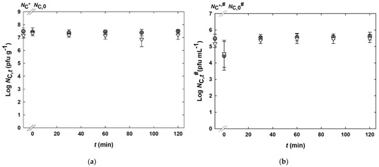
Figure 6.
Static in vitro digestion of bacteriophage K5 capsules composed of alginate and micellar casein () or alginate and whey protein isolate (). (a) Incubation in simulated gastric fluid (SGF). Results are given as bacteriophage titer of solid capsules (NC in pfu g−1). (b) Incubation in simulated intestinal fluid (SIF). Results are given as bacteriophage titer of dissolved capsules (1% in SIF) (NC# in pfu mL−1). NC* and NC*,# indicate the initial bacteriophage titers before any treatment, respectively. The limits of quantification were 103 pfu g−1 and 101 pfu mL−1 for bacteriophage titers in SGF and SIF, respectively.
) or alginate and whey protein isolate (). (a) Incubation in simulated gastric fluid (SGF). Results are given as bacteriophage titer of solid capsules (NC in pfu g−1). (b) Incubation in simulated intestinal fluid (SIF). Results are given as bacteriophage titer of dissolved capsules (1% in SIF) (NC# in pfu mL−1). NC* and NC*,# indicate the initial bacteriophage titers before any treatment, respectively. The limits of quantification were 103 pfu g−1 and 101 pfu mL−1 for bacteriophage titers in SGF and SIF, respectively.
Figure 6a shows that K5 bacteriophage capsules incubated in SGF for 120 min withstand the low pH value of 2.5 and bacteriophages stay intact. The bacteriophage titers show a non-significant reduction of 0.1–0.2 log units after incubation. The same is true for SGF having a pH value of 2.0 (data not shown). Any enzymatic reactions of pepsin did not have a measurable effect on the bacteriophage titer. In general, no differences were observed for the two variants of capsule matrices.
As proven, bacteriophage K5 is quickly inactivated by acidic conditions, especially at a pH value of 3.0 or below (Figure 4). From this, the effect of encapsulation becomes clear, because inadequately encapsulated or free bacteriophages could not have survived the incubation of 120 min.
Figure 6b depicts the release of the bacteriophage K5 capsules from their matrix. Within the first 30 min, the bacteriophage titers were slightly lower (by 0.6–1.0 log units) than the values between 30 and 120 min. This indicates that there was a very quick dissolution of the capsules and release of the K5 bacteriophages into the liquid phase. It should be noted that the given values (log NC,t# in pfu mL−1) show the released bacteriophages in the liquid phase of the experimental approach (after adding 1% capsules to SIF) and are, therefore, lower than the values from Figure 6a (log NC,t in pfu g−1), which represent the surviving bacteriophages in the capsules.
Static in vitro digestion is a method that is frequently used in the literature to investigate the efficacy of different encapsulation methods. In contrast to dynamic in vitro digestion, static systems are less complex, require small quantities of samples and enzymes, and are independent of any specific laboratory devices. However, all average parameters (e.g., pH value) are set at the beginning of the experiment, the dynamic release of enzymes is not feasible, the accumulation of metabolites is not considered, and the complex physiology of the digestive tract is not imitated. Therefore, there is a need for further research using dynamic in vitro digestion protocols in order to come as close as possible to what occurs in vivo [87]. Taking into account the advantages and disadvantages mentioned here, only static systems will be used for discussion in the following section.
Samtlebe et al., investigated the survival of Lactococcus lactis bacteriophage P008 encapsulated in extruded capsules in SGF at pH 2, either with alginate or with alginate and whey protein. The extruded alginate/whey protein capsules protected the bacteriophages over a period of 120 min, whereas the alginate capsules did not protect the bacteriophages sufficiently, because bacteriophages were no longer detectable after 30 min [33]. Similar results were obtained by Tang et al., when comparing alginate capsules with and without whey protein [88]. Ergin et al., determined which concentration ratios of alginate and sodium caseinate must be present for Salmonella bacteriophages Felix O1 to remain intact over a 120 min period in SGF at pH 2. For the optimal ratio (2.0% alginate and 2.69% sodium caseinate), the titer was reduced by 1.25 log units [36]. Shi et al., encapsulated Lactobacillus delbrueckii ssp. bulgaricus cells (formerly referred to as Lactobacillus bulgaricus) into alginate/milk capsules with extrusion. They observed a proportionality between milk concentration and bacteriophage stability in SGF at pH 2.0 [89]. This effect, or the absence of any protein component, was particularly noticeable with Eudragit S100®/trehalose capsules by Vinner et al., which did not provide protection [76]. Again, the important role of a protein component as a bacteriophage protector was shown, analogous to the protection during spray drying, as already discussed in Section 3.4. Since the positively charged proteins (isoelectric point of milk proteins pI ≈ 4.1–5.2) interact with negatively charged alginate molecules, they produce a dense network, preventing the penetration of acid molecules and ions into the core [90]. In addition, the milk proteins can contribute to the protection mechanism with their buffering capacity [91]. Tang et al., produced extruded and air-dried alginate/whey protein capsules. Their Salmonella bacteriophages Felix O1 were not within the capsules when incubated in SGF at pH 2.0. Scanning electron micrographs showed cracks and holes on the surface of these capsules, whereby the authors postulated that they allowed ions to move easily into the matrix [88]. However, the air-drying process in the previous study with a total time of 30 h is not directly comparable to the spray drying process of this study. Furthermore, cracks and holes do not seem to be present or problematic in the present experiments (Figure 5b).
Just as important as protecting the capsules from acidic pH is the subsequent release of the bacteriophages. Most likely, the contact of the bacteriophages with the neutral environment (SIF) results in swelling of the polymer alginate and the proteins. The previously dense capsular matrix expands, causing the K5 bacteriophages to release from the matrix. In addition, pancreatic enzymes are able to diffuse into the matrix in the opposite direction and hydrolyze proteins inside the capsules [33,92]. Compared with the existing literature, the release of the bacteriophages in this study was complete and extremely rapid. Samtlebe et al., found that the release of the extruded alginate/whey protein capsules was fast, within the first minute, and then slow but steadily increasing over the incubation period of 120 min. Furthermore, not all bacteriophages were fully released in the end [33]. Ergin et al., observed 60–80% released bacteriophages after 5 min, but did not observe a full release [36]. The extruded alginate/milk capsules with Lactobacillus delbrueckii ssp. bulgaricus cells from Shi et al., were fully released after 60 min [89]. Tang et al., reported complete release of Salmonella bacteriophages Felix O1 from air-dried alginate/whey protein capsules after more than 100 min [88].
The rapid release of the K5 bacteriophages from the capsules could lead to the bacteriophages being released prematurely in the small intestine and, thus, not reaching their host bacterium in the colon, in view of in vivo conditions. If the bacteriophages are released more slowly, encapsulated bacteriophages may also enter the colon or trigger a kind of long-term effect, whereby bacteriophages are gradually released again and again. Colom et al., reported a long-term effect of alginate/calcium carbonate encapsulated Salmonella enterica ssp. enterica serovar Typhimurium (abbreviated as Salmonella Typhimurium) bacteriophages in an in vivo study in broiler chickens, referring to the mucoadhesive property of the capsules [93]. To counteract the rapid expansion of alginate, a denser or stronger crosslinking of the polymer could be obtained or an additional polymer could be used [94]. This was already shown by Tang et al., by comparing alginate/whey protein matrices containing either 0.8 or 1.5% alginate with a constant protein content of 3%; full bacteriophage release was obtained after 120 or 180 min, respectively [88].
Another possibility to prevent the early release of bacteriophages is a consecutive encapsulation process. The substance or bacteriophage to be encapsulated is entrapped in the first matrix by spray drying. The obtained dry particles are then suspended in a second matrix and spray dried again. This results in a so-called coating of the primary matrix and the bacteriophages are encapsulated twice. LeClair et al., encapsulated a virus-based vaccine in disaccharides (primary matrix) in this way and coated these particles with the polymer Eudragit L100® (secondary matrix) in a second spray drying process [95]. Ma et al., used the polymer chitosan as a coating material for alginate capsules produced using extrusion technology. The bacteriophages encapsulated therein were successively released within 6 h [52]. Further development of the process of this study should pay attention to the size of the resulting particles (should be <100 µm, see Section 3.4) and to the bacteriophage loss during two spray drying steps.
3.6. Good Survival during Storage at 0 and −20 °C
For bacteriophage products (in pure form as powder/compressed into tablets or incorporated into food matrices) to be able to influence the gut microbiota and to achieve a therapeutic effect, their infectivity must not be lost during storage. For this purpose, the capsules were subjected to a storage experiment. The bacteriophage titers for the capsule variant composed of alginate and micellar casein during a storage period of 60 days at 23, 0, and −20 °C are exemplary shown in Table 3.
Table 3.
Survival during storage at 23, 0, and −20 °C of bacteriophage K5 capsules composed of alginate and micellar casein. The limit of quantification was 103 pfu g−1.
A preliminary test (single determination) showed that storage at 23 °C reduces the titer by more than 3 log units within 60 days, which is unacceptable in terms of the quality characteristics and function of such a product. Therefore, this temperature is not recommended. At storage temperatures of 0 and −20 °C, the bacteriophage titers remain stable over 60 days. Accordingly, it would be sufficient to store the product at around 4 °C. It may be assumed that the bacteriophages remain active for a much longer period of time at cool temperatures; however, this needs to be investigated further.
In most studies, a low storage temperature is considered beneficial. Tang et al., stored air-dried extruded alginate/whey protein isolate capsules containing Salmonella bacteriophage Felix O1 or Staphylococcus aureus bacteriophage K for 14 days. They found a reduction of 10–20% at 23 °C, but no reduction at 4 °C [88,90]. Vinner et al., analyzed the storage stability of Salmonella bacteriophages Felix O1 in spray-dried Eudragit S100®/trehalose capsules over a period of 90 days. They observed 1 log unit reduction at 23 °C, but only 0.2 log units reduction at 4 °C [76]. Within a time frame of 6 months at 4 °C, alginate/calcium carbonate encapsulated Salmonella Typhimurium bacteriophages were reduced by around 25% [93]. Leung et al., showed that Pseudomonas bacteriophages encapsulated in trehalose/leucine capsules survived storage at 20 and 4 °C for one year [96]. Storage studies of bacteriophages at freezing conditions showed their persistence over longer periods. For example, Lactococcus spp. bacteriophages (formerly referred to as Streptococcus spp. bacteriophages) stored at −18 °C did not decrease significantly within 2.5 years [97].
Besides the low temperature, a dry ambient air is also beneficial. This is directly correlated to the humidity of the capsules themselves (Section 3.4), since they tend to absorb water from their surroundings. Leung et al., could have taken this into account, as they vacuum packaged their particularly long-lasting capsules [96]. Additionally, the composition of the capsules is important for the survival of bacteriophages. For example, saccharides such as lactose or trehalose are known as efficient auxiliary materials [96,98]. In the end, the bacteriophage species should always be taken into account as well.
3.7. Successful Application of Bacteriophage K5 Capsules in Semi-Solid Cereal Matrices and Unsuitable Application in Milk Products
As an alternative to pure bacteriophage products, i.e., as powder or tablets, the produced capsules should be applied in different food matrices. Initially, the bacteriophages were added to a milk product as “bacteriophage-biotics”, similar to probiotic products, which already exist on the market. However, the preliminary studies (data not shown) indicated that the bacteriophage capsules from this study are not stable in liquid and almost neutral milk products (pH value of milk: ~ 6.6 [99]).
Instead, dry and semi-solid products were then considered. Potential advantages of applying the capsules in semi-solid products include the lower water content, minor influence of the pH value, and decreased diffusion processes. Furthermore, dry storage of the capsules themselves was already promising (see Section 3.5). Cereal cookies, composed of oat flakes and glucose syrup, were used as a model matrix. The stability of the bacteriophage K5 titer over a period of 28 days is shown in Figure 7.
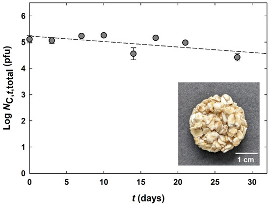
Figure 7.
Stability of bacteriophage K5 titer in a cereal cookie at 23 °C (). The limit of quantification was 2.3 × 10² pfu (related to one cookie with a mass of 2.505 g). A photograph of the cereal cookie is presented in the lower right corner of the picture.
). The limit of quantification was 2.3 × 10² pfu (related to one cookie with a mass of 2.505 g). A photograph of the cereal cookie is presented in the lower right corner of the picture.
The bacteriophage capsules that were incorporated into the cereal cookie were not visible after mixing and drying. After the drying process, the rather bulky structure of oat flakes was firmly held together by the glucose syrup and the surface of the oat flakes appeared slightly glossy. The texture of the cookie remained firm and dry throughout the storage period of 28 days, with a water activity of 0.59 ± 0.02.
The presented total titer of bacteriophage K5 (Figure 7) is referred to one cookie that contained 0.05 g encapsulated bacteriophage K5 in 1.5 g of oat flakes and 1.0 g of glucose syrup and was dissolved in 20 mL MBS for quantification. Over 28 days of storage at 23 °C, there is a slight tendency of a bacteriophage K5 titer reduction by less than 1 log unit. The fluctuations between the values result from the more difficult release of the bacteriophages from the capsules from the oat flakes/glucose matrix. To achieve more constant values, the incubation time in MBS could be increased. Losses are also conceivable due to potential attachment of bacteriophages to the oats during the centrifugation step. In addition, it should be noted that the determination was only twofold. However, what is quite clear is the difference of bacteriophage reduction between storing the pure capsules (Section 3.5) and storing the capsules in cereal cookies, both at 23 °C. After 30 days, the bacteriophage titer was reduced by more than 2 log units in the pure capsules versus less than 1 log unit in the cereal cookies.
When free bacteriophages are placed in a highly concentrated sugar solution, they normally suffer osmotic shock [100,101]. An opposite effect is seen for encapsulated bacteriophages. A strong and restricted water binding of alginate and micellar casein could have counteracted osmotic forces originating from the high glucose concentration in the surrounding syrup. In a study by Tang et al., extruded microcapsules composed of alginate, whey proteins and bacteriophage K were soaked in corn syrup before drying. The procedure was found to maintain bacteriophage titer in capsules during the subsequent drying step [90]. Preservation of bacteriophage capsid protein structure by hydrogen interaction with saccharides was proposed as an underlying mechanism [90,102].
To the best of our knowledge, no studies applying (encapsulated) bacteriophages in cereal products are available. However, encapsulated probiotics were studied for their application in cereal bars. Bampi et al., incorporated encapsulated Lactobacillus acidophilus and Bifidobacterium animalis ssp. lactis in cereal bars with storage viability at 4 °C for at least 90 days [103]. In addition, Bastos et al., introduced encapsulated Saccharomyces boulardii and Lactobacillus acidophilus in cereals bars; however, a reduction of cell viability of 3–4 log units was observed after 49 days [104].
Generally, consumer and market acceptance of functionalized cereal bars was evaluated to be good [104,105]. Additionally, oats are known to comfort human gastrointestinal distress [106,107,108]. Furthermore, the combination of oat sprinkles with a probiotic product, such as yoghurt, could increase product value. In particular, intestinal bacteria should be specifically eliminated by bacteriophages and, in parallel, the niche that has been created should be recolonized with beneficial bacterial species [109]. Therefore, a cereal bar combined with probiotics demonstrates a high potential to be used for bacteriophage capsule applications. This kind of food could be a new trend in nutrition, as also proposed by Połaska and Sokołowska [29].
4. Conclusions
Microencapsulation offers the possibility to use bacteriophages in a versatile way in food or medical applications while protecting the bacteriophages from external extreme conditions. The encapsulation process applied in this study was an in situ complexation of alginate, calcium ions, and milk proteins. With the use of the heat- and pH-sensitive Escherichia coli bacteriophage K5, we wanted to orient ourselves to the weakest representative. Therefore, this process should be transferable to a wide variety of (more resistant) bacteriophage species and mixtures (so called bacteriophage cocktails) should likewise be possible to encapsulate. Although the aim was to achieve the highest possible bacteriophage titers in the capsules and in the food matrices, it is highly dependent on the bacteriophage species and the dose necessitated for application. In order to investigate the functionality of phages in the future, in vivo human experiments will be carried out. In feasibility studies, the non-hazardous nature of the bacteriophage host model chosen in this study (Escherichia coli Nissle 1917 and bacteriophage K5) is highly advantageous. If promising results are shown, capsules with other bacteriophage species, e.g., against pathogens, could easily be produced and applied thereafter. In summary, the encapsulation procedure of this work enables the further step towards modulation of the human gut microbiota and increased health benefits.
Author Contributions
Conceptualization, methodology, data curation, writing—original draft preparation, writing—review and editing, visualization, project administration, C.S.; investigation, data curation, visualization, S.F. and K.D.; data curation, L.T.; writing—review and editing, supervision, Z.A. and J.H. All authors have read and agreed to the published version of the manuscript.
Funding
This research was partly funded by the Ministry of Science, Research and Arts Baden-Württemberg (Germany) in the frame of the State Graduate Scholarships.
Institutional Review Board Statement
Not applicable.
Informed Consent Statement
Not applicable.
Data Availability Statement
Not applicable.
Acknowledgments
The authors would like to thank Theresa Hock, Alina Reti, and Lea Rothenbach for performing part of the experiments. Peter Gschwind and Nora Ruprecht from the Department of Process Engineering and Powder Technology of the University of Hohenheim are acknowledged for the opportunity to use, and the support in dealing with, the Büchi Mini Spray Dryer and other devices. Finally, the authors express their gratitude to Anisa Heck for proofreading the manuscript from the perspective of a native speaker.
Conflicts of Interest
The authors declare no conflict of interest.
References
- Schubert, C.; Fischer, S.; Dorsch, K.; Teßmer, L.; Hinrichs, J.; Atamer, Z. Microencapsulation of Bacteriophages for the Delivery to and Modulation of the Human Gut Microbiota through Milk and Cereal Products. Appl. Sci. 2022, 12, 6299. [Google Scholar] [CrossRef]
- O’Hara, A.M.; Shanahan, F. The Gut Flora as a Forgotten Organ. EMBO Rep. 2006, 7, 688–693. [Google Scholar] [CrossRef] [PubMed]
- Sorboni, S.G.; Moghaddam, H.S.; Jafarzadeh-Esfehani, R.; Soleimanpour, S. A Comprehensive Review on the Role of the Gut Microbiome in Human Neurological Disorders. Clin. Microbiol. Rev. 2022, 35, e00338-20. [Google Scholar] [CrossRef] [PubMed]
- Eckburg, P.B.; Bik, E.M.; Bernstein, C.N.; Purdom, E.; Dethlefsen, L.; Sargent, M.; Gill, S.R.; Nelson, K.E.; Relman, D.A. Diversity of the Human Intestinal Microbial Flora. Science 2005, 308, 1635–1638. [Google Scholar] [CrossRef] [PubMed]
- Turnbaugh, P.J.; Gordon, J.I. The Core Gut Microbiome, Energy Balance and Obesity. J. Physiol. 2009, 587, 4153–4158. [Google Scholar] [CrossRef]
- Lee, J.-H.; Park, J.-H. Host—Microbial Interactions in Metabolic Diseases: From Diet to Immunity. J. Microbiol. 2022, 60, 561–575. [Google Scholar] [CrossRef]
- Rowland, I.; Gibson, G.; Heinken, A.; Scott, K.; Swann, J.; Thiele, I.; Tuohy, K. Gut Microbiota Functions: Metabolism of Nutrients and Other Food Components. Eur. J. Nutr. 2018, 57, 1–24. [Google Scholar] [CrossRef]
- Jernberg, C.; Löfmark, S.; Edlund, C.; Jansson, J.K. Long-Term Impacts of Antibiotic Exposure on the Human Intestinal Microbiota. Microbiology 2010, 156, 3216–3223. [Google Scholar] [CrossRef]
- Dethlefsen, L.; Huse, S.; Sogin, M.L.; Relman, D.A. The Pervasive Effects of an Antibiotic on the Human Gut Microbiota, as Revealed by Deep 16S RRNA Sequencing. PLoS Biol. 2008, 6, e280. [Google Scholar] [CrossRef]
- Schwartz, D.J.; Langdon, A.E.; Dantas, G. Understanding the Impact of Antibiotic Perturbation on the Human Microbiome. Genome Med. 2020, 12, 82. [Google Scholar] [CrossRef]
- Power, S.E.; O’Toole, P.W.; Stanton, C.; Ross, R.P.; Fitzgerald, G.F. Intestinal Microbiota, Diet and Health. Br. J. Nutr. 2014, 111, 387–402. [Google Scholar] [CrossRef] [PubMed]
- McCarville, J.L.; Caminero, A.; Verdu, E.F. Novel Perspectives on Therapeutic Modulation of the Gut Microbiota. Ther. Adv. Gastroenterol. 2016, 9, 580–593. [Google Scholar] [CrossRef] [PubMed]
- Nagpal, R.; Mainali, R.; Ahmadi, S.; Wang, S.; Singh, R.; Kavanagh, K.; Kitzman, D.W.; Kushugulova, A.; Marotta, F.; Yadav, H. Gut Microbiome and Aging: Physiological and Mechanistic Insights. Nutr. Heal. Aging 2018, 4, 267–285. [Google Scholar] [CrossRef] [PubMed]
- Rinninella, E.; Raoul, P.; Cintoni, M.; Franceschi, F.; Miggiano, G.A.D.; Gasbarrini, A.; Mele, M.C. What Is the Healthy Gut Microbiota Composition? A Changing Ecosystem across Age, Environment, Diet, and Diseases. Microorganisms 2019, 7, 14. [Google Scholar] [CrossRef] [PubMed]
- Zuppi, M.; Hendrickson, H.L.; O’Sullivan, J.M.; Vatanen, T. Phages in the Gut Ecosystem. Front. Cell. Infect. Microbiol. 2022, 11, 822562. [Google Scholar] [CrossRef]
- Kim, M.-S.; Park, E.-J.; Roh, S.W.; Bae, J.-W. Diversity and Abundance of Single-Stranded DNA Viruses in Human Feces. Appl. Environ. Microbiol. 2011, 77, 8062–8070. [Google Scholar] [CrossRef]
- Hoyles, L.; McCartney, A.L.; Neve, H.; Gibson, G.R.; Sanderson, J.D.; Heller, K.J.; van Sinderen, D. Characterization of Virus-like Particles Associated with the Human Faecal and Caecal Microbiota. Res. Microbiol. 2014, 165, 803–812. [Google Scholar] [CrossRef]
- Bikel, S.; López-Leal, G.; Cornejo-Granados, F.; Gallardo-Becerra, L.; García-López, R.; Sánchez, F.; Equihua-Medina, E.; Ochoa-Romo, J.P.; López-Contreras, B.E.; Canizales-Quinteros, S.; et al. Gut DsDNA Virome Shows Diversity and Richness Alterations Associated with Childhood Obesity and Metabolic Syndrome. iScience 2021, 24, 102900. [Google Scholar] [CrossRef]
- Myelnikov, D. An Alternative Cure: The Adoption and Survival of Bacteriophage Therapy in the USSR, 1922–1955. J. Hist. Med. Allied Sci. 2018, 73, 385–411. [Google Scholar] [CrossRef]
- Międzybrodzki, R.; Borysowski, J.; Weber-Dabrowska, B.; Fortuna, W.; Letkiewicz, S.; Szufnarowski, K.; Pawełczyk, Z.; Rogóz, P.; Kłak, M.; Wojtasik, E.; et al. Clinical Aspects of Phage Therapy. Adv. Virus Res. 2012, 83, 73–121. [Google Scholar] [CrossRef]
- Malik, D.J.; Sokolov, I.J.; Vinner, G.K.; Mancuso, F.; Cinquerrui, S.; Vladisavljevic, G.T.; Clokie, M.R.J.; Garton, N.J.; Stapley, A.G.F.; Kirpichnikova, A. Formulation, Stabilisation and Encapsulation of Bacteriophage for Phage Therapy. Adv. Colloid Interface Sci. 2017, 249, 100–133. [Google Scholar] [CrossRef] [PubMed]
- Melo, L.D.R.; Oliveira, H.; Pires, D.P.; Dabrowska, K.; Azeredo, J. Phage Therapy Efficacy: A Review of the Last 10 Years of Preclinical Studies. Crit. Rev. Microbiol. 2020, 46, 78–99. [Google Scholar] [CrossRef] [PubMed]
- García, P.; Martínez, B.; Obeso, J.M.; Rodríguez, A. Bacteriophages and Their Application in Food Safety. Lett. Appl. Microbiol. 2008, 47, 479–485. [Google Scholar] [CrossRef] [PubMed]
- Gill, J.J. Practical and Theoretical Considerations for the Use of Bacteriophages in Food Systems. In Bacteriophages in the Control of Food- and Waterborne Pathogens; Sabour, P.M., Griffiths, M.W., Eds.; ASM Press: Washington, DC, USA, 2010; ISBN 9781119737896. [Google Scholar]
- O’Sullivan, L.; Bolton, D.; McAuliffe, O.; Coffey, A. Bacteriophages in Food Applications: From Foe to Friend. Annu. Rev. Food Sci. Technol. 2019, 10, 151–172. [Google Scholar] [CrossRef] [PubMed]
- Amiri, S.; Moghanjougi, Z.M.; Bari, M.R.; Khaneghah, A.M. Natural Protective Agents and Their Applications as Bio-Preservatives in the Food Industry: An Overview of Current and Future Applications. Ital. J. Food Sci. 2021, 33, 55–68. [Google Scholar] [CrossRef]
- Lewis, R.; Hill, C. Overcoming Barriers to Phage Application in Food and Feed. Curr. Opin. Biotechnol. 2020, 61, 38–44. [Google Scholar] [CrossRef]
- Moye, Z.D.; Woolston, J.; Sulakvelidze, A. Bacteriophage Applications for Food Production and Processing. Viruses 2018, 10, 205. [Google Scholar] [CrossRef]
- Połaska, M.; Sokołowska, B. Bacteriophages—A New Hope or a Huge Problem in the Food Industry. AIMS Microbiol. 2019, 5, 324–346. [Google Scholar] [CrossRef]
- Fernández, L.; Gutiérrez, D.; García, P.; Rodríguez, A. The Perfect Bacteriophage for Therapeutic Applications—A Quick Guide. Antibiotics 2019, 8, 126. [Google Scholar] [CrossRef]
- Jończyk-Matysiak, E.; Łodej, N.; Kula, D.; Owczarek, B.; Orwat, F.; Międzybrodzki, R.; Neuberg, J.; Bagińska, N.; Weber-Dąbrowska, B.; Górski, A. Factors Determining Phage Stability/Activity: Challenges in Practical Phage Application. Expert Rev. Anti-Infect. Ther. 2019, 17, 583–606. [Google Scholar] [CrossRef]
- Ergin, F. Effect of Freeze Drying, Spray Drying and Electrospraying on the Morphological, Thermal, and Structural Properties of Powders Containing Phage Felix O1 and Activity of Phage Felix O1 during Storage. Powder Technol. 2022, 404, 117516. [Google Scholar] [CrossRef]
- Samtlebe, M.; Ergin, F.; Wagner, N.; Neve, H.; Küçükçetin, A.; Franz, C.M.A.P.; Heller, K.J.; Hinrichs, J.; Atamer, Z. Carrier Systems for Bacteriophages to Supplement Food Systems: Encapsulation and Controlled Release to Modulate the Human Gut Microbiota. LWT Food Sci. Technol. 2016, 68, 334–340. [Google Scholar] [CrossRef]
- Liu, H.; Cui, S.W.; Chen, M.; Li, Y.; Liang, R.; Xu, F.; Zhong, F. Protective Approaches and Mechanisms of Microencapsulation to the Survival of Probiotic Bacteria during Processing, Storage and Gastrointestinal Digestion: A Review. Crit. Rev. Food Sci. Nutr. 2019, 59, 2863–2878. [Google Scholar] [CrossRef] [PubMed]
- Niamah, A.K.; Al-Sahlany, S.T.G.; Ibrahim, S.A.; Verma, D.K.; Thakur, M.; Singh, S.; Patel, A.R.; Aguilar, C.N.; Utama, G.L. Electro-Hydrodynamic Processing for Encapsulation of Probiotics: A Review on Recent Trends, Technological Development, Challenges and Future Prospect. Food Biosci. 2021, 44, 101458. [Google Scholar] [CrossRef]
- Ergin, F.; Atamer, Z.; Göcer, E.M.C.; Demir, M.; Hinrichs, J.; Kucukcetin, A. Optimization of Salmonella Bacteriophage Microencapsulation in Alginate-Caseinate Formulation Using Vibrational Nozzle Technique. Food Hydrocoll. 2021, 113, 106456. [Google Scholar] [CrossRef]
- Liu, H.; Gong, J.; Chabot, D.; Miller, S.S.; Cui, S.W.; Zhong, F.; Wang, Q. Improved Survival of Lactobacillus Zeae LB1 in a Spray Dried Alginate-Protein Matrix. Food Hydrocoll. 2018, 78, 100–108. [Google Scholar] [CrossRef]
- Batalha, L.S.; Gontijo, M.T.G.; de Carvalho Teixeira, A.V.N.; Boggione, D.M.G.; Lopez, M.E.S.; Eller, M.R.; Mendonça, R.C.S. Encapsulation in Alginate-Polymers Improves Stability and Allows Controlled Release of the UFV-AREG1 Bacteriophage. Food Res. Int. 2021, 139, 109947. [Google Scholar] [CrossRef]
- Zuidam, N.J.; Nedović, V.A. Encapsulation Technologies for Active Food Ingredients and Food Processing; Springer: New York, NY, USA, 2009; ISBN 9781441910073. [Google Scholar]
- Jeyakumari, A.; Zynudheen, A.A.; Parvathy, U. Microencapsulation of Bioactive Food Ingredients and Controlled Release—A Review. MOJ Food Process. Technol. 2016, 2, 214–224. [Google Scholar] [CrossRef]
- Parracho, H.M.R.T.; Burrowes, B.H.; Enright, M.C.; McConville, M.; Harper, D.R. The Role of Regulated Clinical Trials in the Development of Bacteriophage Therapeutics. J. Mol. Genet. Med. 2012, 6, 279–286. [Google Scholar] [CrossRef]
- Górski, A.; Międzybrodzki, R.; Łobocka, M.; Głowacka-Rutkowska, A.; Bednarek, A.; Borysowski, J.; Jończyk-Matysiak, E.; Łusiak-Szelachowska, M.; Weber-Dabrowska, B.; Bagińska, N.; et al. Phage Therapy: What Have We Learned? Viruses 2018, 10, 288. [Google Scholar] [CrossRef]
- Michel, C.; Samtlebe, M.; Wagner, N.; Neve, H.; Franz, C.M.A.P.; Hinrichs, J.; Atamer, Z. Orthogonal Processing Strategies to Create “Phage-Free” Whey–Membrane Filtration Followed by Thermal or Ultraviolet C Treatment for the Reduction of Lactococcus Lactis Bacteriophages. Int. Dairy J. 2021, 122, 105149. [Google Scholar] [CrossRef]
- Adams, M. Bacteriophages; Interscience Publishers: New York, NY, USA, 1959. [Google Scholar]
- Atamer, Z.; Dietrich, J.; Müller-Merbach, M.; Neve, H.; Heller, K.J.; Hinrichs, J. Screening for and Characterization of Lactococcus Lactis Bacteriophages with High Thermal Resistance. Int. Dairy J. 2009, 19, 228–235. [Google Scholar] [CrossRef]
- Müller-Merbach, M.; Rauscher, T.; Hinrichs, J. Inactivation of Bacteriophages by Thermal and High-Pressure Treatment. Int. Dairy J. 2005, 15, 777–784. [Google Scholar] [CrossRef]
- Kessler, H.G. Food and Bio Process Engineering—Dairy Techology, 5th ed.; Verlag A. Kessler: Munich, Germany, 2002; ISBN 3980237850. [Google Scholar]
- Jeoh Zicari, T.; Scher, H.B.; Santa-Maria, M.C.; Strobel, S. Spray Dry Method for Encapsulation of Biological Moieties and Chemicals in Polymers Cross-Linked by Multivalent Ions for Controlled Release Applications. U.S. Patent 9,700,519 B2, 11 July 2017. [Google Scholar]
- Strobel, S.A.; Allen, K.; Roberts, C.; Jimenez, D.; Scher, H.B.; Jeoh, T. Industrially-Scalable Microencapsulation of Plant Beneficial Bacteria in Dry Cross-Linked Alginate Matrix. Ind. Biotechnol. 2018, 14, 138–147. [Google Scholar] [CrossRef]
- Krischer, O.; Kast, W. Trocknungstechnik: Die Wissenschaftlichen Grundlagen der Trocknungstechnik, 3rd ed.; Springer: Berlin/Heidelberg, Germany, 1978; ISBN 9783642618796. [Google Scholar]
- Scott, T.A. Refractive Index of Ethanol-Water Mixtures and Densitiy and Refractive Index of Ethanol-Water-Ethyl Ether Mixtures. J. Phys. Chem. 1946, 50, 406–412. [Google Scholar] [CrossRef]
- Ma, Y.; Pacan, J.C.; Wang, Q.; Xu, Y.; Huang, X.; Korenevsky, A.; Sabour, P.M. Microencapsulation of Bacteriophage Felix O1 into Chitosan-Alginate Microspheres for Oral Delivery. Appl. Environ. Microbiol. 2008, 74, 4799–4805. [Google Scholar] [CrossRef]
- Soykut, E.A.; Tayyarcan, E.K.; Evran, Ş.; Boyacı, İ.H.; Çakır, İ.; Khaaladi, M.; Fattouch, S. Microencapsulation of Phages to Analyze Their Demeanor in Physiological Conditions. Folia Microbiol. 2019, 64, 751–763. [Google Scholar] [CrossRef]
- Troge, A.; Scheppach, W.; Schroeder, B.O.; Rund, S.A.; Heuner, K.; Wehkamp, J.; Stange, E.F.; Oelschlaeger, T.A. More than a Marine Propeller—The Flagellum of the Probiotic Escherichia coli Strain Nissle 1917 Is the Major Adhesin Mediating Binding to Human Mucus. Int. J. Med. Microbiol. 2012, 302, 304–314. [Google Scholar] [CrossRef]
- Gupta, D.S.; Jann, B.; Schmidt, G.; Golecki, J.R.; Ørskov, I.; Ørskov, F.; Jann, K. Coliphage K5, Specific for E. Coli Exhibiting the Capsular K5 Antigen. FEMS Microbiol. Lett. 1982, 14, 75–78. [Google Scholar] [CrossRef]
- Soundararajan, M.; von Bünau, R.; Oelschlaeger, T.A. K5 Capsule and Lipopolysaccharide Are Important in Resistance to T4 Phage Attack in Probiotic E. Coli Strain Nissle 1917. Front. Microbiol. 2019, 10, 2783. [Google Scholar] [CrossRef]
- Scholl, D.; Kieleczawa, J.; Kemp, P.; Rush, J.; Richardson, C.C.; Merril, C.; Adhya, S.; Molineux, I.J. Genomic Analysis of Bacteriophages SP6 and K1-5, an Estranged Subgroup of the T7 Supergroup. J. Mol. Biol. 2004, 335, 1151–1171. [Google Scholar] [CrossRef]
- Molineux, I.J. T7 like Phages (Podoviridae). In Encyclopedia of Virology; Granoff, A., Webster, R.G., Eds.; Academic Press: San Diego, CA, USA; London, UK, 1999; ISBN 9780122270307. [Google Scholar]
- Sambrook, J.; Russel, D.W. Molecular Cloning: A Laboratory Manual; Cold Spring Harbor Laboratory Press: Cold Spring Harbor, NY, USA, 2001; ISBN 9780879695774. [Google Scholar]
- Vörös, Z.; Csík, G.; Herényi, L.; Kellermayer, M. Temperature-Dependent Nanomechanics and Topography of Bacteriophage T7. J. Virol. 2018, 92, e01236-18. [Google Scholar] [CrossRef]
- Pollard, E.C.; Solosko, W. The Thermal Inactivation of T4 and λ Bacteriophage. Biophys. J. 1971, 11, 66–74. [Google Scholar] [CrossRef]
- Pitaksutheepong, C.; Abhisingha, M.; Dumnin, J.; Visessanguan, W. Isolation, Detection and Inactivation of a Myoviridae Bacteriophage Infecting Bacillus amyloliquefaciens FB11. Ann. Microbiol. 2015, 65, 1841–1846. [Google Scholar] [CrossRef]
- Garneau, J.E.; Moineau, S. Bacteriophages of Lactic Acid Bacteria and Their Impact on Milk Fermentations. Microb. Cell Fact. 2011, 10, S20. [Google Scholar] [CrossRef]
- Atamer, Z. An Extraordinary Dairy Phage and Its Properties: Occurrence, Growth, Inactivation, Survival. Int. Dairy J. 2022, 129, 105336. [Google Scholar] [CrossRef]
- Evans, D.F.; Pye, G.; Bramley, R.; Clark, A.G.; Dyson, T.J.; Hardcastle, J.D. Measurement of Gastrointestinal PH Profiles in Normal Ambulant Human Subjects. Gut 1988, 29, 1035–1041. [Google Scholar] [CrossRef]
- Cook, M.T.; Tzortzis, G.; Charalampopoulos, D.; Khutoryanskiy, V.V. Microencapsulation of Probiotics for Gastrointestinal Delivery. J. Control. Release 2012, 162, 56–67. [Google Scholar] [CrossRef]
- Dong, Q.Y.; Chen, M.Y.; Xin, Y.; Qin, X.Y.; Cheng, Z.; Shi, L.E.; Tang, Z.X. Alginate-Based and Protein-Based Materials for Probiotics Encapsulation: A Review. Int. J. Food Sci. Technol. 2013, 48, 1339–1351. [Google Scholar] [CrossRef]
- Dussault, P.; Bourgault, J.M.; Verly, W.G. T7 Coliphage Inactivation by Nitrous Acid. Biochim. Biophys. Acta 1970, 213, 312–319. [Google Scholar] [CrossRef]
- Feng, Y.Y.; Ong, S.L.; Hu, J.Y.; Tan, X.L.; Ng, W.J. Effects of PH and Temperature on the Survival of Coliphages MS2 and Qβ. J. Ind. Microbiol. Biotechnol. 2003, 30, 549–552. [Google Scholar] [CrossRef] [PubMed]
- Capra, M.L.; Quiberoni, A.; Reinheimer, J. Phages of Lactobacillus Casei/Paracasei: Response to Environmental Factors and Interaction with Collection and Commercial Strains. J. Appl. Microbiol. 2006, 100, 334–342. [Google Scholar] [CrossRef] [PubMed]
- Campagna, C.; Villion, M.; Labrie, S.J.; Duchaine, C.; Moineau, S. Inactivation of Dairy Bacteriophages by Commercial Sanitizers and Disinfectants. Int. J. Food Microbiol. 2014, 171, 41–47. [Google Scholar] [CrossRef]
- Salo, R.J.; Cliver, D.O. Effect of Acid PH, Salts, and Temperature on the Infectivity and Physical Integrity of Enteroviruses. Arch. Virol. 1976, 52, 269–282. [Google Scholar] [CrossRef]
- Gharsallaoui, A.; Roudaut, G.; Chambin, O.; Voilley, A.; Saurel, R. Applications of Spray-Drying in Microencapsulation of Food Ingredients: An Overview. Food Res. Int. 2007, 40, 1107–1121. [Google Scholar] [CrossRef]
- Malik, D.J. Bacteriophage Encapsulation Using Spray Drying for Phage Therapy. Curr. Issues Mol. Biol. 2021, 40, 303–316. [Google Scholar] [CrossRef]
- Cal, K.; Sollohub, K. Spray Drying Technique. I: Hardware and Process Parameters. J. Pharm. Sci. 2010, 99, 575–586. [Google Scholar] [CrossRef]
- Vinner, G.K.; Rezaie-Yazdi, Z.; Leppanen, M.; Stapley, A.G.F.; Leaper, M.C.; Malik, D.J. Microencapsulation of Salmonella-Specific Bacteriophage Felix O1 Using Spray-Drying in a PH-Responsive Formulation and Direct Compression Tableting of Powders into a Solid Oral Dosage Form. Pharmaceuticals 2019, 12, 43. [Google Scholar] [CrossRef]
- Hussein, N.; Omer, H.; Ismael, A.; Alhnan, M.A.; Elhissi, A.; Ahmed, W. Spray-Dried Alginate Microparticles for Potential Intranasal Delivery of Ropinirole Hydrochloride: Development, Characterization and Histopathological Evaluation. Pharm. Dev. Technol. 2020, 25, 290–299. [Google Scholar] [CrossRef]
- Hansen, L.T.; Allan-Wojtas, P.M.; Jin, Y.L.; Paulson, A.T. Survival of Ca-Alginate Microencapsulated Bifidobacterium Spp. in Milk and Simulated Gastrointestinal Conditions. Food Microbiol. 2002, 19, 35–45. [Google Scholar] [CrossRef]
- Matinkhoo, S.; Lynch, K.H.; Dennis, J.J.; Finlay, W.H.; Vehring, R. Spray-Dried Respirable Powders Containing Bacteriophages for the Treatment of Pulmonary Infections. J. Pharm. Sci. 2011, 100, 5197–5205. [Google Scholar] [CrossRef] [PubMed]
- Leung, S.S.Y.; Parumasivam, T.; Gao, F.G.; Carrigy, N.B.; Vehring, R.; Finlay, W.H.; Morales, S.; Britton, W.J.; Kutter, E.; Chan, H.-K. Production of Inhalation Phage Powders Using Spray Freeze Drying and Spray Drying Techniques for Treatment of Respiratory Infections. Pharm. Res. 2016, 33, 1486–1496. [Google Scholar] [CrossRef] [PubMed]
- Young, S.L.; Sarda, X.; Rosenberg, M. Microencapsulating Properties of Whey Proteins. 1. Microencapsulation of Anhydrous Milk Fat. J. Dairy Sci. 1993, 76, 2868–2877. [Google Scholar] [CrossRef]
- Rosenberg, M.; Sheu, T.-Y. Microencapsulation of Volatiles by Spray-Drying in Whey Protein-Based Wall Systems. Int. Dairy J. 1996, 6, 273–284. [Google Scholar] [CrossRef]
- Teijeiro, M.; Pérez, P.F.; De Antoni, G.L.; Golowczyc, M.A. Suitability of Kefir Powder Production Using Spray Drying. Food Res. Int. 2018, 112, 169–174. [Google Scholar] [CrossRef]
- Gbassi, G.K.; Vandamme, T.; Ennahar, S.; Marchioni, E. Microencapsulation of Lactobacillus plantarum Spp. in an Alginate Matrix Coated with Whey Proteins. Int. J. Food Microbiol. 2009, 129, 103–105. [Google Scholar] [CrossRef]
- Zhang, Y.; Zhang, H.; Ghosh, D. The Stabilizing Excipients in Dry State Therapeutic Phage Formulations. AAPS PharmSciTech 2020, 21, 133. [Google Scholar] [CrossRef] [PubMed]
- Millqvist-Fureby, A.; Elofsson, U.; Bergenståhl, B. Surface Composition of Spray-Dried Milk Protein-Stabilised Emulsions in Relation to Pre-Heat Treatment of Proteins. Colloids Surfaces B Biointerfaces 2001, 21, 47–58. [Google Scholar] [CrossRef]
- Xavier, A.A.O.; Mariutti, L.R.B. Static and Semi-Dynamic in Vitro Digestion Methods: State of the Art and Recent Achievements towards Standardization. Curr. Opin. Food Sci. 2021, 41, 260–273. [Google Scholar] [CrossRef]
- Tang, Z.; Huang, X.; Baxi, S.; Chambers, J.R.; Sabour, P.M.; Wang, Q. Whey Protein Improves Survival and Release Characteristics of Bacteriophage Felix O1 Encapsulated in Alginate Microspheres. Food Res. Int. 2013, 52, 460–466. [Google Scholar] [CrossRef]
- Shi, L.-E.; Li, Z.-H.; Li, D.-T.; Xu, M.; Chen, H.-Y.; Zhang, Z.L.; Tang, Z.-X. Encapsulation of Probiotic Lactobacillus Bulgaricus in Alginate-Milk Microspheres and Evaluation of the Survival in Simulated Gastrointestinal Conditions. J. Food Eng. 2013, 117, 99–104. [Google Scholar] [CrossRef]
- Tang, Z.; Huang, X.; Sabour, P.M.; Chambers, J.R.; Wang, Q. Preparation and Characterization of Dry Powder Bacteriophage K for Intestinal Delivery through Oral Administration. LWT Food Sci. Technol. 2015, 60, 263–270. [Google Scholar] [CrossRef]
- Salaün, F.; Mietton, B.; Gaucheron, F. Buffering Capacity of Dairy Products. Int. Dairy J. 2005, 15, 95–109. [Google Scholar] [CrossRef]
- Gunasekaran, S.; Ko, S.; Xiao, L. Use of Whey Proteins for Encapsulation and Controlled Delivery Applications. J. Food Eng. 2007, 83, 31–40. [Google Scholar] [CrossRef]
- Colom, J.; Cano-Sarabia, M.; Otero, J.; Aríñez-Soriano, J.; Cortés, P.; Maspoch, D.; Llagostera, M. Microencapsulation with Alginate/CaCO3: A Strategy for Improved Phage Therapy. Sci. Rep. 2017, 7, 41441. [Google Scholar] [CrossRef] [PubMed]
- Agüero, L.; Zaldivar-Silva, D.; Peña, L.; Dias, M. Alginate Microparticles as Oral Colon Drug Delivery Device: A Review. Carbohydr. Polym. 2017, 168, 32–43. [Google Scholar] [CrossRef]
- LeClair, D.A.; Cranston, E.D.; Lichty, B.D.; Xing, Z.; Thompson, M.R. Consecutive Spray Drying to Produce Coated Dry Powder Vaccines Suitable for Oral Administration. ACS Biomater. Sci. Eng. 2018, 4, 1669–1678. [Google Scholar] [CrossRef]
- Leung, S.S.Y.; Parumasivam, T.; Nguyen, A.; Gengenbach, T.; Carter, E.A.; Carrigy, N.B.; Wang, H.; Vehring, R.; Finlay, W.H.; Morales, S.; et al. Effect of Storage Temperature on the Stability of Spray Dried Bacteriophage Powders. Eur. J. Pharm. Biopharm. 2018, 127, 213–222. [Google Scholar] [CrossRef]
- Keogh, B.P.; Pettingill, G. Long-Term Storage of Bacteriophages of Lactic Streptococci. Appl. Microbiol. 1966, 14, 421–424. [Google Scholar] [CrossRef]
- Vandenheuvel, D.; Singh, A.; Vandersteegen, K.; Klumpp, J.; Lavigne, R.; Van Den Mooter, G. Feasibility of Spray Drying Bacteriophages into Respirable Powders to Combat Pulmonary Bacterial Infections. Eur. J. Pharm. Biopharm. 2013, 84, 578–582. [Google Scholar] [CrossRef]
- Mahmood, A.; Usman, S. A Comparative Study on the Physicochemical Parameters of Milk Samples Collected from Buffalo, Cow, Goat and Sheep of Gujrat, Pakistan. Pakistan J. Nutr. 2010, 9, 1192–1197. [Google Scholar] [CrossRef]
- Whitman, P.A.; Marshall, R.T. Characterization of Two Psychrophilic Pseudomonas Bacteriophages Isolated from Ground Beef. Appl. Microbiol. 1971, 22, 463–468. [Google Scholar] [CrossRef] [PubMed]
- Duyvejonck, H.; Merabishvili, M.; Vaneechoutte, M.; de Soir, S.; Wright, R.; Friman, V.P.; Verbeken, G.; De Vos, D.; Pirnay, J.P.; Van Mechelen, E.; et al. Evaluation of the Stability of Bacteriophages in Different Solutions Suitable for the Production of Magistral Preparations in Belgium. Viruses 2021, 13, 865. [Google Scholar] [CrossRef] [PubMed]
- Miao, S.; Mills, S.; Stanton, C.; Fitzgerald, G.F.; Roos, Y.; Ross, R.P. Effect of Disaccharides on Survival during Storage of Freeze Dried Probiotics. Dairy Sci. Technol. 2008, 88, 19–30. [Google Scholar] [CrossRef]
- Bampi, G.B.; Backes, G.T.; Cansian, R.L.; de Matos, F.E.; Ansolin, I.M.A.; Poleto, B.C.; Corezzolla, L.R.; Favaro-Trindade, C.S. Spray Chilling Microencapsulation of Lactobacillus acidophilus and Bifidobacterium animalis Subsp. Lactis and Its Use in the Preparation of Savory Probiotic Cereal Bars. Food Bioprocess Technol. 2016, 9, 1422–1428. [Google Scholar] [CrossRef]
- Bastos, G.A.; Paulo, E.M.; Chiaradia, A.C.N. Aceitabilidade de Barra de Cereais Potencialmente Probiótica (Acceptability of Potentially Probiotic Cereal Bars). Brazilian J. Food Technol. 2014, 17, 113–120. [Google Scholar] [CrossRef]
- Da Silva, D.C.M.N.; Paulino, V.P.D.A.; de Nascimento, C.P.; Pereira, M.D.G. Desenvolvimento de Barra de Cereais Salgada Enriquecida Com Chia e Fos (Development of a Salty Cereal Bar Enriched Wich Chia and FOS). Rev. Científica Ciências Apl. FAIP 2016, 3, 80–97. [Google Scholar]
- Connolly, M.L.; Lovegrove, J.A.; Tuohy, K.M. In Vitro Evaluation of the Microbiota Modulation Abilities of Different Sized Whole Oat Grain Flakes. Anaerobe 2010, 16, 483–488. [Google Scholar] [CrossRef]
- Wilczak, J.; Błaszczyk, K.; Kamola, D.; Gajewska, M.; Harasym, J.P.; Jałosińska, M.; Gudej, S.; Suchecka, D.; Oczkowski, M.; Gromadzka-Ostrowska, J. The Effect of Low or High Molecular Weight Oat Beta-Glucans on the Inflammatory and Oxidative Stress Status in the Colon of Rats with LPS-Induced Enteritis. Food Funct. 2015, 6, 590–603. [Google Scholar] [CrossRef]
- Nie, Y.; Lin, Q.; Luo, F. Effects of Non-Starch Polysaccharides on Inflammatory Bowel Disease. Int. J. Mol. Sci. 2017, 18, 1372. [Google Scholar] [CrossRef]
- Mills, S.; Shanahan, F.; Stanton, C.; Hill, C.; Coffey, A.; Paul Ross, R. Movers and Shakers: Influence of Bacteriophages in Shaping the Mammalian Gut Microbiota. Gut Microbes 2013, 4, 4–16. [Google Scholar] [CrossRef] [PubMed]


Publisher’s Note: MDPI stays neutral with regard to jurisdictional claims in published maps and institutional affiliations. |
© 2022 by the authors. Licensee MDPI, Basel, Switzerland. This article is an open access article distributed under the terms and conditions of the Creative Commons Attribution (CC BY) license (https://creativecommons.org/licenses/by/4.0/).