Article

Impact of Steam Autoclaving on the Mechanical Properties of 3D-Printed Resins Used for Insertion Guides in Orthodontics and Implant Dentistry

1
Department of Orthodontics, University of Dusseldorf, 40225 Dusseldorf, Germany
2
Department of Oral Surgery, University of Dusseldorf, 40225 Dusseldorf, Germany
3
Department of Neurosciences, University of Padua, 35128 Padua, Italy
4
Oral Technology, University of Bonn, 53111 Bonn, Germany
*
Author to whom correspondence should be addressed.
Appl. Sci. 2022, 12(12), 6195; https://doi.org/10.3390/app12126195
Submission received: 30 May 2022 / Revised: 14 June 2022 / Accepted: 15 June 2022 / Published: 17 June 2022
(This article belongs to the Special Issue 3D Printed Materials Dentistry)

Abstract

:
Guided implant placement has been shown to be more accurate than free-handed insertion. Still, implant position deviations occur and could possibly pose risks. Thus, there is a quest to identify factors that might impair the accuracy of implantation protocols using templates. This study aimed to investigate the influence of autoclaving cycles (cycle 1: 121 °C, 1 bar, 20.5 min; cycle 2: 134 °C, 2 bar, 5.5 min) on the Vickers hardness and flexural modulus of five different materials used for 3D-printed insertion guides. The specimens were subjected to Vickers hardness tests, showing significant changes in the Vickers hardness for two and three materials out of five for cycle 1 and 2, respectively. The results of the three-point bending tests (n = 15 specimens per material) showed decreasing flexural moduli after autoclaving. However, changes were significant only for one material, which presented a significant decrease in the flexural modulus after cycle 2. No significant changes were detected after cycle 1. In conclusion, our findings show that autoclaving can alter the mechanical properties of the templates to some extent, especially with cycle 2. Whether these modifications are associated with dimensional changes of the templates and reduced accuracy of the implantation protocols remains to be investigated.

1. Introduction

In recent years, computer-guided surgery in combination with digital backward-planning gained popularity among clinicians in the field of implant dentistry and also in orthodontics for mini-implant placement. Especially in challenging situations, accurate transfer of the virtually planned position of the implants may increase the safety of the intervention, and improve patient comfort as a consequence of reduced operation time and invasiveness [1]. Guided implant surgery using surgical templates was found to be more accurate compared to free-handed implant placement, exhibiting significantly lower angular, coronal, and apical deviations between the intended and the actual implant positions [2,3].
Despite these benefits, a mean coronal deviation of 1.3 mm (95% CI: 1.09 mm; 1.56 mm) with values up to 2.2 mm and a mean apical deviation of 1.5 mm (95% CI: 1.29 mm; 1.62 mm) with values up to 2.5 mm have been reported for computer-guided implant insertion [4]. These distances should be incorporated into the virtual planning to reduce the risk of permanent damage to adjacent anatomical structures. Nevertheless, this risk cannot be fully eliminated and a safe distance between the implants and the anatomical structures should be considered in the planning phase [5]. These circumstances stress the importance of investigating factors that could provoke the loss of accuracy, so that the risk–benefit ratio of computer-guided implantation protocols can be optimized in the future. As stated in the EU Medical Devices Regulation (MDR), it is crucial to minimize all the “known and foreseeable risks and any undesirable side-effects” and weigh them against possible benefits of the chosen protocol [6].
Formerly identified factors influencing the accuracy of guided implantation include the support of the surgical template, favoring tooth- and mucosa-supported surgical templates over bone-supported ones [7]. In the case of mucosa-supported surgical templates, the mucosal thickness at the insertion site was found to affect implant placement accuracy, whereby increased tissue thickness seems to lower the accuracy and may require flap preparation for more accurate results [8,9]. Furthermore, the quality of radiographic image data and the usage of intraoral scanning devices might have an impact on the accuracy of guided implantation protocols [10,11,12,13]. In addition, the fit and the length of the metallic drilling sleeves embedded within the surgical template and the drilling distance could determine the extent of implant position deviations [14,15].
Surgical guides have been widely employed in implant dentistry. Recently, mini-implant insertion templates were introduced in orthodontics to increase the safety and accuracy of the procedure [16]. Few studies confirmed an increased accuracy following guided placement of orthodontic implants [17]. In the anterior palate, insertion templates not only favor ideal mini-implant positioning in accordance with the variable bone height available [18], but also facilitate simultaneous placement of skeletally anchored orthodontic appliances in a digital workflow [19].
Nowadays, guides are usually produced in resin-based materials using additive manufacturing technologies [20]. Recent studies have suggested that the influence of different 3D-printers on the accuracy of the protocol is negligible [21,22], whereas dimensional changes caused by prolonged storage have been reported [23]. Another critical aspect that might alter the accuracy of implant insertion and that has not gained much attention so far is the impact of steam autoclaving. The sterilization of the templates is fundamental, as they can come temporarily in contact with blood. According to international hygiene guidelines, including the Center for Disease Control and Prevention (CDC, USA) and the EU Regulations, they are critical medical devices and have to be sterilized before usage [6,24]. Few recent studies could not find significant changes in template dimension after autoclaving [25,26,27], whereas the impact on biomechanical properties remains unclear. Alteration of the biomechanical properties, such as flexural properties and hardness of the template materials, might lead to inaccuracies in implant insertion. Thus, there is a quest for studies assessing the influence of steam autoclaving parameters, such as temperature, pressure, and duration of autoclaving. Furthermore, the effect of autoclaving on different resin-based materials manufactured with different printing methods, such as stereolithography (SLA), liquid crystal display stereolithography (LCD-SLA), and digital light processing (DLP), remains to be clarified.
Therefore, the aim of this study was to evaluate the influence of steam autoclaving parameters on the Vickers hardness and flexural modulus of 3D-printed resin-based templates, manufactured using different resin materials and printing methods. The null hypotheses were that being subjected to autoclaving did not significantly change the specimen Vickers hardness HV 0.5 and the flexural modulus.

2. Materials and Methods

2.1. 3D-Printed Specimens Preparation

As the test standard DIN EN ISO 178 requires the usage of test pieces with a length (l) height (h) ratio of l/h = 20 for 3-point bending tests on polymer materials, a virtual model of the specimens with the dimensions 2 mm × 25 mm × 40 mm was designed using the software 3D-Builder (Microsoft, Redmond, WA, USA). In total, 75 specimens (i.e., 15 for each of the 5 materials) were printed for 3-point bending tests and Vickers hardness tests. With regard to test groups, two different digital light processing (DLP) printers (group 1: NextDent 5100, Vertex-Dental B.V., Soesteberg, The Netherlands; group 2: ASIGA MAX, Pluradent GmbH & Co. KG, Offenbach, Germany), one desktop stereolithography (SLA) printer (group 3: Form 3, Formlabs Inc., Sommerville, MA, USA), and one liquid crystal display stereolithography (LCD-SLA) printer (group 4: Slash Plus, UniZ Technology LLC., San Diego, CA, USA) were used. The samples were produced using four different 3D printing resins, one for each printing machine (group 1: NextDent SG, Vertex-Dental B.V.; group 2: Optiprint Guide, dentona AG, Dortmund, Germany; group 3: Dental SG, Formlabs Inc.; group 4: zSG Amber, UniZ Technology LLC.). All of the resins mentioned before are authorized by the manufacturer for steam autoclaving and printing with the particular printer utilized. The resin used in group 0 (E-Guide, Envisiontec Inc., Dearborn, MI, USA) is not authorized by the manufacturer for steam autoclaving, but only for immersion disinfection. It was printed with the DLP printer authorized by the manufacturer (Micro Plus XL, Envisiontec Inc.).
All specimens were printed such that the printing layers were parallel to the longer edge and perpendicular to the support structures and to the shorter edges, as recommended by Quintana et al. [28].
Five specimens per group (total n = 25) were not subjected to autoclaving and were used as controls. Ten specimens per group were sterilized by two different vacuum steam autoclaving programs (Vacuklav 44-B, MELAG oHG, Berlin, Germany): 5 specimens at 121 °C, 1 bar, and 20.5 min (cycle 1), while the remaining 5 were sterilized at 134 °C, 2 bar, and 5.5 min (cycle 2). A summary is provided in Table 1.

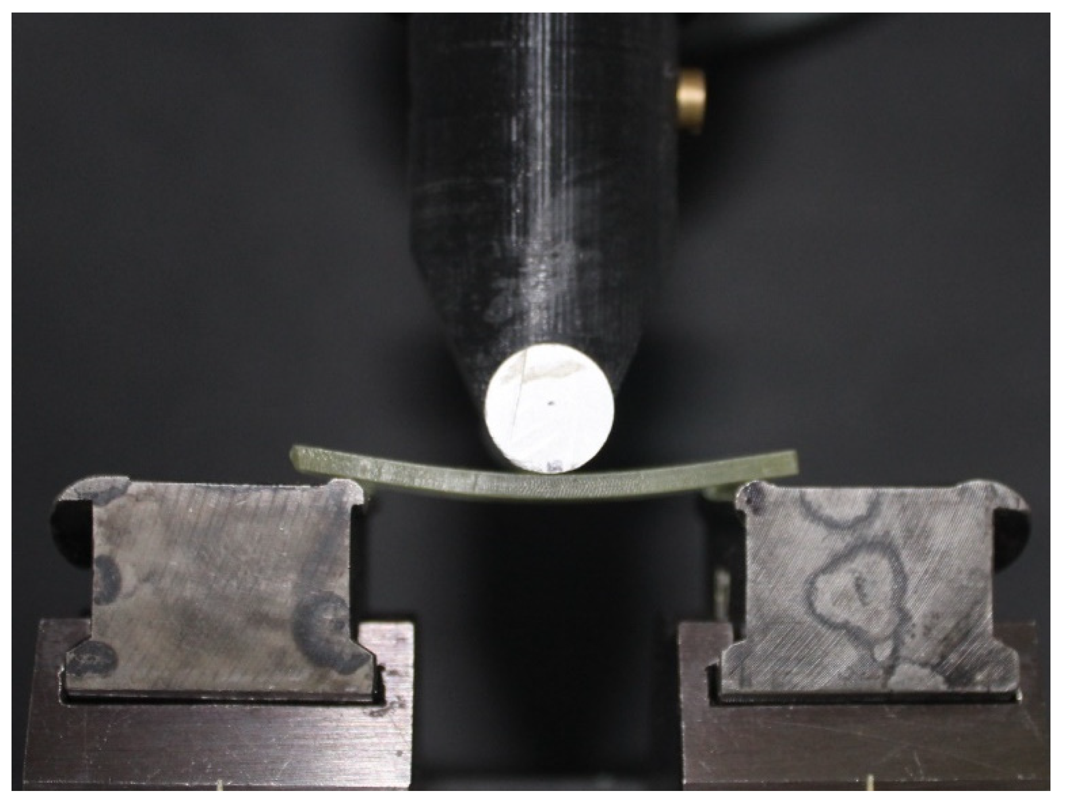
2.2. Mechanical Tests

The Vickers hardness test was performed on three specimens of each subgroup using the hardness testing machine ZHV20/Z2.5 (Zwick-Roell GmbH & Co. KG, Ulm, Germany) and repeated 5 times. Tests were run according to the ISO/TS 19278 norm. A 136° pyramidal indenter was pressed into the material with a force (F) of 4.903 N for 10 s. Images of the resulting impression were acquired using the optical microscope with a magnification of 20:1, that the hardness testing machine ZHV20/Z2.5 is equipped with. The diagonals d1 and d2 of the impression were measured manually using the software testXpert (Zwick-Roell GmbH & Co. KG) (Figure 1). Then, the average diagonal d and Vickers hardness HV0.5 were calculated using the following equations.
d = (d1 + d2)/2
HV0.5 = 0.1891 × F/d2
To evaluate autoclaving-induced changes in terms of flexural properties of the tested materials, a 3-point bending test was performed following the test standard DIN EN ISO 178 on all specimens (n = 75). The tests were performed using the material testing machine ZMART.PRO (Zwick-Roell GmbH & Co. KG) with a testing stamp radius of 5 mm that bends the specimens with a speed of 2 mm/s. The specimens were positioned such that the acquired ratio of the test piece length (l) to the distance between the support points (d) was l/d = 16 (Figure 2). The software testXpert (Zwick-Roell GmbH & Co. KG) was used to detect the flexural moduli.

2.3. Statistical Analysis

The statistical analysis was performed using Excel 2016 (Microsoft). A convenience sample size was determined, based on similar publications in this field [29,30]. For Vickers hardness, repeated measurements on the same specimen were pooled. For each resin/printer combination and autoclaving protocol, the mean ± standard deviation (M ± SD) Vickers hardness and flexural modulus were calculated. The Shapiro–Wilk test was used to assess whether measured data were normally distributed. The normal distribution of residues was validated through Q-Q plots. Homoscedasticity was verified by conducting a Levene test.
The ANOVA and post hoc t-test with Bonferroni correction were used to assess differences among autoclaving protocols for each printer/resin combination. If the assumptions for ANOVA were not met, a Kruskal–Wallis test and a post hoc Bonferroni-corrected Mann–Whitney-U test were utilized instead.
As the measurements of d1 and d2 in Vickers hardness testing were performed manually, the reliability of the test method was assessed using the interclass correlation coefficient (ICC), which was calculated based on the 5 repeated measurements.
Results were found significant at p < 0.05.

3. Results

3.1. Vickers Hardness

The reliability of the Vickers hardness test ranged from moderate (ICC = 0.58) to excellent (ICC = 0.99). The mean Vickers hardness (in HV0.5) and standard deviations ranged from 13.50 ± 2.62 to 29.16 ± 6.83 for the untreated subgroups. After the specimens were autoclaved with cycle 1, the Vickers hardness ranged from 6.94 ± 2.21 to 27.67 ± 5.42. After being subjected to cycle 2, the Vickers hardness ranged from 10.52 ± 1.05 to 25.30 ± 1.65.
The results of the Vickers hardness test are given in Table 2, and corresponding boxplots are provided in Figure 3. In three out of five resin/printer groups, the Kruskal–Wallis test pointed at qualitative differences (group 0, group 2, and group 3). In these groups, a post hoc test was conducted. In group 0, the Vickers hardness of the specimens autoclaved with cycle 1 significantly decreased and almost halved compared to the untreated control (p < 0.001), whereas cycle 2 yielded significantly higher Vickers hardness values (p < 0.001. In group 2, the Vickers hardness of cycle 1 increased slightly (p = 0.359) compared to the untreated control, whereas the Vickers hardness of the cycle 2 subgroup was significantly lower compared to the untreated control and cycle 1 groups (p < 0.001, respectively). In group 3, a higher Vickers hardness was found for both autoclaving protocols (untreated vs. cycle 1: p = 0.010; untreated vs. cycle 2: p < 0.001), whereas no significant differences were found between the two autoclaving cycles.

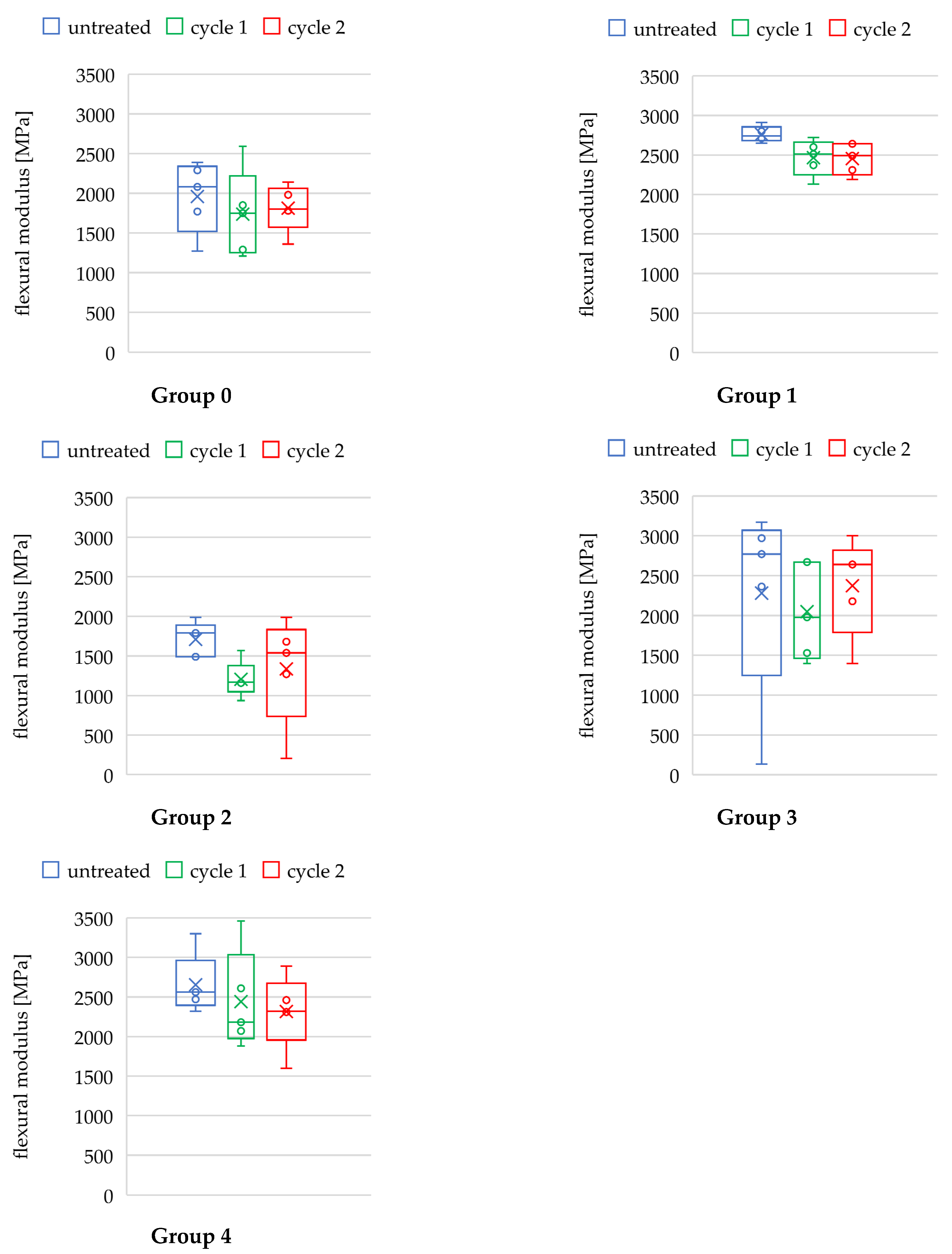
3.2. Flexural Modulus

The results of the three-point bending test are summarized in Table 3, and corresponding boxplots are provided in Figure 4. The mean flexural moduli ranged between 1960 MPa and 2762 MPa for the untreated groups. After autoclaving, the mean flexural modulus was lower for each resin/printer combination, ranging from 1205 MPa to 2466 MPa and from 1337 MPa to 2454 MPa for cycles 1 and 2, respectively. However, in the majority of groups, these data failed significance.
A qualitative difference was noted in group 1 (ANOVA, p = 0.035). The post hoc test revealed a significantly lower flexural modulus following cycle 2 autoclaving compared to the untreated group (t-test, p = 0.045). In contrast, no differences were seen following cycle 1 autoclaving compared to the untreated control (t-test, p = 0.085). However, there were no significant differences in flexural modulus between the two autoclaving protocols (p > 0.999).

4. Discussion

The aim of this study was to evaluate whether autoclaving changes the Vickers hardness and flexural modulus of resin materials used to additively manufacture insertion guides, which could pose a risk for the accuracy of static navigated implantation.
For the majority of specimens, the two autoclaving protocols had a minor impact on the flexural modulus, whereas changes in the Vickers hardness were more pronounced.
Looking at the results of the Vickers hardness testing, it seems that choosing the right autoclaving cycle might decrease the risk of hardness deterioration of the templates. In one out of the three groups exhibiting significant autoclaving-induced changes in the Vickers hardness (group 2), these changes occurred after the cycle 2 sterilization protocol (134 °C, 2 bar for 5.5 min) and not with the so-called “delicate” program cycle 1 (121 °C, 1 bar for 20.5 min). Overall, none of the materials that are authorized for autoclaving by the manufacturers showed signs of decreasing Vickers hardness after being autoclaved with cycle 1. Still, it is questionable whether the increase in hardness could be accompanied by a contraction of the material, which could lead to dimensional changes of the templates.
Our results showed that in most cases, autoclaving does not significantly change the flexural modulus of resin materials used to print the templates. Thus, it seems that autoclaving might not be a major risk factor for implantation accuracy loss caused by significant changes in the flexural modulus. We found no significant changes in the flexural modulus for groups 2, 3, and 4.
Interestingly, a decrease in flexural modulus was also seen in group 0, which was expected to be more prone to heat-induced changes in the flexural properties, as that resin is not authorized for autoclaving. Nevertheless, for most of the groups, the flexural moduli decreased to some extent. In group 1, autoclaving significantly decreased the flexural modulus, independently from the cycle.
Avoiding medical risks related to the contamination of the surgical site is the primary reason that makes autoclaving a must. It has been shown that the immersion in different disinfection solutions such as chlorhexidine digluconate, sodium hypochlorite, sodium perborate, or glutaraldehyde cannot entirely eradicate the microbial contamination of surgical template surfaces made of acrylic resin [31,32]. Ethanol at 70–80% resulted to be most effective among the disinfectants [25,32]. Tallarico et al. found that, after immersion disinfection with 70% ethanol for 15 min, about 16% of colony-forming units (CFU) remained on the resin surfaces [25]. Hence, in accordance with the risk mitigation when utilizing medical devices, as requested by the EU Medical Device Regulation [6], the sole reduction in microorganisms on surgical template surfaces by immersion disinfection seems insufficient and sterilization should be chosen. In light of our findings, the biological benefit of autoclaving the templates outweighs the risk of changes in the flexural properties or Vickers hardness. Nevertheless, some authors suggest cold sterilization protocols for heat-sensitive resin materials, i.e., utilizing ethylene oxide gas [33], which, however, are not commonly utilized in dental practice. The investigation of high-temperature sterilizing protocols such as autoclaving is clinically relevant, as it is one of the most commonly used methods in dentistry [34,35,36,37].
In agreement with our results for groups 1, 2, and 4, a pilot study by Török et al. found that additively manufactured template materials did not significantly change in hardness when subjected to 121 °C autoclaving [27]. Our observations differ to some extent, as we found significant changes of the hardness for group 3 after 121 °C autoclaving. Furthermore, their investigation on specimens that had been autoclaved at 134 °C exhibited significant changes in hardness values compared to specimens that were left untreated. These results are in line with our findings for groups 0 and 2. Despite utilizing similar autoclaving cycles (i.e., 121 °C, 1 bar, 20 min and 134 °C, 2 bar, 10 min), the varying observations could be a consequence of the different specimen preparation. In their work, the specimens were cut out of surgical templates, embedded, and coated with a gold layer to improve the visibility of the indentations prior to testing. In addition, the group conducted their tests with different parameters in terms of load (50 g) and duration (5 s). Furthermore, they investigated the flexural properties by testing for changes in the flexural strength, showing that autoclaving at 134 °C increased the materials stiffness significantly, while there were no significant changes in the flexural properties after autoclaving at 121 °C. Overall, our results are not entirely comparable, as different flexural properties were tested. Still, the results found in our study show similarities to the findings of Török et al., as we found no significant changes in the flexural modulus for 121 °C, while detecting a significant change for one group after autoclaving at 134 °C. However, the limited amount of data does not allow us to draw any firm conclusions about the influence of the autoclaving parameters on the flexural properties. It would be interesting to clarify whether autoclaving-induced changes in flexural properties are minor at lower sterilization temperature and pressure [27].
Most recently, Pop et al. conducted a similar study on the influence of disinfection and autoclaving on the flexural properties of surgical guide materials for additive manufacturing [29]. In contrast to our results, they found significantly increased flexural moduli for both autoclaving cycles that the specimens were subjected to (i.e., 121 °C, 1 bar, 20 min and 134 °C, 2 bar, 10 min). Reasons for the contrasting results could be different post-curing conditions of the specimens, different materials, different loading speed (i.e., 5 mm/s), and possibly different specimen geometry, which, however, remained unspecified. Interestingly, one of the two materials that were examined in their study was used in the present study in group 3 (i.e., Dental SG, Formlabs Inc.), which was the only group that we found to have an increased flexural modulus after being treated at 134 °C, in agreement with the results of Pop et al. [29].
Bayarsaikhan et al. investigated the behavior of resin materials for additive manufacturing when subjected to different temperatures (i.e., 40 °C, 60 °C, and 80 °C) and for varying treatment durations (i.e., 15, 30, 60, 90, and 120 min) after curing. They found that, with increasing temperature and treatment duration, the flexural modulus and Vickers hardness increased [30]. Jindal et al. conducted a resembling study on post-curing treatment, showing that 3D-printed aligner materials treated with higher temperatures endured higher compressive loads without deforming plastically [38].
In the present study, only two sterilization protocols were selected, and this might represent a limitation of the current study. However, the chosen protocols are among the simplest and most effective ones in dental practices and therefore are clinically relevant [39]. Another limitation is that not all the resin/printer combinations available on the market could be tested. Further, as recommended in the literature, specimens designed for mechanical testing were utilized, which is not 100% transferable to clinical settings. In addition, it has to be noted that three resins were printed with DLP, whereas only one resin was printed with SLA and SLA-LCD. It might be of interest to perform chemical analyses of the different materials before and after steam autoclaving to verify if there is any common pattern in autoclaving-induced chemical modifications. In future studies, in addition to the analysis of the mechanical properties, it would also be relevant to further explore the autoclaving-induced dimensional changes of templates fabricated by common resin–printer combinations.

5. Conclusions

In conclusion, and within the limitations of this study, in three groups out of five, a significant change in Vickers hardness was observed following autoclaving. This included the material not authorized for steam autoclaving. Just one material presented a significant decrease in the flexural modulus after cycle 2, whereas none of the materials showed a significant change following cycle 1. Thus, clinicians might consider using lower temperatures and pressures and longer autoclaving durations to sterilize additively manufactured templates. Materials authorized for autoclaving should be preferred. It is not possible to draw firm conclusions on the clinical significance of autoclaving-induced guide changes relying solely on biomechanical data. Indeed, whether and to what extent the autoclaving cycles are also associated with dimensional changes of the templates should be investigated in future studies.

Author Contributions

Conceptualization, K.B.; methodology, A.K., S.D., C.B. and K.B.; formal analysis, A.K. and K.B.; investigation, A.K. and L.K.; resources, S.D., C.B. and K.B.; data curation, K.B.; writing—original draft preparation, A.K., G.B. and K.B.; writing—review and editing, A.K., G.B., D.D. and K.B.; supervision, K.B.; project administration, K.B. All authors have read and agreed to the published version of the manuscript.

Funding

This research received no external funding.

Institutional Review Board Statement

Not applicable.

Informed Consent Statement

Not applicable.

Data Availability Statement

Data will be provided upon reasonable request.

Acknowledgments

The specimens printed with the materials NextDent SG, Vertex-Dental B.V., and Optiprint Guide, dentona AG were provided free of cost by Dentalstudio-Sankt Augustin GmbH. The other specimens were provided by 3D Agency oHG at a reduced cost. The authors would like to thank Viktoria Trelenberg-Stoll for her help in the conception of the study.

Conflicts of Interest

The authors declare no conflict of interest.

References

  1. Louvrier, A.; Marty, P.; Barrabé, A.; Euvrard, E.; Chatelain, B.; Weber, E.; Meyer, C. How useful is 3D printing in maxillofacial surgery? J. Stomatol. Oral Maxillofac. Surg. 2017, 118, 206–212. [Google Scholar] [CrossRef] [PubMed]
  2. Tattan, M.; Chambrone, L.; González-Martín, O.; Avila-Ortiz, G. Static computer-aided, partially guided, and free-handed implant placement: A systematic review and meta-analysis of randomized controlled trials. Clin. Oral Implant. Res. 2020, 31, 889–916. [Google Scholar] [CrossRef] [PubMed]
  3. Chen, S.; Ou, Q.; Lin, X.; Wang, Y. Comparison between a Computer-Aided Surgical Template and the Free-Hand Method: A Systematic Review and Meta-Analysis. Implant Dent. 2019, 28, 578–589. [Google Scholar] [CrossRef] [PubMed]
  4. Tahmaseb, A.; Wu, V.; Wismeijer, D.; Coucke, W.; Evans, C. The accuracy of static computer-aided implant surgery: A systematic review and meta-analysis. Clin. Oral Implant. Res. 2018, 29, 416–435. [Google Scholar] [CrossRef] [PubMed] [Green Version]
  5. Lal, K.; White, G.S.; Morea, D.N.; Wright, R.F. Use of stereolithographic templates for surgical and prosthodontic implant planning and placement. Part I. The concept. J. Prosthodont. 2006, 15, 51–58. [Google Scholar] [CrossRef]
  6. Regulation (EU) 2017/745 of the European Parliament and of the Council of 5 April 2017 on Medical Devices, Amending Directive 2001/83/EC, Regulation (EC) No 178/2002 and Regulation (EC) No 1223/2009 and Repealing Council Directives 90/385/EEC and 93/42/EEC. Annex 1, Chapters 1, 8. Available online: https://eur-lex.europa.eu/eli/reg/2017/745/oj (accessed on 16 February 2022).
  7. Raico Gallardo, Y.N.; da Silva-Olivio, I.; Mukai, E.; Morimoto, S.; Sesma, N.; Cordaro, L. Accuracy comparison of guided surgery for dental implants according to the tissue of support: A systematic review and meta-analysis. Clin. Oral Implant. Res. 2017, 28, 602–612. [Google Scholar] [CrossRef] [PubMed]
  8. Cassetta, M.; Stefanelli, L.V.; Giansanti, M.; Di Mambro, A.; Calasso, S. Depth deviation and occurrence of early surgical complications or unexpected events using a single stereolithographic surgi-guide. J. Stomatol. Oral Maxillofac. Surg. 2011, 40, 1377–1387. [Google Scholar] [CrossRef]
  9. Schnutenhaus, S.; Edelmann, C.; Rudolph, H.; Luthardt, R.G. Retrospective study to determine the accuracy of template-guided implant placement using a novel nonradiologic evaluation method. Oral Surg. Oral Med. Oral Pathol. Oral Radiol. 2016, 121, 72–79. [Google Scholar] [CrossRef]
  10. Makins, S.R. Artifacts interfering with interpretation of cone beam computed tomography images. Dent. Clin. N. Am. 2014, 58, 485–495. [Google Scholar] [CrossRef]
  11. Tadinada, A.; Jalali, E.; Jadhav, A.; Schincaglia, G.P.; Yadav, S. Artifacts in Cone Beam Computed Tomography Image Volumes: An Illustrative Depiction. J. Mass. Dent. Soc. 2015, 64, 12–15. [Google Scholar]
  12. Giménez, B.; Özcan, M.; Martínez-Rus, F.; Pradíes, G. Accuracy of a digital impression system based on active wavefront sampling technology for implants considering operator experience, implant angulation, and depth. Clin. Implant Dent. Relat. Res. 2015, 17, 54–64. [Google Scholar] [CrossRef] [PubMed]
  13. Joda, T.; Zarone, F.; Ferrari, M. The complete digital workflow in fixed prosthodontics: A systematic review. BMC Oral Health 2017, 17, 124. [Google Scholar] [CrossRef] [PubMed]
  14. Cassetta, M.; Giansanti, M.; Di Mambro, A.; Stefanelli, L.V. Accuracy of positioning of implants inserted using a mucosa-supported stereolithographic surgical guide in the edentulous maxilla and mandible. Int. J. Oral Maxillofac. Implant. 2014, 29, 1071–1078. [Google Scholar] [CrossRef] [PubMed] [Green Version]
  15. El Kholy, K.; Janner, S.F.M.; Schimmel, M.; Buser, D. The influence of guided sleeve height, drilling distance, and drilling key length on the accuracy of static computer-assisted implant surgery. Clin. Implant Dent. Relat. Res. 2019, 21, 101–107. [Google Scholar] [CrossRef] [PubMed] [Green Version]
  16. Cassetta, M.; Altieri, F.; Di Giorgio, R.; Barbato, E. Palatal orthodontic miniscrew insertion using a CAD-CAM surgical guide: Description of a technique. Int. J. Clin. Oral Maxillofac. Surg. 2018, 47, 1195–1198. [Google Scholar] [CrossRef] [PubMed]
  17. Jedliński, M.; Janiszewska-Olszowska, J.; Mazur, M.; Ottolenghi, L.; Grocholewicz, K.; Galluccio, G. Guided Insertion of Temporary Anchorage Device in Form of Orthodontic Titanium Miniscrews with Customized 3D Templates—A Systematic Review with Meta-Analysis of Clinical Studies. Coatings 2021, 11, 1488. [Google Scholar] [CrossRef]
  18. Becker, K.; Unland, J.; Wilmes, B.; Tarraf, N.E.; Drescher, D. Is there an ideal insertion angle and position for orthodontic mini-implants in the anterior palate? A CBCT study in humans. Am. J. Orthod. Dentofac. Orthop. 2019, 156, 345–354. [Google Scholar] [CrossRef]
  19. Küffer, M.; Drescher, D.; Becker, K. Application of the Digital Workflow in Orofacial Orthopedics and Orthodontics: Printed Appliances with Skeletal Anchorage. J. Appl. Sci. 2022, 12, 3820. [Google Scholar] [CrossRef]
  20. D’haese, J.; Ackhurst, J.; Wismeijer, D.; De Bruyn, H.; Tahmaseb, A. Current state of the art of computer-guided implant surgery. Periodontology 2000 2017, 73, 121–133. [Google Scholar] [CrossRef]
  21. Wegmüller, L.; Halbeisen, F.; Sharma, N.; Kühl, S.; Thieringer, F.M. Consumer vs. High-End 3D Printers for Guided Implant Surgery-An In Vitro Accuracy Assessment Study of Different 3D Printing Technologies. J. Clin. Med. 2021, 10, 4894. [Google Scholar] [CrossRef]
  22. Herschdorfer, L.; Negreiros, W.M.; Gallucci, G.O.; Hamilton, A. Comparison of the accuracy of implants placed with CAD-CAM surgical templates manufactured with various 3D printers: An in vitro study. J. Prosthet. Dent. 2021, 125, 905–910. [Google Scholar] [CrossRef] [PubMed]
  23. Chen LLin, W.S.; Polido, W.D.; Eckert, G.J.; Morton, D. Accuracy, reproducibility, and dimensional stability of additively manufactured surgical templates. J. Prosthet. Dent. 2019, 122, 309–314. [Google Scholar] [CrossRef] [PubMed]
  24. Kohn, W.G.; Collins, A.S.; Cleveland, J.L.; Harte, J.A.; Eklund, K.J.; Malvitz, D.M.; Centers for Disease Control and Prevention (CDC). Guidelines for infection control in dental health-care settings—2003. MMWR Recomm. Rep. 2003, 52, 1–61. [Google Scholar] [PubMed]
  25. Tallarico, M.; Lumbau, A.I.; Park, C.J.; Puddu, A.; Sanseverino, F.; Amarena, R.; Meloni, S.M. In vitro evaluation of bioburden, three-dimensional stability, and accuracy of surgical templates without metallic sleeves after routinely infection control activities. Clin. Implant Dent. Relat. Res. 2021, 23, 380–387. [Google Scholar] [CrossRef] [PubMed]
  26. Marei, H.F.; Alshaia, A.; Alarifi, S.; Almasoud, N.; Abdelhady, A. Effect of Steam Heat Sterilization on the Accuracy of 3D Printed Surgical Guides. Implant Dent. 2019, 28, 372–377. [Google Scholar] [CrossRef]
  27. Török, G.; Gombocz, P.; Bognár, E.; Nagy, P.; Dinya, E.; Kispélyi, B.; Hermann, P. Effects of disinfection and sterilization on the dimensional changes and mechanical properties of 3D printed surgical guides for implant therapy—Pilot study. BMC Oral Health 2020, 20, 19. [Google Scholar] [CrossRef]
  28. Quintana, R.; Choi, J.W.; Puebla, K.; Wicker, R. Effects of build orientation on tensile strength for stereolithography-manufactured ASTM D-638 type I specimens. Int. J. Adv. Manuf. Technol. 2010, 46, 201–215. [Google Scholar] [CrossRef]
  29. Pop, S.I.; Dudescu, M.; Mihali, S.G.; Păcurar, M.; Bratu, D.C. Effects of Disinfection and Steam Sterilization on the Mechanical Properties of 3D SLA- and DLP-Printed Surgical Guides for Orthodontic Implant Placement. Polymers 2022, 14, 2107. [Google Scholar] [CrossRef]
  30. Bayarsaikhan, E.; Lim, J.H.; Shin, S.H.; Park, K.H.; Park, Y.B.; Lee, J.H.; Kim, J.E. Effects of Postcuring Temperature on the Mechanical Properties and Biocompatibility of Three-Dimensional Printed Dental Resin Material. Polymers 2021, 13, 1180. [Google Scholar] [CrossRef]
  31. Da Silva, F.C.; Kimpara, E.T.; Mancini, M.N.; Balducci, I.; Jorge, A.O.; Koga-Ito, C.Y. Effectiveness of six different disinfectants on removing five microbial species and effects on the topographic characteristics of acrylic resin. J. Prosthodont. 2008, 17, 627–633. [Google Scholar] [CrossRef]
  32. Sennhenn-Kirchner, S.; Weustermann, S.; Mergeryan, H.; Jacobs, H.G.; Borg-von Zepelin, M.; Kirchner, B. Preoperative sterilization and disinfection of drill guide templates. Clin. Oral Investig. 2008, 12, 179–187. [Google Scholar] [CrossRef] [PubMed]
  33. Smith, P.N.; Palenik, C.J.; Blanchard, S.B. Microbial contamination and the sterilization/disinfection of surgical guides used in the placement of endosteal implants. Int. J. Oral Maxillofac. Implant. 2011, 26, 274–281. [Google Scholar]
  34. Smith, A.J.; Bagg, J.; Hurrell, D.; McHugh, S. Sterilization of re-usable instruments in general dental practice. Br. Dent. J. 2007, 203, E16. [Google Scholar] [CrossRef] [PubMed] [Green Version]
  35. Mahasneh, A.M.; Alakhras, M.; Khabour, O.F.; Al-Sa’di, A.G.; Al-Mousa, D.S. Practices of Infection Control among Dental Care Providers: A Cross Sectional Study. Clin. Cosmet. Investig. Dent. 2020, 12, 281–289. [Google Scholar] [CrossRef]
  36. Dagher, J.; Sfeir, C.; Abdallah, A.; Majzoub, Z. Sterilization and Biologic Monitoring in Private Dental Clinics in Lebanon. J. Contemp. Dent. Pract. 2018, 19, 853–861. [Google Scholar]
  37. Röhm-Rodowald, E.; Jakimiak, B.; Chojecka, A.; Zmuda-Baranowska, M.; Kanclerski, K. Ocena procesów dekontaminacji: Mycia, dezynfekcji i sterylizacji w praktyce stomatologicznej w Polsce w latach 2011–2012 [Assessment of decontamination processes: Cleaning, disinfection and sterilization in dental practice in Poland in the years 2011–2012]. Prz. Epidemiol. 2012, 66, 635–641. [Google Scholar]
  38. Jindal, P.; Juneja, M.; Bajaj, D.; Siena, F.L.; Breedon, P. Effects of post-curing conditions on mechanical properties of 3D printed clear dental aligners. Rapid Prototyp. J. 2020, 26, 1337–1344. [Google Scholar] [CrossRef]
  39. Jakubovics, N.; Greenwood, M.; Meechan, J.G. General medicine and surgery for dental practitioners: Part 4. Infections and infection control. Br. Dent. J. 2014, 217, 73–77. [Google Scholar] [CrossRef]
Figure 1. Microscopic image of the impression left by the pyramidal indenter during the Vickers hardness test with markings of diagonals d1 and d2.
Applsci 12 06195 g001
Figure 2. 3-point bending test of a specimen from group 3.
Applsci 12 06195 g002
Figure 3. Boxplots: Vickers hardness of the different groups and subgroups. The x represents the mean, the circles represent data points not covered by the boxplots.
Applsci 12 06195 g003
Figure 4. Boxplots: flexural moduli of the different groups and subgroups. The x represents the mean, the circles represent data points not covered by the boxplots.
Applsci 12 06195 g004
Table 1. Printers and resins used in this study, with details of the number of specimens per group and subgroup.
Group §Printing MethodPrinter, ManufacturerResin, Manufacturer
0DLPMicro Plus XL, Envisiontec Inc.E-Guide, Envisiontec Inc.
1DLPNextDent 5100, Vertex-Dental B.V.NextDent SG, Vertex-Dental B.V.
2DLPASIGA MAX, Pluradent GmbH & Co. KGOptiprint Guide, dentona AG
3SLAForm 3, Formlabs Inc.Dental SG, Formlabs Inc.
4LCD-SLASlash Plus, UniZ Technology LLC.zSG Amber, UniZ Technology LLC.
§ for each of the five groups, 15 samples divided in 3 subgroups (n = 5 per subgroup): untreated; cycle 1 (121 °C, 1 bar, 20.5 min); cycle 2 (134 °C, 2 bar, 5.5 min). DLP: digital light processing; LCD-SLA: liquid crystal display stereolithography; SLA: stereolithography.
Table 2. Results of the Vickers hardness test and statistical analysis.
Vickers Hardness [HV0.5]Kruskal–WallisBonferroni-Corrected Post Hoc Test
(Mann–Whitney-U, If pH < 0.05)
Group §ICCSubgroup §§M ± SDH (2)pHComparisonUzpUr
00.993untreated13.50 ± 2.6235.896<0.001 ***untreated vs. cycle 18−4.334<0.001 ***−0.791
cycle 16.94 ± 2.21untreated vs. cycle 29−4.293<0.001 ***−0.784
cycle 219.57 ± 2.04cycle 1 vs. cycle 21−4.625<0.001 ***−0.844
10.582untreated29.16 ± 6.835.8470.054-----
cycle 127.67 ± 5.42-----
cycle 225.30 ± 1.65-----
20.945untreated14.14 ± 2.3326.303<0.001 ***untreated vs. cycle 175−1.5550.359−0.284
cycle 116.00 ± 3.06untreated vs. cycle 226−3.5880.001 **−0.655
cycle 210.52 ± 1.05cycle 1 vs. cycle 23−4.542<0.001 ***−0.829
30.925untreated17.12 ± 1.9622.659<0.001 ***untreated vs. cycle 142−2.9240.010 *−0.534
cycle 120.80 ± 2.95untreated vs. cycle 21−4.625<0.001 ***−0.844
cycle 223.84 ± 2.24cycle 1 vs. cycle 260−2.1780.088−0.398
40.670untreated15.67 ± 2.411.8440.398-----
cycle 116.96 ± 2.92-----
cycle 214.74 ± 1.25-----
* p < 0.05; ** p < 0.01; *** p < 0.001; § n = 9 specimens for each of the 5 groups; §§ n = 3 specimens for each of the 15 subgroups; pH p-value from Kruskal–Wallis test; pU p-value from Mann–Whitney-U test.
Table 3. Results of the three-point bending test and statistical analysis.
Flexural Modulus Ef [MPa]ANOVABonferroni-Corrected Post Hoc Test
(Students-t, If pA < 0.05)
Group §Subgroup §§M ± SDF (2,12)pFComparisont (8)pt
0untreated1960 ± 4050.3220.731---
cycle 11738 ± 494---
cycle 21812 ± 261---
1untreated2762 ± 884.4920.035 *untreated vs. cycle 12.6710.085
cycle 12466 ± 203untreated vs. cycle 23.0800.045 *
cycle 22454 ± 179cycle 1 vs. cycle 20.089>0.999
2untreated1710 ± 1941.8100.206---
cycle 11205 ± 204---
cycle 21337 ± 613---
3untreated2280 ± 11080.1810.837---
cycle 12050 ± 542---
cycle 22372 ± 551---
4untreated2654 ± 3380.5800.575---
cycle 12440 ± 564---
cycle 22367 ± 536---
* p < 0.05; § n = 15 for each of the 5 groups; §§ n = 5 for each of the 15 subgroups; pF p-value from ANOVA; pt p-value from Student’s t-test.
Publisher’s Note: MDPI stays neutral with regard to jurisdictional claims in published maps and institutional affiliations.

