Article

Prostaglandin D2 Attenuates Lipopolysaccharide-Induced Acute Lung Injury through the Modulation of Inflammation and Macrophage Polarization

by
Ahmad Almatroudi
1,
Mohammed A. Alsahli
1,
Mansoor Ali Syed
2,
Amjad Ali Khan
3 and
Arshad Husain Rahmani
1,*
1
Department of Medical Laboratories, College of Applied Medical Sciences, Qassim University, Buraydah 52571, Saudi Arabia
2
Translational Research Lab, Department of Biotechnology, Faculty of Natural Sciences, Jamia Millia Islamia, New Delhi 110025, India
3
Department of Basic Health Science, College of Applied Medical Sciences, Qassim University, Buraydah 52571, Saudi Arabia
*
Author to whom correspondence should be addressed.
Appl. Sci. 2022, 12(12), 6076; https://doi.org/10.3390/app12126076
Submission received: 12 May 2022 / Revised: 9 June 2022 / Accepted: 9 June 2022 / Published: 15 June 2022
(This article belongs to the Special Issue Asthma and Respiratory Disease: Prediction, Diagnosis and Treatment)

Abstract

:
Acute lung injury (ALI) is a well-known respiratory disease and a leading cause of death worldwide. Despite advancements in the medical field, developing complete treatment strategies against this disease is still a challenge. In the current study, the therapeutic role of prostaglandin D2 (PGD2) was investigated on lipopolysaccharide (LPS)-induced lung injury in mice models and RAW264.7 macrophages through anti-inflammatory, histopathology, immunohistochemistry, and TUNEL staining. The overproduction of cytokines by RAW264.7 macrophages was observed after stimulation with LPS. However, pretreatment with PGD2 decreased the production of cytokines. The level of inflammatory markers was significantly restored in the PGD2 treatment group (TNF-α = 58.6 vs. 78.5 pg/mL; IL-1β = 29.3 vs. 36.6 pg/mL; IL-6 = 75.4 vs. 98.2 pg/mL; and CRP = 0.84 vs. 1.14 ng/mL). The wet/dry weight ratio of the lungs was quite significant in the disease control (LPS-only treatment) group. Moreover, the histological changes as determined by haematoxylin and eosin (H&E) staining clearly showed that PGD2 treatment maintains the lung tissue architecture. The iNOS expression pattern was increased in lung tissues of LPS-treated animals, whereas, in mice treated with PGD2, the expression of iNOS protein decreased. Flow cytometry data demonstrated that LPS intoxication enhanced apoptosis, which significantly decreased with PGD2 treatment. In conclusion, all these observations indicate that PGD2 provides an anti-inflammatory response in RAW264.7 macrophages and in ALI, and they suggest a therapeutic potential in lung pathogenesis.

1. Introduction

Acute lung injury (ALI) is a well-known respiratory disease and a leading cause of mortality worldwide. Despite advancements in treatment approaches and progress in combination therapies, the mortality rate is still high [1]. There are multiple reasons for ALI development, and if untreated, it can worsen to acute respiratory distress syndrome (ARDS). Some factors act as culprits in this pathogenesis, including sepsis, pneumonia, mechanical injury, toxic compound inhalation, and viral and microbial infections [2,3].
The formation of alveolar oedema and neutrophil accumulation, developed by endothelial/epithelial barrier disruption, are well-known and characteristic pathological findings in ALI/ARDS [4]. During inflammation, the lung tissue macrophages play a significant role, and their activation leads to the production of mediators of inflammation, such as interleukin (IL) 1β, IL-6, tumour necrosis factor (TNF)-α, myeloperoxidase (MPO), inducible nitric oxide synthase (iNOS), and cyclooxygenase-2 (COX-2) [5,6]. These inflammatory mediators lead to stimulation of lung tissue cells and can even damage them, resulting in impaired functions and cell necrosis [7,8]. Thus, for the alleviation of ALI, obtaining effective control over the inflammatory mediators is a valuable therapeutic strategy [5,9]. Mitogen-activated protein kinase (MAPK) and nuclear factor (NF)-κB are classical signalling pathways that participate in inflammation. The activation of MAPK and NF-κB can exaggerate the secretion of mediators of inflammation.
Infection by Gram-negative bacteria causes sepsis, the frequent cause of ALI, and lipopolysaccharides (LPS) are well-recognized endotoxins and major components of the bacterial cell wall. So, in experimental models, LPS is commonly used to produce strong immunogenicity and an inflammatory response [5,7,10]. The in-vivo-based study reported that LPS initiates a complex inflammatory response network via ligand–receptor interactions with different immune cells. This interaction leads to the release of an immense array of proinflammatory mediators that orchestrate the acute inflammatory response [11].
Oxidative stress is enhanced by the release of oxygen radicals by the activation of neutrophils. However, the exact mechanism of LPS-mediated ALI is not fully known [12]. Due to their lower toxicity and fewer side-effects, natural products, or their purified bioactive ingredients, have opened a novel therapeutic window in the management of lung pathogenesis.
Prostaglandins (PGs) are the metabolites of cyclooxygenase (COX), especially COX-2, which play a critical role during inflammatory responses. The bronchoalveolar lavage fluid (BALF) from patients suffering from ARDS showed elevated PG levels [13]. In experimental animal models, it has been reported that COX inhibition reduces the development of sepsis, and the survival rate of the animals improves [14].
It has also been confirmed that the inhibition of COX-2 attenuates carrageenan-induced ALI in experimental rats [15]. These observations suggest that PGs produced via COX-2-mediated metabolism are crucial for the initiation, as well as the progression, of inflammation in the lungs. Furthermore, treatment with ibuprofen, a nonselective inhibitor of COX, has been reported with no ARDS reduction in patients who had developed sepsis [16]. Therefore, these findings recommend that PGs perform a multifaceted role as both proinflammatory and anti-inflammatory agents in the pathophysiology of airway inflammation.
In this study, the role of PGD2 in the inflammatory response caused by LPS was determined by using RAW264.7 cells via the evaluation of nitric oxide (NO) and cytokine levels. Furthermore, we aimed to study the protective role of PGD2 in LPS-caused lung injury in animal models.

2. Materials and Methods

2.1. Chemicals and Reagents

Lipopolysaccharides and PGD2 were obtained from Sigma Aldrich, USA. The proinflammatory markers, TNF-α, IL-1β, and IL-6, were determined calorimetrically by using ELISA kits, purchased from Abcam, UK. Foetal bovine serum (FBS) and Dulbecco’s Modified Eagle’s Medium (DMEM) were obtained from Thermo Fisher Scientific, Waltham, MA, USA. In addition, the primary and secondary antibodies used for immunohistochemistry and the TUNEL assay kit were also purchased from Abcam, Cambridge, UK. The supplementary reagents and other chemicals used in this study were obtained from other commercial sources.

2.2. In Vitro Study

2.2.1. Cell Culture

DMEM was used to culture RAW264.7 macrophages complemented with 10% foetal bovine serum (FBS). Streptomycin (100 μg/mL) and penicillin (100 U/mL) were added to cells in a humidified atmosphere supplemented with 5% CO2 and maintained at 37 °C. Tissue culture dishes were used to culture the cells to create a monolayer. These macrophages were incubated with or without PGD2 [17] added 30 min before LPS stimulation.

2.2.2. Cell Viability Analysis

RAW264.7 cells were plated at a density of 1 × 10⁵ cells/mL in 24-well plates at 37 °C. MTT assay was used to determine the cell viability. PGD2 pretreatment (10 μM) was performed on the RAW 264.7 macrophages, and after 30 min, they were exposed to LPS (200 ng/mL) treatment. After a 24-h incubation period, each well was treated with 0.5 mg/mL MTT, and the plates were incubated for an additional 2 h at 37 °C in the presence of 5% CO2. The aspiration of supernatants from each well was performed, and formazan crystals dissolved in 150 μL DMSO were used. A microplate reader was used to determine the absorbance at 540 nm. The optical density of the MTT-formazan that formed in the nontreated cells was considered as 100% viable.

2.2.3. Nitric Oxide Measurement

The cells were cultured in a 24-well plate overnight with approximately 1 × 10⁵ cells/well. Some cells were pretreated with PGD2 (10 μM) for 30 min, and LPS (200 ng/mL) was used for an additional 24 h. The supernatant obtained from every well was used to analyse the NO production (in μM). Griess reagents I and II (100 μL) and 50 μL of cell culture medium were reacted at room temperature for 10 min in a 96-well plate. A microplate reader was used to determine the absorbance at 540 nm.

2.2.4. Proinflammatory Cytokine Determination

A 24-well plate was used to culture RAW264.7 cells with almost 1 × 10⁵ cells/well, and they were treated in the presence of either LPS (200 ng/mL) only or PGD2 with LPS for 24 h. The supernatants were used for the estimation of proinflammatory cytokines, such as IL-6 and TNF-α (in ng/mL), via ELISA.

2.3. In Vivo Study

2.3.1. Animal Model

For this study, male albino mice weighing 25 to 30 g were obtained from King Saud University, Riyadh, Saudi Arabia. The animals were kept in the animal house for one week so as to acclimatize. The room temperature was maintained at 23 ± 2 °C, 45–65% relative humidity, with a 12 h dark/12 h light cycle, and the animals received water and food ad libitum. The experimental designs were planned according to the Ethical Committee of the College of Applied Medical Sciences, Qassim University (No. 0194-Cams1-2020-1-3-I). For the care and experimental use of laboratory animals, the committee follows the guidelines of National Institutes of Health (NIH) of the USA.

2.3.2. Grouping of Animals According to the Treatment Plan

The animals were grouped as described in Table 1 (n = 8/group).
The mortality of the animals was checked every 3 h after LPS treatment for 24 h. After 24 h of LPS treatment, ketamine and xylazine injections (60 mg and 16 mg) were used intraperitoneally to anesthetize the animals [18]. The blood samples from all of the experimental mice were collected and were kept at room temperature for 30 min to initiate clotting. The clotted samples were centrifuged at 3000 rpm for 10 min to obtain the serum.
After sacrifice, the lungs were removed from all the animals and were properly washed with ice-cold saline solution. One lung from each animal was divided into two sections. Potassium phosphate buffer (pH 7.4) was used to homogenize 1 portion of lung from each animal and centrifuged at 4 °C for 10 min at 10,000 rpm. All the supernatants were stored in a deep freezer (−20 °C) as aliquots for biochemical analysis. The second portion of each lung was processed for histopathological examination [20,21].

2.3.3. Harvesting the BALF

All of the experimental mice were sacrificed at the end of the treatment plan. Alcohol was used to disinfect the animals, and after dissecting the chests, the lungs were exposed. Silk thread was used to ligate one lung, and the left lung was lavaged with 0.5 mL PBS. The lavage was repeated 3 times for 30 s each time. The lavage fluid was collected and centrifuged for 5 min at 2000 rpm at 4 °C. The supernatants were stored at −20 °C for further use. PBS was used to resuspend the cell sediment. The total cell count was estimated by haemocytometer.

2.3.4. Determination of Lung Wet/Dry Weight Ratio

The levels of LPS-mediated pulmonary oedema were assessed by measuring the wet and dry weight (W/D) ratio in grams. Directly after sacrificing the animals, 24 h after the LPS administration (5 mg/kg b.w.), the wet weight of all the lung samples was determined. The lungs were then placed in a drying oven (Binder, ED-S) maintained at 80 °C for 24 h. The dry weight was measured specific to each lung, and the W/D weight ratio was calculated accordingly [22].

2.3.5. Determination of Inflammatory Markers

The levels of inflammatory markers, such as interleukin-1β (IL-1β), IL-6, tumour necrosis factor-α (TNF-α) (in pg/mL), and CRP (in ng/mL), were determined by using the lung tissue supernatant of all of the animals by using a kit from Abcam, UK. The manufacturer ‘s instructions were followed, each sample was taken in triplicate, and average values were used for further calculations.

2.3.6. Histopathological Analysis

Haematoxylin–eosin staining was performed so as to examine the tissue architecture. After the staining, the slides were examined under a light microscope, and photographs were taken. Lung injury was quantified and scored as 0 for absent, 1 for mild, 2 for moderate, and 3 for severe injury, as determined by the pathological changes present, including the presence of exudates, congestion, infiltration of neutrophils, and alveolar haemorrhage [23,24].

2.3.7. Immunohistochemical Study

Immunohistochemistry of all samples was performed by using the previous method [25,26]. The expression pattern of iNOS protein was evaluated by using antibodies. The biotinylated secondary antibodies and streptavidin peroxidase were used. Finally, DAB was used, and sections were counterstained with haematoxylin for 30 s. A Leica light microscope with an attached camera was used to visualize the slides, and photographs were taken. The quantification of cytoplasmic positive staining was calculated by a pathologist blinded to the treatment plan. A total of 500 cells were counted manually in 5 selected fields at high power magnification (×400). The positively stained cells were expressed as a percentage of the total cells counted in each case [27]. The cytoplasmic expression of iNOS protein was calculated and scored as negative (0 for <5% positive cells), mild/weak positive (1+ for 5–20% positive cells), intermediate/moderate positive (2+ for 21–50% positive cells), and high/strong positive (3+ for >50% positive cells).

2.3.8. Apoptosis Analysis by TUNEL Assay

A TUNEL (terminal deoxynucleotidyl transferase-mediated dUTP-biotin nick-end labelling) assay was performed on each lung tissue sample to evaluate apoptosis per the manufacturer’s instructions. The stained nuclei were examined under a light microscope for the detection of any DNA fragmentation. The TUNEL-positive nuclei were calculated, and photographs were taken accordingly.

2.3.9. Apoptosis Analysis by Flow Cytometry

The apoptosis of the cells was analysed by flow cytometry by using a FITC/Annexin V Apoptosis detection kit (Miltenyi Biotec, Gergisch Gladbach, Germany). The cells were harvested briefly in a binding buffer following RBC lysis, and the samples were incubated in the dark with FITC/Annexin V and PI at room temperature. The samples were centrifuged, resuspended with the binding buffer, and the cells were analysed by using a MacsQuant Flow Cytometer (Miltenyi Biotec, Gergisch Gladbach, Germany).

2.3.10. Statistical Analysis

The experiments were run independently in triplicate, and the in vitro data were expressed as mean ± SEM. The in vivo data were obtained from eight mice in each group and expressed as mean ± SEM. The statistical analysis was performed by using one-way analysis of variance (ANOVA). The statistical significance of the differences between separate groups was obtained by applying Duncan’s Multiple Range Test using SPSS. All the results were analysed and considered statistically significant at p < 0.05.

3. Results

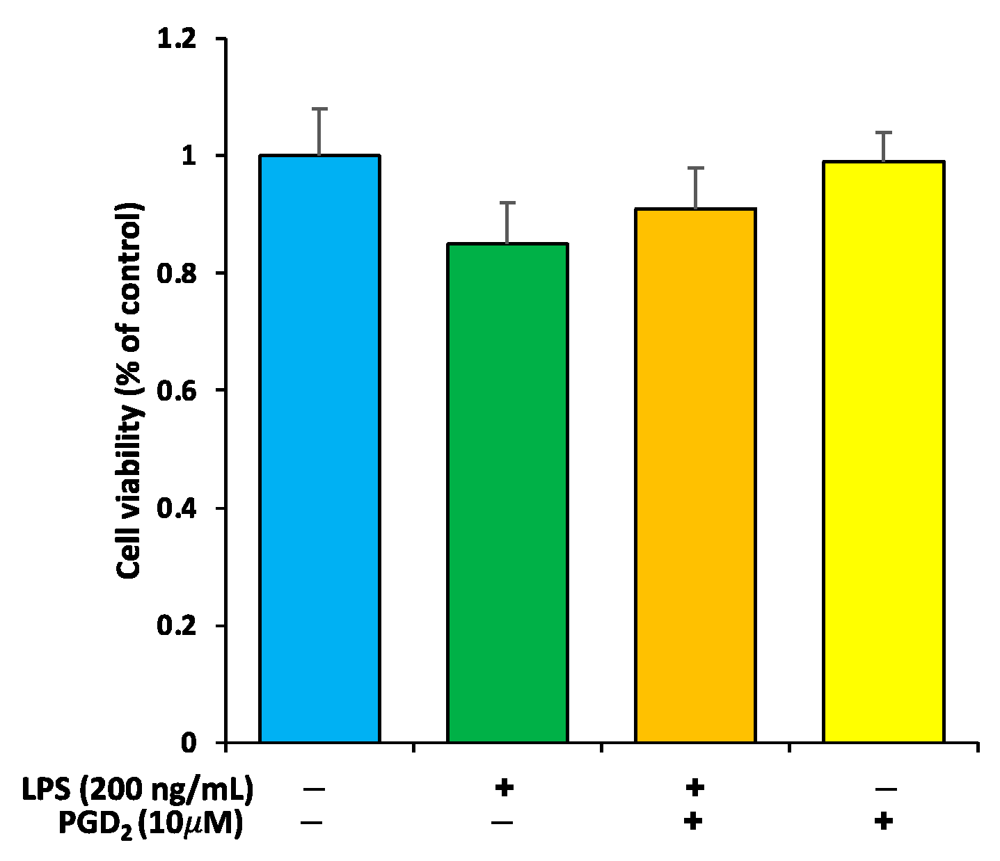
3.1. Viability Check of RAW264.7 Macrophages by PGD2 and LPS Treatment

The MTT colorimetric assay demonstrated that the LPS treatment had no more effect on cell viability as compared to the control that received no treatment (Figure 1). However, in the cells that received PGD2 1 h before the LPS treatment, cell viability was preserved. The cells that only received PGD2 did not show any change in the cell viability.

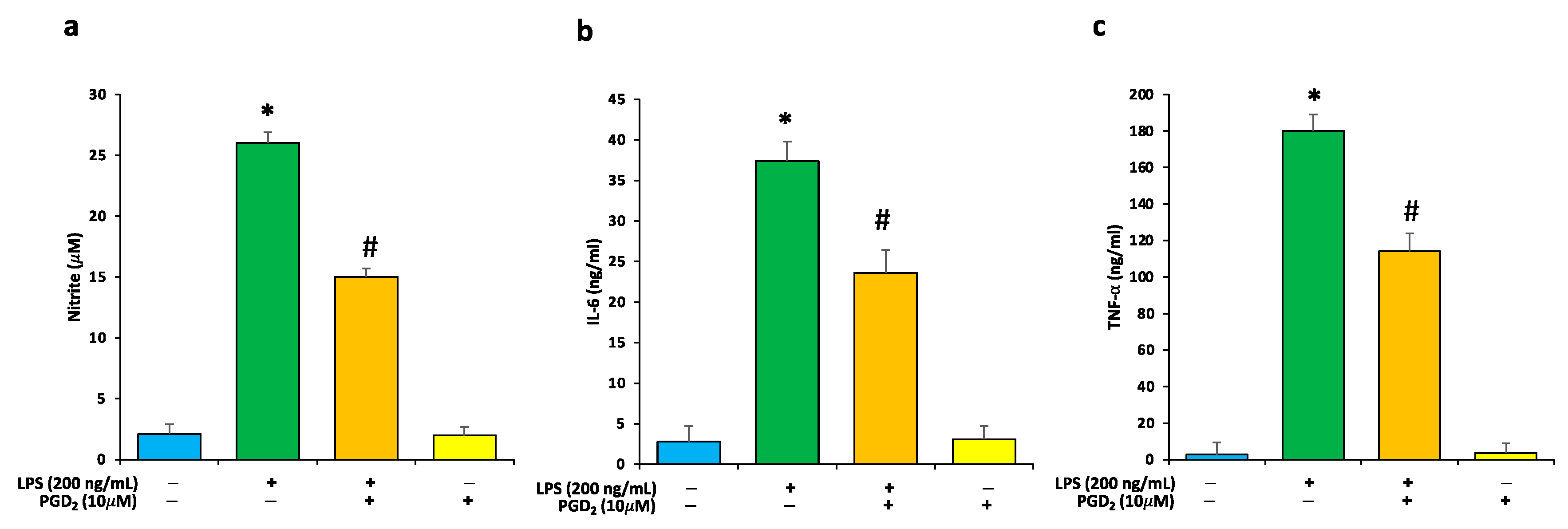
3.2. Effects of LPS and PGD2 Treatment on NO and Proinflammatory Cytokines

The overproduction of proinflammatory cytokines (IL-6 and TNF-α) by RAW264.7 macrophages was observed after 24 h of exposure with LPS. However, pretreatment with PGD2 significantly reduced the level of these proinflammatory cytokines (Figure 2). The NO production was significantly elevated in LPS-treatment-only cells as compared to control samples. However, PGD2 treatment was found to significantly inhibit LPS-promoted NO synthesis.

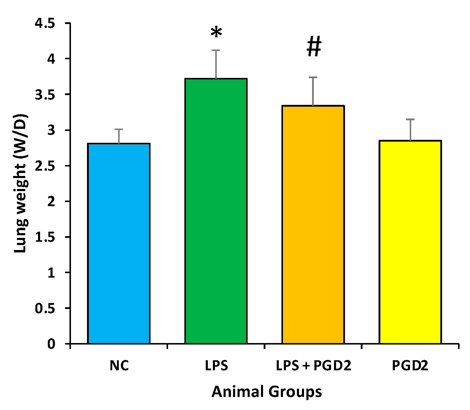
3.3. Effect of PGD2 on Weight (Wet/Dry) Ratio of Lungs

In comparison to the normal control animals, LPS (5 mg/kg b.w.) intoxication considerably enhanced the lung W/D ratio as assessed by using a previous method [28]. The animals treated with PGD2 (100 μg/kg b.w.) prior to LPS treatment showed significantly lower W/D ratios in comparison to those in the disease control group (p < 0.05) (Figure 3).

3.4. Effect of PGD2 on Inflammatory Markers

The protective role of PGD2 against LPS-intoxicated lung injury was examined by estimating the levels of proinflammatory cytokines. The levels of proinflammatory markers IL-1β (36.6 ± 2.3 pg/mL), IL-6 (98.6 ± 6 pg/mL), and TNF-α (78.5 ± 7 pg/mL) were enhanced remarkably in disease control (LPS) mice (Figure 4), while all these parameters were considerably reduced with PGD2 (100 μg/kg b.w.) pretreatment in comparison to the LPS group (p < 0.05). These findings demonstrate that PGD2 lessens the lung inflammation/injury and also minimizes the levels of proinflammatory cytokines provoked by LPS (5 mg/kg b.w.) treatment (Figure 4).

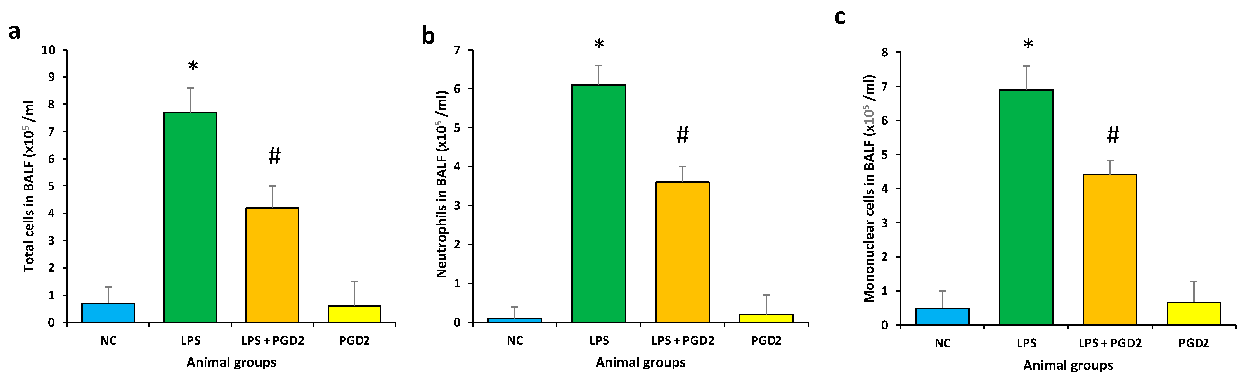
3.5. Effect of PGD2 on Inflammatory Cells Influx in BALF

The number of inflammatory cells, such as neutrophils and mononuclear cells (macrophages and monocytes), was calculated in BALF, and it increased significantly after the LPS (5 mg/kg b.w.) challenge as compared to the control sample (p < 0.05). The number of neutrophils and mononuclear cells in the PGD2 (100 μg/kg b.w.) treatment group, prior to LPS insult, was remarkably reduced overall (Figure 5). LPS exposure remarkably enhanced the total number of cells (7.2 ± 1.1 × 10⁵/mL), neutrophils (6.1 ± 1.7 × 10⁵/mL), and mononuclear cells (6.9 ± 1.7 × 10⁵/mL) in comparison with the control group. However, PGD2 pretreatment of the LPS exposure group resulted in a significant decrease in the total number of cells (3.1 ± 1.1 × 10⁵/mL), neutrophils (1.9 ± 1.3 × 10⁵/mL), and mononuclear cells (5.6 ± 1.7 × 10⁵/mL) (Figure 5).

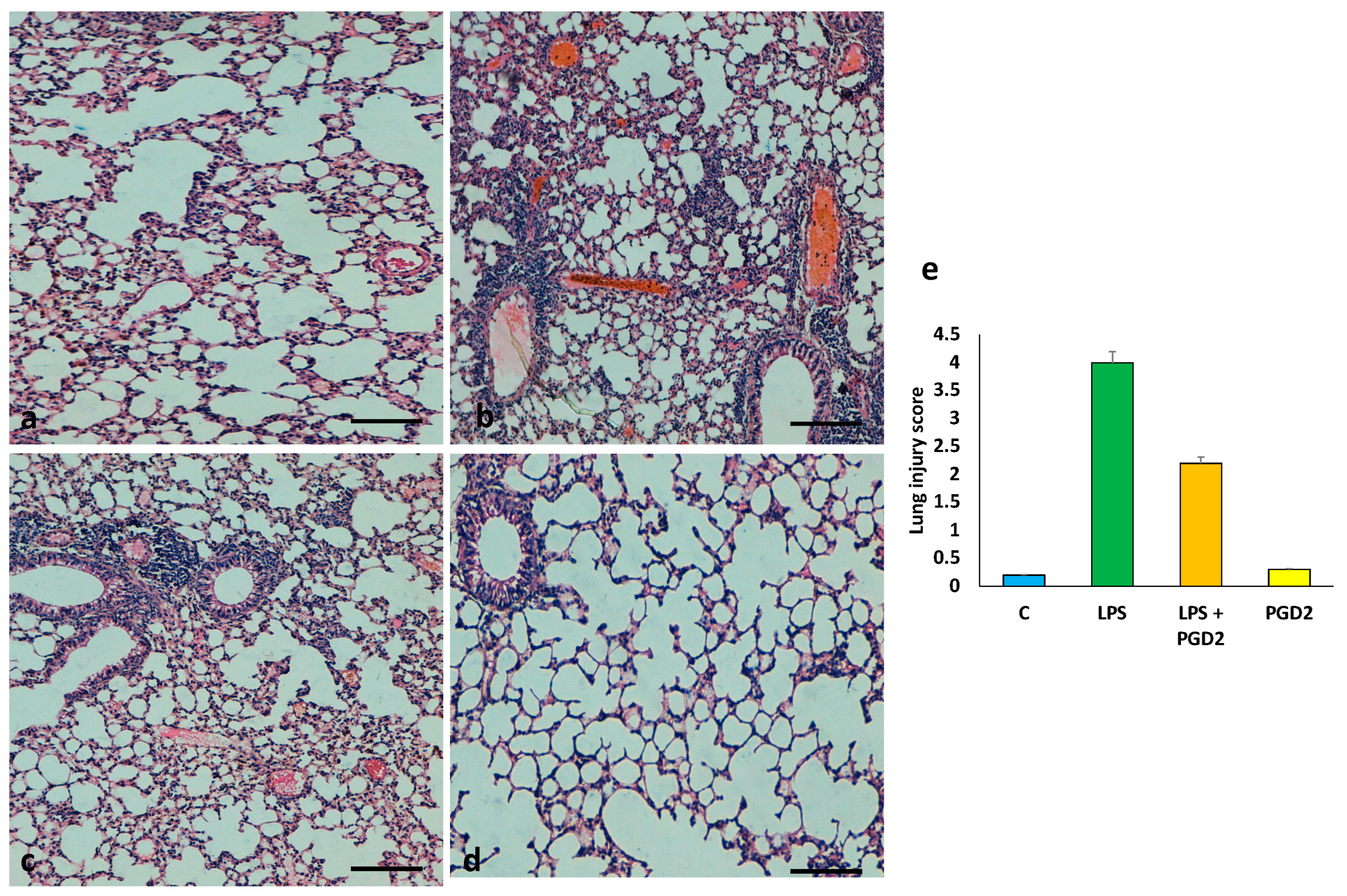
3.6. The Role of PGD2 on the Maintenance of Lung Tissue Architecture

Histopathological analysis of the lung tissue in different animal groups was used to evaluate the lung architecture. In the control group, normal bronchial structure was observed (Figure 6). LPS intoxication leads to lung damage, including interstitial inflammatory cell infiltration, congestion, and oedema. However, the animals treated with PGD2 demonstrated remarkable improvement in lung tissue architecture as compared to the disease control animals (Figure 6).

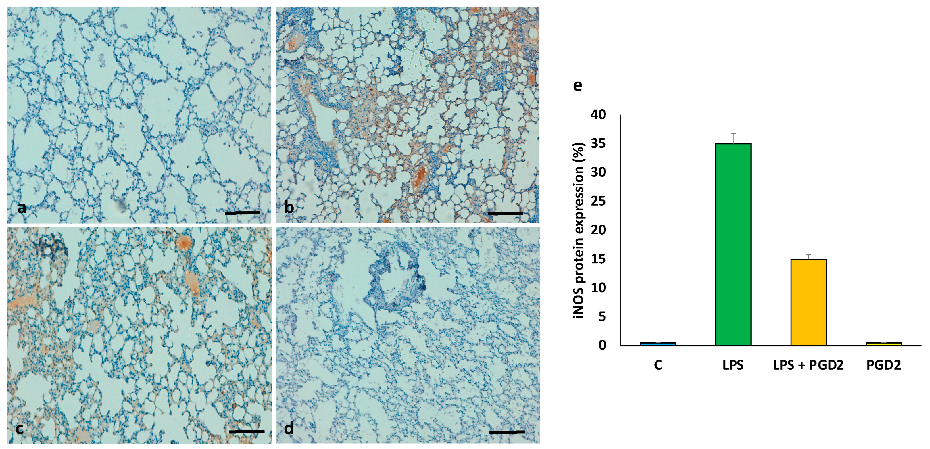
3.7. The Role of PGD2 on the Expression of iNOS

Immunohistochemical (IHC) staining demonstrated that the iNOS expression level was significantly enhanced in the lung tissues of LPS-intoxicated (5 mg/kg b.w.) animals, whereas no expression was found in control animals. The expression of this protein decreased in mice treated with PGD2 (100 μg/kg b.w.) (Figure 7).

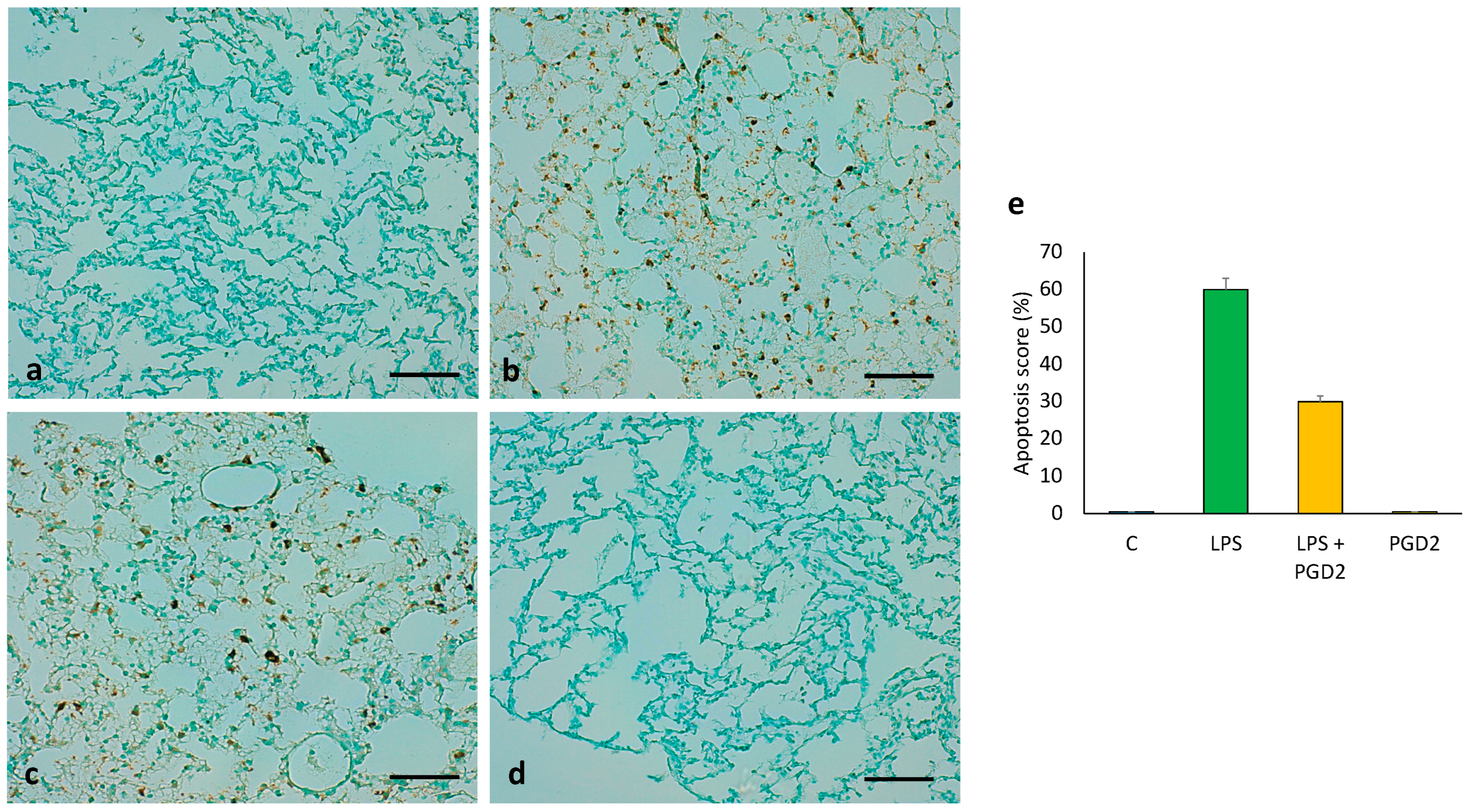
3.8. The Role of PGD2 on LPS-Treated Apoptotic Cell Death of Lung Tissue

In control group animals, the sections did not show any TUNEL-positive cells. However, as compared to the control group, the TUNEL positivity was significantly enhanced in the disease control (LPS-treated only) (5 mg/kg b.w.) group. However, the animals pretreated with PGD2 (100 μg/kg b.w.) demonstrated a low content of TUNEL-positive cells in comparison to the disease control animals. These observations reveal that PGD2 performs a significant role in the attenuation of apoptotic cell death (Figure 8).

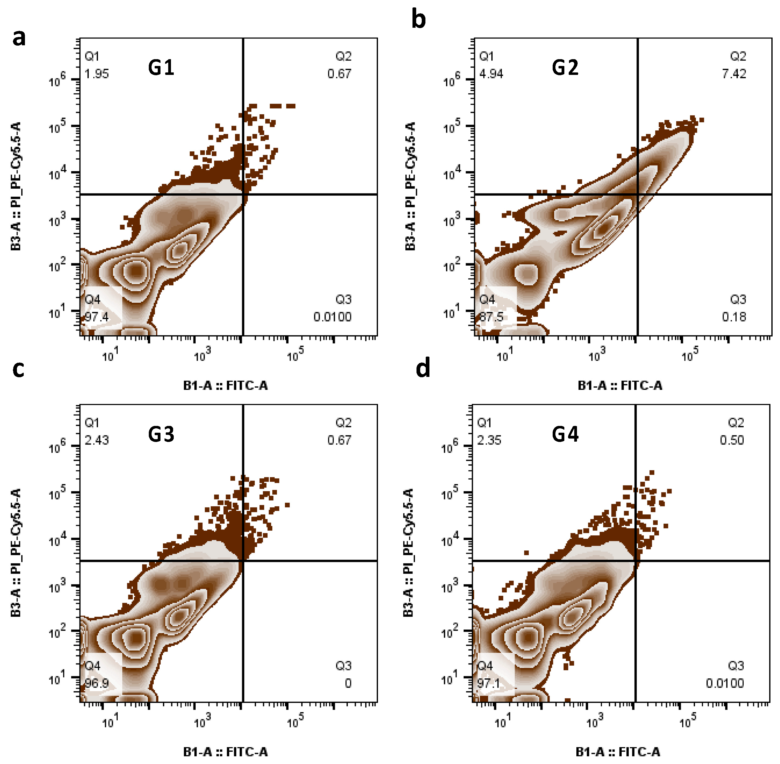
3.9. The Role of PGD2 Treatment on Apoptosis

Flow cytometry analysis showed that apoptotic cell death was increased in disease control (LPS-treated) (5 mg/kg b.w.) animals, exhibiting 87.5% viable cells as compared to the normal control having 97.4% viable cells. The exposure with PGD2 (100 μg/kg b.w.) remarkably minimized the ratio of apoptosis, and the viable cell count was restored to 96.9% (Figure 9).

4. Discussion

ALI is a well-known clinical disease, mainly described by pulmonary inflammation, loss of alveolar-capillary-membrane integrity, oedema, and impairment of arterial oxygenation, followed by apoptosis [29]. The mortality rate of this disease is high at approximately 30–50% [30]. Different types of prostaglandins (PGs) intricately modulate ALI as the pathophysiological function of each PG differs in its specific receptor subtypes, context of activation, and target cells [19].
Different types of PGs have been reported to perform paradoxical roles, both as proinflammatory and anti-inflammatory agents in the case of injured lungs [30]. Major PGs, such as PGE2 and thromboxane A2 (TXA2), function as proinflammatory agents in ALI [31,32]. However, the anti-inflammatory functions of lipoxin A4 and 15d-PGJ2 have also been reported in ALI in experimental mice [33,34]. Some previous reports have proposed a proinflammatory role for PGD2 in some respiratory disorders, arbitrated through specific signalling cascades, while others state that PGD2 have a dual role as tissue-protective in the context of airway inflammation, depending upon the specific receptors involved and the severity of the disease. It is also well-reported that the breakdown product of PGD2 as 15d-PGJ2 possesses anti-inflammatory functions through either PPAR-γ-based or PPAR-γ-independent reactions [19].
In the present study, the mouse model for ALI was successfully established by using 5 mg/kg b.w. LPS through i.p. injection [18]. The administration of LPS significantly enhanced the lung W/D weight ratio. The lung W/D weight ratio indicates oedema accumulation and is a function of the rate of fluid filtration across the lung capillaries.
However, pretreatment with PGD2 demonstrated a significantly lower W/D ratio as compared to the disease control animals. In one study, similar observations were observed in LPS-treated mice and disease control mice treated with continentalic acid [18]. PGD2 treatment attenuates the progression of pulmonary oedema, as presented by the reduction in the lung W/D weight ratio. The activity of MPO, as chiefly found in cytoplasmic granules of neutrophil, reflects the infiltration of these cells within the lung tissues [35].
A complex setup of inflammatory cytokines, such as IL-1β, IL-6, and TNF-α, plays a significant role in LPS-induced ALI and enhances the severity of lung damage [36]. Furthermore, various chemokines are also recruited, and they stimulate the enormous recruitment of neutrophils that ultimately leads to a steep rise in MPO activity. Treatment with PGD2 significantly diminishes the rise in IL-1β, IL-6, and TNF-α after LPS exposure, an outcome consistent with that of the MPO level. Furthermore, the production of other inflammatory markers, such as iNOS, was also diminished by PGD2 treatment.
A number of molecular mechanisms have been proposed for the mediation of the pathogenic events of LPS-induced lung injury, among which oxidative stress plays a major role in mediating the disease [37]. The inflammatory responses lead to respiratory burst and superoxide production. ROS overproduction results in severe pathophysiological events. Excessive LPS-induced ROS production promotes lipid peroxidation [38]. Moreover, LPS-induced oxidative stress is associated with reduced antioxidant enzyme activity. Among the different antioxidant enzymes, catalase, SOD, and GSH are significantly depressed by LPS treatment [39].
Exaggerated inflammation is generally demonstrated as triggering apoptosis, which plays a serious role in the pathological events of inflammatory diseases [40]. The infiltration of inflammatory cells and their accumulation within the alveolar cavity contribute to the apoptosis and necrosis of pulmonary cells [41,42]. Apoptosis is regarded as a major reason for lung injury, and its inhibition demonstrates a reduction in ARDS morbidity [43]. The anti-apoptotic effects were evaluated by TUNEL assay, and the results demonstrated that PGD2 treatment alleviates lung cell apoptosis in vivo. The substantial decrease in apoptosis was also supported by flow cytometry.
Previous studies have reported the antifibrotic role of PGE2, while PGF acts as a pulmonary fibrosis mediator [44]. In the current study, the anti-inflammatory role of PGD2 was demonstrated in mice. The disease control mice showed enhanced inflammation, denoted by the enhanced expression of inflammatory mediators, followed by neutrophil infiltration. Although the disease-stage-dependent role of PGD2 needs to be examined in more depth, as PGD2 has been stated to improve bleomycin-induced fibrosis, partially by suppressing early inflammatory response. In parallel, it has been demonstrated that inflammasome signals suppress lung inflammation, followed by reduced fibrosis [45]. PGD2 locally produced within neutrophils and monocytes may also suppress the infiltration of immune cells [19]. PGD2 from the resident lung cells is accountable for anti-inflammatory responses, including the suppression of vascular permeability in acute lung injury [19].
Besides its anti-inflammatory role, PGD2 might have a direct role on fibroblasts as it attenuates collagen secretion induced by TGF-β in human lung fibroblasts by activating the D-proteinoid receptor [46]. The role of PGD2 in the inhibition of proliferation of mouse lung fibroblasts through the D-prostaglandin receptor has also been reported [47]. It was further confirmed that PGD2 has no role in apoptosis as assessed by TUNEL assay [48]. Thus, the anti-inflammatory and antifibrotic roles of PGD2 signify that it is a potential therapeutic option. In this regard, the protective role of PGD2 in anti-inflammatory action and bleomycin-induced pulmonary fibrosis has been previously verified [48]. In the current study, it was observed that LPS treatment of the mice induced lung injury and various other pathological alterations although the co-administered PGD2 and LPS treatment showed a role in the maintenance of lung architecture. These results were in agreement with previous reports that LPS induction in mice causes various types of pathological changes in the lung tissues, such as inflammatory cell infiltration, congestion and oedema [18,49].
The expression of iNOS is elevated during the inflammatory insults. The IHC staining for iNOS in different animal groups demonstrated that LPS-intoxication significantly enhances iNOS expression. However, PGD2 pretreatment significantly reduces the expression of iNOS. These observations are in agreement with a previous study, which demonstrates that continentalic acid treatment to LPS-intoxicated mice shows decreased iNOS expression [18]. Furthermore, the role of PGs in disease management should be investigated in more depth. The temporal and spatial variations of each PG synthase expression that elucidate their roles is highly significant.
The overall significance of this study is briefly described here, as there are very few studies demonstrating the mechanism of LPS and the protective role of PGD2 in lung pathogenesis. Thus, in this study, we tried to find the possible mechanism of PGD2 in lung protection through the modulation of inflammation. In parallel, the overproduction of cytokines by RAW264.7 macrophages was observed after stimulation with LPS. However, pretreatment with PGD2 decreased the production of cytokines. The number of inflammatory cells increased significantly after LPS treatment, whereas the PGD2 treatment decreased the number of inflammatory cells. The expression pattern of iNOS and the measurement of apoptosis through IHC and TUNNEL assays were also demonstrated. The LPS treatment caused lung injury, and the PGD2 treatment maintained the lung tissue architecture.

5. Conclusions

In this study, we demonstrated the role of PGD2 in alleviating the LPS-induced ALI complications via the investigation of different clinical parameters. The anti-inflammatory role of PGD2 upon LPS-challenged RAW264.7 macrophages demonstrated that PGD2 noticeably suppressed the formation of IL-6 and TNF-α and also inhibited iNOS expression and NO synthesis. In addition, treatment with PGD2 effectively prevented the development of LPS-induced ALI. These observations suggest a potential role for PGD2 and may have potential implications for the treatment of ARDS, a clinical syndrome resulting from ALI in human beings.

Author Contributions

Conceptualization, A.A., M.A.S., A.H.R. and A.A.K.; methodology, A.A., M.A.S., A.A.K., M.A.A. and A.H.R.; investigation, A.A., M.A.A., A.A.K. and A.H.R.; writing—original draft preparation, A.A., M.A.S., A.A.K. and A.H.R.; writing—review and editing, A.A., M.A.S., A.A.K., M.A.A. and A.H.R.; supervision, A.A. and M.A.A. All authors have read and agreed to the published version of the manuscript.

Funding

This work was funded and supported by the Deanship of Scientific Research, Qassim University (Grant No. 10194-Cams1-2020-1-3-I).

Institutional Review Board Statement

The animals were maintained at animal facility of the College of Applied Medical Sciences (CAMS) accordance with the guidelines of the Qassim University on Animal Care. The animal experiments were carried out as per the guidelines of CAMS, Qassim University and approved by the Institutional Animal Ethics Committee (10194-Cams1-2020-1-3-I), CAMS, Qassim University.

Informed Consent Statement

Not applicable.

Data Availability Statement

The data used to support the findings of this study are included within the article.

Conflicts of Interest

All authors declare that they have no conflict of interest.

References

  1. Tao, W.; Su, Q.; Wang, H.; Guo, S.; Chen, Y.; Duan, J.; Wang, S. Platycodin D attenuates acute lung injury by suppressing apoptosis and inflammation in vivo and in vitro. Int. Immunopharmacol. 2015, 27, 138–147. [Google Scholar] [CrossRef] [PubMed]
  2. Dushianthan, A.; Grocott, M.P.; Postle, A.D.; Cusack, R. Acute respiratory distress syndrome and acute lung injury. Postgrad. Med. J. 2011, 87, 612–622. [Google Scholar] [CrossRef] [PubMed] [Green Version]
  3. Butt, Y.; Kurdowska, A.; Allen, T.C. Acute lung injury: A clinical and molecular review. Arch. Pathol. Lab. Med. 2016, 140, 345–350. [Google Scholar] [CrossRef] [Green Version]
  4. Mendez, J.L.; Hubmayr, R.D. New insights into the pathology of acute respiratory failure. Curr. Opin. Crit. Care 2005, 11, 29–36. [Google Scholar] [CrossRef] [PubMed]
  5. Lv, H.; Liu, Q.; Wen, Z.; Feng, H.; Deng, X.; Ci, X. Xanthohumol ameliorates lipopolysaccharide (LPS)-induced acute lung injury via induction of AMPK/GSK3β-Nrf2 signal axis. Redox Biol. 2017, 12, 311–324. [Google Scholar] [CrossRef] [PubMed]
  6. Hou, W.; Hu, S.; Su, Z.; Wang, Q.; Meng, G.; Guo, T.; Zhang, J.; Gao, P. Myricetin attenuates LPS-induced inflammation in RAW 264.7 macrophages and mouse models. Future Med. Chem. 2018, 10, 2253–2264. [Google Scholar] [CrossRef] [PubMed]
  7. Zhong, W.T.; Wu, Y.C.; Xie, X.X.; Zhou, X.; Wei, M.M.; Soromou, L.W.; Ci, X.X.; Wang, D.C. Phillyrin attenuates LPS-induced pulmonary inflammation via suppression of MAPK and NF-Κb activation in acute lung injury mice. Fitoterapia 2013, 90, 132–139. [Google Scholar] [CrossRef]
  8. Dias-Freitas, F.; Metelo-Coimbra, C.; Roncon-Albuquerque, R., Jr. Molecular mechanisms underlying hyperoxia acute lung injury. Respir. Med. 2016, 119, 23–28. [Google Scholar] [CrossRef] [Green Version]
  9. Huang, X.L.; Wei, X.C.; Guo, L.Q.; Zhao, L.; Chen, X.H.; Cui, Y.D.; Yuan, J.; Chen, D.F.; Zhang, J. The therapeutic effects of jaceosidin on lipopolysaccharide-induced acute lung injury in mice. J. Pharmacol. Sci. 2019, 140, 228–235. [Google Scholar] [CrossRef]
  10. Kan, X.; Liu, B.; Guo, W.; Wei, L.; Lin, Y.; Guo, Y.; Gong, Q.; Li, Y.; Xu, D.; Cao, Y.; et al. Myricetin relieves LPS-induced mastitis by inhibiting inflammatory response and repairing the blood-milk barrier. J. Cel. Physiol. 2019, 234, 16252–16262. [Google Scholar] [CrossRef]
  11. Ulich, T.R.; Yin, S.; Remick, D.G.; Russell, D.; Eisenberg, S.P.; Kohno, T. Intratracheal administration of endotoxin and cytokines. IV. The soluble tumor necrosis factor receptor type I inhibits acute inflammation. Am. J. Pathol. 1993, 142, 1335–1338. [Google Scholar] [PubMed]
  12. Faffe, D.S.; Seidl, V.R.; Chagas, P.S.; de Moraes, V.G.; Capelozzi, V.L.; Rocco, P.R.; Zin, W.A. Respiratory effects of lipopolysaccharide-induced inflammatory lung injury in mice. Eur. Respir. J. 2000, 15, 85–91. [Google Scholar] [CrossRef] [PubMed] [Green Version]
  13. Frank, J.A.; Matthay, M.A. Leukotrienes in acute lung injury: A potential therapeutic target? Am. J. Respir. Crit. Care Med. 2005, 172, 261–262. [Google Scholar] [CrossRef] [PubMed] [Green Version]
  14. Hinshaw, L.B.; Solomon, L.A.; Erdös, E.G.; Reins, D.A.; Gunter, B.J. Effects of acetylsalicylic acid on the canine response to endotoxin. J. Pharmacol. Exp. Ther. 1967, 157, 665–671. [Google Scholar] [PubMed]
  15. Cuzzocrea, S.; Mazzon, E.; Sautebin, L.; Dugo, L.; Serraino, I.; De Sarro, A.; Caputi, A.P. Protective effects of Celecoxib on lung injury and red blood cells modification induced by carrageenan in the rat. Biochem. Pharmacol. 2002, 63, 785–795. [Google Scholar] [CrossRef]
  16. Bernard, G.R.; Wheeler, A.P.; Russell, J.A.; Schein, R.; Summer, W.R.; Steinberg, K.P.; Fulkerson, W.J.; Wright, P.E.; Christman, B.W.; Dupont, W.D.; et al. The effects of ibuprofen on the physiology and survival of patients with sepsis. N. Engl. J. Med. 1997, 336, 912–918. [Google Scholar] [CrossRef]
  17. Syed, M.A.; Joo, M.; Abbas, Z.; Rodger, D.; Christman, J.W.; Mehta, D.; Sadikot, R.T. Expression of TREM-1 is inhibited by PGD2 and PGJ2 in macrophages. Exp. Cell Res. 2010, 316, 3140–3149. [Google Scholar] [CrossRef]
  18. Ali, H.; Khan, A.; Ali, J.; Ullah, H.; Khan, A.; Ali, H.; Irshad, N.; Khan, S. Attenuation of LPS-induced acute lung injury by continentalic acid in rodents through inhibition of inflammatory mediators correlates with increased Nrf2 protein expression. BMC Pharmacol. Toxicol. 2020, 21, 1–4. [Google Scholar] [CrossRef]
  19. Murata, T.; Aritake, K.; Tsubosaka, Y.; Maruyama, T.; Nakagawa, T.; Hori, M.; Hirai, H.; Nakamura, M.; Narumiya, S.; Urade, Y.; et al. Anti-inflammatory role of PGD2 in acute lung inflammation and therapeutic application of its signal enhancement. Proc. Natl. Acad. Sci. USA 2013, 110, 5205–5210. [Google Scholar] [CrossRef] [Green Version]
  20. Khalid, S.; Ullah, M.Z.; Khan, A.U.; Afridi, R.; Rasheed, H.; Khan, A.; Ali, H.; Kim, Y.S.; Khan, S. Antihyperalgesic properties of honokiol in inflammatory pain models by targeting of NF-κB and Nrf2 signaling. Front Pharmacol. 2018, 9, 140. [Google Scholar] [CrossRef]
  21. Ullah, M.Z.; Khan, A.U.; Afridi, R.; Rasheed, H.; Khalid, S.; Naveed, M.; Ali, H.; Kim, Y.S.; Khan, S. Attenuation of inflammatory pain by puerarin in animal model of inflammation through inhibition of pro-inflammatory mediators. Int. Immunopharmacol. 2018, 61, 306–316. [Google Scholar] [CrossRef] [PubMed]
  22. Khan, A.; Khan, S.; Ali, H.; Shah, K.U.; Ali, H.; Shehzad, O.; Onder, A.; Kim, Y.S. Anomalin attenuates LPS-induced acute lungs injury through inhibition of AP-1 signaling. Int. Immunopharmacol. 2019, 73, 451–460. [Google Scholar] [CrossRef] [PubMed]
  23. Aziz, M.; Matsuda, A.; Yang, W.L.; Jacob, A.; Wang, P. Milk fat globule-epidermal growth factor-factor 8 attenuates neutrophil infiltration in acute lung injury via modulation of CXCR2. J. Immunol. 2012, 189, 393–402. [Google Scholar] [CrossRef] [PubMed] [Green Version]
  24. Cui, T.; Miksa, M.; Wu, R.; Komura, H.; Zhou, M.; Dong, W.; Wang, Z.; Higuchi, S.; Chaung, W.; Blau, S.A.; et al. Milk fat globule epidermal growth factor 8 attenuates acute lung injury in mice after intestinal ischemia and reperfusion. Am. J. Respir. Crit. Care Med. 2010, 181, 238–246. [Google Scholar] [CrossRef] [Green Version]
  25. Rahmani, A.; Alzohairy, M.; Khadri, H.; Mandal, A.K.; Rizvi, M.A. Expressional evaluation of vascular endothelial growth factor (VEGF) protein in urinary bladder carcinoma patients exposed to cigarette smoke. Int. J. Clin. Exp. Pathol. 2012, 5, 195. [Google Scholar]
  26. Rahmani, A.H.; Babiker, A.Y.; AlWanian, W.M.; Elsiddig, S.A.; Faragalla, H.E.; Aly, S.M. Association of cytokeratin and vimentin protein in the genesis of transitional cell carcinoma of urinary bladder patients. Dis. Mark. 2015, 2015, 204759. [Google Scholar] [CrossRef] [Green Version]
  27. Almatroodi, S.A.; Almatroudi, A.; Anwar, S.; Yousif Babiker, A.; Khan, A.A.; Alsahli, M.A.; Rahmani, A.H. Antioxidant, anti-inflammatory and hepatoprotective effects of olive fruit pulp extract: In vivo and in vitro study. J Taibah Univ Sci. 2020, 14, 1660–1670. [Google Scholar] [CrossRef]
  28. Xie, K.; Yu, Y.; Huang, Y.; Zheng, L.; Li, J.; Chen, H.; Han, H.; Hou, L.; Gong, G.; Wang, G. Molecular hydrogen ameliorates lipopolysaccharide-induced acute lung injury in mice through reducing inflammation and apoptosis. Shock 2012, 37, 548–555. [Google Scholar] [CrossRef] [Green Version]
  29. Matthay, M.A.; Zimmerman, G.A.; Esmon, C.; Bhattacharya, J.; Coller, B.; Doerschuk, C.M.; Floros, J.; Gimbrone, M.A., Jr.; Hoffman, E.; Hubmayr, R.D.; et al. Future research directions in acute lung injury: Summary of a National Heart, Lung, and Blood Institute working group. Am. J. Respir. Crit. Care Med. 2003, 167, 1027–1035. [Google Scholar] [CrossRef] [Green Version]
  30. Gilroy, D.W.; Colville-Nash, P.R.; Willis, D.; Chivers, J.; Paul-Clark, M.J.; Willoughby, D.A. Inducible cyclooxygenase may have anti-inflammatory properties. Nat. Med. 1999, 5, 698–701. [Google Scholar] [CrossRef]
  31. Ishitsuka, Y.; Moriuchi, H.; Hatamoto, K.; Takase, J.; Irikura, M.; Irie, T.; Yang, C.; Golbidi, S. Involvement of thromboxane A2 (TXA2) in the early stages of oleic acid-induced lung injury and the preventive effect of ozagrel, a TXA2 synthase inhibitor, in guinea-pigs. J. Pharm. Pharmacol. 2004, 56, 513–520. [Google Scholar] [CrossRef] [PubMed]
  32. Goggel, R.; Hoffman, S.; Nüsing, R.; Narumiya, S.; Uhlig, S. Platelet-activating factor- induced pulmonary edema is partly mediated by prostaglandin E(2), E-prostanoid 3-receptors, and potassium channels. Am. J. Respir. Crit. Care Med. 2002, 166, 657–662. [Google Scholar] [CrossRef] [PubMed]
  33. Fukunaga, K.; Kohli, P.; Bonnans, C.; Fredenburgh, L.E.; Levy, B.D. Cyclooxygenase 2 plays a pivotal role in the resolution of acute lung injury. J. Immunol. 2005, 174, 5033–5039. [Google Scholar] [CrossRef] [PubMed]
  34. Mochizuki, M.; Ishii, T.; Itoh, K.; Iizuka, T.; Morishima, Y.; Kimura, T.; Kiwamoto, T.; Matsuno, Y.; Hegab, A.E.; Nomura, A.; et al. Role of 15-deoxy delta(12,14) prostaglandin J2 and Nrf2 pathways in protection against acute lung injury. Am. J. Respir. Crit. Care Med. 2005, 171, 1260–1266. [Google Scholar] [CrossRef] [PubMed]
  35. Klebanoff, S.J. Myeloperoxidase: Friend and foe. J. Leukoc. Biol. 2005, 77, 598–625. [Google Scholar] [CrossRef]
  36. Goodman, R.B.; Pugin, J.; Lee, J.S.; Matthay, M.A. Cytokine-mediated inflammation in acute lung injury. Cytokine Growth Factor Rev. 2003, 14, 523–535. [Google Scholar] [CrossRef]
  37. Liu, Q.; Ci, X.; Wen, Z.; Peng, L. Diosmetin alleviates lipopolysaccharide-induced acute lung injury through activating the Nrf2 pathway and inhibiting the NLRP3 inflammasome. Biomol. Ther. 2018, 26, 157–166. [Google Scholar] [CrossRef] [Green Version]
  38. El-Agamy, D.S. Nilotinib ameliorates lipopolysaccharide-induced acute lung injury in rats. Toxicol. Appl. Pharmacol. 2011, 253, 153–160. [Google Scholar] [CrossRef]
  39. El-Agamy, D.S.; Mohamed, G.A.; Ahmed, N.; Elkablawy, M.A.; Elfaky, M.A.; Elsaed, W.M.; Mohamed, S.G.A.; Ibrahim, S.R.M. Protective anti-inflammatory activity of tovophyllin A against acute lung injury and its potential cytotoxicity to epithelial lung and breast carcinomas. Inflammopharmacology 2019, 28, 153–163. [Google Scholar] [CrossRef]
  40. Oyinloye, B.E.; Adenowo, A.F.; Kappo, A.P. Reactive oxygen species, apoptosis, antimicrobial peptides and human inflammatory diseases. Pharmaceuticals 2015, 8, 151. [Google Scholar] [CrossRef]
  41. Jing, W.; Chunhua, M.; Shumin, W. Effects of acteoside on lipopolysaccharide-induced inflammation in acute lung injury via regulation of NF-kappa B pathway in vivo and in vitro. Toxicol. Appl. Pharmacol. 2015, 285, 128–135. [Google Scholar] [CrossRef] [PubMed]
  42. Li, Y.; Xiao, J.; Tan, Y.; Wang, J.; Zhang, Y.; Deng, X.; Luo, Y. Inhibition of PKR ameliorates lipopolysaccharide-induced acute lung injury by suppressing NF-kappa B pathway in mice. Immunopharmacol. Immunotoxicol. 2017, 39, 165–172. [Google Scholar] [CrossRef] [PubMed]
  43. Galani, V.; Tatsaki, E.; Bai, M.; Kitsoulis, P.; Lekka, M.; Nakos, G.; Kanavaros, P. The role of apoptosis in the pathophysiology of Acute Respiratory Distress Syndrome (ARDS): An up-to- date cell-specific review. Pathol. Res. Pract. 2010, 206, 145–150. [Google Scholar] [CrossRef] [PubMed]
  44. Oga, T.; Matsuoka, T.; Yao, C.; Nonomura, K.; Kitaoka, S.; Sakata, D.; Kita, Y.; Tanizawa, K.; Taguchi, Y.; Chin, K.; et al. Prostaglandin F(2alpha) receptor signaling facilitates bleomycin-induced pulmonary fibrosis independently of transforming growth factor-beta. Nat. Med. 2009, 15, 1426–1430. [Google Scholar] [CrossRef] [PubMed]
  45. Gasse, P.; Riteau, N.; Charron, S.; Girre, S.; Fick, L.; Petrilli, V.; Tschopp, J.; Lagente, V.; Quesniaux, V.F.J.; Ryffel, B.; et al. Uric acid is a danger signal activating NALP3 inflammasome in lung injury inflammation and fibrosis. Am. J. Respir. Crit. Care Med. 2009, 179, 903–913. [Google Scholar] [CrossRef] [PubMed]
  46. Ayabe, S.; Kida, T.; Hori, M.; Ozaki, H.; Murata, T. Prostaglandin D2 inhibits collagen secretion from lung fibroblasts by activating the DP receptor. J. Pharmacol. Sci. 2013, 121, 312–317. [Google Scholar] [CrossRef] [Green Version]
  47. Van den Brule, S.; Wallemme, L.; Uwambayinema, F.; Huaux, F.; Lison, D. The D prostanoid receptor agonist BW245C [(4S)-(3-[(3R,S)-3-cyclohexyl-3-hydroxypropyl]-2,5-dioxo)-4-imidazolidine-heptanoi c acid] inhibits fibroblast proliferation and bleomycin-induced lung fibrosis in mice. J. Pharmacol. Exp. Ther. 2010, 335, 472–479. [Google Scholar] [CrossRef]
  48. Kida, T.; Ayabe, S.; Omori, K.; Nakamura, T.; Maehara, T.; Aritake, K.; Urade, Y.; Murata, T. Prostaglandin D2 attenuates bleomycin-induced lung inflammation and pulmonary fibrosis. PLoS ONE 2016, 11, e0167729. [Google Scholar] [CrossRef] [Green Version]
  49. Guo, Z.; Li, Q.; Han, Y.; Liang, Y.; Xu, Z.; Ren, T. Prevention of LPS-induced acute lung injury in mice by progranulin. Mediat. Inflamm. 2012, 2012, 540794. [Google Scholar] [CrossRef]
Figure 1. RAW264.7 macrophage cell viability in the presence of LPS and PGD2 treatments. The treatment of RAW264.7 cells was performed with LPS (200 ng/mL) alone and in the presence of PGD2 (10 μM) for 24 h. MTT colorimetric assay was used to check the cell viability.
Applsci 12 06076 g001
Figure 2. Role of PGD2 as an anti-inflammatory agent on LPS-exposed RAW264.7 macrophages. The macrophages were pretreated with PGD2 (10 μM) for 1 h and exposed to LPS (200 ng/mL) for an extra 24 h. The figure (a) represents NO; (b) IL-6; and (c) TNF-α in different treatment groups. Statistically significant differences (p < 0.05) are marked with (*) between the NC and LPS groups and with (#) between the NC and LPS + PGD2 group.
Applsci 12 06076 g002
Figure 3. W/D ratios of lungs in various groups of animals. The values denote mean ± SEM, with 8 animals in each group. The statistically significant difference (p < 0.05) is illustrated by asterisk (*) between the NC and LPG groups and with (#) between the NC and LPS + PGD2 groups.
Applsci 12 06076 g003
Figure 4. Proinflammatory marker levels in various groups of mice. The values represent the mean ± SEM, with 8 mice in each group. Treatment with LPS (5 mg/kg b.w.) (Group 2) enhanced IL-1β, IL-6, CRP, and TNF-α levels in comparison to NC animals. Management with PGD2 (100 μg/kg b.w.) remarkably reduced the levels of these parameters in Group 3 (LPS + PGD2) animals (p < 0.05). The figure (a) represents IL-1 β; (b) IL-6; and (c) CRP and (d) TNF-α in different treatment groups. Statistically significant differences (p < 0.05) are represented by (*) between the NC and LPS groups and (#) between the NC and LPS + PGD2 groups.
Applsci 12 06076 g004
Figure 5. Effects of LPS challenge on total cell, neutrophil, and mononuclear cell counts in LPS-exposed (5 mg/kg b.w.) and PGD2 (100 μg/kg b.w.)-treated mice. The figure (a) represents total cells; (b) Neutrophils; and (c) Mononuclear cells in different treatment groups The significant changes (p < 0.05) are represented by (*) between the control group and LPS groups and (#) between the control group and LPS + PGD2 groups.
Applsci 12 06076 g005
Figure 6. Histopathological analysis of lung tissue of mice models in different groups: (a) control animal group showing normal lung architecture, alveolar spaces, alveolar tissue, and normal bronchial and blood vessel patterns; (b) LPS-treated (5 mg/kg b.w.) animals demonstrating extensive destruction of alveolar architecture, infiltration of inflammatory cells, oedema, and congestion; (c) damaging alterations were significantly fewer with co-administration of PGD2 (100 μg/kg b.w.); (d) in the PGD2-only treatment group, lung tissues present normal architecture; (e) lung injury scores in different groups. (Scale bar = 100 μm).
Applsci 12 06076 g006
Figure 7. Expressional pattern of iNOS in different experimental mice models: (a) the control mice did not show any iNOS level; (b) in LPS-treated (5 mg/kg b.w.) animals, the expression of this enzyme was significantly high; (c) in treatment group animals (LPS + PGD2), the expression of this protein was decreased; (d) in PGD2 (100 μg/kg b.w.) only treatment animals, no expression of iNOS protein was observed; (e) iNOS percent expression pattern in different groups. (Scale bar = 100 µm).
Applsci 12 06076 g007
Figure 8. The therapeutic effect of PGD2 was analysed through the measurement of apoptosis by TUNEL assay: (a) the control group showed no brown-stained nuclei; (b) LPS-intoxicated (5 mg/kg b.w.) animals showed a significant number of brown-stained nuclei; (c) the animals treated with PGD2 (100 μg/kg b.w.) prior to LPS treatment displayed a significant decrease in apoptosis; (d) in the animals receiving PGD2 only, no brown-staining nuclei were observed; (e) percentage of apoptosis in different groups. (Scale bar = 100 μm).
Applsci 12 06076 g008
Figure 9. Flow cytometry data of PGD2’s effect on apoptosis: (a) control group shows 97.4% viable cells; (b) in the LPS-treatment group (5 mg/kg b.w.), total viable cells decreased to 87.5%; (c) LPS + PGD2 group had a reduced apoptosis ratio of 96.9%; (d) PGD2 (100 μg/kg b.w.)-treated animals displayed no noteworthy comparison in relation to the control animals.
Applsci 12 06076 g009
Table 1. Grouping of animals and treatment plan.
Group NumberGroup DescriptionShort NameTreatment Plan
1Normal controlNCThe mice were treated with 50 μL of PBS i.p. as a vehicle
2Disease controlLPSLPS (5 mg/kg b.w. + vehicle) through i.p. injection [18]
3Disease control + treatmentLPS + PGD2PGD2 (100 μg/kg b.w.) given 30 min before the LPS challenge (5 mg/kg b.w. + vehicle) [19]
4Treatment onlyPGD2Treatment with PGD2 (100 μg/kg b.w.) only
Publisher’s Note: MDPI stays neutral with regard to jurisdictional claims in published maps and institutional affiliations.

