Article

The Facial Reconstruction of a Mesolithic Dog, Muge, Portugal

1
Ortogonline Treinamento em Desenvolvimento Profissional e Consultoria Ltda, 78557-682 Sinop, Brazil
2
Faculdade de Medicina Veterinária, Universidade Lusófona de Humanidades e Tecnologias, 1749-024 Lisbon, Portugal
3
Veterinary and Animal Research Centre (CECAV), Department of Veterinary Sciences, University of Trás-os-Montes e Alto Douro, 5000-801 Vila Real, Portugal
4
Associate Laboratory for Animal and Veterinary Sciences (AL4AnimalS), 2825-466 Setúbal, Portugal
5
Centro de Investigação Interdisciplinar em Sanidade Animal (CIISA), Faculty of Veterinary Medicine, University of Lisbon, 1300-477 Lisbon, Portugal
6
BIOPOLIS-CIBIO-InBIO-Centro de Investigação em Biodiversidade e Recursos Genéticos-ArchGen Group, Universidade do Porto, 4485-661 Vairão, Portugal
7
Departamento de História, UNIARQ-Centro de Arqueologia da Universidade de Lisboa, Faculdade de Letras de Lisboa, Universidade de Lisboa, 1600-214 Lisboa, Portugal
8
Memoriam-Museu Geológico (LNEG), Rua da Academia das Ciências, 1249-280 Lisboa, Portugal
9
LARC/DGPC-Laboratório de Arqueociências, Direcção Geral do Património Cultural, 1300-418 Lisboa, Portugal
*
Authors to whom correspondence should be addressed.
Appl. Sci. 2022, 12(10), 4867; https://doi.org/10.3390/app12104867
Submission received: 7 April 2022 / Revised: 28 April 2022 / Accepted: 29 April 2022 / Published: 11 May 2022
(This article belongs to the Special Issue 3D Virtual Reconstruction for Archaeological Sites)

Abstract

:
This paper presents the facial reconstruction of a Mesolithic dog whose skeleton was recovered from the Muge shell middens (Portugal) in the 19th century. We used the anatomical deformation approach based on a collection of computer tomography images as an attempt to reconstruct the Muge dog’s head appearance. We faced a few challenges due to the level of bone displacement and the absence of some cranium anatomical parts, as well as accurate information on soft tissue thickness for modern dogs. This multidisciplinary study combined anatomical, veterinary, zooarchaeological, artistic and graphic aspects to allow for the facial reconstruction of the Muge dog. Albeit an approximation, it confers a recognition to this prehistoric finding.

1. Introduction

The Muge dog is one of the oldest almost complete dog skeletons found in Portuguese territory. Excavations at Muge started in 1864 by Carlos Ribeiro (Ribeiro, 1867) [1] (Figure 1A), and the Muge dog was uncovered in 1880 when half of the shell mound was excavated in order to show the site to visiting scientists of the 9th International Congress of Prehistoric Anthropology and Archaeology held in Lisbon, Portugal. Like most archaeological faunal remains found at the time, it did not garner much attention from the scientific community. The focus of the excavation campaigns was instead to determine the antiquity of man in the Tagus and Sado rivers’ valleys (Portugal), a research triggered by the recently proposed theories on geology and biological evolution by Charles Lyell and Charles Darwin (Cardoso, 2018) [2].
The Muge dog was discovered in a shell midden located in the Tagus valley—Cabeço da Arruda (Figure 1B,C)—at a depth of 4 m (Detry and Cardoso, 2010) [3]. This specific site was interpreted as a cemetery ground holding a minimum number of 110 human skeletons (Cunha and Cardoso, 2003 [4], Roksandic, 2006 [5]). The Muge shell middens are nowadays considered as a national monument, since they provided one of the largest European osteological collections dated to the Mesolithic period. The Muge dog skeleton was only re-discovered c. 120 years later, undisturbed inside a cabinet drawer at the Museu Geológico de Lisboa (Lisbon, Portugal) (Figure 2A,B). Only then was an extensive odontometric and osteometric study developed by Detry and Cardoso (2010) [3]. It was directly radiocarbon-dated to 7680–7450 years cal BP (Pires et al., 2019) [6]. Today, this specimen is an important piece of the museum’s permanent exhibition and is considered its 16th wonder.
Dog remains dated to the late Palaeolithic are rarely found. However, a few European examples do exist and are directly radiocarbon-dated such as Abri le Morin [9] Kesslerloch [10], Bonn-Oberkassel [11] and Pont d’Ambon [12]. In Iberia, the oldest known canid is a fragmented humerus from Erralla (Gipuzkoa, Spain) [13] dated by context from 12,500–17,000 years BP; in this case, from a late Magdalenian cave. A study conducted by Pioneer-Capitan and colleagues in 2011 [12] showed the small size of South-Western Europe Upper Palaeolithic dogs in contrast to those contemporaneous dogs living in the North-Eastern region of Europe. During the Mesolithic, in Iberia, a series of shell middens proliferated along the Atlantic coast, where burials of dogs in contexts close to human burials were common, demonstrating the unique and close relationship of these two species. From this region and chronology, two almost complete dog skeletons were excavated and studied [3,6]. These Mesolithic dogs are morphologically uniform and of medium size [6]. The importance in reconstructing the face of one of these Mesolithic dogs—the Muge dog—is related to its antiquity (c. 7600 years cal BP), its completeness (almost complete skeleton) and its burial context, e.g. found within a human burial ground, at a depth of 4 m. The dog skeleton was very well preserved, likely meaning that it was buried by humans with care, which is interpreted as a strong emotional bond between humans and their dogs. The reconstruction of this dog can benefit our understanding of the morphology of these animals and help the general public to interpret scientific data that are sometimes cryptic.
Following the work of Detry and Cardoso (Detry and Cardoso, 2010) [3], which describes the Muge dog as an adult medium sized dog—older than 2 years of age at the time of death—with an approximate shoulders height of 48.5–51 cm, this specimen has been the target of other studies, including Pires and colleagues (Pires et al., 2019) [6] who attempted to extract and sequence DNA from its remains. Successfull recovery of mitochondrial DNA allowed its assignment to the dog C clade, which was the dominant clade at the time (Mesolithic) in other parts of Europe. Other attempts to extract and sequence its nuclear DNA to determine its biological sex or coat color, remain unsuccessful. Previously, a radiographic study (Figure 2C) allowed us to attribute a more precise age of approximately 2–6 years old, according to the pulp canal closure ratios (PCCR) of both maxillary and mandibular canine teeth, following the methodology proposed by Nomokonova and colleagues (Nomokonova et al., 2020) [14]. In 2018, in an effort to disseminate this important finding to both the scientific and general public, we produced a short digital movie describing our multidisciplinary study. It is freely available online (The Muge dog—A prehistoric friend, https://drive.google.com/file/d/1xxME2E2xyhjBs2KHakD1rzaY-U5Opi5l/view, accessed on 6 April 2022). The amount of data gathered and the previous research encouraged our team to reconstruct the head and face of the Muge dog, and this work is described in detail the process of reconstruction.

2. Materials and Methods
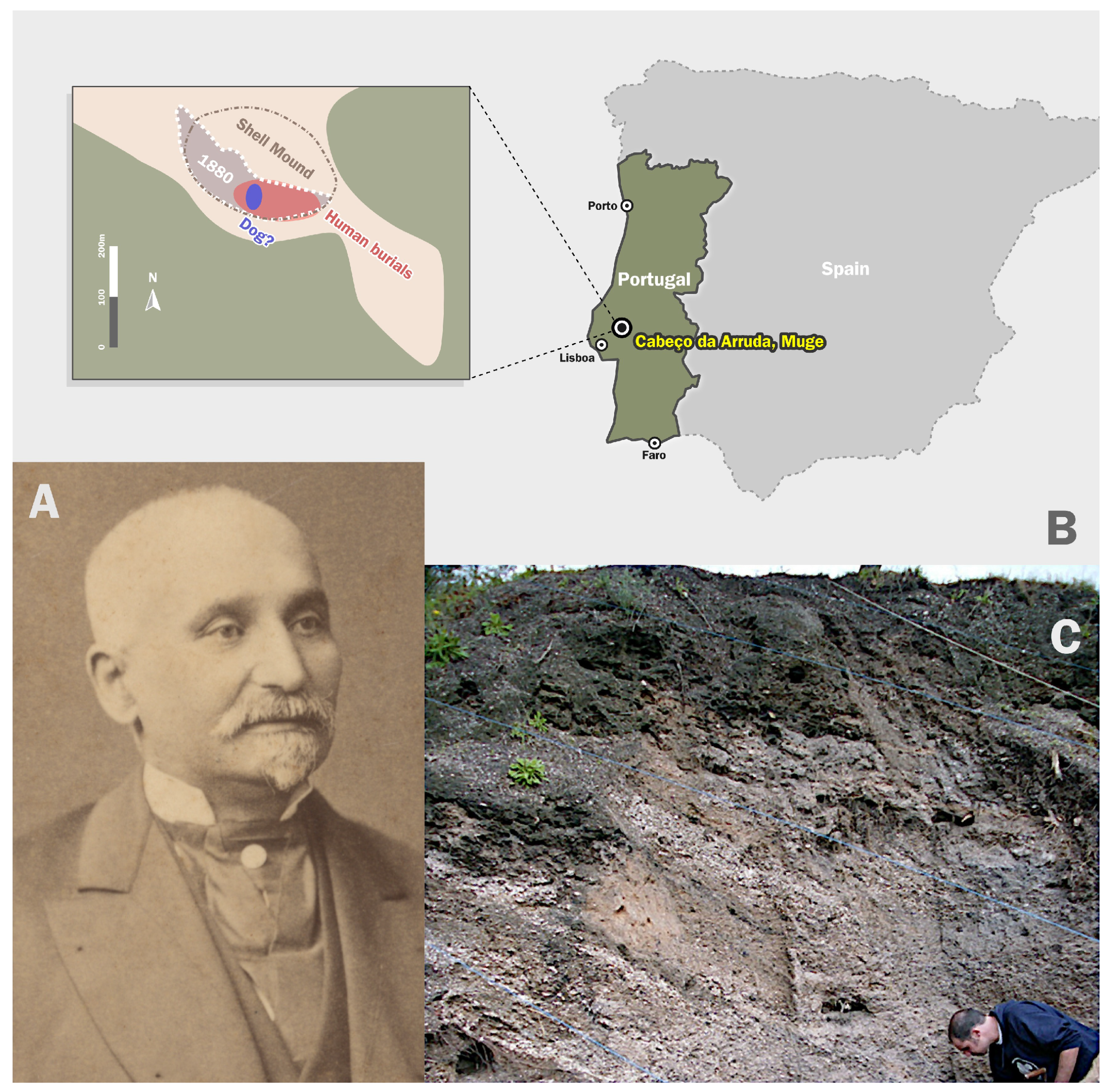
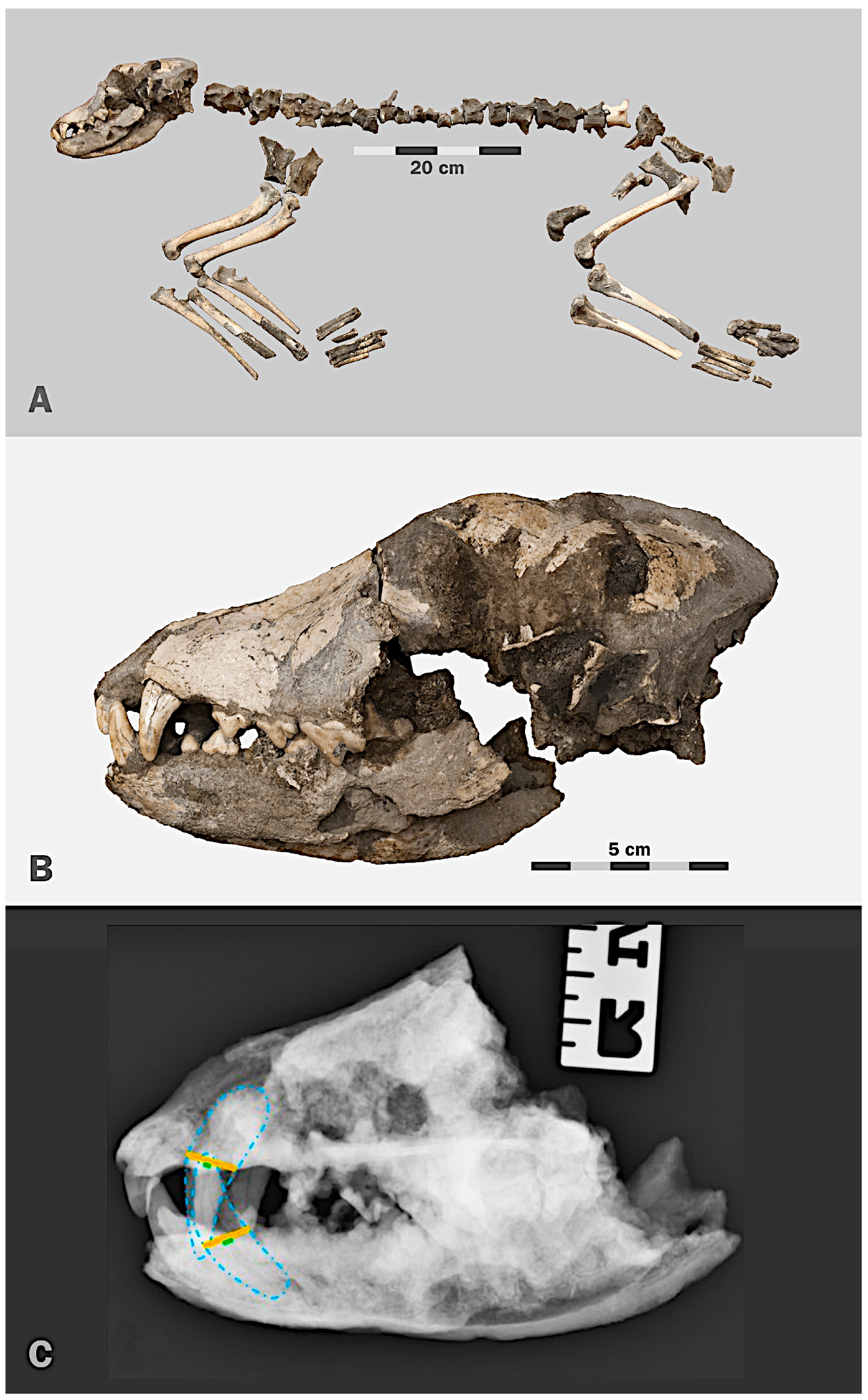
2.1. Sample

The zooarchaeological sample know as the Muge dog, dates to 7680–7450 cal BP, and was used as a case study for the application of a digital strategy enabling its 3D facial reconstruction.

2.2. Computed Tomography (CT) Acquisition

The Muge dog skull was subjected to a computed tomography (CT) scan using a Toshiba Astelion 16-slice CT scanner, at the Faculty of Veterinary Medicine, University of Lisbon (Portugal) (images https://drive.google.com/file/d/1nnIZlh7Vku_ZaCgM44V2C3614EiWv2xI/view?usp=sharing, accessed on 6 April 2022). The following scanning parameters were used: 120 kV, 200 mA; 3.0 mm slice thickness; 0.3 mm interslice distance; slice reconstruction at 0.5 mm; field of view (FOV) of 240. The skull CT images were then analyzed using a DICOM image processing software (Horos 64-bit).

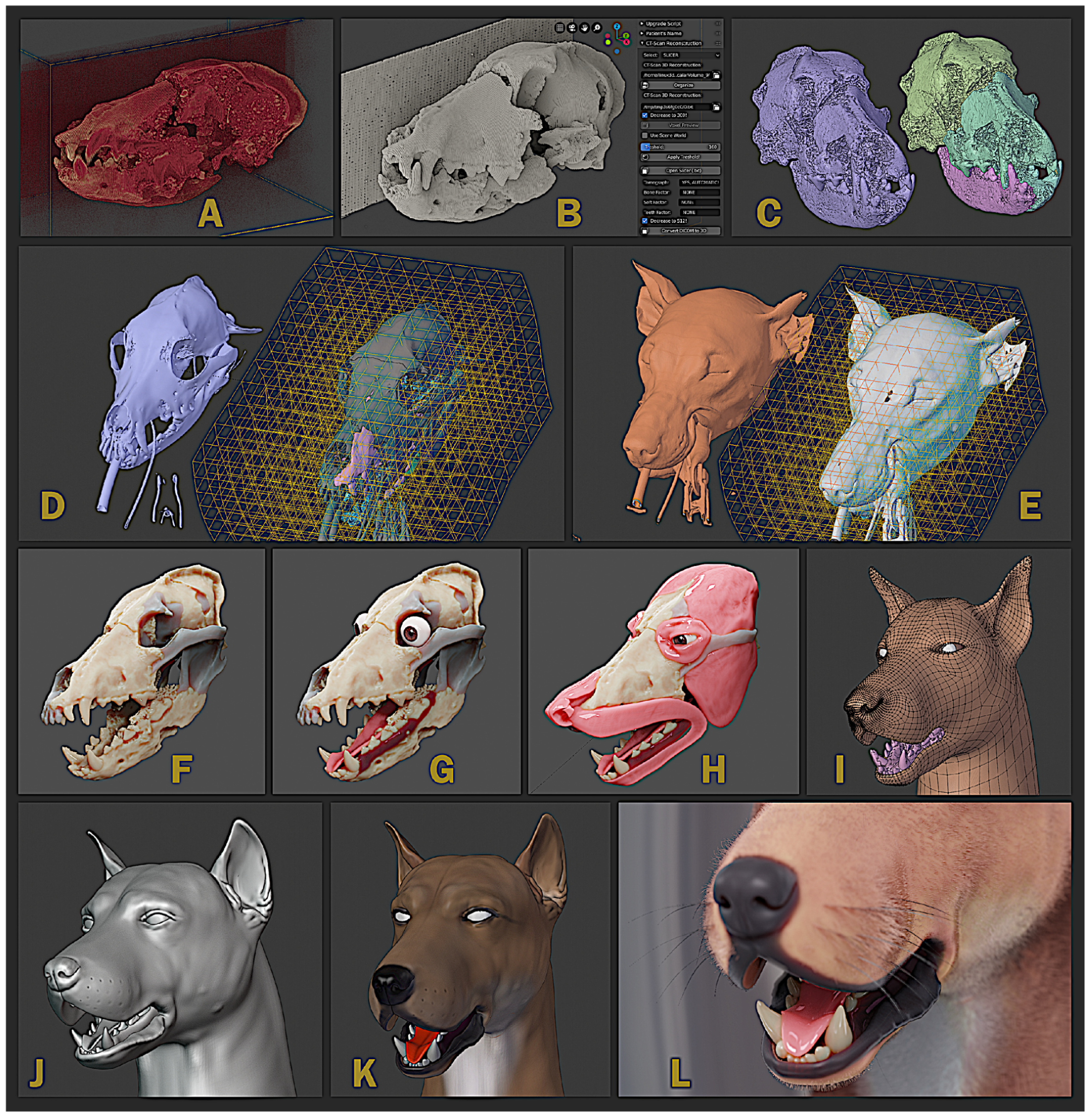
2.3. Virtual Tridimensional Reconstruction of the Skull

Forensic facial reconstruction is an ancillary technique for facial recognition that uses the skull of an individual as a working basis (Taylor, 2000) [15]; (Wilkinson, 2004) [16]. This approach is generally used for the reconstruction of human faces. In the present work, it has been adapted to reveal the Muge dog’s face features. The authors used the modeling platform offered by OrtogOnBlender, a set of scripts and solutions based on free and open source software, which allow working with medical images and facial reconstructions within the Blender 3D software (https://github.com/cogitas3d/OrtogOnBlender, accessed on 6 April 2022) (Moraes, 2020) [17].
Initially, the Muge dog´s CT images in the voxel mode were imported and used to study the structure (Figure 3A) and select the threshold of interest (hounsfield 120℃220) for the reconstruction of the 3D mesh (B). The reconstructed mesh was edited so that bones could be segmented and organized, e.g., rotated, oriented and placed in their expected anatomical position (C). As bones were deformed (due to the burial conditions), it was necessary to use the tomography of a virtual donor or a modern analogue to serve as a guided reference in the organization of the anatomical structures and also to provide the few bone fragments that were absent in the Muge dog. The virtual donor (CT images https://drive.google.com/drive/folders/14Zrztp58JxzCDYUOCfQebM0wnMNr0Me8?usp=sharing, accessed on 6 April 2022) was a dog of the Belgium Shepherd Malinois breed (a male). This specimen was selected since its skull size and conformation matched that of the Muge dog. From the virtual donor reference, missing bone parts (D, on the left) and soft tissues (E, on the left) were reconstructed in the Muge dog. The two meshes resulting from this procedure were subjected to the lattice modifier, which deforms the objects (skull and soft tissue from the donor) inside the structure (skull of the archaeological specimen) according to the adaptations of a three-dimensional grid. Using the fossil (Muge dog) as a reference, soft tissue was hidden and the grid was deformed until the skull of the virtual donor became compatible with the studied skull (D, on the right). Soft tissue was also subjected to the action of the lattice grid, which was checked by making it visible again. The performed deformations were shown, resulting in a face compatible with the fossil (E, on the right). This process allowed missing parts of the skull to be completed with the structure of the virtual donor and, once the bones were segmented, the mandible was rotated to provide the basis for beginning the facial reconstruction (F).
The tongue, eyes and other soft tissue structures were modeled, as well as the teeth, which underwent a remodeling process to mitigate the characteristic irregularities of the fossil, which can still be seen in high resolution images (G). Some major muscles were positioned on the structure (H). Using the deformed mesh of the virtual donor as a base, low-resolution modeling involved the bones and muscles (I). Then, the mesh was subdivided into smaller 3D mesh faces and was detailed with a digital sculpture (J). The resulting sculpture was digitally pigmented (K) and, finally, the hair was distributed along the body surface (L). The obtained final image went through a treatment that converted the color into a gray grade scale. Later, a filter simulating the sepia tone was applied, since accurate information about the animal’s hair texture or coat color remains unknown.
Given the CT images of the Muge dog and the absence of soft tissue data, the use of a virtual donor (or a modern specimen analogue) is the sole approach for its facial reconstruction.

3. Results and Discussion

The Muge dog facial reconstruction is presented in Figure 4 and should be viewed as an approximation only. Extracted files from this reconstruction can be assessed from https://drive.google.com/drive/folders/1oqeCOo_EJjCS9etEdb4CBMWn4iqaNq3y?usp=sharing, accessed on 6 April 2022.
The process described above can be visualized in a short video prepared by our team, (Reconstruction MugeDogFace https://drive.google.com/file/d/1fb_Puu63eT33Ls7g7BJlKkAbdME6JkNX/view?usp=sharing, accessed on 6 April 2022), for the purpose of scientific outreach activities.
To the best of the authors’ knowledge, this is the first attempt of a digital face reconstruction of an early dog that lived around 7600 years ago. As mentioned by Martinez-Labarga and colleagues (Martínez-Labarga et al., 2021) [18], this kind of reconstruction is commonly reported in popular science magazines but less so in scientific publications. This paper tries to bridge that gap. Facial reconstruction results from multidisciplinary techniques and allows the recreation of the physical appearance of an extinct specimen based on its skull parts. Some degree of subjectivity was inevitable but, in this study, it was tentatively reduced to a minimum by restricting it to the reconstruction of the hair coat and to some degree of pigmentation.
A review of the literature relative to dog reconstruction shows a manual reconstruction of two extinct prehistoric dog morphotypes—the wool dog and a village/hunting dog from the New World (Crockford and Pye, 1997) [19]. More recently, the reconstruction—artwork—of an extinct species of wolf, Canis orcensis (from Early Pleistocene), was also published (Martinez-Navarro et al., 2021) [20]. Other approaches, such as sculpture or virtual reconstructions, have also been employed. The first canine forensic reconstruction was recently made public in 2019 and concerns a Neolithic dog dated to 4500 years ago from Scotland’s Orkney Islands. This reconstruction started with a CT-scan of one of the canine skulls and, afterwards, a 3D-print was prepared [21].
Our study attempted a virtual approach to depict a Mesolithic dog. Our approach used the anatomical deformation approach of Subsol and Quatrehomme (2005) [22], as information about the Muge dog’s soft tissues thickness was absent. The advantage of using a reference, such as an extant analogue (same species) provides useful information regarding the localization of the skeletal parts, as well as of the soft tissues. In this case, it was the only option available. Although forensic facial reconstruction is a well-established and well-documented technique (Taylor, 2000 [15]; Wilkinson, 2004 [16]), the need for soft tissue thickness markers (Gietzen et al., 2019) [23] impaired its application to our study. These markers provide robust statistical support by limiting the skin surface of the reconstructed object. However, in this case, a large study that includes dogs of a breed compatible with the Muge dog would be necessary to, for example, obtain reference values of the skin thickness of canine species. Fortunately, new technologies allow us to use modern analogues as references. In this specific case, computed tomography data from a donor were used, which was converted into two three-dimensional meshes with structural data from the bones (skull) and the soft tissues (e.g., skin, muscle).
The anatomical deformation technique was previously tested on animals of different species or genera, such as the conversion of a Pan troglodytes into a Gorilla gorilla and vice versa (Bezzi and Moraes, 2015) [24]. The initial studies using the anatomical deformation approach (using tomographic images as source materials) resulted in an exhibition entitled FACCE (Bezzi et al., 2015) [25] at the Museum of the University of Studies in Padua. Here, the reconstructed faces of dozens of extinct human ancestors were put on display. Another example is the facial reconstruction of a female dated from the Roman period from Falagueira, Portugal (Dias, 2020) [26] and (Rosa, 2021) [27]. In addition to its usefulness to archaeology, the anatomical deformation technique was also applied in the veterinary medicine field for the manufacture of beaks’ prostheses for species of toucan (IE Staff, 2016) [28], goose (Dormehl, 2016) [29], macaw parrot (Hooper, 2016) [30], crow (Duncombe, 2017) [31] and tortoise shell (Thorbecke, 2016) [32], which demand high anatomical rigor. Human medicine (e.g., surgical planning) has also benefited (Façanha de Carvalho et al., 2021) [33], since the anatomical deformation allowed for the adjustment of prostheses using virtual donors or even mirroring healthy parts of the face (cogitas3d, 2016 [34]; Cunha et al., 2020 [35]; Gamarra et al., 2019 [36]; Salazar-Gamarra, 2020) [37]. It is an approach based on real structures and deformations that maintain anatomical proportions.
The Muge dog is dated to the Mesolithic, so this facial reconstruction is extremely important. Using a multidisciplinary approach combining zooarchaeological osteometrics, direct radiocarbon dating, X-ray and CT imaging, computer-assisted techniques and art, we have been able to reassure the importance of this archaeological specimen and provide a three-dimensional facial reconstruction of this ancient dog, without the potential implications (social, legal, religious) of misidentification, as can happen with humans.
In our experience with the short movie previously produced and mentioned above, reconstruction of archaeological specimens into their original form improves the general public’s understanding and illustrates the importance of archaeo-sciences, enabling a more profound bond with our past and increases awareness towards our patrimony. Archaeology and heritage need to be more understandable to the general public in order to be more valued and supported. The reconstruction of ancient pets can assist this purpose. Being animals, and, in particular, pets, they induce more empathy and therefore attract more attention.

4. Conclusions

The anatomical deformation approach was first applied in the archaeology field (e.g., FACCE exhibition), then applied in the clinic (human and veterinary) and now applied to a zooarchaeological remain. The knowledge arising from the practice of facial reconstructions has been used to improve the quality of life (such as the case of the turtle shell) and has helped to construct prosthetics of essential parts of the skeleton that allow wild and domestic animals to keep crucial functions, such as feeding and walking. Another application can be to keep history and prehistory documented. Albeit being an approximation, the portrait of the Muge dog allows for a greater recognition for this truly important archaeological finding. In the future, when genetic information is avaiable we expect to reconstruct the full body of the animal and possibly provide the hair color for a more realistic portrait.

Author Contributions

C.M., formal analysis, software; H.M.P., formal analysis, data curation; J.F.R., roles/writing—original draft; writing—review and editing; L.A., roles/writing—original draft, writing—review and editing; G.A.-P., data acquisition, roles/writing—original draft; S.d.J., data acquisition, roles/writing—original draft; S.G., roles/writing—original draft; C.G., funding acquisition, investigation, project administration, roles/writing—original draft, writing—review and editing; C.D., funding acquisition, investigation, project administration, roles/writing—original draft, writing—review and editing; M.R., data curation; A.E.P., conceptualization, data curation, funding acquisition, investigation, project administration, roles/writing—original draft, writing—review and editing. All authors have read and agreed to the published version of the manuscript.

Funding

This work was funded by National Funds through FCT-Fundação para a Ciência e a Tecnologia in the scope of the project UIDB/50027/2020. The research leading to these results has also received funding from FEDER Funds through the Operational Competitiveness Factors Program “COMPETE”, and by National Funds through FCT in the scope of the projects POCI-01-0145-FEDER-029545—PTDC/HAR—ARQ/29545/2017, UIDB/CVT/00772/2020 and LA/P/0059/2020, as well as from individual grants and contracts DL57/2016/CP1440/CT0029 (AEPires), DL 57/2016/CP1443/CT0029 (CDetry), 2020.02754.CEECIND (CGinja) and CEECINS/00127/2018 (JRequicha).

Institutional Review Board Statement

Not applicable to zooarchaeological specimens.

Informed Consent Statement

Not applicable to zooarchaeological specimens.

Data Availability Statement

Data concerning this study can be assessed from “Dataset” (“Donor dog head CT scans”; “Muge dog head CT scans” and “Reconstruction files” at https://drive.google.com/drive/folders/1q1Dru6WgNL6ffY-YVqJcGXdsWjuXd80g?usp=sharing, accessed on 6 April 2022).

Acknowledgments

Tribute: Miguel Ramalho was a Geologist, Professor at Faculdade de Ciências, at Universidade de Lisboa (Portugal) and Director of the Museu Geológico (Lisbon, Portugal) promoting the Portuguese palaeontological and archaeological patrimony. He had a special affection for the Muge dog skeleton and was his main guardian. Ramalho did everything he could to facilitate our access to this special/protected “wonder”, making its CT scan analysis possible. Without his precious participation, this study could not have been accomplished. We thank David Gonçalves for a critical review of this paper at its early stage; Ryan Schmidt for English revision; and Ludmilla Blaschikoff for helping with the map (Figure 2B) and citations using the reference management software Paperpile.

Conflicts of Interest

The authors declare no conflict of interest.

References

  1. Ribeiro, C. Note sur le terrain quaternaire du Portugal. Bull. Société Géologique Fr. 1867, 692–717. [Google Scholar]
  2. Cardoso, J.L. Primórdios dos estudos pré-históricos em Portugal: Os concheiros mesolíticos de Muge (Salvaterra de Magos) e os seus exploradores. In Memoriam. Estudos de Homenagem a António Augusto Tavares; Cardoso, J.L., Sales, J.C., Eds.; Universidade Aberta: Lisbon, Portugal, 2018. [Google Scholar]
  3. Detry, C.; Cardoso, J.L. On some remains of dog (Canis familiaris) from the Mesolithic shell-middens of Muge, Portugal. J. Archaeol. Sci. 2010, 37, 2762–2774. [Google Scholar] [CrossRef]
  4. Cunha, E.; Cardoso, F. New data on Muge shell middens: A contribution to more accurate numbers and dates. Muge Estud. Arqueol. 2003, 1, 171–184. [Google Scholar]
  5. Roksandic, M. Analysis of burials from the new excavations of the sites Cabeço da Amoreira and Cabeço da Arruda (Muge, Portugal). In Do Epipalaeolítico Ao Calcolítico Na Península Ibérica; Bicho, A.H.V., Ed.; Universidade do Algarve: Faro, Portugal, 2006; pp. 43–54. [Google Scholar]
  6. Pires, A.E.; Detry, C.; Chikhi, L.; Rasteiro, R.; Amorim, I.R.; Simões, F.; Matos, J.; Petrucci-Fonseca, F.; Ollivier, M.; Hänni, C.; et al. The curious case of the Mesolithic Iberian dogs: An archaeogenetic study. J. Archaeol. Sci. 2019, 105, 116–129. [Google Scholar] [CrossRef]
  7. Cardoso, J.L. (Ed.) Carlos Ribeiro (1813–1882) geólogo e arqueólogo. Homenagem da Câmara Municipal de Oeiras e da Academia das Ciências de Lisboa nos 200 anos do seu nascimento. In Estudos Arqueológicos de Oeiras; Câmara Municipal de Oeiras: Oeiras, Portugal, 2013; Volume 20, 800p. [Google Scholar]
  8. Cardoso, J.L.; Rolão, J.M. Prospecções e escavações nos concheiros mesolíticos de Muge e de Magos (Salvaterra de Magos): Contribuição para a história dos trabalhos arqueológicos efectuados. Estud. Arqueol. Oeiras 2000, 8, 83–240. [Google Scholar]
  9. Boudadi-Maligne, M.; Mallye, J.-B.; Langlais, M.; Barshay-Szmidt, C. Des restes de chiens magdaléniens à l’abri du Morin (Gironde, France). Implications socio-économiques d’une innovation zootechnique. Paléo 2012, 23, 39–54. [Google Scholar] [CrossRef]
  10. Baumann, C.; Pfrengle, S.; Münzel, S.C.; Molak, M.; Feuerborn, T.R.; Breidenstein, A.; Reiter, E.; Albrecht, G.; Kind, C.-J.; Verjux, C.; et al. A refined proposal for the origin of dogs: The case study of Gnirshöhle, a Magdalenian cave site. Sci. Rep. 2021, 11, 1–14. [Google Scholar] [CrossRef] [PubMed]
  11. Street, M.; Napierala, H.; Janssens, L. The late Palaeolithic dog from Bonn-Oberkaassel in context. In The Late Glacial Burial from Oberkassel Revisited; Giemsch, L., Schmitz, R.W., Eds.; Verlag Philipp von Zabern GmbH: Darmstadt, Germany, 2015; pp. 253–274. ISBN 978-3-8053-4970-3. [Google Scholar]
  12. Pionnier-Capitan, M.; Bemilli, C.; Bodu, P.; Célérier, G.; Ferrié, J.-G.; Fosse, P.; Garcià, M.; Vigne, J.-D. New evidence for Upper Palaeolithic small domestic dogs in South-Western Europe. J. Archaeol. Sci. 2011, 38, 2123–2140. [Google Scholar] [CrossRef]
  13. Vigne, J.D. L’humérus de chien magdalénien de Erralla (Gipuzkoa, Espagne) et la domestication tardiglaciaire du loup en Europe. Munibe 2005, 51, 279–287. [Google Scholar]
  14. Nomokonova, T.; Losey, R.J.; McLachlin, K.; Bachura, O.P.; Gusev, A.V.; Kosintsev, P.A.; Fedorova, N.V.; Sablin, M.V. Age estimation of archaeological dogs using pulp cavity closure ratios. J. Archaeol. Sci. 2020, 123, 105252. [Google Scholar] [CrossRef]
  15. Taylor, K.T. Forensic Art and Illustration; CRC Press: Boca Raton, FL, USA, 2000. [Google Scholar] [CrossRef]
  16. Wilkinson, C. The history of facial reconstruction. In Forensic Facial Reconstruction; Cambridge University Press: Cambridge, UK, 2004; pp. 39–68. [Google Scholar] [CrossRef]
  17. Moraes, C. OrtogOnBlender: Documentação Oficial. Brazil: Sinop/MT. 2020. Available online: http://www.ciceromoraes.com.br/doc/pt_br/OrtogOnBlender/index.html (accessed on 5 April 2022).
  18. Martínez-Labarga, C.; Carbone, R.; Ridolfi, V.; Parrino, M.; Vitali, S.; Bravi, L.; Rickards, O.; Falconi, M. Craniofacial reconstruction of Raphael Sanzio from Urbino: Face and features of a “mortal god”. Digit. Appl. Archaeol. Cult. Herit. 2021, 22, e00190. [Google Scholar] [CrossRef]
  19. Crockford, S.J.; Pye, C.J. Forensic reconstruction of prehistoric dogs from the northwest coast. Can. J. Archaeol./J. Can. D’Archéol. 1997, 21, 149–153. [Google Scholar]
  20. Martinez-Navarro, B.; Lucenti, S.B.; Palmqvist, P.; Ros-Montoya, S.; Madurell-Malapeira, J.; Patrocinio Espigares, M. A new species of dog from the Early Pleistocene site of Venta Micena (Orce, Baza Basin, Spain). Comptes Rendus Palevol 2021. [Google Scholar] [CrossRef]
  21. The Guardian. Neolithic Dog Reveals Tales behind Orkney’s Monuments. The Guardian. 2018. Available online: https://www.theguardian.com/uk-news/2019/apr/13/neolithic-dog-reveals-tales-behind-orkney-monuments (accessed on 22 April 2022).
  22. Subsol, G.; Quatrehomme, G. Automatic 3D facial reconstruction by feature-based registration of a reference head. In Computer-Graphic Facial Reconstruction; Clement, J.G., Marks, M.K., Eds.; Academic Press: Cambridge, MA, USA, 2005; Volume 5, pp. 79–101. [Google Scholar]
  23. Gietzen, T.; Brylka, R.; Achenbach, J.; Zum Hebel, K.; Schömer, E.; Botsch, M.; Schwanecke, U.; Schulze, R. A method for automatic forensic facial reconstruction based on dense statistics of soft tissue thickness. PLoS ONE 2019, 14, e0210257. [Google Scholar] [CrossRef] [PubMed]
  24. Bezzi, L.; Moraes, C. Archaeological forensic facial reconstruction with FLOSS, in: 43 Rd Computer Applications and Quantitative Methods in Archaeology. In Proceedings of the Computer Applications and Quantitative Methods in Archaeology, Siena, Italy, 30 March–3 April 2015; p. 416. [Google Scholar]
  25. Bezzi, A.; Bezzi, L.; Moraes, C.; Carrara, N.; Pievani, T.; Tiziani, M. Facce. I Molti Volti Della Storia Umana. UNA Mostra Open Source. 2015. Available online: http://www.archcalc.cnr.it/indice/Suppl_8/35_Bezzi_et_al.pdf (accessed on 5 April 2022).
  26. Dias, G.E.A. Povoamento em época Romana na Amadora—resultados de um projeto pluridisciplinar. In Arqueologia Em Portugal—2020 Estado Da Questão; José Morais, A., Neves, C., Martins, A., Eds.; Associação dos Arqueólogos Portugueses e CITCEM: Porto, Portugal, 2020; pp. 1361–1370. [Google Scholar]
  27. Rosa, G.P. A Dama Romana da Amadora; National Geographic Portugal: Porto, Portugal, 2021; pp. 70–73. [Google Scholar]
  28. IE Staff. Toucan Found Injured on Roadside Gets New 3D-Printed Beak. Inside Edition. 2016. Available online: https://www.insideedition.com/15976-toucan-found-injured-on-roadside-gets-new-3d-printed-beak (accessed on 5 April 2022).
  29. Dormehl, L. Team Uses 3D Printing to Save the Life of Injured Goose Named Victoria. Digital Trends. 2016. Available online: https://www.digitaltrends.com/cool-tech/3d-printed-goose-beak-brazil-animal-avengers/ (accessed on 5 April 2022).
  30. Hooper, B. Injured Macaw Gets 3D-Printed Titanium Beak in Brazil. UPI. 2016. Available online: https://www.upi.com/Odd_News/2016/02/25/Injured-macaw-gets-3D-printed-titanium-beak-in-Brazil/4481456433334/ (accessed on 5 April 2022).
  31. Duncombe, E. Hood You Believe It—Build a Beak—Storytrender. 2017. Available online: http://www.storytrender.com/12088/hood_believe-build-beak/ (accessed on 5 April 2022).
  32. Thorbecke, C. Tortoise Burned in Fire Gets Custom 3-D-Printed Shell. ABC News. 2016. Available online: https://abcnews.go.com/International/tortoise-burned-fire-custom-3d-printed-shell/story?id=39306133 (accessed on 5 April 2022).
  33. Façanha de Carvalho, E.; Alkmin Paiva, G.L.; Yonezaki, F.; Machado, G.G. Computer-Aided Surgical Simulation in Severe Atrophic Mandibular Fractures: A New Method for Guided Reduction and Temporary Stabilization Before Fixation. J. Oral Maxillofac. Surg. 2021, 79, 892.e1–892.e7. [Google Scholar] [CrossRef] [PubMed]
  34. cogitas3d. Low Cost Human Face Prosthesis with the Aid of Blender and 3D Printing. 2016. Available online: https://www.blendernation.com/2016/12/21/low-cost-human-face-prosthesis-aid-blendern-3d-printing/ (accessed on 5 April 2022).
  35. Cunha, H.S.; da Costa Moraes, C.A.; de Faria Valle Dornelles, R.; da Rosa, E.L.S. Accuracy of three-dimensional virtual simulation of the soft tissues of the face in OrtogOnBlender for correction of class II dentofacial deformities: An uncontrolled experimental case-series study. Oral Maxillofac. Surg. 2020, 25, 319–335. [Google Scholar] [CrossRef] [PubMed]
  36. Gamarra, R.; Seelaus, R.; da Silva, J.V.L.; Dib, L.; Moraes, C. Introdução à metodologia “mais identidade”: Próteses faciais 3D com a utilização de tecnologias acessíveis para pacientes sobreviventes de câncer no rosto. In Comunicação Científica E Técnica Em Odontologia; Atena Editora: Ponta Grossa, Brazil, 2019; pp. 251–272. [Google Scholar]
  37. Salazar-Gamarra, R.; Moraes, C.A.; Costa, D.; Rosa, E.L.S.D.; Seelaus, R.; Silva, J.V.L.D.; Dib, L. Multiresolution and displacement: Modifier attribution to enhance realistic 3D photogrammetry for models of the face. Biomed. J. Sci. Tech. Res. 2020, 29, 22812–22817. [Google Scholar] [CrossRef]
Figure 1. (A) A portrait of the archaeologist Carlos Ribeiro, who excavated the Muge dog in 1880 (photograph kindly shared by Prof JL Cardoso [7]); (B) map of the Iberian Peninsula displaying the Cabeço da Arruda shell middens location with detail about the place where human burials and the Muge dog were found. There is some imprecision regarding the exact place from where the dog was excavated. Adapted from (Cardoso and Rolão 2000) [8]; (C) section of the Cabeço da Arruda stratigraphic complex profile (photograph kindly shared by Anabela Joaquinito), since it shows how the superficial layer and the others below lie in different directions. Additionally, the layers have been disturbed by animal burrows.
Applsci 12 04867 g001
Figure 2. (A) Almost complete skeleton of the Muge dog (photograph kindly shared by JPRuas) (Detry and Cardoso, 2010) [3]; (B) skull showing deformations, fractures and calcareum concretions (photograph kindly shared by JPRuas) (Detry and Cardoso, 2010) [3]; (C) radiographic image of the Muge dog skull indicating the pulp canal (in blue) versus tooth widths (yellow) for both maxillary and mandibular canine teeth (surrounded by a blue dotted line), parameters that are useful for the estimation of age at death.
Applsci 12 04867 g002
Figure 3. Snapshots of the process for the virtual tridimensional reconstruction of the Muge dog skull and, finally, the face (CC BY-SA 4.0). (A) Muge dog´s CT images in the voxel model. (B) Threshold of interest for the reconstruction (hounsfield 120-220). (C) Reconstructed mesh with bones in their expected anatomical position. (D,E) Bone and soft tissue deformation using the tomography of a virtual donor, and applying the lattice modifier. (F) Obtained model used as base for facial reconstruction. (G) Modelling of the teeth, tongue, eyes and other soft tissue structures. (H) Modelling of muscles in the correct position. (I) Low resolution modeling involved the bones and muscles, using the deformed mesh. (J) Digital sculpture obtained from smaller 3D mesh faces. (K) Digital pigmentation. (L) Final result, including the hair distributed along the body surface.
Applsci 12 04867 g003
Figure 4. The picture of the Muge dog 3D model in sepia tones due to the lack of genetic information regarding its coat texture and color (CC BY-SA 4.0).
Applsci 12 04867 g004
Publisher’s Note: MDPI stays neutral with regard to jurisdictional claims in published maps and institutional affiliations.

