Review

Important Roles of Oligo- and Polysaccharides against SARS-CoV-2: Recent Advances

by
Siavash Iravani
1,* and
Rajender S. Varma
2,*
1
Faculty of Pharmacy and Pharmaceutical Sciences, Isfahan University of Medical Sciences, Isfahan 81746-73461, Iran
2
Regional Centre of Advanced Technologies and Materials, Palacký University in Olomouc, Šlechtitelů 27, 783 71 Olomouc, Czech Republic
*
Authors to whom correspondence should be addressed.
Appl. Sci. 2021, 11(8), 3512; https://doi.org/10.3390/app11083512
Submission received: 20 March 2021 / Revised: 9 April 2021 / Accepted: 12 April 2021 / Published: 14 April 2021
(This article belongs to the Special Issue COVID-19: Impact on Human Health and Behavior)

Abstract

:
The severe acute respiratory syndrome coronavirus 2 (SARS-CoV-2)-initiated outbreak of COVID-19 has spread rapidly around the world, posing a huge threat to public health. Natural oligo- and polysaccharides with low toxicity, good sustainability, high biocompatibility, respectable safety, immune regulation, and antiviral activity can be employed as promising candidates for the prevention and inhibition of viral infections, especially COVID-19. Glycosaminoglycans, marine polysaccharides, terrestrial plant polysaccharides, and some others have exhibited potential antiviral activity against pathogenic viruses, in the format of polysaccharide-centered vaccine adjuvants, nano-based structures, drug conveyance platforms, etc. In this review, significant recent advancements pertaining to the antiviral applications of oligo- and polysaccharides against SARS-CoV-2 are highlighted, including important challenges and future perspectives.

1. Introduction

Various types of antiviral drugs are being examined against severe acute respiratory syndrome coronavirus 2 (SARS-CoV-2), but unfortunately, several of these candidates are often toxic and have several side effects [1,2]. Therefore, it is imperative to search for virucidal drugs with low or no toxicity that irreversibly inhibit viruses. In this regard, various naturally occurring compounds have been evaluated for their potential inhibitory activities against viral infections [2,3]. Among them, natural polysaccharides are effective inhibitors of assorted pathogenic viruses, such as influenza A virus, human cytomegalovirus (HCMV), herpes simplex virus (HSV), human immunodeficiency virus (HIV), dengue virus (DENV), respiratory syncytial virus (RSV), human metapneumovirus (hMPV), severe acute respiratory syndrome coronavirus (SARS-CoV), and Middle East respiratory syndrome-related coronavirus (MERS-CoV) [4,5,6]. For instance, sulfated polysaccharides were examined as topical microbicidal agents in medical trials to thwart viruses-triggered sexually transmitted diseases (STDs). Accordingly, various polysulfate compounds have shown promising potentials for inhibition of the virus replication by obstructing the virion attachment to the host cell [4,7]. However, the efficacy of these compounds in vivo against these viral infections has some important challenges and drawbacks, including poor absorption, failure to reach target tissues, noxious side effects, and anticoagulant characteristics. Moreover, there is a demand for probing new materials, especially for the drug resistance to antiviral agents during therapy [4,7].
Importantly, the intricacy of marine polysaccharides and their derivatives and their structural variety are responsible for their antiviral actions in diverse stages of various viral infection procedures (Figure 1) [8]. Different types of seaweed polysaccharides, comprising agarans, ulvans, fucoidans, carrageenans, laminarins, and alginates, have exhibited appropriate antiviral effects, and their promising potentials for therapeutic appliances are garnering remarkable attention [8]. These materials with distinctive structures exert antiviral effects via interference at various phases of the viral infection cycle. Additionally, polysaccharides and, especially, sulfated polysaccharides are endowed with significant polyanionic attributes and can, thus, be employed for blocking the cationic charge on the surface of cells to prevent the virus assault or even adsorption [9]. These sulfated polysaccharides have significant therapeutic potentials, as they can imitate sugar-rich molecules, glycosaminoglycans, prevailing in the membranes of the cells. These glycosaminoglycans such as dermatan sulfate, chondroitin sulfate, and heparan sulfate are vital in mammalian compositions. As an example, receptors of heparan sulfate on the surfaces of the cells are critical in various pathological and physiological processes, besides being necessary for the entry of the virus into vulnerable cells. It has been noted that sulfated polysaccharides compete for binding sites usually used by glycosaminoglycans, thus inhibiting these actions [10,11].
The preventive action of natural sulfated polysaccharides varies with the compound and the virus. In one study, three different forms of carrageenan G3d and the dl-galactan hybrid C2S-3 (two homogeneous sulfated polysaccharides), accessible from the red seaweeds Gymnogongrus griffithsiae and Cryptonemia crenulata, were evaluated for their virucidal potentials against four serotypes of DENV in various host cell types [12]; both seaweed derivatives had specific inhibitory effects against DENV-2 multiplication in Vero cells (selectivity indices >1000 and half maximal inhibitory concentration (IC50 = 1 μg mL−1)). However, these materials showed inferior antiviral effects for DENV-3 (IC50 = 13.9–14.2 μg mL−1) and an even poorer effect for DENV-4 (IC50 = 29.3 to >50 μg mL−1), and they were completely ineffective versus DENV-1 [12]. These compounds were inhibitors for DENV-2 when simply added simultaneously with the virus or at an early infection stage, and both initial processes of virus adhesion and incorporation were the major aims of these compounds. Consequently, the virucidal action of polysaccharides hinges on the host cell and viral serotype, which can be altered in the cell–virus interface leading to the entry of virus [12].
Green chemistry provokes the application of natural and biorenewable resource-based feedstocks to prepare valuable materials [13,14,15]. Polysaccharides with their good antiviral activities, low toxicity, good biocompatibility, high biodegradability, renewability, and safety can be utilized as promising anti-CoVs agents. Further, polysaccharide adjuvants can improve the antiviral effects of vaccines and immunity and may engage in imperative functions in antiviral nanomaterials and transport systems; the argument for the development of a vaccine for COVID-19 by the fusion of nanotechnology and polysaccharides should be heeded, carefully. Oligo- and polysaccharides can fight against the pathogenic viruses by the direct interaction with the virus (negative charge on the surface of viruses), inhibition of the invasion and adsorption of viruses, and also the obstruction of the viral transcription and replication via direct interference with viral reproduction-associated enzymes and targets in host cells) [16]. As an example, the virucidal action of seaweed-derived sulfated polysaccharides was predominantly exercised during DENV-2 adhesion and incorporation [17]. Further, N-sulfonated poly (allylamine) hydrochloride-derived iota-carrageenan demonstrated remarkable virucidal action versus hMPV, a type of respiratory infection RNA virus, by obstructing the discharge of viruses from the membrane of cells and prohibiting the viral adhesion [18]. In this review, significant recent advancements pertaining to the antiviral effects of oligo- and polysaccharides against SARS-CoV-2 are highlighted, including important challenges and future perspectives.

2. Oligo- and Polysaccharides against SARS-CoV-2

Polysaccharides are polymeric carbohydrates that are formed naturally by various identical or different monosaccharide units (Cn(H2O)n) connected via glycosidic bonds [5,19]. SARS-CoV-2 deploys its vastly glycosylated trimeric spike protein to attach to the angiotensin-converting enzyme 2 (ACE2) glycoprotein on the cell surface receptor for facilitating entry into the host cell [20]; the transmembrane spike glycoprotein of CoVs exists as a homotrimeric form, and it is covered with N-linked glycans. S glycoprotein is recognized as the main target of antibodies with a neutralizing potency, and it has been examined as an attractive target for therapeutic or vaccine development. The targeting of N-linked glycans of the S glycoprotein envelope of CoVs through carbohydrate-binding agents could serve as an attractive therapeutic strategy for developing innovative antivirals. Carbohydrate-binding agents from natural resources like lectins from plants, and marine algae and prokaryotes and lectin mimics like Pradimicin-A have exhibited antiviral performances against CoVs and other enveloped viruses. However, the potential application of carbohydrate-binding agents and lectins has been restricted because of unfavorable responses such as immunogenicity, mitogenicity, hemagglutination, inflammatory activity, and cellular toxicity [21]. The mammalian’s cell surface is coated with intricate glycans or polysaccharides where many viruses become attached [22]. These glycans on the cell surface, namely heparan sulfate proteoglycan, can enhance the host cells infection. Additionally, glycans on such cell surfaces function as receptors implicated in the transfer of signals induced by endocytosis or in the initiation of blending among the cell membrane and the viral envelope for certain viruses. Alternatively, the viral proteins can be glycosylated as viruses exploit the machinery of the host cell, thus affecting the function and stability of the viral glycoprotein while making an entry into the host cell. Further, it has been shown that viral glycoprotein glycosylation is implicated in viral antigenicity accountable for virus’s immune avoidance [23].
Natural polysaccharides are sustainable, renewable, abundant, and environmentally friendly biopolymers readily available from diverse natural bioresources, namely animals (e.g., gelatin/collagen, silk, chitosan, hyaluronic acid, or hyaluronan), plants (e.g., pectin, cellulose, and starch), algae (e.g., agar and alginate), and microorganisms (e.g., xanthan gum, dextran, and pullulan) [24,25,26,27]. Polysaccharides from natural/edible origins can help modulate the immunity against SARS-CoV-2 via several pathways (Figure 2); glucans and chitosan can be utilized for the delivery of antigens on various platforms and in vaccine developments [28]. These polysaccharides retain different chemical structures, including branched or linear arrangements, diverse charge states (neutral, negatively or positively charged), varying chemical compositions (α and/or β-glycosidic bonds among assorted monosaccharides units), and a wide range of molecular masses [29]. Polysaccharides represent promising potentials for pharmaceutical and biomedical applications, in terms of the chemical diversity of their structures, physicochemical and biological features, safety, high chemical reactivity, biocompatibility, renewability, low toxicity, and biodegradability. The high biocompatibility and biodegradability, as well as low/non-toxicity, of these materials contribute toward the enhancement of their biomedical appliances as immunogenicity depends on the individual structures [30,31].
Polysaccharides have exhibited several attractive anti-CoVs effects, and they can be utilized in the form of vaccine adjuvants that are polysaccharide- and nano-based structures, and innovative drug conveyance systems versus these pathogenic viruses [8,32,33,34]. Importantly, they have exhibited directly and indirectly antiviral effects against SARS-CoV-2 [16]; some important mechanistic aspects proposed by Zhao et al. are described in Figure 3 [16]. It was reported that marine natural compounds have displayed potential inhibitory effects against SARS-CoV-2, and these can be employed for the better management of COVID-19 [6]. In an impressive study, these polysaccharides were examined against COVID-19, encompassing four marine sulfated polysaccharides that have been evaluated for their inhibitory activity against SARS-CoV-2, comprising fucoidan from brown algae, sulfated polysaccharide from sea cucumber, chondroitin sulfate C from sharks, and iota-carrageenan from red algae [35]. Accordingly, carrageenan, sea cucumber sulfated polysaccharide, and fucoidan had remarkable antiviral performances at 3.90–500 μg mL−1; the sea cucumber sulfated polysaccharide showed a robust inhibitory activity with IC50 of 9.10 μg mL−1. Additionally, an evaluation by applying the S glycoprotein-equipped pseudotype virus established that sea cucumber sulfated polysaccharide could attach to the S glycoprotein for the prevention of the entrance of SARS-CoV-2 into host cell [35]. In another study, Panavir®, a higher molecular-weight portion of polysaccharides obtained from the shoots of Solanum tuberosum (potato), showed potential against SARS-CoV-2 infection [36]. Besides, polysaccharides obtained from Ecklonia kurome Okam seaweed could inhibit/block the 3CLpro (3-chymotrypsin-like protease) enzyme, which plays important roles in viral replication/transcription; these mixtures of polysaccharides demonstrated efficient anti-SARS-CoV-2 activity (in vitro), and they can be considered as potential candidates in antiviral drug developments and research [37].
Sulfated polysaccharides can be employed as selective inhibitors of several enveloped and nonenveloped viruses, as they mainly act by obstructing the binding or internalization of virus into the host cells [19]. The human ACE2 and SARS-CoV-2 spike glycoproteins are two important targets for preventing and treating COVID-19; heparan sulfate on the host cell surface is assumed to network with spike glycoproteins from SARS-CoV-2 to expedite entry into the host cell. Additionally, the glucuronomannan and sulfated galactofucan demonstrated remarkable fastening competence to SARS-CoV-2 spike glycoproteins, proposing such polysaccharides as promising alternatives for prevention and/or treatment of SARS-CoV-2 [38]. It has been shown that sulfated polysaccharides extracted from Porphyridium sp. (marine red alga) are suitable antiviral agents against respiratory viruses from the family of CoV, and they can be promising agents against SARS-CoV-2 [19]. The initial stage for the virus to enter a cell is to attach to the surface of the cell surface via a receptor, namely heparan sulfated proteoglycan, adorning their surface or by electrostatic means. Importantly, sulfated polysaccharides can attach securely in vitro to the SARS-CoV-2′s S-protein, thus serving as a trap to restrict the attachment of the S-protein to the heparan sulfate co-receptor in tissues of the host, blocking the infection from the virus [5]. It appears that such polysaccharides with low cytotoxicity have propitious in vitro antiviral potentials, and they should be evaluated further for clinical applications [5].

2.1. Chitosan

Chitosan is a naturally occurring cationic polysaccharide polymer with a high biodegradability, significant biocompatibility, and low toxicity, comprising β-(1-4) linkages of glucosamine units, which occurs rarely in nature, but is commonly generated from extensive N-deacetylation of chitin, a homopolymer of N-acetyl-glucosamine present in the exoskeletons of shrimp, shellfish and crabs, cuticles of insects, and cell walls of fungi [39,40,41]. This low-cost polysaccharide is environmentally benign, biodegradable, biocompatible, abundant, mucoadhesive, and is a positively charged nontoxic biopolymer, which displays antiviral [42], antimicrobial [43], and immunoadjuvant effects, making it suitable for various medical and biomedical applications [41,44,45]. The amine groups (NH2) in chitosan-based materials are responsible for their various characteristics, including their cationic nature, in situ gelation, controlled drug release, antimicrobial, and mucoadhesion/permeation enhancement. Chitosan and its derivatives have been employed for cell encapsulation/delivery [46], 3D bioprinting [47], tissue engineering [48,49], and delivery of biomolecule cargo (stimuli response release and targeted drug/gene delivery) [50,51,52]. Further, chitosan has been deployed for the encapsulation and coating of assorted nanoparticles (NPs), thereby fabricating diverse materials with appropriate potentials for the detection of diseases (e.g., cancers) [53,54]. Additionally, it can be employed against pathogenic viruses, as has been reported in the case of curcumin chitosan nanocomposite (~29–39.5 nm) with their potential anti-hepatitis C virus effects, or the incitement of the inherent immune system by the dispensation of chitosan via the nasal route for the protection of BALB/c mice from deadly contagion by the H7N9 virus (influenza A virus subtype H7N9) [55,56]. Besides, chitosan NPs have been used as drug delivery systems/carriers for antiviral drugs, because of their good stability, low toxicity, and providing of versatile routes for their safe administration. As an example, these NPs were utilized for the delivery of didanosine (a medication typically used for the treatment of HIV/AIDS) for improvement of the systemic and brain targeting efficiency of this drug after intranasal administration [57].
SARS-CoV-2 via contact with human ACE2 can invade human respiratory epithelial cells [58]. In an impressive study, it has been illustrated that β-chitosan prevented the attachment, degradation, and internalization of ACE2 with SARS-CoV-2S-receptor-binding domain (RBD), and also inhibited the stimulation of inflammatory gesturing routes; β-chitosan’s inhibitory effect on SARS-CoV-2S-RBD-triggered inflammation has been evaluated [58]. It was revealed that β-chitosan could effectively attach to SARS-CoV-2S-RBD or ACE2, and the ensuing conjugate of ACE2 and β-chitosan could not attach to SARS-CoV-2S-RBD anymore [58]. Remarkably, detection by immunofluorescence means on human ACE2 mice and Vero E6 cells showed that β-chitosan exhibited remarkable preventive and curative effects on SARS-CoV-2S-RBD attachment; this binding could stimulate the inflammation gesturing routes of mice and cells, but β-chitosan could noticeably subdue the SARS-CoV-2S-RBD-triggered inflammations. The conclusions from this study revealed that β-chitosan had an analogous antibody role, which can deactivate SARS-CoV-2S-RBD and can also be successfully applied for blocking the binding between SARS-CoV-2S-RBD and ACE2. The stimulation of ADAM17 by β-chitosan diminished the catalytic activity of angiotensin II and improved the cleavage of the ACE2 extracellular domain followed by the release of the extracellular region into the extracellular surroundings [58]. Sharma et al. [59] described that β-chitosan and its related derivatives can obstruct the infection by SARS-CoV-2 via the inhibition of the spike protein binding to ACE2 receptors, and also chitosan oligosaccharides can downregulate the expression of CD147, which is important in the invasion of viruses via binding to the spike protein (Figure 4) [59].

2.2. Heparin or Heparin-Like Materials

Heparin, an assorted collection of anionic linear-chain mucopolysaccharides, is a component of a glycosaminoglycan family of molecules that comprise hyaluronic acid, keratan sulfate, chondroitin sulfate, and heparan sulfate [60]. The main sugars, among others, present in heparin include β-D-glucuronic acid, α-L-iduronic acid, α-L-iduronic acid 2-sulfate, 2-deoxy-2-sulfamino-α-D-glucose-6-sulfate, and 2-acetamido-2-deoxy-α-D-glucose. Glycosaminoglycan entities are articulated all over the body, with different biological functions, and they are typically related to extracellular matrices (ECM), respiratory and endothelial cell surfaces, and basement membranes. Indeed, various pathogenic bacteria and viruses rest on communications with proteoglycans, namely heparan sulfate articulated on an array of human tissue exteriors, for the sticking to and incursion of host tissues. It was revealed that heparin competed with heparan sulfate for the bonding to bacteria and viruses, thus preventing or restricting the pathogenic invasions [60]. Recently, the antiviral effect of heparin and enoxaparin derivatives, including enoxaparin (IC50 = 1.08 mg L−1), unfractionated heparin (IC50 = 5.99 μg L−1), 6-O-desulfated unfractionated heparin (IC50 = 1.77 μg L−1), and 6-O-desulfated enoxaparin (IC50 = 5.86 mg L−1) has been reported against SARS-CoV-2; it was revealed that the binding of the SARS-CoV-2 spike glycoprotein to glycosaminoglycans (such as heparan sulfate) could be the reason for these inhibitory effects [61].
The viral invasion is typically related to the virus endocytosis, the virus’s fusion with the cell membrane, and its translocation [9]. Heparin or its mimic substances endowed with wide-spread spectrum antiviral characteristics were utilized to imitate the cell surface carbohydrates accountable for the first viral bonding, such as carrageenan and heparan sulfate [9]. Innovatively, the glycosaminoglycan-binding motif was inserted at the S1/S2 proteolytic cleavage site and additional such motifs within the SARS-CoV-2 spike glycoprotein, and it was proposed that glycosaminoglycans present on the cell surfaces of the host might liaise with SARS-CoV-2 spike glycoproteins to accelerate entry into the host cell [62]. It was uncovered that both the trimeric and monomeric SARS-CoV-2 spike glycoproteins could attach more firmly to the immobilized heparin than the MERS-CoV and SARS-CoV spike glycoproteins could. In one study, by adding heparin to Vero cells in the range of 6.25–200 μg mL−1, the incursion by SARS-CoV-2 was inhibited to the extent of 44 and 80%; heparin could bind to the binding domain of the Spike (S1) protein receptor and promote a conformational modification [63]. Additionally, Au NPs, with no detectable cytotoxicity, either in a sulfonated form or coated with heparin, had wide-spectrum antiviral effects versus some pathogenic viruses (RSV, HPV, HSV, lentivirus, and DENV), in vitro [64]. Therefore, future investigations may focus on the anti-CoVs activity of these nontoxic antiviral nanosystems with appropriate virucidal inhibitory efficacy.
Brown seaweeds-originated sulfated polysaccharides have a structural similarity with glycosaminoglycans [5]. Assorted glycosaminoglycans, heparan sulfates, and heparin, along with additional sulfated polysaccharides and fucoidan, have been evaluated to analyze their affinity to attach to the SARS-CoV-2 S-protein. Intricate sulfated polysaccharides (fucoidans) acquired from the seaweed Saccharina japonica, RPI-27 and RPI-28, non-fractionated USP-heparin by itself, and chemo-enzymatically produced trisulfated heparin (TriS) were capable of competition with heparin to bind to the S-protein. Additionally, RPI-28 and RPI-27 did not show toxicity at the highest concentration in Vero cells via usual water-soluble tetrazolium salt-1 (WST-1) assay [5]. The structure–activity relationship has been examined by using surface plasmon resonance (SPR) for polysaccharides obtained from Saccharina japonica on their binding capabilities to pseudotype particles, including ACE2 and SARS-CoV-2 spike glycoproteins. Glucuronomannan and sulfated galactofucan exhibited significant inhibitory effects against the interface amongst heparin and SARS-CoV-2 spike glycoproteins, whereas they demonstrated insignificant inhibitory effects against the interaction among SARS-CoV-2 spike glycoproteins and ACE2 [38]. At the host cell membrane, heparan sulfate plays an important role as a vital co-factor for the binding of SARS-CoV-2 to the ACE2. Results obtained from molecular analyses showed that the heparan sulfate interacted with RBD at the S1 subunit of the SARS-CoV-2 trimeric S-protein, which can facilitate the S-protein conformation opening processes for the binding to ACE2 (Figure 5) [65]. Kalra et al. [66] described in detail the central role of heparan sulfate in facilitating/promoting the opening of S-protein conformation for ACE2 binding, which can potentiate SARS-CoV-2 infection (Figure 6).

2.3. Carrageenan

Carrageenan is a natural polysaccharide extracted from special species of red seaweed (Rhodophyta), with attractive features such as high molecular weight, superior viscosity, and gelling properties [67,68]. This polysaccharide has desirable physicochemical features for the drug delivery and can be employed as a matrix in prolonged-release tablets. The ionic binding between the sulfonic acid group of negatively charged carrageenan and hydrogen of the positively charged drug is suitable for producing carrageenan-drug complexes; the controlled drug release and improved bioavailability of model drugs are important advantages of carrageenan-drug complexes [69]. Structurally associated entities of carrageenan may act as suitable adjuvants for improving the effectiveness of peptide-based vaccines via immune improvement [70]. Carrageenans are high-molecular-weight sulphated D-galactans and comprise recurring disaccharide units with 3,6-anhydro-β-galactopyranose, 4-linked-β-galactopyranose, or 3-linked-β-D-galactopyranose [71]; kappa, iota, and lambda are the most important forms of carrageenan utilized for biomedical and pharmaceutical applications. The glycopeptides generated by combining peptides with the appropriate polysaccharides, and the peptide vaccines encompassing polysaccharide adjuvants, may have promising potentials for obstructing CoVs.
Iota-carrageenan, a high-molecular-weight sulfated polysaccharide, has proven to be a promising antiviral agent with suitable interaction with the viral surface [72]; it can be utilized for the effective and safe treatment and prophylaxis of SARS-CoV-2 [73], as it was evaluated for its potential inhibitory effects against the cell entry of SARS-CoV-2 Spike Pseudotyped Lentivirus with successful deactivation attained with an IC50 value of 2.6 µg mL−1 [73]. Similar IC50 values were obtained for in vitro results on iota-carrageenan against different Rhino- and CoVs and were quickly translated into clinical usefulness, wherein iota-carrageenan comprising nasal spray exhibited a reduction in severity and length of indications of the common cold triggered by different respiratory viruses [73]. Additionally, abundant marine algae-derived fucoidan and carrageenan oligosaccharide with good attributes of biodegradability, and nontoxicity, can be utilized as safe reductants for the greener production of gold NPs [74,75]. It has been proposed that the S or N protein (from CoVs) can be stacked onto Au NPs capped by polysaccharides in protein corona generation for CoV NPs vaccine applications [16].

2.4. Cyclodextrins

Cyclic oligosaccharides comprising cyclodextrins are created by 6 (α-CD), 7 (β-CD), or 8 (γ-CD) α-1,4-d-glucopyranoside units, generating a structure with exterior hydrophilic rims and interior hydrophobic cavities of 0.5 to 1.0 nm in diameter [76]. Cyclodextrins can be employed for enhancing the water-solubility and bioavailability of pharmaceutical products, and also for the prevention and reduction of ocular and gastrointestinal irritations, the elimination of displeasing smells or tastes, and the prevention of interactions among drugs/drug additives in different formulations [77,78]. These oligosaccharides have several promising advantages such as low toxicity, cost-effectiveness, elevated biocompatibility, muted immunogenicity, and ready accessibility, which make them attractive for various important fields to improve solubility and absorption, drug stability, regulate drug discharge, conceal disagreeable odors and tastes, reduce systemic and local noxiousness, and enhance the penetrability of drugs through biological blockades [79,80,81].
Importantly, cyclodextrin structures can be altered and employed as antiviral agents or for the control of infections [82]. Mercaptoundecane sulfonic acid-modified cyclodextrins have been developed to imitate heparan sulfates, offering a vital nontoxic antiviral activity [83]. These macromolecules were broad-spectrum, biocompatible, and endowed with in vitro antiviral activity at micromolar concentrations against various pathogenic viruses (such as HSV, RSV, DENV, and Zika virus). Additionally, these materials were suitable ex vivo versus both laboratory and clinical strains of HSV-2 and RSV in vaginal tissue culture and respiratory models, respectively; prior to intravaginal HSV-2 inoculation in mice, they also had inhibitory effects [83]. It appears that they can be evaluated further against SARS-CoV-2, as it has been indicated that when combined with flavonoids agents, β-cyclodextrins could offer suitable adjunctive therapy for reducing the viral load of nasopharyngeal microbiota and saliva, comprising possible SARS-CoV-2 carriage [81]. Additionally, it was revealed that these naturally derived substances could reduce the infectivity of CoVs via the interference with lipid-reliant bonds to host cells in humans [32]. Cyclodextrins can be employed as adjuvants for the stabilization of proteins or additional molecules implicated in the infection, encapsulating agents for virucidal drugs, vaccine adjuvants, to trap cholesterol for the destabilization of the virus capsid, as transporters for RNA treatments, and also with antiviral and suitable anticoagulant efficacy [33].

2.5. Cellulosic Materials

Cellulosic materials have unique features such as cost-effectiveness, renewability, biodegradability, and sustainability, and can be applied for viral filtration, the viral analysis of water, and the virus clearance of biopharmaceuticals [84,85,86]. Cellulose is a natural linear polysaccharide, and a complex carbohydrate containing a large number of cyclic glucose molecules [87]. Based on the source of this biopolymer, its flat ribbon-like structural conformation contains a chain of some hundred to several thousands of β(1→4)-linked D-glucose units [9,88]. Cellulose and its various modified forms are utilized in drug delivery systems, fundamentally for the modification of the solubility and gelation of assorted drugs, resulting in the control of the same release profiles [89]. The cellulosic-based filter media have been employed for the adsorptive filtration and size-exclusion of pathogenic viruses from biopharmaceutical and contaminated waters [86]. Additionally, cellulose nanomaterials with mechanical strength and hydrophilicity have demonstrated promising potentials for the utilization infiltration of viral-laden liquid samples, affording membranes for filtration with remarkable water permeability, resistance to biofouling, mechanical strength, and surface hydrophilicity [86].
Importantly, viral filtration is typically applied to remove viruses in the plasma products manufacturing. However, the rapid processing of huge quantities of plasma-derived bio-therapeutic products requires high-capacity and low-cost filters [90]. Conventionally, the viral separation of huge amounts of a human intravenous immunoglobulin feed liquid can be time-consuming, requiring a sizeable filter area with a significant resulting total cost of separation [90]. In one study, a nanocellulose-based filter paper was created for virus removal aimed for the processing of human intravenous immunoglobulin feed solution, providing cost-efficient viral removal; this nanocellulose-based filter paper may be appropriate for the processing of recombinant monoclonal antibodies and plasma-derived immunoglobulins [90]. In addition, these materials have been deployed to produce a test kit for some pathogenic viruses like HIV virus, Ebola virus, and hepatitis C virus [91]. Therefore, it appears that these materials have unique potentials and should be investigated further and utilized against CoVs, especially in the case of the COVID-19 pandemic.

3. Conclusions and Future Perspectives

Natural oligo- and polysaccharides can be utilized as suitable alternatives for the prevention and inhibition of viral infections, which comprise sulfated heteropolysaccharides, sulfated homopolysaccharides, sulfoglycolipids, fucoidans, carrageenans, etc., resulting in a pandemic as a result of SARS-CoV-2, which is proliferating around the world at an unparalleled speed. These days, the focus has been on oligo- and polysaccharides obtained from medicinal plants and algae, because of their remarkable bioactivities, including antioxidant, antiviral, and immunomodulatory effects. Sulfated polysaccharides from natural sources have promising potentials against pathogenic viruses, although their medical applications for human viral infections are yet far from being reasonable. The medicinal viewpoints of these compounds should be improved with innovative formulations for clinical practices, including novel drug delivery systems (e.g., the encapsulation in liposomes or NPs); the improvement of the in vivo efficacy and reduction of the undesirable consequences of polysulfates should be noted.
Undeniably, oligo- and polysaccharides with good biocompatibility, renewability, biodegradability, and safety can be employed as broad-spectrum antiviral agents by inhibiting either the penetration or adsorption of viruses and their straight extermination, inhibiting the virus propagation, and stimulating the immune system; they have demonstrated their virucidal effects by obstructing the virus’s life cycle, as well as by improving and galvanizing the antiviral immunomodulatory system of the host against the pathogenic viruses. These natural and biorenewable resources can be utilized for the production of unique antiviral nanomaterials and delivery platforms for prevention, diagnosis, and treatment of pathogenic viruses, especially for SARS-CoV-2. However, polysaccharides have several shortcomings that constrain their growth. As an example, some of them may have anticoagulant effects, and thus, quality control evaluations should be undertaken to ensure their safe applications. It appears that more systematic and analytical studies are still needed, especially for the comprehensive analysis of biocompatibility, efficacy, and cytotoxicity for clinical applications. Remarkably, in terms of the deployment of oligo- and polysaccharides against SARS-CoV-2, the following critical future perspectives need to be considered:
(1)
The identification of innovative formulations with antiviral properties and new delivery systems using polysaccharides with improved efficacy and low toxicity.
(2)
Research on antiviral oligosaccharides/polysaccharides and derivatives to find the relationship between the structure and related bioactivities; research on the utilization of these natural materials as suitable platforms for producing vaccines, as well as their application as co-adjuvants because of their capabilities for the simple binding to different receptor of the cells.
(3)
Understanding the effect and underlying mechanism implicated in the antiviral effects of oligosaccharides/polysaccharides and derivates; structure–activity relationship analyses and mechanisms involved in the anti-CoVs activity of oligo- and polysaccharides can provide insights into future research direction.
(4)
The clinical efficacy needs to be specifically probed to provide solutions; clinical trials regarding the application of oligo- and polysaccharides (e.g., in a formulation containing drugs) should be planned, comprehensively.
(5)
The functionalization of natural-based nanostructures and polymers should be analytically explored, as well as their therapeutic effects and biological activities; modern and innovative strategies/technologies for the effective extraction/isolation of native oligosaccharides/polysaccharides and related active ingredients from natural resources (e.g., biowastes, plants, and microorganisms) should be recognized.
(6)
Uncovering the related biochemical pathways of immune response modulation/activation by oligo- and polysaccharides, and the investigation to find effective ways to further stimulate/activate the immune systems, as the effective roles of polysaccharides for balancing the cytokines production and maintaining the hemostasis of cells have been illustrated. These studies should be focused on the effects of polysaccharides for modulating the immune systems, especially via cytokines (TNF-α and IL-6) release, increased phagocytosis of macrophages, production of nitrous oxide (NO), reactive oxygen species (ROS) formation, and signaling pathways activation (e.g., toll-like 4, type A hijacker receptor, NF-κB, and glucan receptor) [28,92].
Overall, polysaccharides have displayed significant antiviral performances by interfering with the life cycle of viruses, while being endowed with good biodegradability and biosafety issues, and therefore, they can play critical roles in antiviral nanomaterial and delivery systems. Further, polysaccharide adjuvants can improve the immunity and antiviral effects of vaccines, and thus, COVID-19 vaccines combining polysaccharides and nanotechnology can be suggested for future developments. Additionally, some of the polysaccharides such as glucans and chitosan can be employed in different technologies for transferring antigens. Undeniably, the utilization of nano-based systems and NPs for formulating vaccines can enhance the immunogenicity and stability of antigens, and can provide targeted delivery and sustained-release behaviors.

Author Contributions

S.I. and R.S.V. contributed to writing of original draft, conceptualization, writing-review, and editing. All authors have read and agreed to the published version of the manuscript.

Funding

This research received no external funding.

Institutional Review Board Statement

Not applicable.

Informed Consent Statement

Not applicable.

Data Availability Statement

Not applicable.

Conflicts of Interest

The authors declare no conflict of interest.

References

  1. Jiang, S.; Hillyer, C.; Du, L. Neutralizing antibodies against SARS-CoV-2 and other human coronaviruses. Trends Immunol. 2020, 41, 355–359. [Google Scholar] [CrossRef]
  2. Jamalipour Soufi, G.; Hekmatnia, A.; Nasrollahzadeh, M.; Shafiei, N.; Sajjadi, M.; Iravani, P.; Fallah, S.; Iravani, S.; Varma, R.S. SARS-CoV-2 (COVID-19): New Discoveries and Current Challenges. Appl. Sci. 2020, 10, 3641. [Google Scholar] [CrossRef]
  3. Nasrollahzadeh, M.; Sajjadi, M.; Jamalipour Soufi, G.; Iravani, S.; Varma, R.S. Nanomaterials and Nanotechnology-Associated Innovations against Viral Infections with a Focus on Coronaviruses. Nanomaterials 2020, 10, 1072. [Google Scholar] [CrossRef] [PubMed]
  4. Wang, W.; Wang, S.-X.; Guan, H.-S. The Antiviral Activities and Mechanisms of Marine Polysaccharides: An Overview. Mar. Drugs 2012, 10, 2795–2816. [Google Scholar] [CrossRef] [PubMed]
  5. Kwon, P.S.; Oh, H.; Kwon, S.-J.; Jin, W.; Zhang, F.; Fraser, K.; Hong, J.J.; Linhardt, R.J.; Dordick, J.S. Sulfated polysaccharides effectively inhibit SARS-CoV-2 in vitro. Cell Discov. 2020, 6, 1–4. [Google Scholar] [CrossRef] [PubMed]
  6. Tahir Khan, M.; Ali, A.; Wang, Q.; Irfan, M.; Khan, A.; Tariq Zeb, M.; Zhang, Y.-J.; Chinnasamy, S.; Wei, D.-Q. Marine natural compounds as potents inhibitors against the main protease of SARS-CoV-2—a molecular dynamic study. J. Biomol. Struct. Dyn. 2020. [Google Scholar] [CrossRef]
  7. de Gomes Sousa Cardozo, F.T.; Camelini, C.M.; Mascarello, A.; Rossi, M.J.; Nunes, R.J.; Barardi, C.R.M.; Mendonça, M.M.d.; Simões, C.M.O. Antiherpetic activity of a sulfated polysaccharide from Agaricus brasiliensis mycelia. Antivir. Res. 2011, 92, 108–114. [Google Scholar] [CrossRef] [Green Version]
  8. Shi, Q.; Wang, A.; Lu, Z.; Qin, C.; Hu, J.; Yin, J. Overview on the antiviral activities and mechanisms of marine polysaccharides from seaweeds. Carbohydr. Res. 2017, 453–454, 1–9. [Google Scholar] [CrossRef]
  9. Chen, L.; Huang, G. The antiviral activity of polysaccharides and their derivatives. Int. J. Biol. Macromol. 2018, 115, 77–82. [Google Scholar] [CrossRef] [PubMed]
  10. Jinno, A.; Park, P.W. Role of Glycosaminoglycans in Infectious Disease. Methods Mol. Biol. 2015, 1229, 567–585. [Google Scholar]
  11. Mulloy, B. The specificity of interactions between proteins and sulfated polysaccharides. An. Acad. Bras. Cienc. 2005, 77, 651. [Google Scholar] [CrossRef] [Green Version]
  12. Talarico, L.B.; Pujol, C.A.; Zibetti, R.G.M.; Faría, P.C.S.; Noseda, M.D.; Duarte, M.E.R.; Damonte, E.B. The antiviral activity of sulfated polysaccharides against dengue virus is dependent on virus serotype and host cell. Antivir. Res. 2005, 66, 103–110. [Google Scholar] [CrossRef]
  13. Rahman, O.u.; Shi, S.; Ding, J.; Donglin, W.; Ahmad, S.; Yu, H. Lignin nanoparticles: Synthesis, characterization and their corrosion protection performance. New J. Chem. 2018, 42, 3415–3425. [Google Scholar] [CrossRef]
  14. Lievonen, M.; Valle-Delgado, J.J.; Mattinen, M.-L.; Hult, E.-L.; Lintinen, K.; Kostiainen, M.A.; Paananen, A.; Szilvay, G.R.; Setälä, H.; Österberg, M. A simple process for lignin nanoparticle preparation. Green Chem. 2016, 18, 1416–1422. [Google Scholar] [CrossRef] [Green Version]
  15. Myint, A.A.; Lee, H.W.; Seo, B.; Son, W.-S.; Yoon, J.; Yoon, T.J.; Park, H.J.; Yu, J.; Yoon, J.; Lee, Y.-W. One pot synthesis of environmentally friendly lignin nanoparticles with compressed liquid carbon dioxide as an antisolvent. Green Chem. 2016, 18, 2129–2146. [Google Scholar] [CrossRef]
  16. Chen, X.; Han, W.; Wang, G.; Zhao, X. Application prospect of polysaccharides in the development of anti-novel coronavirus drugs and vaccines. Int. J. Biol. Macromol. 2020, 164, 331–343. [Google Scholar] [CrossRef] [PubMed]
  17. Pujol, C.A.; Ray, S.; Ray, B.; Damonte, E.B. Antiviral activity against dengue virus of diverse classes of algal sulfated polysaccharides. Int. J. Biol. Macromol. 2012, 51, 412–416. [Google Scholar] [CrossRef] [PubMed]
  18. Ciejka, J.; Botwina, P.; Nowakowska, M.; Szczubialka, K.; Pyrc, K. Synthetic sulfonated derivatives of poly (allylamine hydrochloride) as inhibitors of human metapneumovirus. PLoS ONE 2019, 14, e0214646. [Google Scholar] [CrossRef] [PubMed] [Green Version]
  19. Nagle, V.; Gaikwad, M.; Pawar, Y.; Dasgupta, S. Marine Red Alga Porphyridium sp. as a Source of Sulfated Polysaccharides (SPs) for Combating Against COVID-19. Preprints 2020, 2020040168, 1–18. [Google Scholar]
  20. Zhao, P.; Praissman, J.L.; Grant, O.C.; Cai, Y.; Xiao, T.; Rosenbalm, K.E.; Aoki, K.; Kellman, B.P.; Bridger, R.; Barouch, D.H.; et al. Virus-Receptor Interactions of Glycosylated SARS-CoV-2 Spike and Human ACE2 Receptor. Cell Host Microbe 2020, 28, 586–601. [Google Scholar] [CrossRef]
  21. Gupta, R.K.; Apte, G.R.; Lokhande, K.B.; Mishra, S.; Pal, J.K. Carbohydrate-Binding Agents: Potential of Repurposing for COVID-19 Therapy. Curr. Protein Pept. Sci. 2020, 21, 1085–1096. [Google Scholar] [CrossRef] [PubMed]
  22. Vankadari, N.; Wilce, J.A. Emerging WuHan (COVID-19) coronavirus: Glycan shield and structure prediction of spike glycoprotein and its interaction with human CD26. Emerg. Microbes Infect. 2020, 9, 601–604. [Google Scholar] [CrossRef]
  23. Raman, R.; Tharakaraman, K.; Sasisekharan, V.; Sasisekharan, R. Glycan–protein interactions in viral pathogenesis. Curr. Opin. Struct. Biol. 2016, 40, 153–162. [Google Scholar] [CrossRef] [PubMed] [Green Version]
  24. Sinha, V.R.; Kumria, R. Polysaccharides in colon-specific drug delivery. Int. J. Pharm. 2001, 224, 19–38. [Google Scholar] [CrossRef]
  25. Baran, T.; Nasrollahzadeh, M. Cyanation of aryl halides and Suzuki-Miyaura coupling reaction using palladium nanoparticles anchored on developed biodegradable microbeads. Int. J. Biol. Macromol. 2020, 148, 565–573. [Google Scholar] [CrossRef] [PubMed]
  26. Hebbalalu, D.; Lalley, J.; Nadagouda, M.N.; Varma, R.S. Greener techniques for the synthesis of silver nanoparticles using plant extracts, enzymes, bacteria, biodegradable polymers, and microwaves. ACS Sustain. Chem. Eng. 2013, 1, 703–712. [Google Scholar] [CrossRef]
  27. Nasrollahzadeh, M.; Shafiei, N.; Nezafat, Z.; Bidgoli, N.S.S.; Soleimani, F. Recent progresses in the application of cellulose, starch, alginate, gum, pectin, chitin and chitosan based (nano) catalysts in sustainable and selective oxidation reactions: A review. Carbohydr. Polym. 2020, 241, 116353. [Google Scholar] [CrossRef] [PubMed]
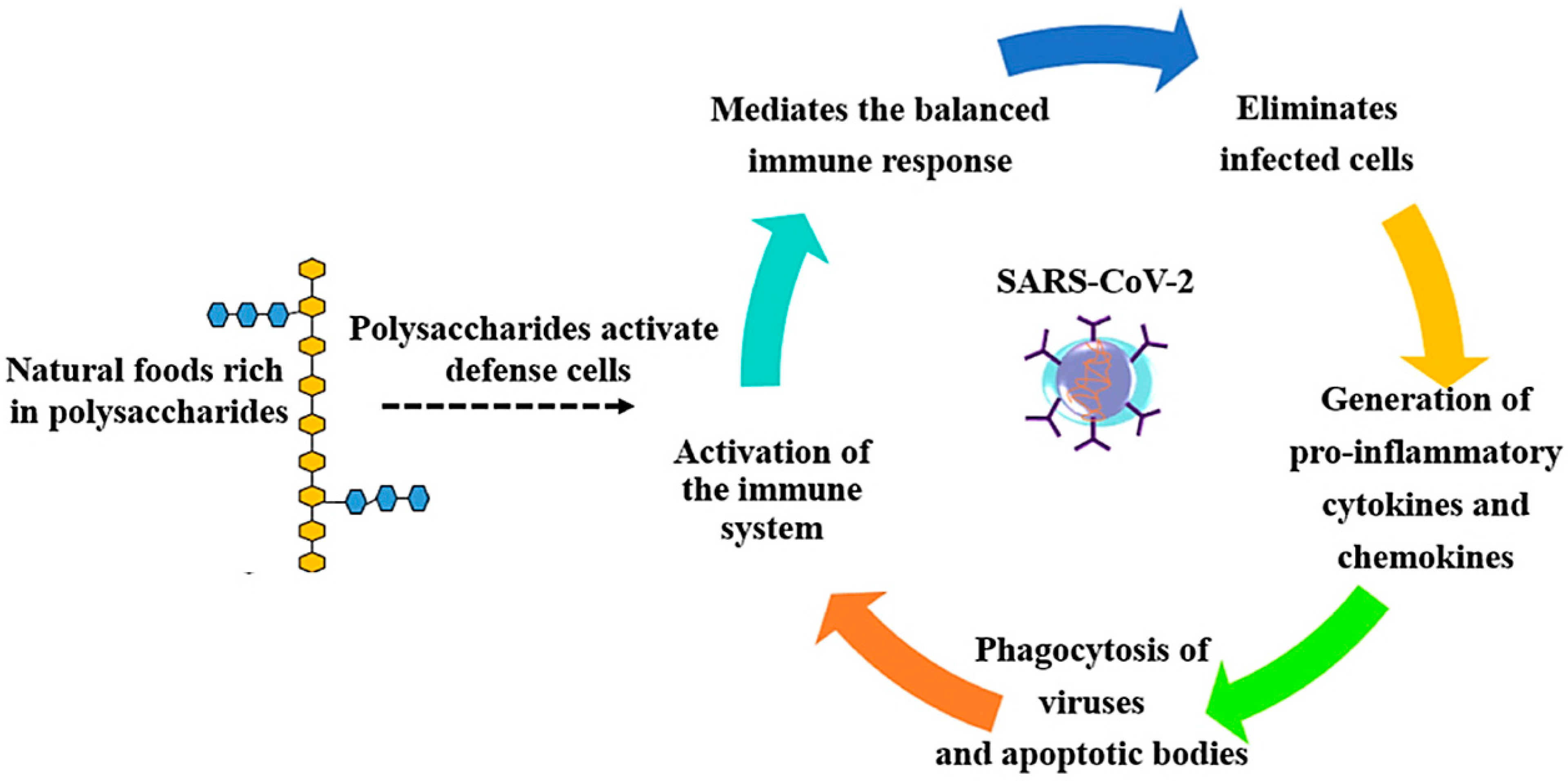
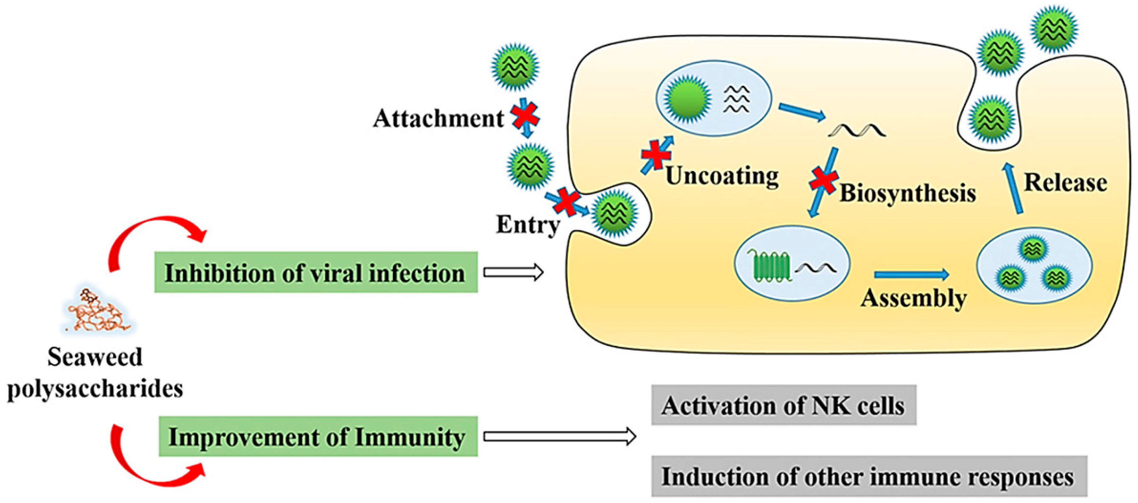
  28. Barbosa, J.R.; Junior, R.N.d.C. Polysaccharides obtained from natural edible sources and their role in modulating the immune system: Biologically active potential that can be exploited against COVID-19. Trends Food Sci. Technol. 2021, 108, 223–235. [Google Scholar] [CrossRef]
  29. Bemiller, J.N. Carbohydrates. In Kirk-Othmer Encyclopedia of Chemical Technology; John Wiley & Sons: Hoboken, NJ, USA, 2004. [Google Scholar]
  30. Ahmed, S.; Ahmad, M.; Swami, B.L.; Ikram, S. A review on plants extract mediated synthesis of silver nanoparticles for antimicrobial applications: A green expertise. J. Adv. Res. 2016, 7, 17–28. [Google Scholar] [CrossRef] [Green Version]
  31. Kaliaraj, G.S.; Subramaniyan, B.; Manivasagan, P.; Kim, S.-K. Green synthesis of metal nanoparticles using seaweed polysaccharides. In Seaweed Polysaccharides; Elsevier: Amsterdam, The Netherlands, 2017; pp. 101–109. [Google Scholar]
  32. Baglivo, M.; Baronio, M.; Natalini, G.; Beccari, T.; Chiurazzi, P.; Fulcheri, E.; Petralia, P.P.; Michelini, S.; Fiorentini, G.; Miggiano, G.A.; et al. Natural small molecules as inhibitors of coronavirus lipid-dependent attachment to host cells: A possible strategy for reducing SARS-COV-2 infectivity? Acta Biomed. 2020, 91, 161–164. [Google Scholar]
  33. Garrido, P.F.; Calvelo, M.; Blanco-González, A.; Veleiro, U.; Suárez, F.; Conde, D.; Cabezón, A.; Piñeiro, Á.; Garcia-Fandino, R. The Lord of the NanoRings: Cyclodextrins and the battle against SARS-CoV-2. Int. J. Pharm. 2020, 588, 119689. [Google Scholar] [CrossRef] [PubMed]
  34. Das, G.; Ghosh, S.; Garg, S.; Ghosh, S.; Jana, A.; Samat, R.; Mukherjee, N.; Roya, R.; Ghosh, S. An overview of key potential therapeutic strategies for combat in the COVID-19 battle. RSC Adv. 2020, 10, 28243–28266. [Google Scholar] [CrossRef]
  35. Song, S.; Peng, H.; Wang, Q.; Liu, Z.; Dong, X.; Wen, C.; Ai, C.; Zhang, Y.; Wang, Z.; Zhu, B. Inhibitory activities of marine sulfated polysaccharides against SARS-CoV-2. Food Funct. 2020, 11, 7415–7420. [Google Scholar] [CrossRef] [PubMed]
  36. Kalinina, T.S.; Zlenko, D.V.; Kiselev, A.V.; Litvin, A.A.; Stovbun, S.V. Antiviral activity of the high-molecular-weight plant polysaccharides (Panavir®). Int. J. Biol. Macromol. 2020, 161, 936–938. [Google Scholar] [CrossRef] [PubMed]
  37. Zhang, S.; Pei, R.; Li, M.; Sun, H.; Su, M.; Ding, Y.; Chen, X.; Du, Z.; Jin, C.; Huang, C.; et al. Structural characterization of cocktail-like targeting polysaccharides from Ecklonia kurome Okam and their anti-SARS-CoV-2 activities in vitro. bioRxiv 2021. [Google Scholar] [CrossRef]
  38. Jin, W.; Zhang, W.; Mitra, D.; McCandless, M.G.; Sharma, P.; Tandon, R.; Zhang, F.; Linhardt, R.J. The structure-activity relationship of the interactions of SARS-CoV-2 spike glycoproteins with glucuronomannan and sulfated galactofucan from Saccharina japonica. Int. J. Biol. Macromol. 2020, 163, 1649–1658. [Google Scholar] [CrossRef]
  39. Xu, C.; Nasrollahzadeh, M.; Sajjadi, M.; Maham, M.; Luque, R.; Puente-Santiago, A.R. Benign-by-design nature-inspired nanosystems in biofuels production and catalytic applications. Renew. Sustain. Energy Rev. 2019, 112, 195–252. [Google Scholar] [CrossRef]
  40. Xu, C.; Nasrollahzadeh, M.; Selva, M.; Issaabadi, Z.; Luque, R. Waste-to-wealth: Biowaste valorization into valuable bio (nano) materials. Chem. Soc. Rev. 2019, 48, 4791–4822. [Google Scholar] [CrossRef]
  41. Peniche, H.; Peniche, C. Chitosan nanoparticles: A contribution to nanomedicine. Polym. Int. 2011, 60, 883–889. [Google Scholar] [CrossRef]
  42. Alarcón, B.; Lacal, J.C.; Fernández-Sousa, J.; Carrasco, L. Screening for new compounds with antiherpes activity. Antivir. Res. 1984, 4, 231–244. [Google Scholar] [CrossRef]
  43. Qin, C.; Li, H.; Xiao, Q.; Liu, Y.; Zhu, J.; Du, Y. Water-solubility of chitosan and its antimicrobial activity. Carbohydr. Polym. 2006, 63, 367–374. [Google Scholar] [CrossRef]
  44. Doostmohammadi, M.; Ameri, A.; Mohammadinejad, R.; Dehghannoudeh, N.; Banat, I.M.; Ohadi, M.; Dehghannoudeh, G. Hydrogels for peptide hormones delivery: Therapeutic and tissue engineering applications. Drug Des. Devel. Ther. 2019, 13, 3405–3418. [Google Scholar] [CrossRef] [PubMed] [Green Version]
  45. Marón, L.B.; Covas, C.P.; Da Silveira, N.P.; Pohlmann, A.; Mertins, O.; Tatsuo, L.N.; Sant’ Anna, O.A.B.; Moro, A.M.; Takata, C.S.; De Araujo, P.S. LUVs recovered with chitosan: A new preparation for vaccine delivery. J. Liposome Res. 2007, 17, 155–163. [Google Scholar] [CrossRef] [PubMed]
  46. Alinejad, Y.; Adoungotchodo, A.; Hui, E.; Zehtabi, F.; Lerouge, S. An injectable chitosan/chondroitin sulfate hydrogel with tunable mechanical properties for cell therapy/tissue engineering. Int. J. Biol. Macromol. 2018, 113, 132–141. [Google Scholar] [CrossRef]
  47. Intini, C.; Elviri, L.; Cabral, J.; Mros, S.; Bergonzi, C.; Bianchera, A.; Flammini, L.; Govoni, P.; Barocelli, E.; Bettini, R. 3D-printed chitosan-based scaffolds: An in vitro study of human skin cell growth and an in-vivo wound healing evaluation in experimental diabetes in rats. Carbohydr. Polym. 2018, 199, 593–602. [Google Scholar] [CrossRef] [PubMed]
  48. Wieckiewicz, M.; W Boening, K.; Grychowska, N.; Paradowska-Stolarz, A. Clinical application of chitosan in dental specialities. Mini Rev. Med. Chem. 2017, 17, 401–409. [Google Scholar] [CrossRef]
  49. Abasi, S.; Aggas, J.R.; Guiseppi-Elie, A. Physiochemical and morphological dependent growth of NIH/3T3 and PC-12 on polyaniline-chloride/chitosan bionanocomposites. Mater. Sci. Eng. C 2019, 99, 1304–1312. [Google Scholar] [CrossRef] [PubMed]
  50. Nadimi, A.E.; Ebrahimipour, S.Y.; Afshar, E.G.; Falahati-Pour, S.K.; Ahmadi, Z.; Mohammadinejad, R.; Mohamadi, M. Nano-scale drug delivery systems for antiarrhythmic agents. Eur. J. Med. Chem. 2018, 157, 1153–1163. [Google Scholar] [CrossRef] [PubMed]
  51. Mu, M.; Li, X.; Tong, A.; Guo, G. Multi-functional chitosan-based smart hydrogels mediated biomedical application. Expert Opin. Drug Deliv. 2019, 16, 239–250. [Google Scholar] [CrossRef]
  52. Ashrafizadeh, M.; Ahmadi, Z.; Mohamadi, N.; Zarrabi, A.; Abasi, S.; Dehghannoudeh, G.; Tamaddondoust, R.N.; Khanbabaei, H.; Mohammadinejad, R.; Thakur, V.K. Chitosan-based advanced materials for docetaxel and paclitaxel delivery: Recent advances and future directions in cancer theranostics. Int. J. Biol. Macromol. 2020, 145, 282–300. [Google Scholar] [CrossRef] [PubMed]
  53. Yhee, J.Y.; Son, S.; Kim, S.H.; Park, K.; Choi, K.; Kwon, I.C. Self-assembled glycol chitosan nanoparticles for disease-specific theranostics. J. Control. Release 2014, 193, 202–213. [Google Scholar] [CrossRef] [PubMed]
  54. Swierczewska, M.; Han, H.S.; Kim, K.; Park, J.H.; Lee, S. Polysaccharide-based nanoparticles for theranostic nanomedicine. Adv. Drug Deliv. Rev. 2016, 99, 70–84. [Google Scholar] [CrossRef] [PubMed] [Green Version]
  55. Loutfy, S.A.; Elberry, M.H.; Farroh, K.Y.; Mohamed, H.T.; Mohamed, A.A.; Mohamed, E.B.; Faraag, A.H.I.; Mousa, S.A. Antiviral Activity of Chitosan Nanoparticles Encapsulating Curcumin Against Hepatitis C Virus Genotype 4a in Human Hepatoma Cell Lines. Int. J. Nanomed. 2020, 15, 2699–2715. [Google Scholar] [CrossRef] [PubMed] [Green Version]
  56. Zheng, M.; Qu, D.; Wang, H.; Sun, Z.; Liu, X.; Chen, J.; Li, C.; Li, X.; Chen, Z. Intranasal Administration of Chitosan Against Influenza A (H7N9) Virus Infection in a Mouse Model. Sci. Rep. 2016, 6, 28729. [Google Scholar] [CrossRef] [PubMed] [Green Version]
  57. Al-Ghananeem, A.M.; Saeed, H.; Florence, R.; Yokel, R.A.; Malkawi, A.H. Intranasal drug delivery of didanosine-loaded chitosan nanoparticles for brain targeting; an attractive route against infections caused by aids viruses. J. Drug Target. 2010, 18, 381–388. [Google Scholar] [CrossRef]
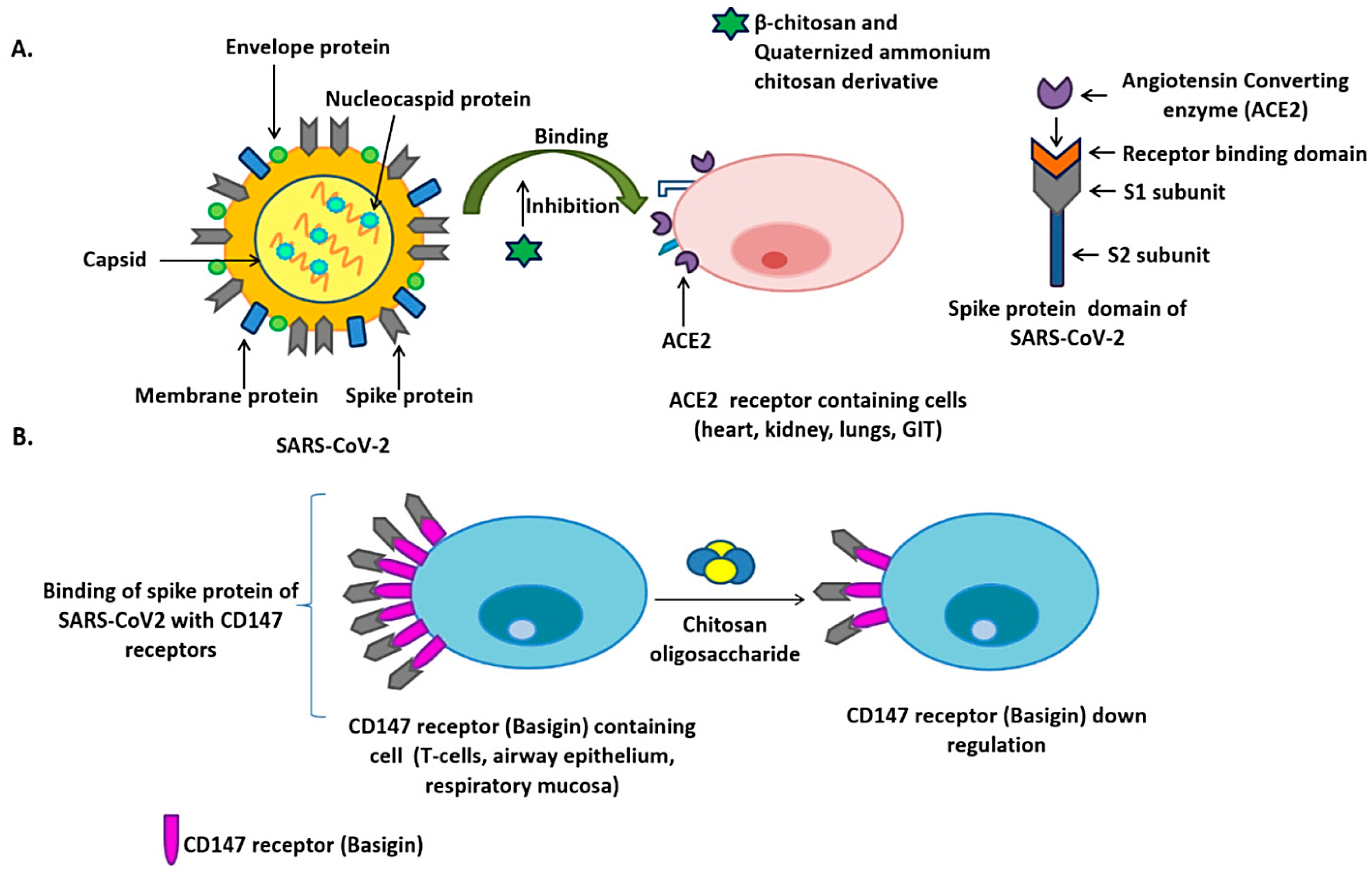
  58. Alitongbieke, G.; Li, X.-m.; Wu, Q.-C.; Lin, Z.-C.; Huang, J.-F.; Xue, Y.; Liu, J.-N.; Lin, J.-M.; Pan, T.; Chen, Y.-X.; et al. Study on β-Chitosan against the binding of SARS-CoV-2S-RBD/ACE2. bioRxiv 2020. [Google Scholar] [CrossRef]
  59. Sharma, N.; Modak, C.; Singh, P.K.; Kumar, R.; Khatri, D.; Singh, S.B. Underscoring the immense potential of chitosan in fighting a wide spectrum of viruses: A plausible molecule against SARS-CoV-2? Int. J. Biol. Macromol. 2021, 179, 33–44. [Google Scholar] [CrossRef]
  60. van Haren, F.M.P.; Page, C.; Laffey, J.G.; Artigas, A.; Camprubi-Rimblas, M.; Nunes, Q.; Smith, R.; Shute, J.; Carroll, M.; Tree, J.; et al. Nebulised heparin as a treatment for COVID-19: Scientific rationale and a call for randomised evidence. Crit. Care 2020, 24, 454. [Google Scholar] [CrossRef]
  61. Tandon, R.; Sharp, J.S.; Zhang, F.; Pomin, V.H.; Ashpole, N.M.; Mitra, D.; McCandless, M.G.; Jin, W.; Liu, H.; Sharma, P.; et al. Effective Inhibition of SARS-CoV-2 Entry by Heparin and Enoxaparin Derivatives. J. Virol. 2021, 95, e01987-20. [Google Scholar]
  62. Kim, S.Y.; Jin, W.; Sood, A.; Montgomery, D.W.; Grant, O.C.; Fuster, M.M.; Fu, L.; Dordick, J.S.; Woods, R.J.; Zhang, F.; et al. Characterization of heparin and severe acute respiratory syndrome-related coronavirus 2 (SARS-CoV-2) spike glycoprotein binding interactions. Antivir. Res. 2020, 181, 104873. [Google Scholar] [CrossRef]
  63. Mycroft-West, C.J.; Su, D.; Pagani, I.; Rudd, T.R.; Elli, S.; Guimond, S.E.; Miller, G.; Meneghetti, M.C.Z.; Nader, H.B.; Li, Y.; et al. Heparin inhibits cellular invasion by SARS-CoV-2: Structural dependence of the interaction of the surface protein (spike) S1 receptor binding domain with heparin. Thromb. Haemost. 2020, 120, 1700–1715. [Google Scholar] [PubMed]
  64. Cagno, V.; Andreozzi, P.; D’Alicarnasso, M.; Jacob Silva, P.; Mueller, M.; Galloux, M.; Goffic, R.L.; Jones, S.T.; Vallino, M.; Hodek, J.; et al. Broad-spectrum non-toxic antiviral nanoparticles with a virucidal inhibition mechanism. Nat. Mater. 2017, 17, 195–203. [Google Scholar] [CrossRef]
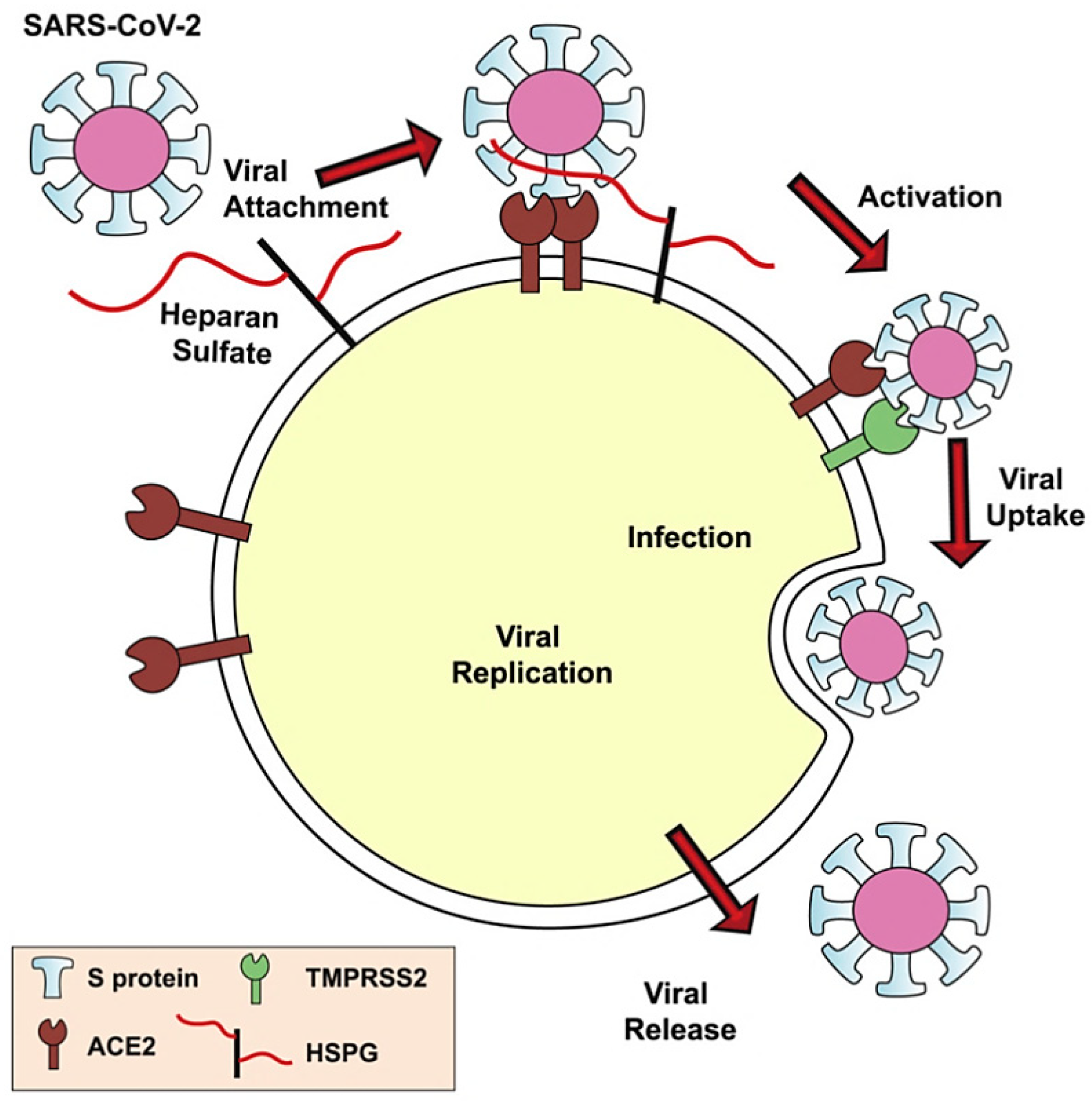
  65. Clausen, T.M.; Sandoval, D.R.; Spliid, C.B.; Pihl, J.; Perrett, H.R.; Painter, C.D.; Narayanan, A.; Majowicz, S.A.; Kwong, E.M.; McVicar, R.N.; et al. SARS-CoV-2 Infection Depends on Cellular Heparan Sulfate and ACE2. Cell 2020, 183, 1043–1057.e1015. [Google Scholar] [CrossRef] [PubMed]
  66. Kalra, R.S.; Kandimalla, R. Engaging the spikes: Heparan sulfate facilitates SARS-CoV-2 spike protein binding to ACE2 and potentiates viral infection. Signal Transduct. Target. Ther. 2021, 6, 39. [Google Scholar] [CrossRef] [PubMed]
  67. Coviello, T.; Matricardi, P.; Marianecci, C.; Alhaique, F. Polysaccharide hydrogels for modified release formulations. J. Control. Release 2007, 119, 5–24. [Google Scholar] [CrossRef] [PubMed]
  68. Liu, J.; Zhan, X.; Wan, J.; Wang, Y.; Wang, C. Review for carrageenan-based pharmaceutical biomaterials: Favourable physical features versus adverse biological effects. Carbohydr. Polym. 2015, 121, 27–36. [Google Scholar] [CrossRef] [PubMed]
  69. Bonferoni, M.C.; Chetoni, P.; Giunchedi, P.; Rossi, S.; Ferrari, F.; Burgalassi, S.; Caramella, C. Carrageenan-gelatin mucoadhesive systems for ion-exchange based ophthalmic delivery: In vitro and preliminary in vivo studies. Eur. J. Pharm. Biopharm. 2004, 57, 465–472. [Google Scholar] [CrossRef]
  70. Zhang, Y.-Q.; Tsai, Y.-C.; Monie, A.; Hung, C.-F.; Wu, T.C. Carrageenan as an adjuvant to enhance peptide-based vaccine potency. Vaccine 2010, 28, 5212–5219. [Google Scholar] [CrossRef] [Green Version]
  71. Kalitnik, A.A.; Byankina Barabanova, A.O.; Nagorskaya, V.P.; Reunov, A.V.; Glazunov, V.P.; Solov’eva, T.F.; Yermak, I.M. Low molecular weight derivatives of different carrageenan types and their antiviral activity. J. Appl. Phycol. 2013, 25, 65–72. [Google Scholar] [CrossRef]
  72. Morokutti-Kurz, M.; Graf, C.; Prieschl-Grassauer, E. Amylmetacresol/2,4-dichlorobenzyl alcohol, hexylresorcinol, or carrageenan lozenges as active treatments for sore throat. Int. J. Gen. Med. 2017, 10, 53–60. [Google Scholar] [CrossRef] [PubMed] [Green Version]
  73. Morokutti-Kurz, M.; Graf, P.; Grassauer, A.; Prieschl-Grassauer, E. SARS-CoV-2 in-vitro neutralization assay reveals inhibition of virus entry by iota-carrageenan. bioRxiv 2020. [Google Scholar] [CrossRef]
  74. Iravani, S. Green synthesis of metal nanoparticles using plants. Green Chem. 2011, 13, 2638–2650. [Google Scholar] [CrossRef]
  75. Iravani, S.; Jamalipour Soufi, G. Gold Nanostructures in Medicine and Biology. In Nanoparticles in Medicine; Shukla, A.K., Ed.; Springer Nature: Singapore, 2019; pp. 175–183. [Google Scholar]
  76. Fan, Q. Polyolefin nanocomposite fibers and films. In Polyolefin Fibres, Industrial and Medical Applications; Series in Textiles; Woodhead Publishing: Cambridge, UK, 2009; pp. 341–362. [Google Scholar]
  77. Saokham, P.; Muankaew, C.; Jansook, P.; Loftsson, T. Solubility of Cyclodextrins and Drug/Cyclodextrin Complexes. Molecules 2018, 23, 1161. [Google Scholar] [CrossRef] [PubMed] [Green Version]
  78. Jambhekar, S.S.; Breen, P. Cyclodextrins in pharmaceutical formulations II: Solubilization, binding constant, and complexation efficiency. Drug Discov. Today 2016, 21, 1161. [Google Scholar] [CrossRef] [PubMed]
  79. Tiwari, G.; Tiwari, R.; Rai, A.K. Cyclodextrins in delivery systems: Applications. J. Pharm. Bioallied Sci. 2010, 2, 72–79. [Google Scholar] [CrossRef] [PubMed]
  80. Loftsson, T.; Jarho, P.; Másson, M.; Järvinen, T. Cyclodextrins in drug delivery. Expert Opin. Drug Deliv. 2005, 2, 335–351. [Google Scholar] [CrossRef] [PubMed]
  81. Carrouel, F.; Conte, M.P.; Fisher, J.; Gonçalves, L.S.; Dussart, C.; Llodra, J.C.; Bourgeois, D. COVID-19: A Recommendation to Examine the Effect of Mouthrinses with β-Cyclodextrin Combined with Citrox in Preventing Infection and Progression. J. Clin. Med. 2020, 9, 1126. [Google Scholar] [CrossRef] [PubMed] [Green Version]
  82. Braga, S.S. Cyclodextrins: Emerging Medicines of the New Millennium. Biomolecules 2019, 9, 801. [Google Scholar] [CrossRef] [Green Version]
  83. Jones, S.T.; Cagno, V.; Janecek, M.; Ortiz, D.; Gasilova, N.; Piret, J.; Gasbarri, M.; Constant, D.A.; Han, Y.; Vukovi, L.; et al. Modified cyclodextrins as broad-spectrum antivirals. Sci. Adv. 2020, 6, eaax9318. [Google Scholar] [CrossRef] [Green Version]
  84. Moon, R.J.; Martini, A.; Nairn, J.; Simonsenf, J.; Youngblood, J. Cellulose nanomaterials review: Structure, properties and nanocomposites. Chem. Soc. Rev. 2011, 40, 3941–3994. [Google Scholar] [CrossRef]
  85. Mokhena, T.C.; John, M.J. Cellulose nanomaterials: New generation materials for solving global issues. Cellulose 2020, 27, 1149–1194. [Google Scholar] [CrossRef]
  86. Junter, G.-A.; Lebrun, L. Cellulose-based virus-retentive filters: A review. Rev. Environ. Sci. Biotechnol. 2017, 16, 455–489. [Google Scholar] [CrossRef] [PubMed]
  87. Trache, D.; Hussin, M.H.; Haafiz, M.M.; Thakur, V.K. Recent progress in cellulose nanocrystals: Sources and production. Nanoscale 2017, 9, 1763–1786. [Google Scholar] [CrossRef] [PubMed] [Green Version]
  88. Martin-Martinez, F.J.; Jin, K.; López Barreiro, D.; Buehler, M.J. The Rise of Hierarchical Nanostructured Materials from Renewable Sources: Learning from Nature. ACS Nano 2018, 12, 7425–7433. [Google Scholar] [CrossRef]
  89. Sun, B.; Zhang, M.; Shen, J.; He, Z.; Fatehi, P.; Ni, Y. Applications of cellulose-based materials in sustained drug delivery systems. Curr. Med. Chem. 2019, 26, 2485–2501. [Google Scholar] [CrossRef] [PubMed]
  90. Wu, L.; Manukyan, L.; Mantas, A.; Mihranyan, A. Nanocellulose-Based Nanoporous Filter Paper for Virus Removal Filtration of Human Intravenous Immunoglobulin. ACS Appl. Nano Mater. 2019, 2, 6352–6359. [Google Scholar] [CrossRef]
  91. Jorfi, M.; Foster, E.J. Recent advances in nanocellulose for biomedical applications. J. Appl. Polym. Sci. 2015, 132, 41719. [Google Scholar] [CrossRef]
  92. Ray, B.; Schütz, M.; Mukherjee, S.; Jana, S.; Ray, S.; Marschall, M. Exploiting the Amazing Diversity of Natural Source-Derived Polysaccharides: Modern Procedures of Isolation, Engineering, and Optimization of Antiviral Activities. Polymers 2021, 13, 136. [Google Scholar] [CrossRef] [PubMed]
Figure 1. Antiviral effects of marine polysaccharides (from seaweeds) against pathogenic viruses, with the proposed mechanisms; NK cells: Natural Killer Cells. Reproduced with permission from Ref. [8].
Applsci 11 03512 g001
Figure 2. Important roles of polysaccharides in immune system modulation against severe acute respiratory syndrome coronavirus 2 (SARS-CoV-2). Reproduced with permission from Ref. [28].
Applsci 11 03512 g002
Figure 3. Some proposed mechanisms for antiviral performances of polysaccharides against SARS-CoV-2. EGFR: epidermal growth factor receptor; HSPG: heparan sulfate proteoglycan; RBD: receptor-binding domain. Reproduced with permission from Ref. [16].
Applsci 11 03512 g003
Figure 4. (A,B) Possible pathways for chitosan and related derivatives to fight against SARS-CoV-2. Reproduced with permission from Ref. [59].
Applsci 11 03512 g004
Figure 5. The dependence of SARS-CoV-2 infection on ACE2 and cellular heparan sulfate. TMPRSS2: transmembrane protease, serine 2; HSPG: heparan sulfate proteoglycans. Reproduced with permission from Ref. [65].
Applsci 11 03512 g005
Figure 6. (ad) The important role of heparan sulfate in the binding of the SARS-CoV-2 spike protein (S-protein) to ACE2 and the associated viral infection. Reproduced from Ref. [66], (CC BY 4.0).
Applsci 11 03512 g006
Publisher’s Note: MDPI stays neutral with regard to jurisdictional claims in published maps and institutional affiliations.

