Article

Effects of Custom-Made Insole Materials on Frictional Stress and Contact Pressure in Diabetic Foot with Neuropathy: Results from a Finite Element Analysis

by
Muhammad Nouman
1,
Tulaya Dissaneewate
2,
Desmond Y. R. Chong
3 and
Surapong Chatpun
1,4,*
1
Department of Biomedical Sciences and Biomedical Engineering, Faculty of Medicine, Prince of Songkla University, Hatyai 90110, Songkhl, Thailand
2
Department of Rehabilitation Medicine, Faculty of Medicine, Prince of Songkla University, Hatyai 90110, Songkhla, Thailand
3
Engineering Cluster, Singapore Institute of Technology, Singapore 138683, Singapore
4
Institute of Biomedical Engineering, Faculty of Medicine, Prince of Songkla University, Hatyai 90110, Songkhla, Thailand
*
Author to whom correspondence should be addressed.
Appl. Sci. 2021, 11(8), 3412; https://doi.org/10.3390/app11083412
Submission received: 3 March 2021 / Revised: 5 April 2021 / Accepted: 8 April 2021 / Published: 10 April 2021
(This article belongs to the Section Applied Biosciences and Bioengineering)

Abstract

:

Featured Application

Finite element analysis to assess the effect of materials used for a custom-made insole to provide information for the suitable material selection in the conventional practice of insole fabrication for the diabetic foot with neuropathy.

Abstract

Offloading plantar pressure in a diabetic foot with neuropathy is challenging in conventional clinical practice. Custom-made insole (CMI) materials play an important role in plantar pressure reduction, but the assessment is costly and time-consuming. Finite element analysis (FEA) can provide an efficient evaluation of different insoles on the plantar pressure distribution. This study investigated the effect of CMI materials and their combinations on plantar pressure reduction for the diabetic foot with neuropathy using FEA. The study was conducted by constructing a three-dimensional foot model along with CMI to study the peak contact pressure between the foot and CMI. The softer material (E = 5 MPa) resulted in a better reduction of peak contact pressure compared with the stiffer material (E = 11 MPa). The plantar pressure was well redistributed with softer material compared with the stiffer material and its combination. In addition, the single softer material resulted in reduced frictional stress under the first metatarsal head compared with the stiffer material and the combination of materials. The softer material and its combination have a beneficial effect on plantar pressure reduction and redistribution for a diabetic foot with neuropathy. This study provided an effective approach for CMI material selection using FEA.

1. Introduction

Diabetic peripheral neuropathy is the major risk factor for the development of plantar foot ulcerations. The lifetime risk of diabetic foot ulceration ranges from 15% to 25% [1,2]. Annually, 26 million people with diabetes are affected by diabetic foot ulcers and its prevalence is 6.3% worldwide [3]. A foot ulcer may require the resection of distal osseous and soft tissue structure, surgical debridement, daily dressings, strict glycemic control and intravenous antibiotic treatment for disinfection [4,5,6]. Diabetic foot complications, including limited joint mobility, foot deformities, and previous history of ulceration, play a major role in the ulcer development [7,8]. The abnormal plantar pressure distribution in the presence of neuropathy causes prolong severe foot complications [9]. It is estimated that 85% of all amputations are preceded by foot ulceration and re-ulceration [10,11] and it can be prevented with appropriate foot care.
There are various factors that contribute to the ulcer healing including glycated hemoglobin level and peak plantar pressure. Adequate levels of glycated hemoglobin is an important factor especially during the healing of an ulcer [12,13,14]. Other factors, including repetitive high plantar pressure, increased friction between foot and footwear, especially during walking activities, contribute to ulcer formation that can be reduced using appropriate insoles [15,16,17]. To prevent ulceration and re-ulceration, the custom-made insole (CMI) is commonly prescribed to redistribute the peak plantar pressure under bony prominences in a diabetic foot with neuropathy [18]. The CMI is considered ideal and is associated with the reduction of peak plantar pressure for a diabetic foot with neuropathy. In experimental studies, plantar pressure sensors have been used to measure the plantar pressure to better understand the biomechanics of the diabetic foot and factors contributing to foot ulcers during gait. However, the experimental procedures are time consuming with varying uncontrolled variables, which has made it difficult for studies to acquire reliable results for a diabetic foot with neuropathy [19,20].
The prolonged trial and error experimental procedures in a clinical setting with a wide variety of materials for insole fabrication is a challenge for both the clinician and patients. To attain ideal results of CMI, it seems impossible to go through each material and foot complication with all possible age-dependent daily living activities. In comparison with experimental methods, finite element analysis (FEA) is a numerical tool providing solutions to complex mathematical problems to determine various stress, strain, deformation and contact pressure [21,22]. Nowadays, FEA is a popular tool for studying biomechanics and for evaluating various protocols without prolonged patient involvement. Subject-specific FE foot models offer a huge amount of data that can be beneficial in both clinical application and footwear designs. It also contributes to understanding the behavior of foot biomechanics and the effects of different interventions [23]. Most researchers focused on a single material used to fabricate an insole with varying hardness. Others used inappropriate materials selected for a diabetic foot. In some cases, the thickness of the CMIs was not well controlled and clinically inapplicable [21,24]. An appropriate material with controlled thickness of CMIs that is clinically prescribed for a diabetic foot with neuropathy is still lacking.
Material stiffness, CMI thickness and insole contour to the foot have an effect on plantar pressure distribution [16,25]. Increasing the material stiffness and thickness of the insole can enhance the stability during gait. However, the cushioning is reduced while increasing the insole stiffness and the stiff insoles are contraindicated towards the diabetic foot [26,27]. Cushioning is an important factor as it acts as a shock absorber, reducing the impact on the joints, especially the ankle, knee and the lower back [28]. There is a vast variety of commercial materials available claiming to reduce the peak plantar pressure. However, very few materials can be prescribed to the diabetic foot with neuropathy. CMIs prescribed in a real clinical scenario are mostly fabricated from multi-density materials for a diabetic foot with neuropathy, while most researchers have focused on single materials used to fabricate an insole with varying hardness and others have used materials selected for a diabetic foot that are inappropriate.
To improve the plantar pressure management for the diabetic foot with neuropathy, it is important to select appropriate materials for the CMI fabrication. In addition, the effect of the combination of various materials, which reflect real clinical situations towards the prescription of CMIs for the diabetic foot with neuropathy, would also be studied. This current study focused on insole materials from softer to stiffer materials used to fabricate a CMI for a diabetic foot with neuropathy. In addition, the effect of the combination of various materials that reflect the real clinical situation towards a prescription of CMI for a diabetic foot with neuropathy would also be studied. Therefore, this study aimed to investigate the effect of various materials and their combinations on the regional plantar pressure reduction and frictional stress under bony prominences for a diabetic foot with neuropathy during balance standing. A three-dimensional finite element (FE) model of the foot geometry and soft tissues was developed in contact with different CMIs and the ground. It is important to improve the selection of appropriate materials using the FE approach for a diabetic foot with neuropathy. The outcome of this study will provide an effective material selection for a desirable plantar pressure reduction and redistribution for a diabetic foot with neuropathy.

2. Materials and Methods

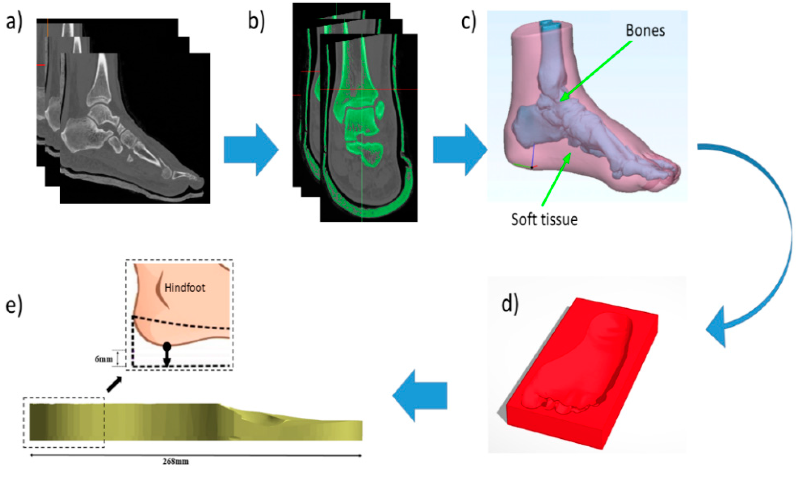
2.1. Finite Element Model Generation

A three-dimensional (3D) FEA model of a foot was constructed to study the contact pressure between the foot and CMI. Two types of CMIs were designed with overall similar thickness; single layer (6 mm) and two layers with top layer (3 mm) and base layer (3 mm). The thickness of CMI was measured from the lowest point of the hindfoot to the ground. Using computer-aided design, the subtraction technique was performed to obtain CMI with an overall thickness of 6 mm. This study used the images from a human subject to reconstruct the foot model. The protocol was approved by the research ethics committee (EC-63-219-25-2), Faculty of Medicine, Prince of Songkla University. The computerized tomography scan images of a left foot (male subject, 57 years old, 84 kg) were imported to the image processing software Mimics version 20 (Materialise, Leuven, Belgium), and a 3D model of foot bones with encapsulated soft tissues were developed. The foot is a complex structure, as it comprises 26 bones, 33 joints, muscles, and numerous ligaments [29]. In this study, the FEA foot model was simplified to reduce the modeling cost and to improve the feasibility of optimization. Therefore, 26 bones were merged together as one whole body. The relative motion between the segments was neglected, and the effects of muscles and ligaments were also neglected [30]. The models were assembled in Ansys SpaceClaim (ANSYS Inc., Canonsburg, PA, USA). The CMI and ground were then created following the geometry of the foot, as shown in Figure 1.

2.2. Material Properties

The 3D reconstructed foot model was imported to a finite element solver Ansys 2019 R1 (ANSYS Inc., Pennsylvania, PA, USA) for analysis and prediction of the plantar pressure distribution with CMI. The foot bones and soft tissues were assumed to be isotropic and assigned with linear elastic material properties. Three materials that were most commonly used in the real clinical setup were selected to design a custom-made insole and their properties are listed in Table 1. Additionally, the combination of materials for a custom-made insole is presented in Table 2.

2.3. Meshing

The complete 3D model of a foot along with a custom-made insole and ground was meshed with linear tetrahedron elements. A mesh convergence analysis was performed to ensure that the predicted FE results were insensitive to the element size. The mesh convergence study was conducted based on the peak contact pressure for five different element sizes ranging from 6 to 4 mm with an interval of 0.5 mm. The mesh density was gradually increased until the deviation in the estimated peak contact pressure was less than 5% [38].

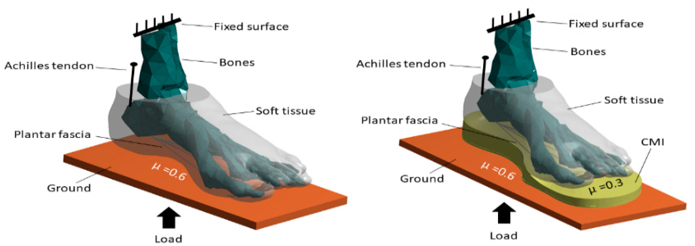
2.4. Loading and Boundary Conditions

The bones and soft tissues were bonded together. The frictional coefficient (μ) of 0.6 was set between the foot and ground in the case of without CMI and between CMI and ground in the case with CMI [39]. Additionally, a frictional coefficient (μ) of 0.3 was set between the foot and CMI [40]. The plantar fascia was represented with 5 tension-only link elements with their origin and insertion sites based on their anatomical location [41].
The top surfaces of the tibia and fibula were fixed, and a 420 N force as the ground reaction force was exerted beneath the ground (Figure 2). The Achilles tendon force in the upward direction was applied in the FEA model. The peak contact pressure between the foot and CMI was computationally evaluated.

2.5. Validation

Validation is one of the most important stages in FEA. A 59-year-old male diabetic subject with neuropathy weighing 84 kg was recruited and half of that weight was prescribed in the FEA model as a ground reaction force of 420 N on a single foot during balanced standing. The experimental averaged peak plantar pressure from a neuropathic diabetic patient measured by the Pedar® system during a balanced standing on a level ground with CMI fabricated from EVA was compared with the simulated peak contact pressure for validation.

3. Results

The 3D FE foot model with CMI was developed to analyze the effect of various materials and their combinations on plantar pressure distribution. Using CMIs with soft materials tends to reduce the peak contact pressure effectively when compared to the stiffer CMIs. CMIs fabricated from a combination of soft and stiff materials resulted in a more even distribution of the contact pressure. The softer top layer provided better distribution of peak contact pressure compared with the stiffer top layer with the same base layer material.

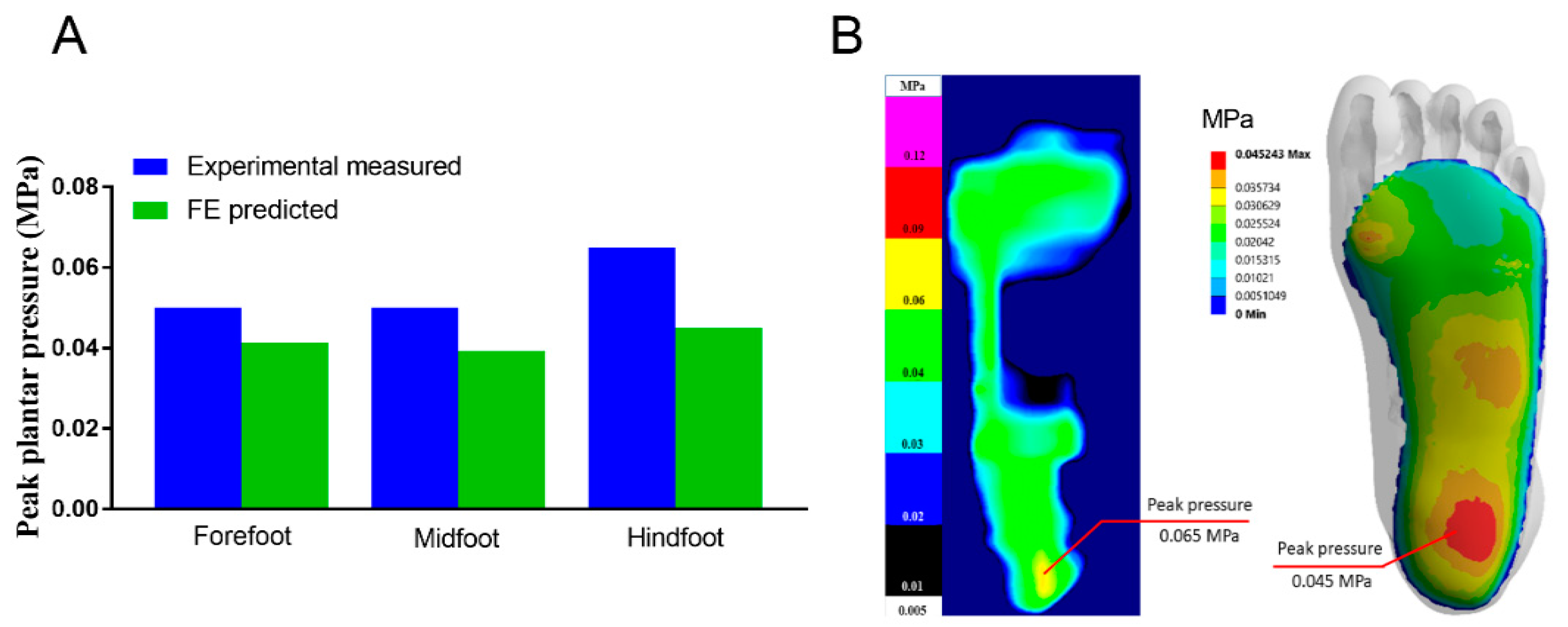
3.1. Mesh Convergence and Model Validation

A negligible difference (of less than 2%) in the peak contact pressure was observed from the element size of 5 to 4.5 mm and further to 4.0 mm. According to the aforementioned convergence criteria, the element size of 5 mm (203,482 elements) was considered to be a computationally optimal mesh size for further analysis.
Using CMI, it was found that the peak contact pressure at the hindfoot with EVA was higher compared with forefoot and midfoot. The trend of predicted pressure distribution from the regions of the foot was similar to the peak plantar pressure measured from the experiment (Figure 3A). Therefore, it was a good agreement of the predicted FE results and experimental peak plantar pressure mapping especially at hindfoot during balanced standing with the increase in contact area with CMI fabricated from EVA (Figure 3B).

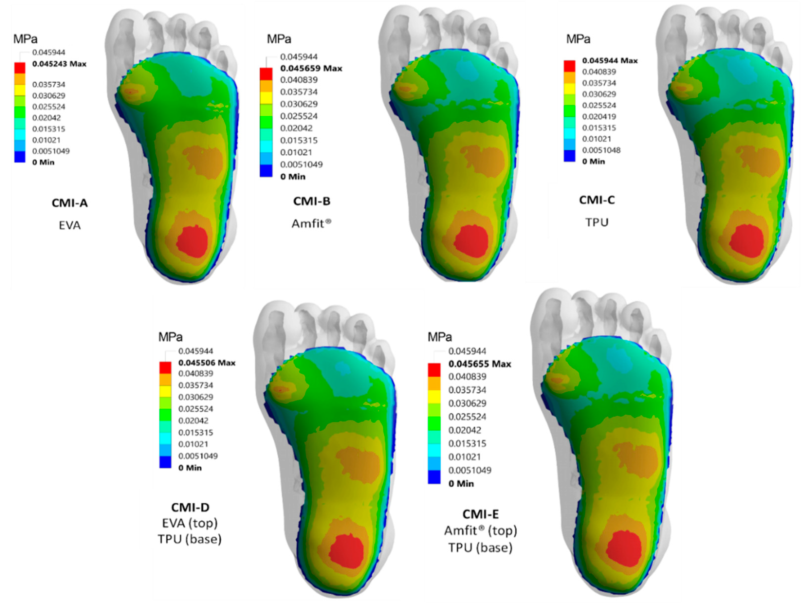
3.2. Contact Pressure Distribution

The FE predictions for distribution of contact pressure corresponding to five custom-made insoles (CMI-A to CMI-E) are shown in Figure 4. The peak contact pressure was highest with CMI-C, especially at the hindfoot compared with other CMI materials and their combinations. The peak contact pressure was distributed more to the medial and lateral sides and it increased from softer to stiffer materials, especially at the hindfoot. The contact pressure contour showed that CMI-A resulted in a 1.52% reduction of peak contact pressure at the hindfoot compared with CMI-C. Moreover, the combination of materials, CMI-D resulted in a 0.95% reduction of peak contact pressure at hindfoot compared with CMI-C. There was also an increase of contact pressure proximal to the metatarsal heads indicating offloading of the peak contact pressure under the first metatarsal head with CMI-D compared with CMI-A.

3.3. Regional Peak Contact Pressure

The peak contact pressure was highest without CMI at the forefoot and hindfoot (Table 3) during balance standing. The peak contact pressure was reduced by 9.44% from the forefoot compared to the hindfoot with CMI-A fabricated from a soft material. Moreover, there was a remarkable difference of 15.62% between forefoot and hindfoot with CMI-C fabricated from a stiffer material. The combination of softer and stiffer materials as CMI-D, presented a 10.17% difference between forefoot and hindfoot, which was closer to the value presented by CMI-A. The maximum contact pressure difference between midfoot and hindfoot was 16.50% from CMI-C fabricated from a single stiffer material.

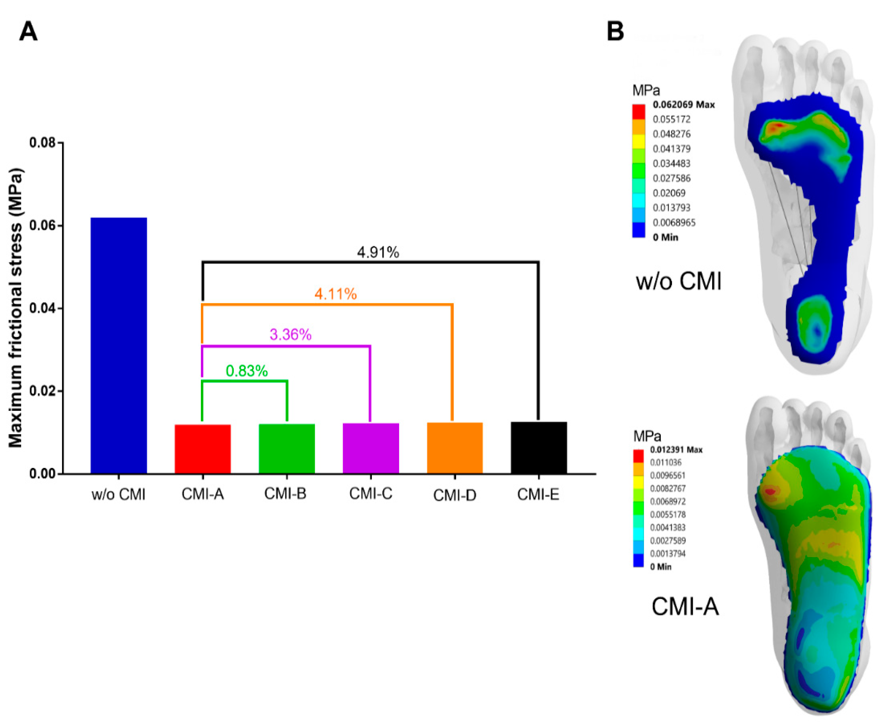
3.4. Frictional Stress

The maximum frictional stress was highest under the first metatarsal head without CMI and with CMI fabricated from various materials and their combinations (Figure 5). All CMIs showed reduction of maximum frictional stress compared to without CMI. Among the CMIs the maximum reduction was observed with CMI-A fabricated from EVA. As CMI-A being a softer material than CMI-C (TPU), the frictional stress was lowered by 3.36% under the first metatarsal head. However, the combination of EVA and TPU (CMI-D) resulted in an increase of the maximum frictional stress by 4.11% compared to CMI-A during balanced standing.

4. Discussion

In this study, a subject-specific 3D FEA foot model with CMI assigned with different materials and their combinations was developed to investigate the effects of materials on peak contact pressure in a diabetic foot with neuropathy. FEA results revealed that the peak contact pressure was reduced with CMI fabricated from all materials used in this study compared to without CMI. Specifically, using CMI with soft material (EVA) tends to reduce the peak contact pressure effectively compared with CMI with stiffer material (TPU). The higher regional contact pressure shifted to the midfoot and the peak contact pressure was reduced from hindfoot and forefoot with CMI. There was a notable reduction of frictional stress with softer material under the first metatarsal head compared with stiffer material.
The predicted results from FEA were validated with the experimental study using the Pedar® system by comparing the peak contact pressure during balanced standing with CMI fabricated from EVA. In this comparison, the FEA results were in agreement with the experimental results. The present FEA study predicted that all the CMIs showed a reduction of peak contact pressure compared to without CMI. The CMI fabricated either from a single soft material or a combination of softer and stiffer materials resulted in a reduction of peak contact pressure compared to without CMI. Similar reduction of peak contact pressure was found while using different cushioning materials and conformity compared with the flat insole [42]. Other studies reported a 30% to 40% reduction of peak contact pressure using CMI [43,44].
The current FE model indicates increased peak contact pressure at the forefoot and hindfoot without CMI. However, the ratio of peak contact pressure between forefoot and hindfoot was less than two showing lower severity of ulceration (Table 3). Researchers found that a severe neuropathic group had a FF/HF ratio higher than two during gait; however, mild to moderate neuropathic groups still had this ratio of less than two [45]. The FF/HF ratio was reduced from 1.07 to 0.87 with CMI fabricated from Multiform, Plastazote® and microcellular rubber compared to without CMI during gait [16,17]. A similar reduction of contact pressure ratio was found in this study during balanced standing with CMI fabricated from EVA. CMIs are strongly dependent on the mechanical characteristics of the material used to provide cushioning [46,47]. The peak plantar pressure reduces with a customized insole, especially from the forefoot compared with a flat insole [48]. Based on our results, the CMI with a softer material was able to redistribute plantar pressure better as compared to stiffer materials and their combinations. Nevertheless, the addition of a base layer of stiffer material has a minimal effect on plantar pressure distribution. The stiffer top layer tends to increase the peak contact pressure compared with the soft top layer material. Similar results were found in our study using softer CMI, as other studies found it was effective in reducing the plantar pressure up to 53% compared to the painful forefoot region of the foot [49,50]. Therefore, CMI with a softer material can be prescribed to people with prolonged standing activities. However, soft materials are not prescribed in a clinical setup as various offloading structures such as metatarsal pads, metatarsal bars or medial arch support because they cannot withstand the repetitive subject’s load during daily living activities. In addition, clinicians would most probably design the CMI with a multi-density material combination to achieve clinical goals from the subjects with different foot complications. These modifications provide comfort by reduction of peak contact pressure, especially at the forefoot and provide stability to the foot, which is similarly found in the previous studies with an improvement in gait [51,52].
The FE modeling allowed a systematic analysis of the CMI fabricated from various materials and their combinations on peak contact pressure in a diabetic foot with neuropathy. The unique aspect of this work is to compare the frictional stress among different materials and their combinations that are useful for clinical practice toward diabetic foot with neuropathy. The peak plantar pressure is frequently addressed in the subjects with foot complications. However, the measurement of frictional stress using in-shoe pressure sensors is rarely addressed and are still challenging in real clinical scenarios. The high rate of ulceration is related to abnormal plantar pressure distribution and frictional stresses under bony prominences. These frictional stresses lead to callus formation and ulceration. Previous studies focused on the attachment of a sensor to the callus sites to measure the forces that cause the callus formation [53,54]. In our current study, a real scenario representing a balanced standing position without CMI in a comparison with different CMI materials was analyzed. FEA is a powerful tool that can provide insights for the clinician to select appropriate insole materials and their combinations for the diabetic foot with neuropathy to reduce the frictional stresses between the foot and CMI.
The repetitive maximum frictional stresses lead to callus formation, causing ulceration under the metatarsal heads. These ulcerations can be prevented with the use of CMI as it shows a remarkable reduction of frictional stress with CMI compared to without CMI. The maximum frictional stress is reduced especially under the first metatarsal head using soft material. However, due to rigidity of the base layer, the addition of a stiffer base layer with softer material increased the maximum frictional stress. As a result, maximum frictional stress can be identified as the secondary factor causing ulceration as the primary causative factor is considered as the imbalance ratio between forefoot and hindfoot in a diabetic foot with neuropathy [55].
Our previous experimental study demonstrated how CMI material and its variations dramatically reduced the peak contact pressure at the forefoot and hindfoot, including a contact pressure shifting to the midfoot [16]. Furthermore, the combination of multi-density materials to fabricate CMI provides comfort and stability to the diabetic neuropathic foot. The information from the FE results is to shorten the trial and error process for the podiatrist, but the clinical consideration should be made after a detailed foot examination of each case. Some limitations were presented in the current study in investigating the use of CMI materials. At present, only a one-foot model was simulated with different types of CMI, and the foot model was simplified and assigned with linear elastic material properties obtained from the literature. The Achilles tendon was the only muscle force considered while other intrinsic and extrinsic muscle forces were not simulated. Moreover, the validation was not done on the same subject as the experimental study, but the plantar pressure measurement was obtained with a subject of similar weight and age.

5. Conclusions

Using FEA, it revealed that CMI materials and their combinations played an important role in the reduction of peak contact pressure during balanced standing for a diabetic foot with neuropathy. Our study demonstrated how CMI materials and their combinations can dramatically alter peak contact pressure and frictional stress under the first metatarsal head. The redistribution of pressure was more significant using a softer material and their combination with a stiffer material compared with only a stiff material. However, it is important to have some preliminary experimental studies that will give the range of the plantar pressure and can be compared with the FE results. Therefore, it is important to go through these important steps of FEA; mesh convergence study, validation with experimental study or with previous literature published with similar boundary conditions. After the clinical diagnosis, this work will provide tangible support for the clinicians during CMI prescription to effectively select CMI materials by eliminating the prolonged trial and error procedure for a desirable plantar pressure reduction and redistribution for a diabetic foot with higher plantar pressure and offloading of plantar foot ulcers. The hyperelastic material properties of the foot’s soft tissues and insole materials should be further considered. In addition, it is necessary to study various insole designs and insole material combinations to reduce the chances of ulceration and re-ulceration for a diabetic foot with neuropathy.

Author Contributions

Conceptualization, M.N., T.D., D.Y.R.C. and S.C.; formal analysis, M.N., D.Y.R.C. and S.C.; methodology, M.N.; supervision, T.D., D.Y.R.C. and S.C.; writing—original draft, M.N.; writing—review and editing, D.Y.R.C. and S.C. All authors have read and agreed to the published version of the manuscript.

Funding

This research was funded by the Graduate School and Faculty of Medicine, Prince of Songkla University, Thailand.

Institutional Review Board Statement

The study was conducted according to the guidelines of the Declaration of Helsinki, and approved by the Ethics Committee of Faculty of Medicine, Prince of Songkla University (REC.58-161-20-2 was approved on 21 December 2015 and REC.63-219-25-2 was approved on 13 July 2020).

Informed Consent Statement

Informed consent was obtained from all subjects involved in the study (REC.58-161-20-2) and patient consent in REC.63-219-25-2 was waived due to no patient’s identification on CT images.

Acknowledgments

We would like to thank the Graduate School and Faculty of Medicine, Prince of Songkla University for the financial support. We would also like to thank Thailand’s Education Hub for Southern Region of ASEAN countries (TEH-AC) scholarship from the Graduate School, Prince of Songkla University, offered to Muhammad Nouman.

Conflicts of Interest

The authors declare no conflict of interest.

References

  1. Yazdanpanah, L.; Shahbazian, H.; Nazari, I.; Arti, H.R.; Ahmadi, F.; Mohammadianinejad, S.E.; Cheraghian, B.; Hesam, S. Incidence and Risk Factors of Diabetic Foot Ulcer: A Population-Based Diabetic Foot Cohort (ADFC Study)-Two-Year Follow-Up Study. Int. J. Endocrinol. 2018, 2018, 7631659. [Google Scholar] [CrossRef] [Green Version]
  2. Yazdanpanah, L.; Nasiri, M.; Adarvishi, S. Literature review on the management of diabetic foot ulcer. World J. Diabetes 2015, 6, 37–53. [Google Scholar] [CrossRef]
  3. Armstrong, D.G.; Boulton, A.J.M.; Bus, S.A. Diabetic Foot Ulcers and Their Recurrence. N. Engl. J. Med. 2017, 376, 2367–2375. [Google Scholar] [CrossRef] [PubMed]
  4. Pemayun, T.G.D.; Naibaho, R.M. Clinical profile and outcome of diabetic foot ulcer, a view from tertiary care hospital in Semarang, Indonesia. Diabet Foot Ankle 2017, 8, 1312974. [Google Scholar] [CrossRef] [Green Version]
  5. Markakis, K.; Bowling, F.L.; Boulton, A.J. The diabetic foot in 2015: An overview. Diabetes Metab. Res. Rev. 2016, 32 (Suppl. 1), 169–178. [Google Scholar] [CrossRef]
  6. Schaper, N.C.; Van Netten, J.J.; Apelqvist, J.; Lipsky, B.A.; Bakker, K. International Working Group on the Diabetic Foot. Prevention and management of foot problems in diabetes: A Summary Guidance for Daily Practice 2015, based on the IWGDF Guidance Documents. Diabetes Metab. Res. Rev. 2016, 32 (Suppl. 1), 7–15. [Google Scholar] [CrossRef] [PubMed] [Green Version]
  7. Boulton, A.J.; Kirsner, R.S.; Vileikyte, L. Clinical practice. Neuropathic diabetic foot ulcers. N. Engl. J. Med. 2004, 351, 48–55. [Google Scholar] [CrossRef] [Green Version]
  8. Frykberg, R.G. Diabetic foot ulcers: Pathogenesis and management. Am. Fam. Phys. 2002, 66, 1655–1662. [Google Scholar]
  9. Grimm, A.; Kastenbauer, T.; Sauseng, S.; Sokol, G.; Irsigler, K. Progression and distribution of plantar pressure in Type 2 diabetic patients. Diabetes Nutr. Metab. 2004, 17, 108–113. [Google Scholar] [PubMed]
  10. Cheng, Q.; Lazzarini, P.A.; Gibb, M.; Derhy, P.H.; Kinnear, E.M.; Burn, E.; Graves, N.; Norman, R.E. A cost-effectiveness analysis of optimal care for diabetic foot ulcers in Australia. Int. Wound J. 2017, 14, 616–628. [Google Scholar] [CrossRef] [PubMed]
  11. Norman, G.; Westby, M.J.; Vedhara, K.; Game, F.; Cullum, N.A. Effectiveness of psychosocial interventions for the prevention and treatment of foot ulcers in people with diabetes: A systematic review. Diabet Med. 2020, 37, 1256–1265. [Google Scholar] [CrossRef] [PubMed]
  12. Zimny, S.; Schatz, H.; Pfohl, M. Determinants and estimation of healing times in diabetic foot ulcers. J. Diabetes Complicat. 2002, 16, 327–332. [Google Scholar] [CrossRef]
  13. Martin, I.d.S.; Beraldo, A.A.; Passeri, S.M.; Freitas, M.C.F.; Pace, A.E. Causas referidas para o desenvolvimento de úlceras em pés de pessoas com diabetes mellitus. Acta Paul. Enferm. 2012, 25, 218–224. [Google Scholar] [CrossRef] [Green Version]
  14. Vella, L.; Gatt, A.; Formosa, C. Does Baseline Hemoglobin A1c Level Predict Diabetic Foot Ulcer Outcome or Wound Healing Time? J. Am. Podiatr. Med. Assoc. 2017, 107, 272–279. [Google Scholar] [CrossRef]
  15. Hanson, D.; Langemo, D.K.; Anderson, J.; Thompson, P.; Hunter, S. Friction and shear considerations in pressure ulcer development. Adv. Ski. Wound Care 2010, 23, 21–24. [Google Scholar] [CrossRef] [PubMed]
  16. Nouman, M.; Dissaneewate, T.; Leelasamran, W.; Chatpun, S. The insole materials influence the plantar pressure distributions in diabetic foot with neuropathy during different walking activities. Gait Posture 2019, 74, 154–161. [Google Scholar] [CrossRef]
  17. Nouman, M.; Leelasamran, W.; Chatpun, S. Effectiveness of Total Contact Orthosis for Plantar Pressure Redistribution in Neuropathic Diabetic Patients During Different Walking Activities. Foot Ankle Int. 2017, 38, 901–908. [Google Scholar] [CrossRef]
  18. Cavanagh, P.R. Therapeutic footwear for people with diabetes. Diabetes Metab. Res. Rev. 2004, 20 (Suppl. 1), S51–S55. [Google Scholar] [CrossRef] [PubMed]
  19. Bus, S.A.; Ulbrecht, J.S.; Cavanagh, P.R. Pressure relief and load redistribution by custom-made insoles in diabetic patients with neuropathy and foot deformity. Clin. Biomech. (BristolAvon) 2004, 19, 629–638. [Google Scholar] [CrossRef]
  20. Praet, S.F.; Louwerens, J.W. The influence of shoe design on plantar pressures in neuropathic feet. Diabetes Care 2003, 26, 441–445. [Google Scholar] [CrossRef] [PubMed] [Green Version]
  21. Cheung, J.T.; Zhang, M. A 3-dimensional finite element model of the human foot and ankle for insole design. Arch. Phys. Med. Rehabil. 2005, 86, 353–358. [Google Scholar] [CrossRef]
  22. Fontanella, C.G.; Carniel, E.L.; Forestiero, A.; Natali, A.N. Investigation of the mechanical behaviour of the foot skin. Ski. Res. Technol. 2014, 20, 445–452. [Google Scholar] [CrossRef] [PubMed]
  23. Dian, W.; Ping, C. Finite Element Analysis of the Expression of Plantar Pressure Distribution in the Injury of the Lateral Ligament of the Ankle. Nano Biomed. Eng. 2019, 11, 290–296. [Google Scholar] [CrossRef]
  24. Ghassemi, A.; Mossayebi, A.R.; Jamshidi, N.; Naemi, R.; Karimi, M.T. Manufacturing and finite element assessment of a novel pressure reducing insole for Diabetic Neuropathic patients. Australas Phys. Eng. Sci. Med. 2015, 38, 63–70. [Google Scholar] [CrossRef] [PubMed]
  25. Gerrard, J.M.; Bonanno, D.R.; Whittaker, G.A.; Landorf, K.B. Effect of different orthotic materials on plantar pressures: A systematic review. J. Foot Ankle Res. 2020, 13, 35. [Google Scholar] [CrossRef] [PubMed]
  26. Ahmed, S.; Barwick, A.; Butterworth, P.; Nancarrow, S. Footwear and insole design features that reduce neuropathic plantar forefoot ulcer risk in people with diabetes: A systematic literature review. J. Foot Ankle Res. 2020, 13, 30. [Google Scholar] [CrossRef]
  27. Chatzistergos, P.; Gatt, A.; Formosa, C.; Farrugia, K.; Chockalingam, N. Optimised cushioning in diabetic footwear can significantly enhance their capacity to reduce plantar pressure. Gait Posture 2020, 79. [Google Scholar] [CrossRef]
  28. Sun, X.; Lam, W.K.; Zhang, X.; Wang, J.; Fu, W. Systematic Review of the Role of Footwear Constructions in Running Biomechanics: Implications for Running-Related Injury and Performance. J. Sports Sci. Med. 2020, 19, 20–37. [Google Scholar]
  29. Flores, D.V.; Mejia Gomez, C.; Fernandez Hernando, M.; Davis, M.A.; Pathria, M.N. Adult Acquired Flatfoot Deformity: Anatomy, Biomechanics, Staging, and Imaging Findings. Radiographics 2019, 39, 1437–1460. [Google Scholar] [CrossRef] [Green Version]
  30. Natali, A.N.; Forestiero, A.; Carniel, E.L.; Pavan, P.G.; Dal Zovo, C. Investigation of foot plantar pressure: Experimental and numerical analysis. Med. Biol. Eng. Comput. 2010, 48, 1167–1174. [Google Scholar] [CrossRef]
  31. Brilakis, E.; Kaselouris, E.; Xypnitos, F.; Provatidis, C.G.; Efstathopoulos, N. Effects of Foot Posture on Fifth Metatarsal Fracture Healing: A Finite Element Study. J. Foot Ankle Surg. 2012, 51, 720–728. [Google Scholar] [CrossRef]
  32. Chen, W.P.; Ju, C.W.; Tang, F.T. Effects of total contact insoles on the plantar stress redistribution: A finite element analysis. Clin. Biomech. (BristolAvon) 2003, 18, S17–S24. [Google Scholar] [CrossRef]
  33. Chen, Y.N.; Chang, C.W.; Li, C.T.; Chang, C.H.; Lin, C.F. Finite element analysis of plantar fascia during walking: A quasi-static simulation. Foot Ankle Int. 2015, 36, 90–97. [Google Scholar] [CrossRef] [PubMed]
  34. Lewis, G. Finite element analysis of a model of a therapeutic shoe: Effect of material selection for the outsole. Bio-Med. Mater. Eng. 2003, 13, 75–81. [Google Scholar]
  35. Lo, W.T.; Yick, K.; Ng, Z.; Yip, J. Numerical simulation of orthotic insole deformation for diabetic foot. J. Fiber Bioeng. Inform. 2015, 8, 401–411. [Google Scholar] [CrossRef] [Green Version]
  36. Frick, A.; Rochman, A. Characterization of TPU-elastomers by thermal analysis (DSC). Polym. Test. 2004, 23, 413–417. [Google Scholar] [CrossRef]
  37. Su, S.; Mo, Z.; Guo, J.; Fan, Y. The Effect of Arch Height and Material Hardness of Personalized Insole on Correction and Tissues of Flatfoot. J. Healthc. Eng. 2017, 2017, 8614341. [Google Scholar] [CrossRef] [Green Version]
  38. Qian, Z.; Ren, L.; Ding, Y.; Hutchinson, J.R.; Ren, L. A dynamic finite element analysis of human foot complex in the sagittal plane during level walking. PLoS ONE 2013, 8, e79424. [Google Scholar] [CrossRef] [Green Version]
  39. Zhang, M.; Mak, A.F. In vivo friction properties of human skin. Prosthet. Orthot. Int. 1999, 23, 135–141. [Google Scholar] [CrossRef] [Green Version]
  40. Tang, L.; Wang, L.; Bao, W.N.; Zhu, S.Y.; Li, D.C.; Zhao, N.X.; Liu, C.Z. Functional gradient structural design of customized diabetic insoles. J. Mech. Behav. Biomed. 2019, 94, 279–287. [Google Scholar] [CrossRef]
  41. Mates, M. Atlas of anatomy: General anatomy and musculoskeletal system. Occup. Ther. Health Care 2008, 22, 76–77. [Google Scholar] [CrossRef]
  42. Goske, S.; Erdemir, A.; Petre, M.; Budhabhatti, S.; Cavanagh, P.R. Reduction of plantar heel pressures: Insole design using finite element analysis. J. Biomech. 2006, 39, 2363–2370. [Google Scholar] [CrossRef]
  43. Albert, S.; Rinoie, C. Effect of custom orthotics on plantar pressure distribution in the pronated diabetic foot. J. Foot Ankle Surg. 1994, 33, 598–604. [Google Scholar]
  44. Kitaoka, H.B.; Luo, Z.P.; Kura, H.; An, K.N. Effect of foot orthoses on 3-dimensional kinematics of flatfoot: A cadaveric study. Arch. Phys. Med. Rehabil. 2002, 83, 876–879. [Google Scholar] [CrossRef]
  45. Caselli, A.; Pham, H.; Giurini, J.; Armstrong, D.; Veves, A. The Forefoot-to-Rearfoot Plantar Pressure Ratio Is Increased in Severe Diabetic Neuropathy and Can Predict Foot Ulceration. Diabetes Care 2002, 25, 1066–1071. [Google Scholar] [CrossRef] [PubMed] [Green Version]
  46. Cheung, J.T.; Zhang, M. Parametric design of pressure-relieving foot orthosis using statistics-based finite element method. Med. Eng. Phys. 2008, 30, 269–277. [Google Scholar] [CrossRef]
  47. Nicolopoulos, C.S.; Black, J.; Anderson, E.G. Foot orthoses materials. Foot 2000, 10, 1–3. [Google Scholar] [CrossRef]
  48. Hsu, Y.C.; Gung, Y.W.; Shih, S.L.; Feng, C.K.; Wei, S.H.; Yu, C.H.; Chen, C.S. Using an optimization approach to design an insole for lowering plantar fascia stress--a finite element study. Ann. Biomed. Eng. 2008, 36, 1345–1352. [Google Scholar] [CrossRef] [PubMed]
  49. Davia-Aracil, M.; Hinojo-Pérez, J.J.; Jimeno-Morenilla, A.; Mora-Mora, H. 3D printing of functional anatomical insoles. Comput. Ind. 2018, 95, 38–53. [Google Scholar] [CrossRef] [Green Version]
  50. Leber, C.; Evanski, P.M. A comparison of shoe insole materials in plantar pressure relief. Prosthet. Orthot. Int. 1986, 10, 135–138. [Google Scholar] [CrossRef] [PubMed] [Green Version]
  51. Fauli, A.C.; Andres, C.L.; Rosas, N.P.; Fernandez, M.J.; Parreno, E.M.; Barcelo, C.O. Physical evaluation of insole materials used to treat the diabetic foot. J. Am. Podiatr. Med. Assoc. 2008, 98, 229–238. [Google Scholar] [CrossRef] [PubMed]
  52. Foto, J.G.; Birke, J.A. Evaluation of Multidensity Orthotic Materials Used in Footwear for Patients with Diabetes. Foot Ankle Int. 1998, 19, 836–841. [Google Scholar] [CrossRef]
  53. Amemiya, A.; Noguchi, H.; Oe, M.; Takehara, K.; Ohashi, Y.; Suzuki, R.; Yamauchi, T.; Kadowaki, T.; Sanada, H.; Mori, T. Shear Stress-Normal Stress (Pressure) Ratio Decides Forming Callus in Patients with Diabetic Neuropathy. J. Diabetes Res. 2016, 2016, 3157123. [Google Scholar] [CrossRef] [PubMed] [Green Version]
  54. Cong, Y.; Cheung, J.T.; Leung, A.K.; Zhang, M. Effect of heel height on in-shoe localized triaxial stresses. J. Biomech. 2011, 44, 2267–2272. [Google Scholar] [CrossRef] [PubMed]
  55. Doupis, J.; Gaber Amin, N. Diabetic foot disease: From the evaluation of the "foot at risk" to the novel diabetic ulcer treatment modalities. World J. Diabetes 2016, 7. [Google Scholar] [CrossRef]
Figure 1. Finite element model generation through various steps including (a) the computerized tomography (CT) scan images of foot, (b) processed CT images with segmentation, (c) reconstructed foot model with bones and soft tissue, (d) subtracted foot from box to construct a custom-made insole (CMI) and (e) CMI dimensions following the foot morphology and length.
Applsci 11 03412 g001
Figure 2. Finite element analysis models showing the foot without and with a custom-made insole (CMI). The boundary and loading conditions prescribed in the simulation were illustrated.
Applsci 11 03412 g002
Figure 3. Validation of plantar pressure: (A) peak contact pressure results and experimental results from forefoot, midfoot and hindfoot during balanced standing on CMI fabricated from EVA and (B) plantar pressure mapping from the experimental measured and finite element predicted values with CMI fabricated from EVA.
Applsci 11 03412 g003
Figure 4. The contact pressure distribution with soft material to stiffer material and its combinations.
Applsci 11 03412 g004
Figure 5. (A) The finite element analysis (FEA) results for the maximum frictional stress obtained from different CMI materials varied from soft to hard compared to without CMI, and (B) the comparison of frictional stress obtained from FEA in the case of without and with CMI fabricated from EVA using a single layer of softer material.
Applsci 11 03412 g005
Table 1. Material properties of bone, soft tissue, plantar fascia and custom-made insole in the finite element model.
ComponentsYoung’s Modulus (MPa)Poisson’s RatioReferences
Bone73000.3Brilakis et al. [31]
Soft tissue0.190.49Chen et al. [32]
Plantar fascia3500.35Chen et al. [33]
EVA (CMI-A 6 mm)50.40Lewis et al. [34]
Amfit® (CMI-B 6 mm)8.970.39Lo et al. [35]
TPU (CMI-C 6 mm)110.45Frick et al. [36]
Ground2,100,0000.3Su et al. [37]
Table 2. The combination of materials for 2-layer custom-made insole.
TypeTop LayerBase Layer
CMI-DEVA (3 mm)TPU (3 mm)
CMI-EAmfit® (3 mm)TPU (3 mm)
Table 3. Peak contact pressure without and with CMI fabricated from various materials and their combinations.
CMIsPeak Contact Pressure (kPa)Ratio
FFMFHFMF/FFHF/FFMF/HF
w/o275.0-279.0-0.98-
CMI-A41.339.345.20.950.910.87
CMI-B40.239.545.60.980.880.87
CMI-C39.739.445.90.990.860.86
CMI-D41.339.245.50.950.900.86
CMI-E40.139.445.60.980.870.86
w/o = without CMI; FF = forefoot; MF = midfoot; HF = hindfoot.
Publisher’s Note: MDPI stays neutral with regard to jurisdictional claims in published maps and institutional affiliations.

