A Fully Automated Pipeline for a Robust Conjunctival Hyperemia Estimation

 ,
,  , ,
, ,  ,
,  , and
, and
Abstract
1. Introduction
2. Materials and Methods
2.1. Patient Selection
2.2. Slit Lamp Images
2.3. Clinical Scoring of Images
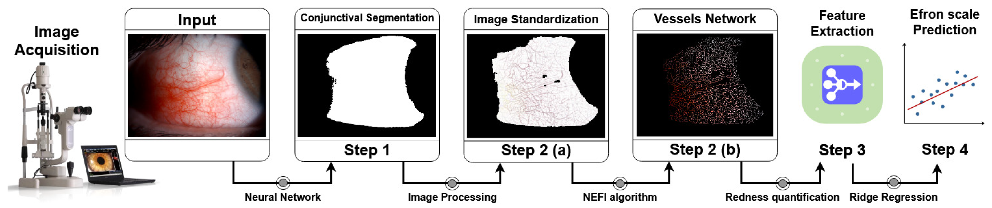
2.4. Image Processing Pipeline
- conjunctiva segmentation;
- vessels network segmentation;
- vessels network features extraction and color features extraction;
- Efron scale values prediction.
2.4.1. Step 1—Conjunctiva Segmentation
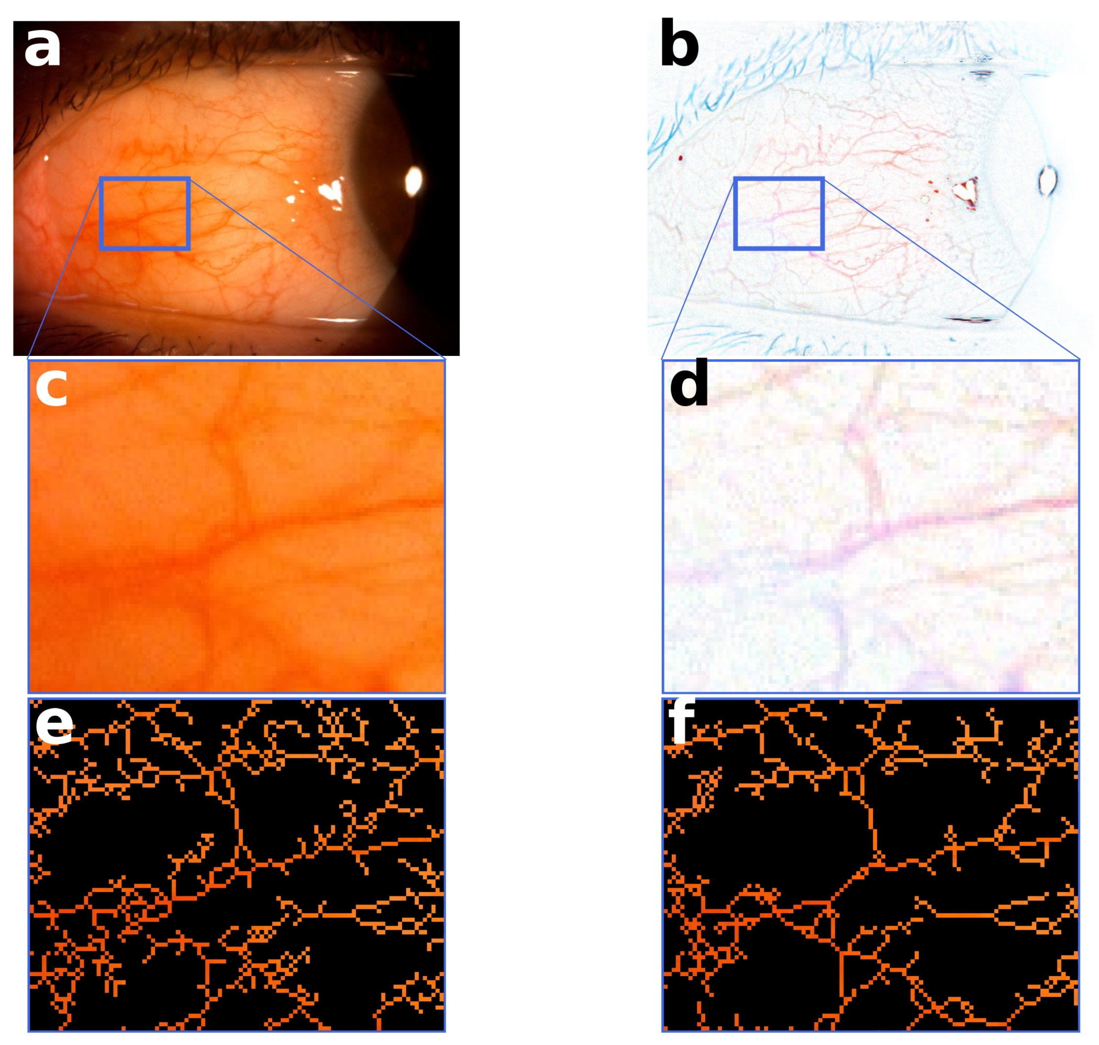
2.4.2. Step 2—Vessels Network Segmentation
2.4.3. Step 3—Redness Features
RGB Redness
HSV Redness
Fractal Analysis
Color Measures
2.4.4. Step 4—Regression Pipeline
3. Results
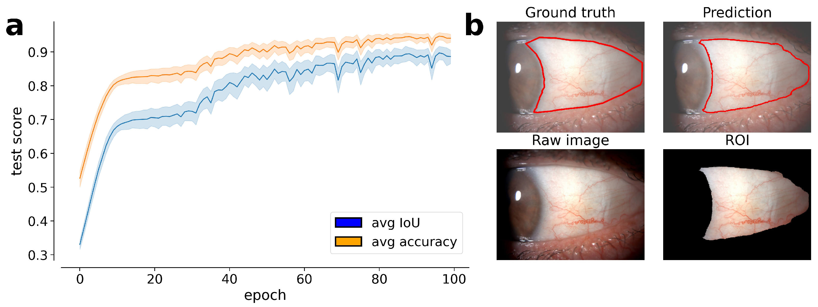
3.1. Conjunctival Segmentation
3.2. Vessels Network Segmentation
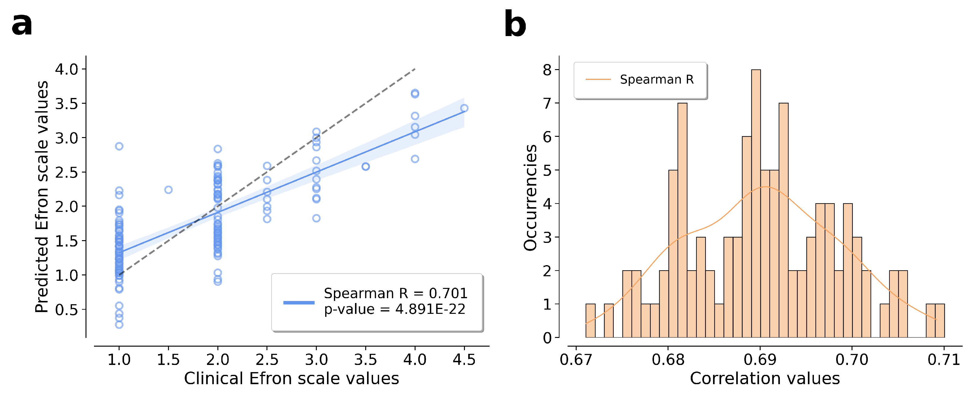
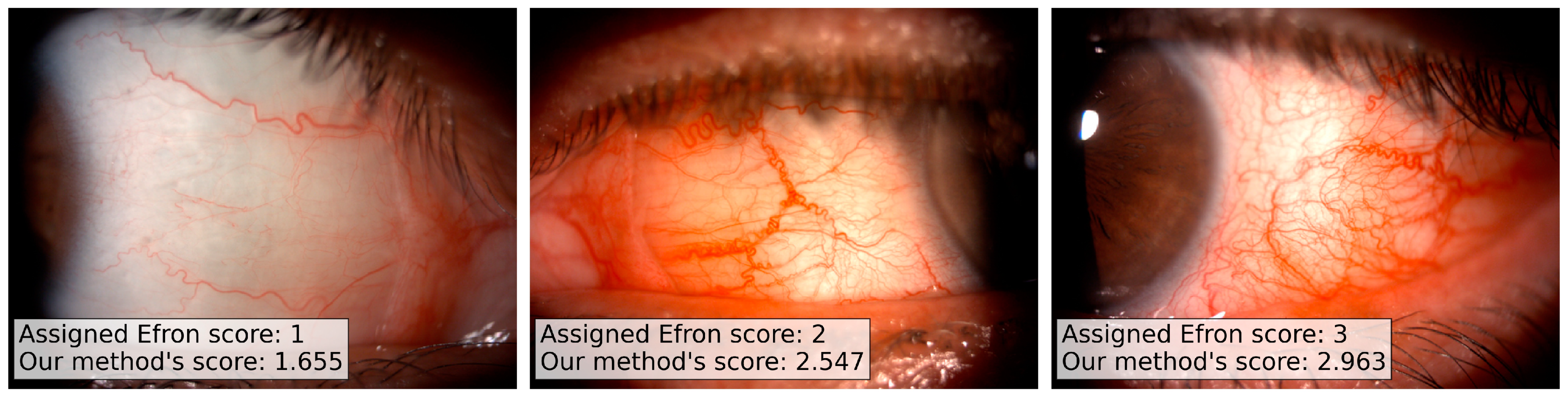
3.3. Efron Scale Prediction
4. Discussion
Author Contributions
Funding
Institutional Review Board Statement
Informed Consent Statement
Data Availability Statement
Conflicts of Interest
References
- Efron, N. Grading scales for contact lens complications. Ophthalmic Physiol. Opt. 1998, 18, 182–186. [Google Scholar] [CrossRef] [Scilit]
- Efron, N.; Morgan, P.B.; Katsara, S.S. Validation of grading scales for contact lens complications. Ophthalmic Physiol. Opt. 2001, 21, 17–29. [Google Scholar] [CrossRef] [PubMed]
- Rodriguez, J.; Johnston, P.; Ousler, G.R.; Smith, L.; Abelson, M.B. Automated grading system for evaluation of ocular redness associated with dry eye. Clin. Ophthalmol. 2013, 7, 1197–1204. [Google Scholar] [CrossRef] [Scilit] [PubMed]
- Schulze, M.M.; Hutchings, N.; Simpson, T.L. Grading Bulbar Redness Using Cross-Calibrated Clinical Grading Scales. Investig. Ophthalmol. Vis. Sci. 2011, 52, 5812–5817. [Google Scholar] [CrossRef] [Scilit] [PubMed]
- Peterson, R.C.; Wolffsohn, J.S. Sensitivity and reliability of objective image analysis compared to subjective grading of bulbar hyperaemia. Br. J. Ophthalmol. 2007, 91, 1464–1466. [Google Scholar] [CrossRef] [Scilit]
- Alexovic, M.; Urban, P.L.; Tabani, H.; Sabo, J. Recent advances in robotic protein sample preparation for clinical analysis and other biomedical applications. Clin. Chim. Acta 2020, 507, 104–116. [Google Scholar] [CrossRef] [Scilit]
- Bunya, V.Y.; Chen, M.; Zheng, Y.; Massaro-Giordano, M.; Gee, J.; Daniel, E.; O’Sullivan, R.; Smith, E.; Stone, R.A.; Maguire, M.G. Development and Evaluation of Semiautomated Quantification of Lissamine Green Staining of the Bulbar Conjunctiva From Digital Images. JAMA Ophthalmol. 2017, 135, 1078–1085. [Google Scholar] [CrossRef] [Scilit]
- Amparo, F.; Wang, H.; Emami-Naeini, P.; Karimian, P.; Dana, R. The Ocular Redness Index: A novel automated method for measuring ocular injection. Investig. Ophthalmol. Vis. Sci. 2013, 54, 4821–4826. [Google Scholar] [CrossRef] [Scilit] [PubMed]
- Park, I.K.; Chun, Y.S.; Kim, K.G.; Yang, H.K.; Hwang, J.M. New Clinical Grading Scales and Objective Measurement for Conjunctival Injection. Investig. Ophthalmol. Vis. Sci. 2013, 54, 5249–5257. [Google Scholar] [CrossRef] [Scilit] [PubMed]
- Schulze, M.M.; Hutchings, N.; Simpson, T.L. The Use of Fractal Analysis and Photometry to Estimate the Accuracy of Bulbar Redness Grading Scales. Investig. Ophthalmol. Vis. Sci. 2008, 49, 1398–1406. [Google Scholar] [CrossRef] [Scilit] [PubMed]
- Papas, E.B. Key Factors in the Subjective and Objective Assessment of Conjunctival Erythema. Investig. Ophthalmol. Vis. Sci. 2000, 41, 687–691. [Google Scholar]
- Fieguth, P.; Simpson, T. Automated Measurement of Bulbar Redness. Investig. Ophthalmol. Vis. Sci. 2002, 43, 340–347. [Google Scholar]
- Huntjens, B.; Basi, M.; Nagra, M. Evaluating a new objective grading software for conjunctival hyperaemia. Contact Lens Anterior Eye J. Br. Contact Lens Assoc. 2020, 43, 137–143. [Google Scholar] [CrossRef] [Scilit]
- Koprowski, R.; Wilczynski, S.; Olczyk, P.; Nowinska, A.; Weglarz, B.; Wylegala, E. A quantitative method for assessing the quality of meibomian glands. Comput. Biol. Med. 2016, 75, 130–138. [Google Scholar] [CrossRef] [Scilit] [PubMed]
- Wu, X.; Liu, L.; Zhao, L.; Guo, C.; Li, R.; Wang, T.; Yang, X.; Xie, P.; Liu, Y.; Lin, H. Application of artificial intelligence in anterior segment ophthalmic diseases: Diversity and standardization. Ann. Transl. Med. 2020, 8. [Google Scholar] [CrossRef] [Scilit]
- Brea, M.L.S.; Rodríguez, N.B.; González, A.M.; Evans, K.; Pena-Verdeal, H. Defining the Optimal Region of Interest for Hyperemia Grading in the Bulbar Conjunctiva. Comput. Math. Methods Med. 2016, 2016. [Google Scholar] [CrossRef] [Scilit]
- Derakhshani, R.; Saripalle, S.K.; Doynov, P. Computational methods for objective assessment of conjunctival vascularity. In Proceedings of the 2012 Annual International Conference of the IEEE Engineering in Medicine and Biology Society, San Diego, CA, USA, 28 August–1 September 2012; pp. 1490–1493. [Google Scholar] [CrossRef] [Scilit]
- Shin, S.Y.; Lee, S.; Yun, I.D.; Lee, K.M. Deep vessel segmentation by learning graphical connectivity. Med. Image Anal. 2019, 58, 101556. [Google Scholar] [CrossRef] [Scilit] [PubMed]
- Dirnberger, M.; Kehl, T.; Neumann, A. NEFI: Network Extraction From Images. Sci. Rep. 2015, 5. [Google Scholar] [CrossRef] [Scilit]
- Liu, X.; Bi, L.; Xu, Y.; Feng, D.D.F.; Kim, J.; Xu, X. Robust deep learning method for choroidal vessel segmentation on swept source optical coherence tomography images. Biomed. Opt. Express 2019, 10, 1601. [Google Scholar] [CrossRef] [Scilit] [PubMed]
- Radu, P.; Ferryman, J.; Wild, P. A robust sclera segmentation algorithm. In Proceedings of the 2015 IEEE 7th International Conference on Biometrics Theory, Applications and Systems (BTAS), Arlington, VA, USA, 8–11 September 2015; pp. 1–6. [Google Scholar] [CrossRef] [Scilit]
- Naqvi, R.; Loh, W.K. Sclera-Net: Accurate Sclera Segmentation in Various Sensor Images Based on Residual Encoder and Decoder Network. IEEE Access 2019, 7, 98208–98227. [Google Scholar] [CrossRef] [Scilit]
- Rot, P.; Emersic, Ž.; Struc, V.; Peer, P. Deep Multi-class Eye Segmentation for Ocular Biometrics. In Proceedings of the 2018 IEEE International Work Conference on Bioinspired Intelligence (IWOBI), San Carlos, Costa Ricam, 18–20 July 2018; pp. 1–8. [Google Scholar] [CrossRef] [Scilit]
- Hassan, G.; El-Bendary, N.; Hassanien, A.E.; Fahmy, A.; Snasel, V. Retinal Blood Vessel Segmentation Approach Based on Mathematical Morphology. Procedia Comput. Sci. 2015, 65, 612–622. [Google Scholar] [CrossRef] [Scilit]
- Huang, G.; Liu, Z.; van der Maaten, L.; Weinberger, K.Q. Densely Connected Convolutional Networks. In Proceedings of the IEEE Conference on Computer Vision and Pattern Recognition (CVPR), Honolulu, HI, USA, 21–26 July 2017. [Google Scholar] [CrossRef] [Scilit]
- Falk, T.; Mai, D.; Bensch, R.; Cicek, O.; Abdulkadir, A.; Marrakchi, Y.; Bohm, A.; Deubner, J.; Jackel, Z.; Seiwald, K.; et al. U-Net: Deep learning for cell counting, detection, and morphometry. Nat. Methods 2019, 16. [Google Scholar] [CrossRef] [Scilit] [PubMed]
- Zhiping, L.; Hua, W.; Hong, J.; Rosa, G.G.; Jianhua, W. Quantitative analysis of conjunctival microvasculature imaged using optical coherence tomography angiography. Eye Vis (Lond.) 2019, 6. [Google Scholar] [CrossRef] [Scilit]
- Tamuli, J.; Jain, A.; Dhan, A.V.; Bhan, A.; Dutta, M.K. An image processing based method to identify and grade conjunctivitis infected eye according to its types and intensity. In Proceedings of the 2015 Eighth International Conference on Contemporary Computing (IC3), Noida, India, 20–22 August 2015; pp. 88–92. [Google Scholar] [CrossRef] [Scilit]
- Guo, Z.; Hall, R.W. Parallel Thinning with Two-Subiteration Algorithms. Commun. ACM 1989, 32, 359–373. [Google Scholar] [CrossRef] [Scilit]
- Xu, Z.; Jiang, H.; Tao, A.; Wu, S.; Yan, W.; Yuan, J.; Liu, C.; DeBuc, D.C.; Wang, J. Measurement variability of the bulbar conjunctival microvasculature in healthy subjects using functional slit lamp biomicroscopy (FSLB). Microvasc. Res. 2015, 101, 15–19. [Google Scholar] [CrossRef] [Scilit] [PubMed]
- Hang, S.; Mingzhou, Z.; Xiaodan, H.; Kaixiu, L.; Xiaodan, J.; Yan, L.; Huibin, L.; Xuemin, L. Correlation Analysis of Ocular Symptoms and Signs in Patients with Dry Eye. J. Ophthalmol. 2017, 2017. [Google Scholar] [CrossRef] [Scilit]
- Chen, W.; Deng, Y.; Jiang, H.; Wang, J.; Zhong, J.; Li, S.; Peng, L.; Wang, B.; Yang, R.; Zhang, H.; et al. Microvascular abnormalities in dry eye patients. Microvasc. Res. 2018, 118, 155–161. [Google Scholar] [CrossRef] [Scilit] [PubMed]




| Grading Dataset | |||
|---|---|---|---|
| # of Samples | Average Age | Average Efron | |
| Male | 23 | ||
| Female | 47 | ||
| Total | 70 | ||
| Feature | Spearman’s Correlation |
|---|---|
| score | 0.642 |
| 0.612 | |
| −0.564 | |
| −0.505 | |
| −0.480 | |
| score | 0.408 |
| pixel-counting | −0.395 |
| 0.314 | |
| 0.267 | |
| box-counting | −0.113 |
Publisher’s Note: MDPI stays neutral with regard to jurisdictional claims in published maps and institutional affiliations. |
© 2021 by the authors. Licensee MDPI, Basel, Switzerland. This article is an open access article distributed under the terms and conditions of the Creative Commons Attribution (CC BY) license (http://creativecommons.org/licenses/by/4.0/).
Share and Cite
Curti, N.; Giampieri, E.; Guaraldi, F.; Bernabei, F.; Cercenelli, L.; Castellani, G.; Versura, P.; Marcelli, E. A Fully Automated Pipeline for a Robust Conjunctival Hyperemia Estimation. Appl. Sci. 2021, 11, 2978. https://doi.org/10.3390/app11072978
Curti N, Giampieri E, Guaraldi F, Bernabei F, Cercenelli L, Castellani G, Versura P, Marcelli E. A Fully Automated Pipeline for a Robust Conjunctival Hyperemia Estimation. Applied Sciences. 2021; 11(7):2978. https://doi.org/10.3390/app11072978
Chicago/Turabian StyleCurti, Nico, Enrico Giampieri, Fabio Guaraldi, Federico Bernabei, Laura Cercenelli, Gastone Castellani, Piera Versura, and Emanuela Marcelli. 2021. "A Fully Automated Pipeline for a Robust Conjunctival Hyperemia Estimation" Applied Sciences 11, no. 7: 2978. https://doi.org/10.3390/app11072978
APA StyleCurti, N., Giampieri, E., Guaraldi, F., Bernabei, F., Cercenelli, L., Castellani, G., Versura, P., & Marcelli, E. (2021). A Fully Automated Pipeline for a Robust Conjunctival Hyperemia Estimation. Applied Sciences, 11(7), 2978. https://doi.org/10.3390/app11072978