Article

Plasma Treatment of Fish Cells: The Importance of Defining Cell Culture Conditions in Comparative Studies

1
Department of Cell Biology, Rostock University Medical Center, Schillingallee 69, 18057 Rostock, Germany
2
Fraunhofer-Research Institution for Marine Biotechnology and Cell Technology, Mönkhofer Weg 239a, 23562 Lübeck, Germany
3
Institute of Genome Biology, Leibniz Institute for Farm Animal Biology (FBN), Wilhelm-Stahl-Allee 2, 18196 Dummerstorf, Germany
*
Author to whom correspondence should be addressed.
Recent address: Bluu GmbH, Schönhauser Allee 176, Building 21, 10119 Berlin, Germany.
Appl. Sci. 2021, 11(6), 2534; https://doi.org/10.3390/app11062534
Submission received: 28 January 2021 / Revised: 19 February 2021 / Accepted: 9 March 2021 / Published: 12 March 2021
(This article belongs to the Special Issue Application of Plasma Technology in Bioscience and Biomedicine)

Abstract

:
The present study provides the fundamental results for the treatment of marine organisms with cold atmospheric pressure plasma. In farmed fish, skin lesions may occur as a result of intensive fish farming. Cold atmospheric plasma offers promising medical potential in wound healing processes. Since the underlying plasma-mediated mechanisms at the physical and cellular level are yet to be fully understood, we investigated the sensitivity of three fish cell lines to plasma treatment in comparison with mammalian cells. We varied (I) cell density, (II) culture medium, and (III) pyruvate concentration in the medium as experimental parameters. Depending on the experimental setup, the plasma treatment affected the viability of the different cell lines to varying degrees. We conclude that it is mandatory to use similar cell densities and an identical medium, or at least a medium with identical antioxidant capacity, when studying plasma effects on different cell lines. Altogether, fish cells showed a higher sensitivity towards plasma treatment than mammalian cells in most of our setups. These results should increase the understanding of the future treatment of fish.

1. Introduction

The primary motivation of the present study was to accelerate wound healing in aquaculture fish by using cold atmospheric pressure plasma (CAP). CAP has been demonstrated to be beneficial for wound healing processes by reducing the microbial load, suppressing inflammatory events, reducing oxidative stress, and enhancing angiogenesis [1,2,3,4] in human and other vertebrate models [5,6]. These CAP-induced benefits support the natural regenerative capacity of the integument, but have not yet been tested on aquatic organisms.
The integument of vertebrates consists of the three layers epidermis, dermis and hypodermis [7,8], which represent a solid barrier against a potentially detrimental environment. To encounter the different spectra of terrestrial and aquatic environmental influences, the skin of land-living tetrapods and amphibians or fish, respectively, has specific adaptations [9]. Since lesions of the skin may result in a loss of homeostasis, a complex regeneration program is initiated immediately after traumata [10]. This wound-healing program involves several well-orchestrated events, including blood clotting, inflammation, re-epithelialisation and the generation and remodelling of tissue [11], which display class- and species-specific characteristics [12,13]. Skin injuries in farmed fish frequently occur as a consequence of intensive fish farming, e.g., due to fungal, bacterial or parasitic infections [14,15], aggressive conspecific interactions, overstocking-related abrasions or inadequate handling [16,17,18]. The immediate treatment of skin wounds rapidly reduces the wound size and stimulates the healing process to prevent bacterial secondary/co-infections, maintain fish welfare and eventually prevent considerable economic losses [19]. However, there are hardly any treatment approaches for this problem so far. While wound healing processes have been investigated in a few in-vitro models from bony fish [20,21,22], to the best of our knowledge therapeutic approaches involving CAP have never been analysed in fish and fish cell lines to date.
Cold atmospheric pressure plasma is an ionized gas formed by the input of high energy and consists of ions, electrons, radicals, ultraviolet photons and uncharged atoms or molecules. CAP can be generated using various plasma devices, such as plasma jets or a dielectric barrier discharge [23]. CAP generates short- and longer-lived molecules, such as reactive oxygen and nitrogen species (RONS), one of which is hydrogen peroxide. Longer-lived species are generated especially by the interaction of CAP with liquids [23,24]. In mammalian cell models, the treatment with CAP is conducted either directly, or indirectly via liquids that were previously treated with plasma. Our former studies proved that CAP mainly affects the cells via the surrounding medium. We showed that the efficacy of plasma-activated medium (PAM) is maintained for up to 21 days when stored at standardized conditions of 37 °C [25,26]. This medium-mediated effect of CAP was also observed by other authors for different fluids (cell culture medium, phosphate buffered saline, water, Ringer’s solution) [27,28,29,30,31]. In general, similar experimental setups are recommended [32,33] to allow for discrimination of cell type-specific reactions from medium-mediated effects, particularly when comparing tumour cells with normal cells.
We wanted to find out whether plasma exerts an effect on fish cells and how this differs from mammalian cells. In our study, we compared different culture conditions and their influence on cell viability. To this end, we conducted parallel studies on cells from different origins in PAM prepared from the same culture medium or in PAM prepared from medium with different antioxidant potential to test whether these parameters influence the cellular sensitivity towards cold atmospheric pressure plasma.

2. Materials and Methods
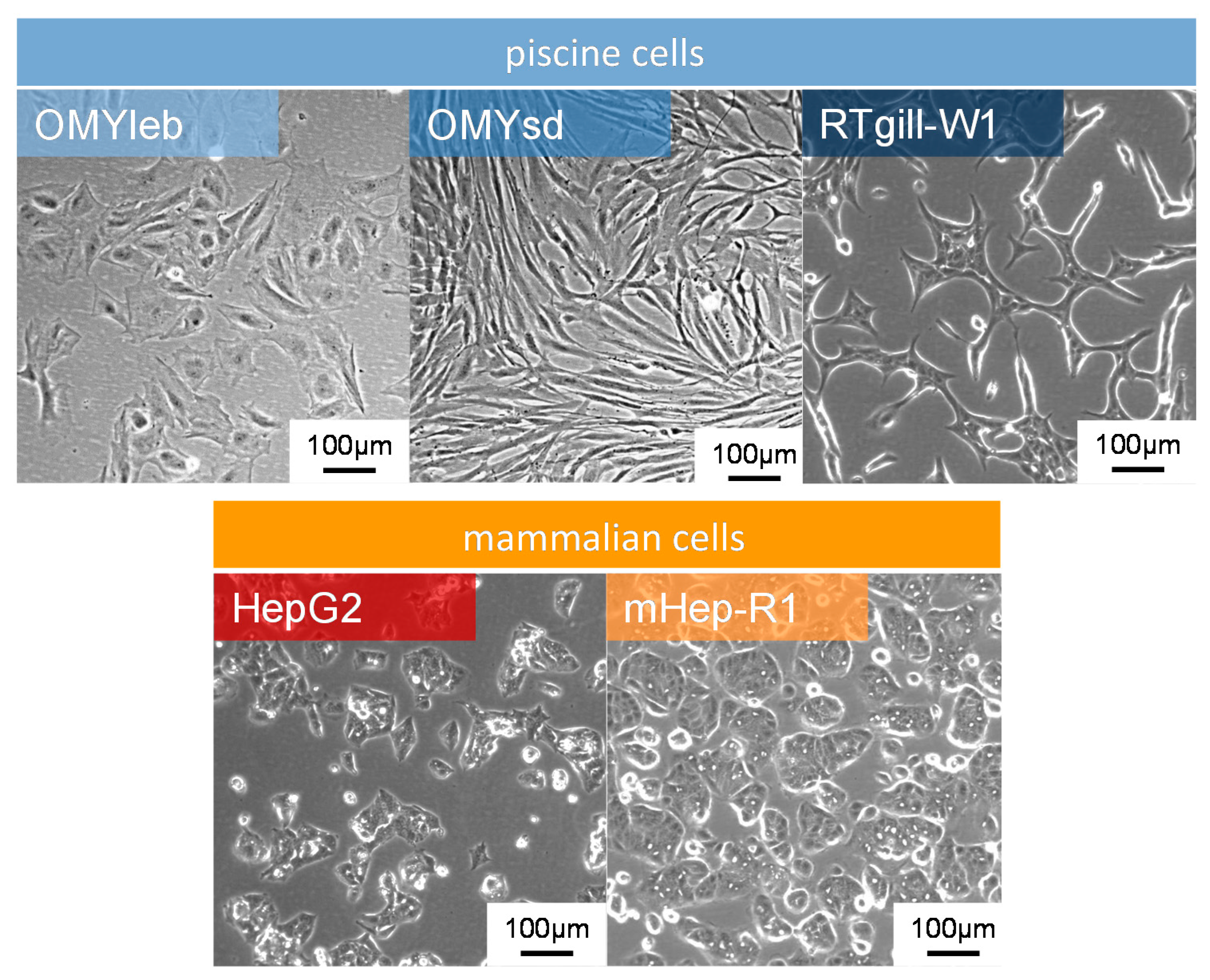
2.1. Cells

We used the following three fish cell lines: OMYleb (liver cell line of rainbow trout Oncorhynchus mykiss), OMYsd (skin-derived cell line from O. mykiss)—both developed by S. Rakers at the Fraunhofer EMB and available from the Cryo-Brehm cell bank, www.cryo-brehm.de [34,35]—as well as RTgill-W1 (gill cell line from O. mykiss, ATCC Id: CRL-2523). For comparative studies, we used the mammalian cells HepG2 (liver cancer cell line from human; ATCC Id: HB-8065) and the mHep-R1 cell line (derived from SV40 transgenic mice [36] with characteristics of hepatocytes), which was described in detail by Henning et al. [37] and has been established in our lab for many years [38,39].
All cell lines were cultured in Dulbecco’s modified Eagle’s medium (DMEM; Thermo Fisher Scientific, Waltham, MA, USA, Cat.no. 10569) or Leibovitz’s L-15 medium (Thermo Fisher (Gibco), Cat.no. 11415), both supplemented with 10% fetal calf serum (FCS; FCS Superior; Biochrom GmbH, Berlin, Germany) and 1% gentamicin (Ratiopharm, Ulm, Germany). An overview of the different cell lines is given in Table S1 and the composition of the two media is listed in Table S2. Importantly, the formulations of these media differ with respect to the content of pyruvate, glucose or glutamine depending on the company selling these media. During culturing, the medium was exchanged every second day and the cells were passaged every 5 days. The mammalian cells HepG2 and mHep-R1 were cultivated at 37 °C with 5% CO2 and fish cells were cultured at 20 °C with 5% CO2 in a humidified atmosphere. For one analysis (cf. Section 3.3), the cell lines OMYsd and OMYleb were cultured in L-15 medium without the addition of CO2. Cells were monitored using the AxioScope A.1 microscope (Carl Zeiss AG, Oberkochen, Germany) and cell size was measured with the ImageJ software (version 1.51j8, National Institutes of Health, USA, 2017).

2.2. Plasma Treatment
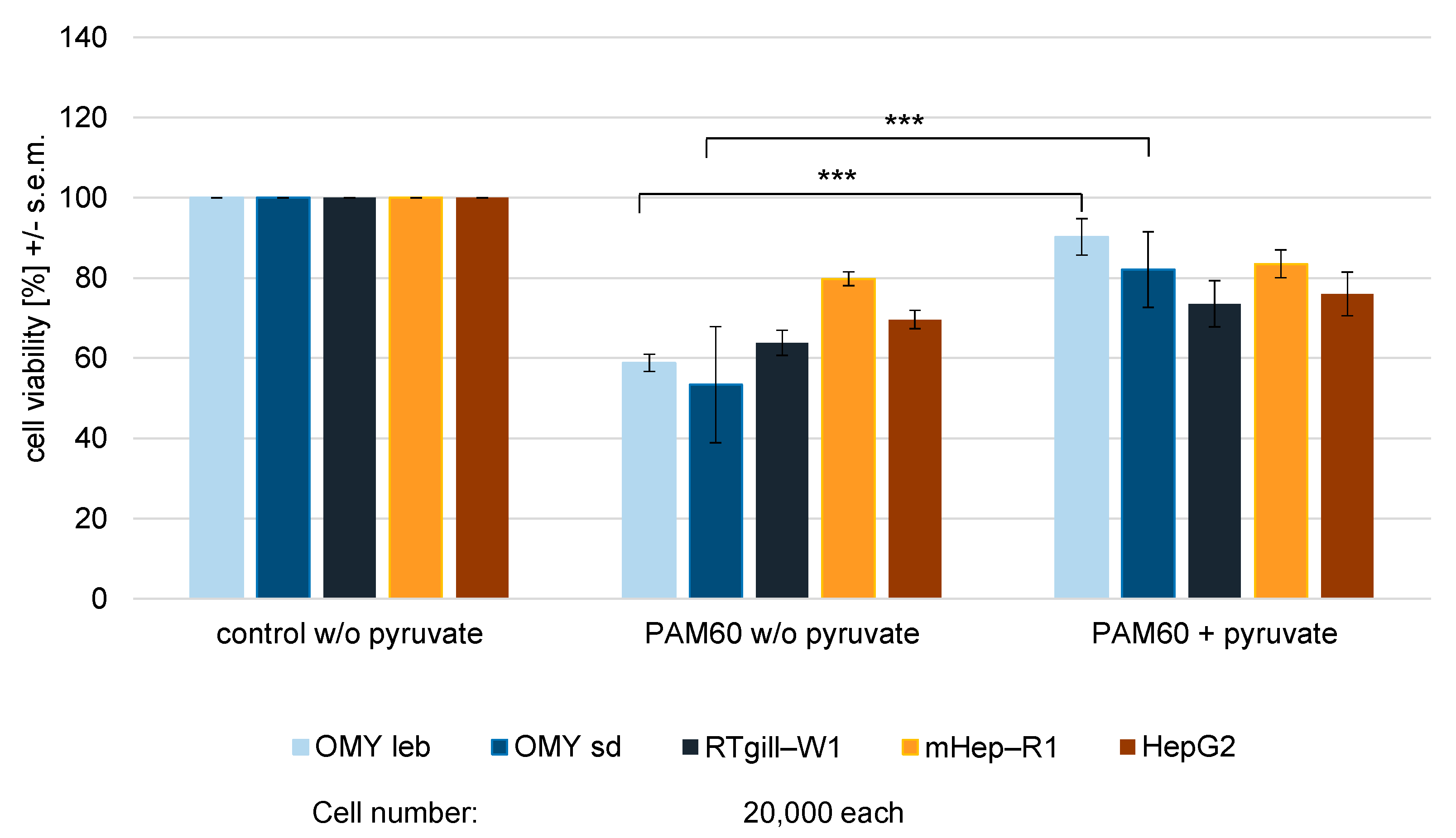
The plasma jet kINPen09 (Neoplas Tools GmbH, Greifswald, Germany) was used for all experiments. This plasma source was characterized in detail previously [25,40]. A gas flow of 1.9 slm argon was used as the feed gas. A gas discharge is ignited at the tip of the high-voltage needle, exciting the argon gas. In this way, low-temperature plasma is generated and blown out of the capillary. The so-called plasma jet outside the nozzle has a length of 12–14 mm and is about 1 mm wide. The temperature at the visible tip of the plasma jet does not exceed 50 °C. To generate PAM, 100 µL of the culture medium were added per well to a plate with 96 wells (Greiner Bio-One, Kremsmünster, Austria). The kINPen09 was mounted vertically on a precision positioning table (OWIS GmbH, Staufen im Breisgau, Germany, control via software OWISoft v. 2.9.1.2, 2018) and the quartz capillary of the jet was positioned centrally at the upper edge of each well in the 96-well plate without exceeding a distance of 1 mm. The medium was treated with the plasma jet for 15 s, 30 s, 60 s, or 120 s. The resulting plasma activated medium (PAM) is hereafter referred to as PAM15, PAM30, PAM60 and PAM120, respectively. Argon (Ar) gas treatment using the kINPen09 without igniting the plasma was used as a control. The treated medium was stored at 37 °C for 24 h to ensure that the oxygen and hydrogen peroxide concentration balanced to normal conditions [25].

2.3. Cell Viability

An MTS assay (CellTiter 96 Aqueous One Solution Cell Proliferation Assay, Promega, Madison, WI, USA) was performed to study the influence of the plasma treatment on the cell’s viability and metabolism. Cells were seeded into 96-well plates for 24 h. After that time, the medium was aspirated and replaced by 100 µL of the plasma-treated medium. The cells were incubated for a further 24 h. Then, 20 µL MTS solution was added and after a 2-h incubation, the medium was transferred into a new 96-well plate and the spectrophotometric absorption was analysed by an ELISA reader (Anthos 2010, Anthos Labtec Instruments, Salzburg, Austria) at 490 nm (see Figure 1 for an overview of the experimental setup). Since the sole plasma treatment of the medium also leads to a slight colour change of the MTS solution, a separate blank was carried along for each treatment time and medium. This value was subtracted from the respective cell sample. The calculated absorbance is proportional to the metabolic activity of the cells. The viability of the cells was calculated in relation to that of the cells in the untreated control medium (100%). Each treatment condition was tested in triplicate in at least three individual experiments.

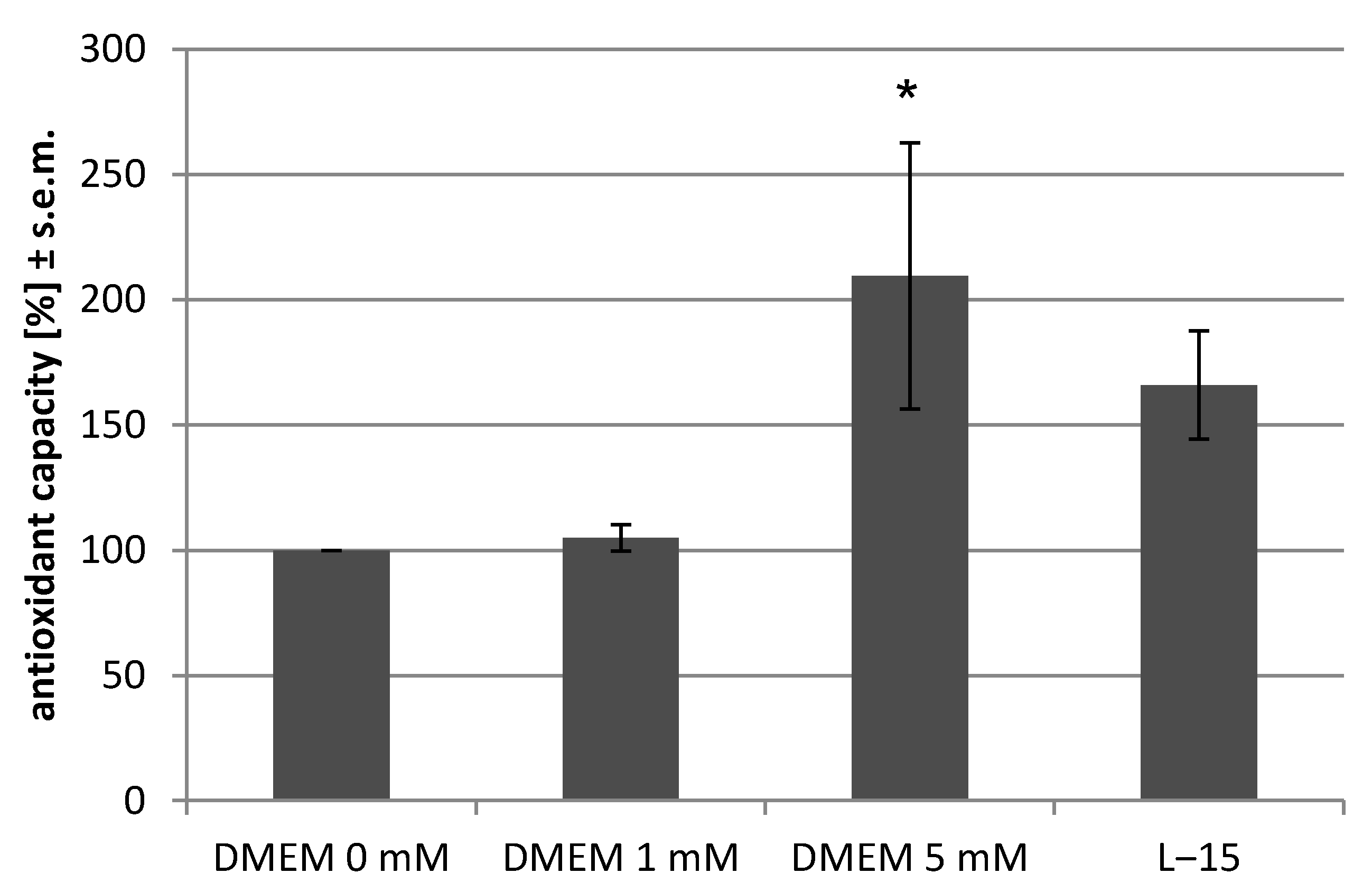
2.4. Antioxidant Assay

The Antioxidant Assay Kit (Cayman Chemical, Ann Arbor, MI, USA) was applied to assess the antioxidant capacity of the medium. This assay measures the combined antioxidant activities of all medium components including vitamins, proteins, lipids, glutathione, etc. Antioxidants in the medium inhibit the oxidation of ABTS (2,2′-Azino-di-[3-ethylbenzthiazoline sulphonate]) to ABTS•+ by metmyoglobin. The amount of ABTS•+ produced is monitored by measuring the absorbance in a plate reader (Tecan Infinite 200, Tecan Trading AG, Switzerland) at 750 nm. The measurements were performed with DMEM either without pyruvate (Cat. No. 10556; Thermo Fisher Scientific) or with 1 mM pyruvate (Cat. No. 10569; Thermo Fisher Scientific) or with 5 mM pyruvate. DMEM with 5 mM pyruvate was prepared from DMEM with 1 mM pyruvate by the addition of sodium pyruvate (cell-culture grade, 44 mg/100 mL; AppliChem GmbH, Darmstadt, Germany). All media contained 10% FCS. Each sample was measured in duplicate.

2.5. Statistics

Statistical analysis of the data was performed using the software GraphPad Prism (v. 8.0 GraphPad Software, Inc., San Diego, CA, USA). Data are presented as mean ± standard error of the mean and analysed using one-way ANOVA or Friedman test with posthoc Dunns test for the antioxidant assay. Differences were considered statistically significant at p < 0.05 (* p < 0.05, ** p < 0.01, *** p < 0.001).

3. Results

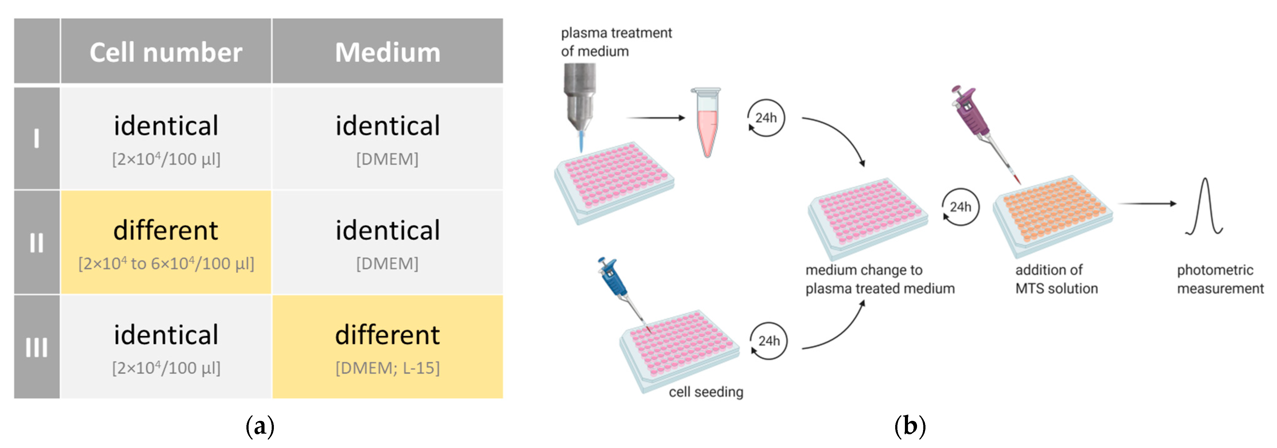
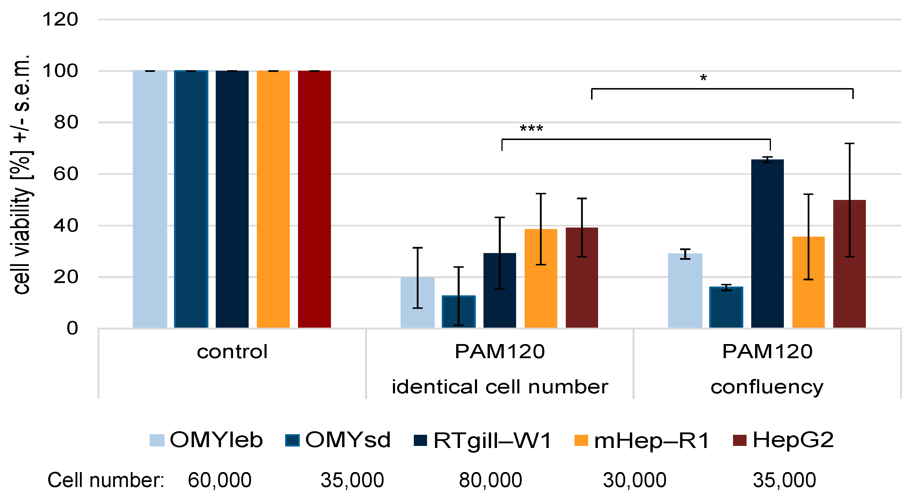
3.1. Experimental Setup I: Identical Cell Number Seeded in Identical Medium

Cell viability served as the criterion to evaluate whether fish cells react to plasma treatment with a similar sensitivity as mammalian cells. For an initial analysis, we seeded 2 × 10⁴ cells of each of the three piscine cell lines (OMYleb, OMYsd, RTgill-W1) and the two mammalian cell lines (HepG2, mHep-R1) (overview in Table S1) and cultured them in DMEM. This cell number was considered optimal in previous studies [26,33]. After 24 h, the cells were stimulated for 24 h either with PAM (PAM15, PAM30, PAM60 and PAM120) or with Ar-treated medium. The cell viability of fish as well as of mammalian cells (Figure 2) decreased only slightly after treatment with PAM15, PAM30 and PAM60. In contrast, PAM120 had a clear inhibitory effect on the viability of all cell lines investigated. The fish cell lines OMYleb (viability of 12.6%) and OMYsd (19.6%) were found to be extremely sensitive, while the mammalian cell lines HepG2 and mHep-R1 were comparably less sensitive (viability of 39.2% and 38.6%). RTgill-W1 cells displayed a viability of 29.3% after treatment with PAM120. Ar-treated medium—as a control—did not affect the viability of the cells investigated (data not shown).
Based on these observations, we assumed that fish cells apparently react more sensitively to plasma treatment than mammalian cells. We tested this hypothesis with a second experiment, which considered the cell type-specific densities.

3.2. Experimental Setup II: Different Cell Number Seeded in Identical Medium

In our initial experiment (Section 3.1), we had used cells, which differed considerably in their morphology and size (Figure 3). Among the cell lines investigated, HepG2 were the smallest cells in their area with a rounded morphology (284 µm2 ± 45 µm2) while OMYsd were relatively large in their area but elongated (1222 µm2 ± 404 µm2). This is of relevance when simulating a similar degree of coverage in the cell cultures across the cell lines investigated. The cell-to-cell contact is an important stimulus for the cellular function, especially for epithelial cells.
Thus, we adjusted a comparable level of confluency (~80%) prior to the treatment (i.e., 24 h post seeding) and observed remarkable differences in the viability outcome (Figure 4, cell numbers are indicated below the graph). The percentage viability of the piscine RTgill-W1 cells—seeded at a 4-fold higher density—increased by 36.4% after PAM120 treatment. Similarly, the percentage viability of the mammalian cell line HepG2 increased by 10% after elevating the cell density 1.75-fold. The fish cell lines OMYleb (+9.3%) and OMYsd (only +3.4%) were again the most sensitive to PAM120. We detected, that the piscine OMYleb cells did not reach the same level of viability as mammalian mHep-R1 cells, although twice as many OMYleb cells were seeded. The viability of mHep-R1 cells was almost unchanged compared with the previous experiment, even at a 1.5-fold higher density. From this observation we concluded, that the optimal cell density alone most likely does not provide a higher degree of resilience to plasma treatment.

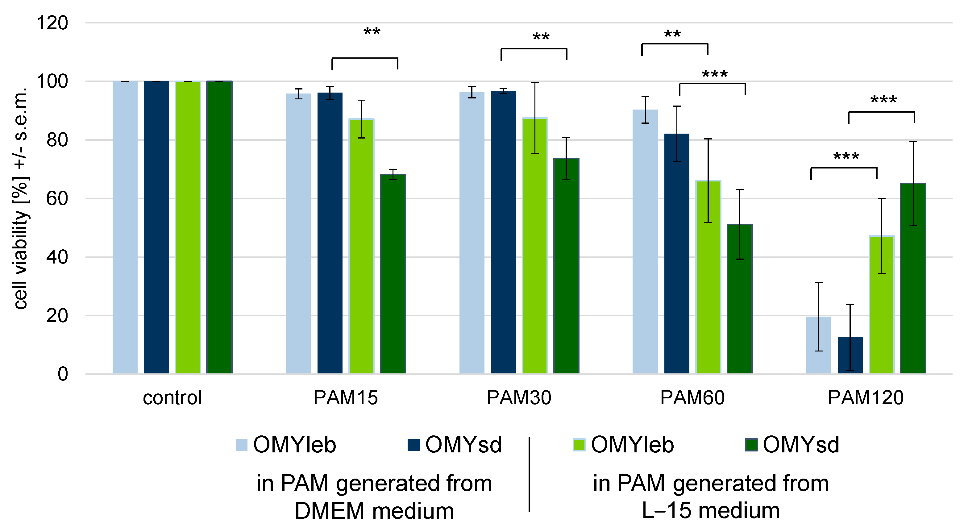
3.3. Experimental Setup III: Identical Cell Number Seeded in Different Media

The effects of CAP on the cells are mediated by the plasma-activated medium. To address the question to what extent the choice of medium influences these effects, we cultured OMYleb and OMYsd cells in DMEM medium or L-15 medium (Figure 5). L-15 medium has been used for decades for culturing cells of salmonid origin [41,42]. The composition of the two media used is listed in Table S2.
The cultivation in L-15 medium previously treated with plasma for 15 s, 30 s and 60 s resulted in a decrease of viability, while cells grown in DMEM—treated for 15 s, 30 s and 60 s—were hardly affected. However, treatment with PAM120 generated from DMEM drastically reduced the viability of OMYleb (−80.4% compared with the untreated control) and OMYsd (−87.4%). In contrast, the same cells in PAM120 generated from L-15 medium were reduced in their viability by only −52.8% and −34.9%, respectively. Thus, L-15 medium had a protective effect against CAP for both fish cell lines.
Since the effect of the plasma is mediated via the liquid, the varying compositions of the different media used may influence the effectiveness of the PAM. The comparison of the antioxidant capacity of the two media (Figure 6) revealed that the L-15 medium (containing 5 mM pyruvate) has a higher oxidative capacity than DMEM medium (which originally contains 1 mM pyruvate). To determine if the oxidative capacity of DMEM depends on the pyruvate content, we added different pyruvate concentrations to DMEM whose pyruvate content was adjusted to that of the L-15 medium and then re-measured the oxidative capacity of these media. We found that DMEM without or with 1 mM pyruvate shares a similar degree of oxidative capacity. After supplementation of 5 mM pyruvate, the antioxidative capacity of DMEM increased enormously (+110%). The capacity of the DMEM medium with 5 mM pyruvate exceeded that of the L-15 medium, which also contains 5 mM pyruvate.
In addition, we cultured the two mammalian and three piscine cell lines in DMEM without pyruvate. Omitting the pyruvate from the medium did not alter the general viability of the cells (data not shown). However, the viability was in part significantly impaired when the cells were stimulated with plasma-treated pyruvate-free medium (Figure 7). A treatment time of only 60 s of pyruvate-free DMEM significantly reduced the viability of OMYleb and OMYsd cells compared with 60-s treatment of DMEM containing pyruvate. By contrast, the measured viability of the other cells (RTgill-W1, mHep-R1 and HepG2) was unchanged in pyruvate-free PAM compared with PAM containing pyruvate.

4. Discussion

4.1. Fish Cells Respond Differently to Plasma Treatment than Mammalian Cells

As a first step on the way to a CAP-mediated therapy of farm fish, we wanted to clarify how piscine cells react to CAP treatment, as this was previously unknown. For this reason, we performed cell viability assays to evaluate the individual responsiveness of gill epithelial cells and fibroblast-like cells from rainbow trout and liver epithelial cells of the three species rainbow trout, mouse, and human. The respective cells were chosen because the use of primary non-cancerous human cells in cell culture often requires highly supplemented media adapted to the specific needs of that particular cell line. Considering that our main experimental principle was to use an identical medium to deliver an identical cocktail of plasma-generated reactive species to the different cell lines, the use of cells requiring a specific medium had to be avoided. Therefore, only cells that could be cultured in the same medium were selected. This allowed us to draw conclusions for the newly used fish cells compared to the already published mammalian cells. Our aim was to provide an objective comparison to place the fish cells in this known context.
The results of an experiment depend, among other variables, on the careful choice of the cell number. To allow for comparisons across different cell lines, the design of an experiment should involve tuned cell numbers. In addition to the adjustment of the cell number, proliferation rates, metabolic status and cell migration effects should be taken into account [32,43]. In line with this, we cultured the piscine cells and the mammalian cells at equal cell numbers in identical medium and found that the fish cells reacted much more sensitively to CAP treatment than the mammalian cells. Subsequently, we changed the experimental conditions with regard to cell density to ensure a similar degree of confluency prior to the plasma treatment. This took into account the size of the individual cells as well as their proliferation rate. The results did not provide clear indications for the interdependence of confluency and cellular sensitivity to CAP. While one piscine cell line (RTgill-W1) showed a higher robustness to PAM after the careful adjustment of the optimal confluency, the sensitivity of another confluent cell line from fish (OMYsd) remained almost unchanged. These differences could be due to specific structural and functional adaptations, as the cell lines were derived from different tissues of rainbow trout (liver, skin and gill) and display different morphological characteristics (see Figure 3).
The serum used to supplement the cell culture media may also have influenced the sensitivity of the individual cell lines towards PAM. We used serum from fetal calves (FCS), which has been used as a universal supplement for more than six decades [44] to culture approximately 95% of all cell lines including insect cells [45]. Essentially, the supplementation of cell media with FCS ensures the supply with growth factors, which are evolutionarily well conserved [46]. However, the exact composition of FCS has not yet been clarified [47], which is also due to the different origins of the cattle stocks. Therefore, we used a serum for our experiments that is composed in a lot-independent manner (FCS Superior, Biochrom GmbH). Since serum contains thousands of different metabolites [48], it is not yet possible to assess whether and how specific serum factors differentially affect cellular signaling cascades and survival pathways in fish or mammals.
Although we tried to keep the cultivation conditions as similar as possible for the five cell lines investigated, the cultivation temperature was a key parameter that had to be kept within the tolerance range of the donor species rainbow trout, mouse, and human. Rainbow trout are explicitly cold-water fish and grow at temperatures between 8 °C and 22 °C [49]. Consequently, we cultured the fish cells at 20 °C and not at 37 °C like their mammalian counterparts. Previous studies revealed that culturing fish cells at 37 °C produces a pronounced heat-stress response [50]. Nonetheless, it is well established that temperature determines the activity of metabolic processes. The fact that fish cells were cultivated in a 17 °C colder environment is expected to have influenced the cellular metabolism. The van-t-Hoff rule states that temperature reduction significantly slows down chemical processes [51,52]. In addition, temperature defines the fluidity of lipid bilayers and therefore the membrane competency including ion transport, enzyme function and the formation of signalling complexes [53]. In the present study, we observed that fish cells reacted more sensitively to oxygen radicals compared with mammalian cells. We can only speculate that distinct temperature-dependent kinetics of the involved anti-oxidative enzymes and the relevant signalling cascades may have influenced the fate of the mammalian and fish cells. In summary, in all experimental setups, the piscine cell lines (OMYleb, OMYsd and RTgill-W1) were the most sensitive to plasma-treated liquids. Therefore, we can conclude that the treatment of fish cells requires carefully adjusted plasma treatment times. As a first step, we performed some experiments on the wound healing potential under our experimental conditions (Figure S1). However, since we found that the sensitivity of the fish cells was very high in our experiments, we were unable to measure any positive effect on wound healing, even with the shortest treatment times. The establishment of suitable plasma treatment regimes in terms of reactive species composition and application protocols will be a task for future research.

4.2. The Choice of Culture Conditions Is Highly Relevant as Changes in the Antioxidant Capacity Make a Difference

The effects of CAP are mediated via the cell culture medium. Therefore, we wanted to address the question to what extent a change in culture conditions modulates the cellular response to CAP treatment. The optimal composition of the cell culture medium has been tailored for the broad range of secondary cell lines and primary culture. Consequently, many different media are commercially available with DMEM (Dulbecco’s modified Eagle medium)/EMEM (Eagle’s minimum essential medium) and RPMI (Roswell Park Memorial Institute) as the most frequently used growth media [54]. Furthermore, various additives such as serum, amino acids, d-glucose or phenol red are added in different concentrations. All of these components may interfere with the eventual effects of physical plasma.
We studied the influence of two different media on the PAM effects measured on the five cell lines. Since L-15 medium is the common medium for the culture of salmonid cells [41,42], we used it for our comparative experiments. The direct comparison to the cultivation and treatment in PAM generated from DMEM revealed that the fish cells were more sensitive to PAM from L-15 medium treated with CAP for short periods and more tolerant to PAM from L-15 treated with CAP for a longer period.
These diverging effects are likely to result from the different composition of the media. The concentration of potentially anti-oxidative compounds might be of crucial importance. We stored the PAM for 24 h before adding it to the cells because this allowed us to exclude the possibility that the observed effects were due to high hydrogen peroxide concentrations or oxygen depletion during plasma treatment. Thus, our study provides evidence that the observed effects are produced by other reactive species (NO2, NO3 and others).
Wende et al. investigated the effect of CAP on human keratinocytes cultivated in IMDM medium (Iscove’s modified Dulbecco’s medium), which is a strong radical scavenger, and in RPMI medium, which is a weak radical scavenger. The viability of cells in IMDM decreased to 50% after 30 s of CAP, while the number of cells in RPMI reduced to 15% after identical treatment [27]. These studies are further evidence that the effect of CAP is mainly mediated by oxygen radicals and their longer-lasting reaction products. In general, the L-15 medium, in contrast to DMEM, is based on a CO2-independent buffer system and therefore contains higher concentrations of phosphates and various amino acids but lacks sodium bicarbonate (Table S2). In addition, the L-15 medium contains 5 mM pyruvate, whereas the DMEM medium used here contains 1 mM pyruvate only. It has been shown that high concentrations of pyruvate can have a protective effect on human lung cancer and osteosarcoma cells or murine liver cells by reacting with reactive oxygen species (ROS), which in turn reduces the pyruvate concentration [33,55,56]. Measurements of the hydrogen peroxide concentration revealed low concentrations of hydrogen peroxide after plasma treatment of a low-pyruvate medium and storage of the medium for 24 h [33]. However, when using a medium with 5 mM pyruvate (either DMEM or L-15), we could not detect hydrogen peroxide even with prolonged treatment times of 240 s (see Supplementary Materials Figure S2). Therefore, in our view, it is not sufficient to measure the hydrogen peroxide concentration in the media, at least not for media with a high pyruvate content. For the exact comparison, we suggest measuring the total antioxidant potential.
We analysed the antioxidative potential of DMEM and L-15 medium using a commercial assay that quantifies the overall amount of antioxidative species in the solution. The combined action of all the antioxidants present in a given medium provide synergistic protection against reactive oxygen or nitrogen species. Therefore, we determined the total antioxidant capacity of each medium instead of adding up the antioxidant capacities of the individual components. Special attention was paid to pyruvate, as former experiments pointed to its role in neutralizing CAP-generated reactive oxygen species [33].
We did prove, however, that the overall antioxidative capacity of a medium is not only dependent on the pyruvate content. The antioxidative capacity of the DMEM medium was higher than in the L-15, although both media contained 5 mM pyruvate. To take the role of pyruvate into account, we also varied this parameter in our experiments and carried out cell culture experiments with PAM generated from a DMEM medium, which lacked pyruvate. After assessing the general vitality of the cells in a pyruvate-free medium, we carried out the plasma treatment, which reduced the viability of two of the three trout cell lines (OMYleb and OMYsd cells). This effect was not observed in RTgill-W1, mHep-R1 and HepG2 cells. This suggests that the cellular responses can vary greatly depending on the cell type. Some cell types (OMYleb, OMYsd) react more sensitively to the loss of an antioxidant agent in PAM, while other cells are able to compensate for this, at least over the relatively short period of investigation (24 h). In comparative experiments on the action of CAP or PAM, it is therefore mandatory to ensure a congruent media composition, as even small deviations (e.g., in pyruvate content) can lead to an altered cell sensitivity.
Other potentially antioxidant substances may be included in the cell culture media used. These include above all phenol red, amino acids [57,58], proteins, carbohydrates [59] and vitamins such as pantothenic acid (VitB5), riboflavin (VitB2) and folic acid [60,61,62]. RONS formed in the medium by the action of CAP can react with these media components. In this process, some compounds are more reactive (e.g., aromatic and sulfur-containing amino acids) than others and therefore tend to act more readily as radical scavengers [57,58]. Carbohydrates such as glucose can be degraded to short-chain organic acids by the action of CAP [59]. Table S2 lists the components of the media used in this study with potentially antioxidative compounds printed in bold.
Our study provides further evidence that pyruvate is one, but not the only important factor contributing to the anti-oxidative potential of a solution. Due to the different composition of the media, the elimination of oxygen radicals can already vary in the liquid phase. Comparative studies on the action of CAP or PAM on cells should ensure that the medium used has a comparable antioxidative potential. It is not sufficient to pay attention to an identical proportion of pyruvate.

5. Conclusions

We found that the plasma treatment of fish cells requires lower dosages than of mammalian cells. The mortality of fish cells was in most of our setups higher than that of mammalian cells regardless of the plasma-treated media used (DMEM vs. L-15).
A major shortcoming of many previous in vitro experiments is their experimental design, which has led to misconclusions regarding the sensitivity of the cells to plasma treatment. In light of the results of our current study, we suggest using similar cell densities, identical medium, or at least medium with identical antioxidant potential when studying plasma effects on different cells.
Future studies on fish cell regeneration should involve not only viability studies but also wound healing assays and migration studies under consideration of short plasma treatment times. Plasma treated liquids could be a promising therapeutic tool for aquatic cultures in the future.

Supplementary Materials

The following are available online at https://www.mdpi.com/2076-3417/11/6/2534/s1, Table S1: Overview of the cell lines used in this study. Table S2: List of the components of the cell culture media L-15 (Leibovitz’s L-15 medium; Thermo Fisher Scientific; Cat.no. 11415) and DMEM (Dulbecco’s modified Eagle’s medium; Thermo Fisher Scientific, Cat.no. 10569) according to the manufacturers’ protocol. Potentially antioxidative compounds are printed in bold. Figure S1: Wound-healing assay with OMYleb cells under the influence of short plasma treatment times. Exemplary images are shown in (a). The size of the gap between two near-confluent cell layers reduced after 24 h growth. We could not detect a difference between untreated cells or cells growing in PAM (b). n = 4 independent experiments in DMEM containing 1 mM pyruvate. Figure S2: Measurement of hydrogen peroxide in the different media used in this study at the time they were given to the cells. Plasma treatment was performed for the indicated times. Only L-15 medium and DMEM 5 mM pyruvate medium were treated for 240 s, but even at these long treatment times no peroxide was detectable (argon-treated medium (AAM) served as control; Quantofix peroxide, Macherey Nagel, Düren, Germany).

Author Contributions

Conceptualization, H.R. and A.R.; methodology, H.R. and C.B.; formal analysis, H.R.; investigation, H.R.; resources, S.R. and B.N.; data curation, H.R.; writing—original draft preparation, H.R. and A.R.; writing—review and editing, H.R., C.B., S.R., B.N. and A.R.; visualization, H.R.; funding acquisition, B.N. All authors have read and agreed to the published version of the manuscript.

Funding

We acknowledge the support the European Social Fund (ESF), reference: ESF/14-BM-A55-0002/18, and the Ministry of Education, Science and Culture of Mecklenburg-Vorpommern, Germany for the joint research project ONCOTHER-H.

Institutional Review Board Statement

Not applicable.

Informed Consent Statement

Not applicable.

Data Availability Statement

Not applicable.

Acknowledgments

The authors would like to thank Petra Seidel and Marie Sawade (University Medical Center Rostock) for qualified technical assistance. We kindly thank Tomáš Korytář (University of South Bohemia in České Budějovice; Faculty of Fisheries and Protection of Waters) for the help with the schematic illustration.

Conflicts of Interest

The authors declare no conflict of interest. The funders had no role in the design of the study; in the collection, analyses, or interpretation of data; in the writing of the manuscript, or in the decision to publish the results.

References

  1. He, R.; Li, Q.; Shen, W.; Wang, T.; Lu, H.; Lu, J.; Lu, F.; Luo, M.; Zhang, J.; Gao, H.; et al. The efficacy and safety of cold atmospheric plasma as a novel therapy for diabetic wound in vitro and in vivo. Int. Wound J. 2020, 17, 851–863. [Google Scholar] [CrossRef] [PubMed]
  2. Kaushik, N.K.; Ghimire, B.; Li, Y.; Adhikari, M.; Veerana, M.; Kaushik, N.; Jha, N.; Adhikari, B.; Lee, S.J.; Masur, K.; et al. Biological and medical applications of plasma-activated media, water and solutions. Biol. Chem. 2018, 400, 39–62. [Google Scholar] [CrossRef]
  3. Semmler, M.L.; Bekeschus, S.; Schäfer, M.; Bernhardt, T.; Fischer, T.; Witzke, K.; Seebauer, C.; Rebl, H.; Grambow, E.; Vollmar, B.; et al. Molecular Mechanisms of the Efficacy of Cold Atmospheric Pressure Plasma (CAP) in Cancer Treatment. Cancers 2020, 12, 269. [Google Scholar] [CrossRef] [Green Version]
  4. Daeschlein, G.; Napp, M.; von Podewils, S. In vitro susceptibility of multidrug resistant skin and wound pathogens against low temperature atmospheric pressure plasma jet (APPJ) and dielectric barrier discharge plasma (DBD). Plasma Proc. Pol. 2014, 11, 175–183. [Google Scholar] [CrossRef]
  5. Schmidt, A.; Bekeschus, S.; Wende, K.; Vollmar, B.; von Woedtke, T. A cold plasma jet accelerates wound healing in a murine model of full-thickness skin wounds. Exp. Dermatol. 2017, 26, 156–162. [Google Scholar] [CrossRef] [PubMed]
  6. Bernhardt, T.; Semmler, M.L.; Schäfer, M.; Bekeschus, S.; Emmert, S.; Boeckmann, L. Plasma Medicine: Applications of Cold Atmospheric Pressure Plasma in Dermatology. Oxid. Med. Cell. Longev. 2019, 3873928. [Google Scholar] [CrossRef] [PubMed] [Green Version]
  7. Faílde, L.D.; Bermúdez, R.; Vigliano, F.; Coscelli, G.A.; Quiroga, M.I. Morphological, immunohistochemical and ultrastructural characterization of the skin of turbot (Psetta maxima L.). Tissue Cell 2014, 46, 334–342. [Google Scholar] [CrossRef]
  8. Kanitakis, J. Anatomy, histology and immunohistochemistry of normal human skin. Eur. J. Dermatol. 2002, 12, 390–399. [Google Scholar] [PubMed]
  9. Abe, G.; Hayashi, T.; Yoshida, K.; Yoshida, T.; Kudoh, H.; Sakamoto, J.; Konishi, A.; Kamei, Y.; Takeuchi, T.; Tamura, K.; et al. Insights regarding skin regeneration in non-amniote vertebrates: Skin regeneration without scar formation and potential step-up to a higher level of regeneration. Semin. Cell Dev. Biol. 2020, 100, 109–121. [Google Scholar] [CrossRef] [PubMed]
  10. van Winkle, W. The epithelium in wound healing. Surg. Gynecol. Obstet. 1968, 127, 1089–1115. [Google Scholar] [PubMed]
  11. Kawasumi, A.; Sagawa, N.; Hayashi, S.; Yokoyama, H.; Tamura, K. Wound healing in mammals and amphibians: Toward limb regeneration in mammals. Curr. Top. Microbiol. Immunol. 2013, 367, 33–49. [Google Scholar] [CrossRef]
  12. Rebl, A.; Goldammer, T. Under control: The innate immunity of fish from the inhibitors’ perspective. Fish Shellfish. Immunol. 2018, 77, 328–349. [Google Scholar] [CrossRef]
  13. Rodrigues, M.; Kosaric, N.; Bonham, C.A.; Gurtner, G.C. Wound Healing: A Cellular Perspective. Physiol. Rev. 2019, 99, 665–706. [Google Scholar] [CrossRef]
  14. Carvalho, L.A.; Whyte, S.K.; Braden, L.M.; Purcell, S.L.; Manning, A.J.; Muckle, A.; Fast, M.D. Impact of co-infection with Lepeophtheirus salmonis and Moritella viscosa on inflammatory and immune responses of Atlantic salmon (Salmo salar). J. Fish Dis. 2020, 43, 459–473. [Google Scholar] [CrossRef] [PubMed]
  15. Pickering, A.D.; Willoughby, L.G. Epidermal lesions and fungal infection on the perch, Perca fluviatilis L., in Windermere. J. Fish Biol. 1977, 11, 349–354. [Google Scholar] [CrossRef]
  16. Almazán-Rueda, P.; Schrama, J.W.; Verreth, J.A. Behavioural responses under different feeding methods and light regimes of the African catfish (Clarias gariepinus) juveniles. Aquaculture 2004, 231, 347–359. [Google Scholar] [CrossRef]
  17. Dror, M.; Sinyakov, M.S.; Okun, E.; Dym, M.; Sredni, B.; Avtalion, R.R. Experimental handling stress as infection-facilitating factor for the goldfish ulcerative disease. Vet. Immunol. Immunopathol. 2006, 109, 279–287. [Google Scholar] [CrossRef]
  18. Tørud, B.; Håstein, T. Skin lesions in fish: Causes and solutions. Acta Vet. Scand. 2008, 50, S7. [Google Scholar] [CrossRef] [Green Version]
  19. Costello, M.J. The global economic cost of sea lice to the salmonid farming industry. J. Fish Dis. 2009, 32, 115–118. [Google Scholar] [CrossRef] [PubMed]
  20. Kalaiselvi Sivalingam, N.N.; Seepoo, A.M.; Gani, T.; Selvam, S.; Azeez Sait, S.H. Zebrafish fin-derived fibroblast cell line: A model for in vitro wound healing. J. Fish Dis. 2019, 42, 573–584. [Google Scholar] [CrossRef]
  21. Richardson, R.; Slanchev, K.; Kraus, C.; Knyphausen, P.; Eming, S.; Hammerschmidt, M. Adult Zebrafish as a Model System for Cutaneous Wound-Healing Research. J. Invest. Dermatol. 2013, 133, 1655–1665. [Google Scholar] [CrossRef] [Green Version]
  22. Sveen, L.; Karlsen, C.; Ytteborg, E. Mechanical induced wounds in fish—A review on models and healing mechanisms. Rev. Aquacult. 2020, 12, 2446–2465. [Google Scholar] [CrossRef]
  23. von Woedtke, T.; Reuter, S.; Masur, K.; Weltmann, K.-D. Plasmas for medicine. Phys. Rep. 2013, 530, 291–320. [Google Scholar] [CrossRef]
  24. Graves, D.B. Oxy-nitroso shielding burst model of cold atmospheric plasma therapeutics. Clin. Plasma Med. 2014, 2, 38–49. [Google Scholar] [CrossRef] [Green Version]
  25. Hoentsch, M.; Bussiahn, R.; Rebl, H.; Bergemann, C.; Eggert, M.; Frank, M.; von Woedtke, T.; Nebe, B. Persistent effectivity of gas plasma-treated, long time-stored liquid on epithelial cell adhesion capacity and membrane morphology. PLoS ONE 2014, 9, e104559. [Google Scholar] [CrossRef] [PubMed]
  26. Bergemann, C.; Hoppe, C.; Karmazyna, M.; Höntsch, M.; Eggert, M.; Gerling, T.; Nebe, B. Physicochemical analysis of argon plasma-treated cell culture medium. In Plasma Science and Technology—Progress in Physical States and Chemical Reactions; Mieno, T., Ed.; InTech: Rijeka, Croatia, 2016; pp. 155–172. ISBN 978-953-51-2280-7. [Google Scholar]
  27. Wende, K.; Straßenburg, S.; Haertel, B.; Harms, M.; Holtz, S.; Barton, A.; Masur, K.; von Woedtke, T.; Lindequist, U. Atmospheric pressure plasma jet treatment evokes transient oxidative stress in HaCaT keratinocytes and influences cell physiology. Cell Biol. Int. 2014, 38, 412–425. [Google Scholar] [CrossRef] [PubMed]
  28. Kalghatgi, S.; Kelly, C.M.; Cerchar, E.; Torabi, B.; Alekseev, O.; Fridman, A.; Friedman, G.; Azizkhan-Clifford, J. Effects of Non-Thermal Plasma on Mammalian Cells. PLoS ONE 2011, 6, e16270. [Google Scholar] [CrossRef] [PubMed] [Green Version]
  29. Tanaka, H.; Nakamura, K.; Mizuno, M.; Ishikawa, K.; Takeda, K.; Kajiyama, H.; Utsumi, F.; Kikkawa, F.; Hori, M. Non-thermal atmospheric pressure plasma activates lactate in Ringer’s solution for anti-tumor effects. Sci. Rep. 2016, 6, 36282. [Google Scholar] [CrossRef] [PubMed] [Green Version]
  30. Turrini, E.; Laurita, R.; Simoncelli, E.; Stancampiano, A.; Catanzaro, E.; Calcabrini, C.; Carulli, G.; Rousseau, M.; Gherardi, M.; Maffei, F.; et al. Plasma-activated medium as an innovative anticancer strategy: Insight into its cellular and molecular impact on in vitro leukemia cells. Plasma Proc. Pol. 2020, 17, e2000007. [Google Scholar] [CrossRef]
  31. Sergeichev, K.F.; Lukina, N.A.; Sarimov, R.M.; Smirnov, I.G.; Simakin, A.V.; Dorokhov, A.S.; Gudkov, S.V. Physicochemical Properties of Pure Water Treated by Pure Argon Plasma Jet Generated by Microwave Discharge in Opened Atmosphere. Front. Phys. 2021, 8, 596. [Google Scholar] [CrossRef]
  32. Biscop, E.; Lin, A.; Van Boxem, W.; Van Loenhout, J.; De Backer, J.; Deben, C.; Dewilde, S.; Smits, E.; Bogaerts, A. The Influence of Cell Type and Culture Medium on Determining Cancer Selectivity of Cold Atmospheric Plasma Treatment. Cancers 2019, 11, 1287. [Google Scholar] [CrossRef] [PubMed] [Green Version]
  33. Bergemann, C.; Rebl, H.; Otto, A.; Matschke, S.; Nebe, B. Pyruvate as a cell-protective agent during cold atmospheric plasma treatment in vitro: Impact on basic research for selective killing of tumor cells. Plasma Process Polym. 2019, 16, 1900088. [Google Scholar] [CrossRef]
  34. Rakers, S.; Klinger, M.; Kruse, C.; Gebert, M. Pros and cons of fish skin cells in culture: Long-term full skin and short-term scale cell culture from rainbow trout, Oncorhynchus mykiss. Eur. J. Cell Biol. 2011, 90, 1041–1051. [Google Scholar] [CrossRef] [PubMed]
  35. Rakers, S.; Imse, F.; Gebert, M. Real-time cell analysis: Sensitivity of different vertebrate cell cultures to copper sulfate measured by xCELLigence(®). Ecotoxicology 2014, 23, 1582–1591. [Google Scholar] [CrossRef]
  36. Paul, D.; Hohne, M.; Pinkert, C.; Piasecki, A.; Ummelmann, E.; Brinster, R.L. Immortalized differentiated hepatocyte lines derived from transgenic mice harboring SV40 T-antigen genes. Exp. Cell Res. 1988, 175, 354–362. [Google Scholar] [CrossRef]
  37. Henning, W.; Bohn, W.; Nebe, B.; Knopp, A.; Rychly, J.; Strauss, M. Local increase of beta 1-integrin expression in cocultures of immortalized hepatocytes and sinusoidal endothelial cells. Eur. J. Cell Biol. 1994, 65, 189–199. [Google Scholar]
  38. Nebe, B.; Rychly, J.; Knopp, A.; Bohn, W. Mechanical induction of 1-integrin mediated calcium signaling in a hepatocyte cell line. Exp. Cell Res. 1995, 218, 479–484. [Google Scholar] [CrossRef]
  39. Hoentsch, M.; von Woedtke, T.; Weltmann, K.D.; Nebe, J.B. Time-dependent effects of low-temperature atmospheric pressure argon plasma on epithelial cell attachment, viability and tight junction formation in vitro. J. Phys. D Appl. Phys. 2012, 45, 025206. [Google Scholar] [CrossRef]
  40. Weltmann, K.-.D.; Kindel, E.; Brandenburg, R.; Meyer, C.; Bussiahn, R.; Wilke, C.; von Woedtke, T. Atmospheric Pressure Plasma Jet for Medical Therapy: Plasma Parameters and Risk Estimation. Contrib. Plasma Phys. 2009, 49, 631–640. [Google Scholar] [CrossRef]
  41. Etlinger, H.M.; Hodgins, H.O.; Chiller, J.M. Rainbow trout leukocyte culture: A simplified method. In Vitro 1976, 12, 599–601. [Google Scholar] [CrossRef]
  42. Siegl, E.; Albrecht, S.; Lüdtke, B. Long-term liquid culture of haematopoietic precursor cells from the head kidney and spleen of the rainbow trout (Oncorhynchus mykiss). Comp. Haematol. Int. 1993, 3, 168. [Google Scholar] [CrossRef]
  43. Dai, X.; Zhang, Z.; Zhang, J.; Ostrikov, K. Dosing: The key to precision plasma oncology. Plasma Process Polym. 2020, 17, e1900178. [Google Scholar] [CrossRef]
  44. Puck, T.T.; Cieciura, S.J.; Robinson, A. Genetics of somatic mammalian cells III. Long-term cultivation of euploid cells from human and animal subjects. J. Exp. Med. 1958, 108, 945–956. [Google Scholar] [CrossRef] [Green Version]
  45. Maranga, L.; Coroadinha, A.S.; Carrondo, M.J. Insect cell culture medium supplementation with fetal bovine serum and bovine serum albumin: Effects on baculovirus adsorption and infection kinetics. Biotechnol. Prog. 2002, 18, 855–861. [Google Scholar] [CrossRef]
  46. Van Zoelen, E.J.J.; Stortelers, C.; Lenferink, A.E.; Van de Poll, M.L. The EGF domain: Requirements for binding to receptors of the ErbB family. Vitam Horm. 2000, 59, 99–131. [Google Scholar] [CrossRef]
  47. van der Valk, J.; Bieback, K.; Buta, C.; Cochrane, B.; Dirks, W.G.; Fu, J.; Hickman, J.J.; Hohensee, C.; Kolar, R.; Liebsch, M.; et al. Fetal Bovine Serum (FBS): Past—Present—Future. ALTEX 2018, 35, 99–118. [Google Scholar] [CrossRef] [PubMed] [Green Version]
  48. Gstraunthaler, G.; Lindl, T.; van der Valk, J. A plea to reduce or replace fetal bovine serum in cell culture media. Cytotechnology 2013, 65, 791–793. [Google Scholar] [CrossRef] [PubMed] [Green Version]
  49. Hokanson, K.E.F.; Kleiner, C.F.; Thorslund, T.W. Effects of Constant Temperatures and Diel Temperature Fluctuations on Specific Growth and Mortality Rates and Yield of Juvenile Rainbow Trout, Salmo gairdneri. J. Fish. Res. Board Can. 1977, 34, 639–648. [Google Scholar] [CrossRef]
  50. Swirplies, F.; Wuertz, S.; Baßmann, B.; Orban, A.; Schäfer, N.; Brunner, R.M.; Hadlich, F.; Goldammer, T.; Rebl, A. Identification of molecular stress indicators in pikeperch Sander lucioperca correlating with rising water temperatures. Aquaculture 2019, 501, 260–271. [Google Scholar] [CrossRef]
  51. van‘t Hoff, J.H. Études de Dynamique Chimique/Studies in Dynamic Chemistry; F. Muller & Co: Amsterdam, The Netherlands, 1884. [Google Scholar]
  52. Vickery, L.; Prentice, A.E.J.; Hobbs, J.K.; Mulholland, A.J.; Van der Kamp, M.W.; Pudney, C.R.; Parker, E.J.; Schipper, L.A. On the Temperature Dependence of Enzyme-Catalyzed Rates. Biochemistry 2016, 55, 1681–1688. [Google Scholar] [CrossRef]
  53. García-Sáez, A.J.; Schwille, P. Stability of lipid domains. FEBS Lett. 2010, 584, 1653–1658. [Google Scholar] [CrossRef] [PubMed] [Green Version]
  54. Arora, M. Cell Culture Media: A Review. Mater. Methods 2013, 3, 175. [Google Scholar] [CrossRef]
  55. Adachi, T.; Tanaka, H.; Nonomura, S.; Hara, H.; Kondo, S.; Hori, M. Plasma-activated medium induces A549 cell injury via a spiral apoptotic cascade involving the mitochondrial-nuclear network. Free Radic. Biol. Med. 2015, 79, 28–44. [Google Scholar] [CrossRef] [PubMed]
  56. Tornin, J.; Mateu-Sanz, M.; Rodríguez, A.; Labay, C.; Rodríguez, R.; Canal, C. Pyruvate Plays a Main Role in the Antitumoral Selectivity of Cold Atmospheric Plasma in Osteosarcoma. Sci. Rep. 2019, 9, 10681. [Google Scholar] [CrossRef] [Green Version]
  57. Takai, E.; Kitamura, T.; Kuwabara, J.; Ikawa, S.; Yoshizawa, S.; Shiraki, K.; Kawasaki, H.; Arakawa, R.; Kitano, K. Chemical modification of amino acids by atmospheric-pressure cold plasma in aqueous solution. J. Phys. D Appl. Phys. 2014, 47, 285403. [Google Scholar] [CrossRef]
  58. Stadtman, E.R.; Levine, R.L. Free radical-mediated oxidation of free amino acids and amino acid residues in proteins. Amino Acids 2003, 25, 207–218. [Google Scholar] [CrossRef]
  59. Li, Y.; Friedman, G.; Fridman, A.; Ji, H.F. Decomposition of sugars under non-thermal dielectric barrier discharge plasma. Clin. Plasma Med. 2014, 2, 56–63. [Google Scholar] [CrossRef]
  60. Yan, D.; Nourmohammadi, N.; Bian, K.; Murad, F.; Sherman, J.; Keidar, M. Stabilizing the cold plasma-stimulated medium by regulating medium’s composition. Sci. Rep. 2016, 6, 26016. [Google Scholar] [CrossRef] [PubMed]
  61. Halliwell, B. Cell culture, oxidative stress, and antioxidants: Avoiding pitfalls. Biomed. J. 2014, 37, 99–105. [Google Scholar] [CrossRef]
  62. Joshi, R.; Adhikari, S.; Patro, B.S.; Chattopadhyay, S.; Mukherjee, T. Free radical scavenging behavior of folic acid: Evidence for possible antioxidant activity. Free Radic. Biol. Med. 2001, 30, 1390–1399. [Google Scholar] [CrossRef]
Figure 1. Overview of the experimental setup. (a) gives a summary of the different cell culture conditions, while (b) shows a schematic workflow of the experiment (created with BioRender.com accessed on 26 January 2021).
Applsci 11 02534 g001
Figure 2. The influence of plasma treatment on cell viability in cultures with similar cell numbers. Cells (at a density of 2 × 10⁴) were grown under identical medium conditions (n = 4). Short treatment times up to 60 s resulted in a marginal reduction of cell viability. Cell viability reduced significantly after treatment times of 120 s. The percentage cell viability is given on the ordinate, the different treatments are indicated along the abscissa. The cell lines investigated are colour-coded according to the legend below the graph.
Applsci 11 02534 g002
Figure 3. Light-microscope images of the piscine and mammalian cell lines used in this assay. The scale bar represents 100 μm in all images.
Applsci 11 02534 g003
Figure 4. The influence of plasma treatment (PAM) on cell viability at different cell numbers. Fish and mammalian cells were seeded in different numbers (2 × 10⁴ to 6 × 10⁴) to reach the same degree of confluency (~80%) at the time of stimulation. The increase of cell number had a strong effect on RTgill-W1, but not on OMYsd cells. Cells were grown under identical medium conditions (n = 3).
Applsci 11 02534 g004
Figure 5. Cell viability after plasma treatment (PAM) when identical numbers of cells were seeded initially. Cells are grown under different medium conditions (DMEM vs. L-15 medium). Note the protective effect of L-15 against the strong PAM120 disturbance (n = 3).
Applsci 11 02534 g005
Figure 6. Antioxidant capacity of DMEM (containing 0 mM, 1 mM or 5 mM pyruvate) and L-15 medium (containing 5 mM pyruvate). The values of DMEM 0 mM were set as 100% (n = 4).
Applsci 11 02534 g006
Figure 7. Cell viability of plasma-treated cells cultured in DMEM without pyruvate compared with DMEM with pyruvate (1 mM). Fish cells OMYleb and OMYsd react much more sensitively to PAM without pyruvate, while the other cell lines are less affected (n = 3).
Applsci 11 02534 g007
Publisher’s Note: MDPI stays neutral with regard to jurisdictional claims in published maps and institutional affiliations.

