Review

Physical Activity and Redox Balance in the Elderly: Signal Transduction Mechanisms

1
Anatomy Unit, Department of Medicine and Surgery, University of Parma, Via Gramsci 14, 43126 Parma, Italy
2
University Hospital of Parma, AOU-PR, Via Gramsci 14, 43126 Parma, Italy
3
Curtin Health Innovation Research Institute, Curtin University, Kent St., Bentley 6102, Australia
4
Curtin Medical School, Faculty of Health Sciences, Curtin University, Kent St., Bentley 6102, Australia
*
Authors to whom correspondence should be addressed.
Daniela Galli and Cecilia Carubbi contributed equally to this work.
Appl. Sci. 2021, 11(5), 2228; https://doi.org/10.3390/app11052228
Submission received: 9 February 2021 / Revised: 25 February 2021 / Accepted: 26 February 2021 / Published: 3 March 2021
(This article belongs to the Special Issue New Trends in Sport and Exercise Medicine)

Abstract

:
Reactive Oxygen Species (ROS) are molecules naturally produced by cells. If their levels are too high, the cellular antioxidant machinery intervenes to bring back their quantity to physiological conditions. Since aging often induces malfunctioning in this machinery, ROS are considered an effective cause of age-associated diseases. Exercise stimulates ROS production on one side, and the antioxidant systems on the other side. The effects of exercise on oxidative stress markers have been shown in blood, vascular tissue, brain, cardiac and skeletal muscle, both in young and aged people. However, the intensity and volume of exercise and the individual subject characteristics are important to envisage future strategies to adequately personalize the balance of the oxidant/antioxidant environment. Here, we reviewed the literature that deals with the effects of physical activity on redox balance in young and aged people, with insights into the molecular mechanisms involved. Although many molecular pathways are involved, we are still far from a comprehensive view of the mechanisms that stand behind the effects of physical activity during aging. Although we believe that future precision medicine will be able to transform exercise administration from wellness to targeted prevention, as yet we admit that the topic is still in its infancy.

1. Introduction

In 2018, the World Health Organization proposed healthy aging as “creating the environments and opportunities that enable people to be and do what they value throughout their lives”. In western countries, aged people are numerous, and the pathologies associated with aging are widely studied. Besides pathogens, excessive Reactive Oxygen Species (ROS) can be considered as an effective cause of age-associated diseases [1].
ROS (i.e., hydrogen peroxide, nitric oxide radicals, hypochlorite) are molecules naturally produced in several metabolic cell processes. However, if their levels are above the physiologic threshold, they cause oxidative stress and cell damage. In fact, if ROS are too high, the cellular antioxidant machinery rises to bring their levels back below the threshold [2]. Thus, to promote healthy aging, adequate interventions should constrain ROS in their physiological boundaries and prevent their deleterious effects [3].
Physical activity is a double-edged sword in terms of oxidant/antioxidant balance, and whenever sport is practised for health—which should be the most common condition—it should be programmed on a personal basis and on the basis of validated evidence. Data in this field are quite contradictory, often making difficult their translation into practical settings, particularly for the elderly, where the effects of aging on the oxidant/antioxidant balance intersect those of exercise. In fact, during exercise, ROS are produced in at least two biochemical pathways: (1) the mitochondrial electron transport chain; and (2) the system of xanthine oxidase that is responsible in the formation of peroxynitrite. With the aim of contrasting ROS effects, all tissues use their reserve of antioxidants, vitamins and glutathione [2,4].
Keeping in mind that oxidative stress is lower in fertile women because of oestrogen protection [5], a high quantity of ROS is generally induced by exercise that provokes modifications of antioxidant activity, both in cardiac and skeletal muscle [6]. Interestingly, it seems that both aerobic and anaerobic training stimulate the antioxidant system with respect to what happens in untrained subjects. Thus, it seems there are no specific exercise effects on redox balance. Moreover, the activation of antioxidant capacity is the same between the rest conditions of the three groups (aerobically, anaerobically and untrained people), suggesting that the antioxidant system is activated only transiently by exercise [6]. From a molecular point of view, DNA analysis of oxidative damage has shown that immediately after exercise, regardless of intensity, there are no signs of oxidative damage. Additionally, acute or extended moderate exercise does not induce DNA damage. Instead, it seems to be associated with decreased levels of oxidation. Finally, extended intense exercise increase DNA modifications [7].
More recently, studies performed in hypoxic conditions have shown that low–moderate exercise exerts a positive influence to protect against altitude/hypoxia-induced oxidative stress, while higher-intensity exercise increases oxidative stress [8]. These observations should be considered for training adaptations in athletes, to hypoxic or high-altitude conditions.
Finally, moderate exercise has been shown to be important to reduce the risks of cardiovascular diseases. In fact, acute cardiovascular exercise increases the oxidative stress, while regularly performed cardiovascular exercise increases the antioxidant capacity, of the cells [9]. Thus, it seems that the body’s antioxidant responses are proportional to exercise intensity, but too-high-intensity exercise produces inflammation and cell damage, both in young and elderly subjects. On the other hand, de Sousa et al. (2017) [10] showed that, independently of intensity, volume, type of exercise and population, physical exercise always has an antioxidant effect. In conclusion, moderate-intensity exercise should be the right compromise to balance oxidant effects.
Interestingly, different scenarios are present in athletes with different levels of ability: while in elite athletes, physical exercise is not enough to maintain the oxidant/antioxidant balance, making an anti-oxidant supplementation necessary, in amateur and master athletes, oxidant/antioxidant activity is kept balanced by exercise, protecting the muscle from damage [11].
Of note, physical exercise also shows positive effects on the redox environment in non-muscular systems such as the cerebral [12,13] and vascular [14] systems. In fact, de Souza et al. (2019) [12] studied the effect of exercise volume on antioxidant enzyme levels in various brain regions. They found that 30–60 min of exercise increased Catalase (CAT) activity, while 8 weeks of training increased Superoxide Dismutase (SOD). More than 8 weeks of training increased both ROS and antioxidant activity. Thus, four to eight weeks of moderate exercise promote a healthy balance of antioxidant enzymes in the rodent brain. Recently, Pinho et al. (2019) [13] reviewed the effects of resistance training to counteract oxidative stress in the brain. In particular, they showed evidence that Insulin Growth Factor-1 (IGF-1) in muscle activates the Protein Kinase B (Akt) signalling pathway at brain level. However, further investigations are necessary, since some observations are only speculative.
In 2016 Park et al. [14] studied the positive effects of exercise on redox balance in vessels. They found that arteries from trained mice presented higher levels of antioxidant markers such as: (i) Peroxisome Proliferative Activated Receptor-coactivator-1 (PPARgamma), (ii) Cytochrome-C Oxidase Subunit IV isoform 1 (COX4I1), and (iii) Isocitrate Dehydrogenase 2 (Idh-2). Moreover, the trained mice showed higher respiratory capacity and improved oxidant/antioxidant balance compared to untrained mice. These results sustain the hypothesis that physical activity can be vasculo-protective. Unfortunately, further studies are necessary.
The aim of this review is to unravel the relationship between redox balance, physical activity and aging, analysing what is known of the involved molecular pathways. It has therefore been organized into five sections: (1) Introduction, (2) Physical activity and redox balance in the young; (3) Physical activity and redox balance in the elderly; (4) Insights into signal transduction; and (5) Conclusions.
Figure 1 reports the diagram of the process that we apply to select papers to review.

2. Physical Activity and Redox Balance in the Young and Adults

In young people, the majority of papers correlated obesity with oxidative stress. In fact, obesity is often associated with high levels of free fatty acids. This increases the levels of NADH that generate high oxidative stress. In obese young subjects (males of 20–26 years), ROS levels are significantly higher than in non-obese, while (SOD) antioxidant activity is significantly lower [15]. After training (aerobic: 40 min treadmill, three times/week for 8 weeks; 70% Heart Rate reserve), ROS decreases and SOD increases. Interestingly, the authors also found a significant increase in Brain-Derived Neurotrophic Factor (BDNF), but not in Glial-cell-line-Derived Neurotrophic Factor (GDNF) or in Nerve Growth Factor (NGF) after aerobic training. At the same time, blood markers of blood-brain barrier (BBB) damage decreased. Notably, BDNF is lower in obese subjects, compared to non-obese. In non-obese subjects, the authors did not find significant changes in the levels of neurotrophines. Finally, Di Liegro et al. (2019) [16] reviewed the effect of physical activity on brain health. They found that BDNF is produced in the periphery of the nervous system in a physical-activity-dependent manner.
A very recent review [17] showed that overweight/obesity does not affect exercise lipid-oxidation systems, suggesting that physical activity could have a positive effect, not directly on lipid oxidation, but on blood-brain barrier damage. Interestingly, three months of Crossfit training increased BDNF level in active men [18]. In particular, they observed improvements in endurance performance (expressed by VO2max) and the levels of plasma antioxidant markers such as SOD, glutathione reductase (GSH), Uric Acid (Uric Acid), the ferric reducing ability of plasma and BDNF. Altogether, these data suggest that physical activity exerts a positive effect on brain health, both reducing damages that can occur to the blood-brain barrier and increasing the levels of the antioxidant BDNF.
Besides physical activity, antioxidant supplementation is widely distributed to counteract oxidative stress. In fact, the antioxidant effect of polyphenolic compounds is related to the inhibition/inactivation of several pathways such as the Nuclear-factor kappa light-chain enhancer pathway of activated B cells (Nf-kB), and the kappa kinase/c-Jun amino-terminal kinase pathway (IKK/JNK), that are involved in oxidation [19]. Moreover, polyphenolic molecules inhibit the activity of the enzymes responsible for ROS production, such as cyclooxygenase, lipoxygenase and xanthine oxidase. Of note, the effects of polyphenols can affect the activation of antioxidant pathways by exercise. For example, Cavarretta et al. (2018) [20] studied the effect of cocoa polyphenols in elite football players practising intense physical exercise. After 30 days of chocolate intake, the elite football players showed a significant decrease in their levels of muscle-damage markers such as Creatin Kinase (CK), Lactate dehydrogenase (LDH) and Myoglobin and an increased antioxidant power, suggesting that polyphenols positively modulated redox balance and reduced muscle tissue injury in elite football players.
On the other hand, activation of antioxidant mechanisms in Glucose-6-Phosphate Dehydrogenase (G6PDH) patients (adults, 33–43 years old), with the Alpha-Lipoic Acid (ALA) supplementation, increases the antioxidant defence without modifying the effects of exercise. In fact, administration of ALA (600 mg/day) or placebo for 4 weeks did not affect their performance with 45 min of treadmill, at 70–75% VO2max and then 90% until exhaustion [21]. Thus, administration of compounds like polyphenols seems to reinforce the antioxidant effect of exercise.
Additionally, some authors have checked the possibility that training level could be associated with different effects on oxidative stress. For example, in adolescent swimmers, it has been shown that high-intensity exercise modifies redox balance without inducing prolonged oxidative stress [22]. Similarly, ultra-marathon swimming did not induce oxidative stress in well-trained swimmers [23], suggesting that well-trained swimmers are able to regulate redox balance. These observations could be important in scheduling training programs for ultra-marathon swimmers.
These results, however, apparently depend on sport intensity. In fact, in amateur women gymnasts, low–moderate-intensity training increases total antioxidant activity after 48 h [24]. Instead, 48 h’ recovery after high-intensity training is not enough to restore redox balance. A modest ROS increase is necessary for normal force generation, while higher ROS levels induce damage in a dose- and time-dependent manner [25]. Thus, a diet rich in antioxidants and sessions of low–moderate intensity training should be recommended to amateur gymnasts after high-intensity training [24].
Interestingly, antioxidant supplementation produces divergent results on exercise performance. For example, vitamin C and E supplementation and resistance activity induce a decrease in protein ubiquitination [26]. Moreover, vitamin supplementation reduces the phosphorylation of p38 Mitogen-Activated Protein Kinase (p38MAPK), of Extracellular signal-Regulated protein Kinases 1 and 2 (ERK1/2), and of p70S6 kinase [26,27]. Similarly, in rats trained with aerobic exercise, the antioxidant vitamin C blunts exercise-induced adaptations such as mitochondrial biogenesis and degradation of worn-out mitochondrial proteins [28].
Thus, the combination of antioxidant and exercise can induce contrasting effects, suggesting that further investigations are required on this subject.

3. Physical Activity and Redox Balance in the Adult and the Elderly

Aging is associated with a decrease in the activity of anti-oxidant systems, while exercise reduces the levels of oxidative stress markers, thus activating the antioxidant enzymes [29], suggesting that the greater the physical exercise, the smaller the effects of aging. Although this hypothesis could be very intriguing, it is generally supposed that low–moderate physical activity induces adaptations that increase resistance to oxidative stress, since exercise training decreases the risk for several diseases associated with oxidative stress. Many studies have been published on this topic, but conflicting results have been reported, likely due to different parameters and measurement methods used: many groups have reported protective effects of exercise on antioxidant systems, while others have reported negative effects of training.
What is commonly accepted is that markers of oxidative stress are significantly lower in trained subjects with respect to untrained ones, similar to the conditions observed in young people. For example, 8 weeks of walking does not induce oxidative stress in aged subjects [30]. In fact, Low-Density Lipoprotein (LDL) oxidation and nitration levels are not affected by walking. Instead, LDL nitration is modified by acute moderate activity. Thus, it seems important to practice intense exercise to stimulate antioxidant enzymes, rather than low–moderate activity. This condition appears also after acute oxidative challenge. In fact, fit aged subjects show lower oxidative stress than unfit age-matched subjects at baseline conditions and after an acute oxidative challenge such as forearm ischemia-reperfusion [29]. However, this is not due to lower levels of circulating antioxidant molecules. Moreover, reduced oxidative damage that has been shown in fit individuals cannot be attributed to physiological parameters like adiposity or High-Density Lipoprotein (HDL). Instead, it is likely related to differences in antioxidant enzyme activity. Thus, to be physically fit appears an effective strategy for fighting age-related oxidative damage [29].
In 2016, Done and Traustadottir [31] showed that sedentary middle-aged adults could increase their resistance to oxidative stress (forearm Ischemia Reperfusion (IR)) through whole-body aerobic training (45-min sessions three times per week, with an intensity of 70–85% maximal heart rate). In fact, the level of the oxidative marker F2-isoprostanes was significantly decreased after exercise, but not at the baseline pre-IR [31].
Age-related dysfunctions of redox balance also determine endothelial alterations that have been associated with cardiovascular disease. Ten days of treadmill training (1 h at 70% of VO2max) significantly increase flow-mediated dilation of arteries and Vascular Endothelial Growth Factor Receptor-positive (KDR+) circulating cells, while not affecting the level of SOD mRNA, intracellular nitric oxide (NO), ROS, endothelial Nitric Oxide Synthase (eNOS), NADPH oxidase 2 and neutrophil cytosolic factor 1 in healthy older adult subjects. Thus, these data suggest that short-term aerobic training can reduce risk factors for cardiovascular disease [32].
As for young subjects, the effect of antioxidant molecules, alone or in combination with exercise, has been tested for muscle physiology and function. For muscle tissue, a good antioxidant has not been found yet, although the effect of plant extract and molecules like resveratrol and vitamins have been investigated [33,34,35,36,37]. For example, a waste-water polyphenolic mixture (LACHI MIX HT) containing hydroxytirosol (HT), gallic acid and homovanillic acid induced functional amelioration in the skeletal muscles of 27-month-old rats [33]. Moreover, a decrease of oxidative stress marker levels was observed [33]. The resveratrol had been used, with positive effects, in 18-month-old mice in combination with exercise for 4 weeks [34]. In particular, decreases in blood lactate, free fatty-acid levels, and gastrocnemius-muscle lipid peroxidation were observed, as well as an increase in the activity of antioxidant enzymes such as SOD and catalase [34]. Interestingly, the up-regulation of peroxisome proliferator-activated receptor gamma coactivator-1α (PGC-1α) mRNA was also observed [34], suggesting the involvement of mitochondrial biogenesis and function. Similar effects have been obtained with plant (Rhus Coriaria) extract in human myoblasts, where Najjaret et al. (2017) [36] observed an increase of the antioxidant enzymes SOD2 and catalase, together with increased viability and adhesion of human myoblasts. Finally, oral ingestion of ascorbic acid increases muscle blood flow and oxygen consumption in older adults practising rhythmic handgrip exercises [37]. Antioxidant supplements have been tested for the treatments of sarcopenia which is a muscle disease known to be associated with the oxidant/antioxidant balance in the elderly. Specifically, sarcopenia is a condition characterized by the progressive loss of muscle mass and function, a decrease in number of motor units, and wasting of muscle fibres. Among the complex biological mechanisms associated with sarcopenia, there is a pronounced imbalance between oxidant and antioxidant species. As yet, it is not clear if antioxidant supplements have a protective effect against the development of this disease [38]. On the other hand, physical exercise could have a role in counteracting this disease, because at a low–moderate intensity, it stimulates antioxidant pathways [39].
Of note, resistance exercise increases satellite cell number, and reverses muscle atrophy in aging by increasing the number of fast fibres that are reduced in the elderly [40,41]. For example, cycles of 12 weeks of resistance training, 12 weeks of de-training and 12 weeks of re-training induce amelioration of physiological and cellular parameters of muscle in aged men [42]. The training increases power and strength in knee-extension exercises, and reduces the number of both 2a and 2x fast fibres. The de-training induces a modest loss of power and strength. Finally, 12 weeks of re-training produce a significant increase in type-II fibre hypertrophy, satellite cell number and myonuclei [42]. On the other hand, 12 weeks of aerobic training in sedentary healthy subjects increase the cross-sectional area of both type-I and type-II fibres, while satellite activation and nuclei addition are found only in slow type-I fibres, suggesting that a differential regulation concerning myonuclear condition occurs [43]. Since the slow fibres have a higher oxidative content than do fast type-II fibres, resistance training could represent a better training method to counteract aging effects on muscle mass and function, but this point is still under debate. As the increase of antioxidant species was observed in many studies, and the reduction of oxidant species (malondyaldehyde) was observed in type-II diabetic patients performing moderate aerobic exercise three times/week [44], with no changes in the control group, it suggests that there is a specific effect of exercise in diabetic patients on lipid peroxidation levels and on the susceptibility of DNA to oxidative damage.
Although it is not directly influenced by physical activity, nervous tissue has the highest metabolic rates in the organism [45] and this elevated oxygen consumption promotes ROS generation, making the nervous tissue more susceptible to developing oxidative stress [45]. Moreover, antioxidant levels are lower in the brain than in other parts of the body, due to the low permeability of the blood-brain barrier to most endogenous antioxidant molecules [45]. It is well known that aging and neurodegenerative diseases show increased levels of ROS and reduction of BNDF and NGF: oxidative stress modulates the activity of neurotrophins that then become unable to save neurons from cell death and to promote neuroplasticity [46].
Lifestyle strongly influences the oxidative stress state, and as a consequence, it could directly affect the developmental and clinical aspects of neurodegenerative disease. It has been reported that diets rich in antioxidants may delay the development of neurodegenerative disease [46]. Polyphenols show potential positive effects against Alzheimer’s Disease (AD), preventing ROS accumulation in brain tissue and clearing neurofibrillary tangles. For instance, curcumin can upregulate anti-tau BAG2, generating a putative beneficial factor against AD-associated tauopathy [47].
Several different physical-activity interventions in the elderly affected by degenerative diseases have been reported. However, the conclusions of the different extensive and systematic analyses reported in several reviews are not always optimistic [48].
Trials suffer from a small number of enrolled subjects, different and short follow-up times, and great differences in type, frequency, intensity and duration of physical-activity protocols. Moreover, one’s sedentary status is defined and measured differently. Finally, many different cognitive tests and a plethora of fitness assays are used, making comparison more difficult.

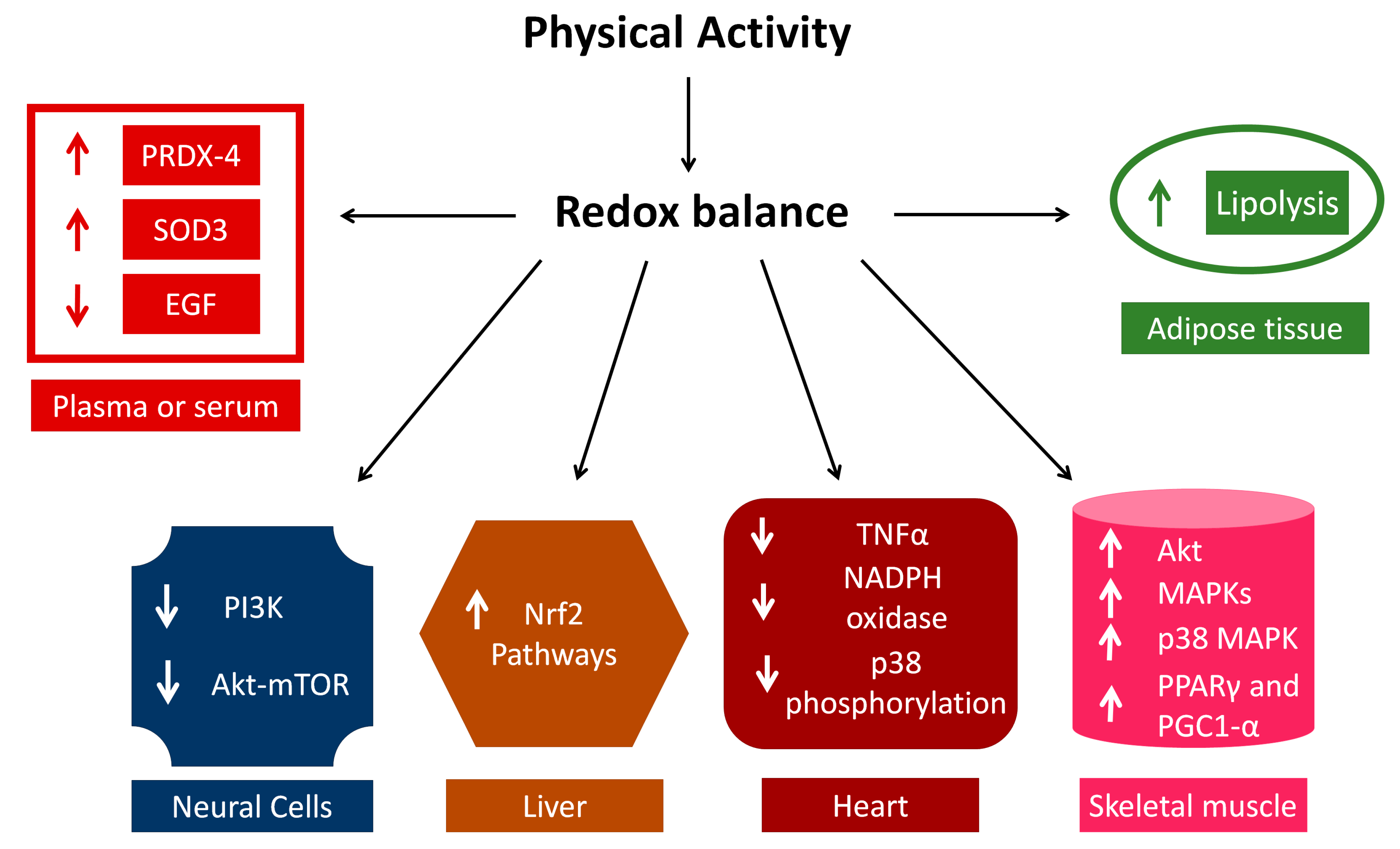
4. Insights into Signal Transduction

The role of physical activity as an antioxidant to counteract the negative effects of aging and chronic disease has been widely studied, and we summarize below the main results obtained so far. First, it is worth noting, however, that we are still far from a structured comprehensive view of this complex topic. Besides, in this field, several studies have been done in the mouse model, and the translation to humans is far away.
Several different signalling pathways have been implicated in the control of redox balance by physical activity, also depending on the analysed organs or systems.

4.1. Plasma and Adipose Tissue

Increased oxidative stress is related to abnormal activation of the renin-angiotensin-aldosterone system [49]. Resistance exercise increases the level of Triacylglycerol Lipase Activity (TGLA), the level of plasma glycerol and the level of oxidative stress in obese with respect to lean subjects [49,50]. This is associated with higher levels of serum non-esterified fatty acid and increased lipolysis in adipose tissue [50]. The relationship between exercise and obesity is under study in a clinical trial (current controlled trials, ISRCTN95488515). In particular, they are investigating the effects of endurance and high-intensity intermittent endurance exercise on plasma concentrations of glycerol, Atrial Natriuretic Peptide (ANP) and Brain Natriuretic Peptide (BNP). In fact, ANP and BNP are potent lipolytic agents. The results of this trial will be useful for optimizing training protocols for the prevention and treatment of obesity [51].
Contrasting results have been obtained on the role of exercise on plasma levels of cytokines [52,53,54]. For example, triathlon competitions like Ironman or Half Ironman significantly increase the levels of oxidative stress markers, Interleukin-6 (IL-6), Interleukin-1 (IL-1) and Tumour Necrosis Factor alpha (TNF-alpha). However, the training hours spent before the competitions were inversely correlated with the level of IL-6 found, suggesting that only IL-6 is related to the time of training [52]. In aged subjects affected by rheumatoid arthritis, 10 weeks of a walking-based high-intensity interval training (HIIT) increased ROS production, but there were no significant changes in the levels of inflammatory markers such as IL-1, IL-6, TNF-alpha and Interleukin-10 (IL-10) [53]. On the other hand, a study performed in post-menopausal women observed a significant decrease in IL-2, IL-4, IL-6 and TNF-alpha levels, both after 3 months of aerobic training (60–70% of maximal heart rate on treadmill, with low speed and without inclination) and after 3 months of resistance training with better results in terms of BMI and quality-of-life in subjects performing aerobic training [54]. In contrast, a single short bout of resistance exercise in healthy untrained men increased catecholamines, Epidermal Growth Factor (EGF), IL-2 and TNF-alpha, with the levels of IL-1α, IL-1β, IL-6, IL-8 and IL-10 maintained constant levels [55]. Additionally, Accattato et al. (2017) [49] observed that a single bout of physical activity does not induce changes in plasma cytokine levels. Instead, they observed a decrease of EGF and an increase of the antioxidant enzyme Glutathione Reductase (GR).
Eccentric exercise of the elbow flexors does not induce significant change in plasma cytokine levels, even in the presence of significant muscle damage as demonstrated by the increase in Creatine Kinase (CK) activity and myoglobin [56]. In any case, a transient increase in oxidative stress and cytokines is considered relevant for improvements induced by regular exercise, in a process called mitohormesis, because mitochondria are necessary for energy production and associated with oxidative stress induction [57]. All these data suggest that, since the role of cytokines in response to oxidative stress and exercise has not been definitively clarified, the aerobic, resistance, endurance and strength training sessions should be carefully set up within physical-activity programs for adults and elderly people in order to avoid undesirable effects in inflammation-signalling pathways.

4.2. Nervous System

In the nervous system, ROS are involved in neurogenesis, proliferation and differentiation of neuronal stem cells; however, if the ROS concentrations exceed threshold levels, it can lead to neurodegeneration [58]. Physical activity increases neurogenesis by inducing neurotrophic factors. Moreover, it protects DNA from damage by stimulating antioxidant systems [58]. A recent review by Vilela et al. (2020) [59] summarizes the effects of physical exercise on brain synaptic plasticity and memory through different pathways. In particular, studies performed in rat hippocampi suggest that aerobic training induces N-Methyl-D-Aspartate phosphorylate (pNMDA) and Postsynaptic Density protein 95 (PSD-95) activity, which increases memory. Instead, the strength training ameliorates the results of memory tests by upregulating Protein Kinase C alpha (PKC-alpha) and the cytokines IL-1 and TNF-alpha. Moreover, aerobic exercise ameliorates spatial memory through BDNF signalling [60]. Interestingly, in older adults affected by mild subcortical ischaemic vascular cognitive impairment, 6 months of aerobic exercise on the treadmill improved efficiency of the affected brain areas [60].
Feter et al. (2019) [61] investigated how different training models affect memory and redox balance. Resistance training induced a significant increase in lipid peroxidation and ROS levels, compared to the sedentary group. The moderate-intensity continuous-training group and physical-activity (running wheel) group showed a higher level of the antioxidant enzyme catalase, compared to the sedentary group. Moreover, the moderate-intensity continuous-training group showed better recognition memory, suggesting that performing physical activity, with a moderate but continuous intensity, could represent a non-pharmacological strategy to counteract the symptoms of Alzheimer’s disease. Of note, the role of physical activity as a strategy for counteracting Alzheimer’s Disease (AD) has been reviewed elsewhere [62]. In particular, although the pathogenesis of this disease has been clarified, the exact mechanisms are still under study. The role of physical activity seems to be correlated with microRNA alterations associated with impaired autophagy. This process is mediated by the Phosphatidyl Inositol 3-Kinase (PI3Kinase)/Akt-mammalian Target Of Rapamycin (mTOR) signalling pathway [62]. In detail, physical activity, through mTOR signalling activation, downregulates the production of abnormal micro-RNAs such as miR-130a, miR106b and miRlet7c, and stimulates neurogenesis, synaptic plasticity and memory, although the exact mechanism is still vague. The mTOR activation leads to dysfunctional autophagy, and consequently, to Tau hyperphosphorylation, β-amyloid (Aβ) accumulation and neurofibrillary tangles that are characteristics of Alzheimer’s disease [62]. Discovering how physical activity can exert this effect in the central nervous system and interfere with the abnormal synthesis of miRNAs in AD pathogenesis is an intriguing challenge that requires further investigation.

4.3. Liver

Nuclear factor erythroid-2-related factor 2 (Nrf2) and its antioxidant responsive elements play a prominent role in the reaction to oxidative stress. Acute aerobic exercise activates nuclear Nrf2 independently of exercise intensity, while higher-intensity physical activity increases the levels of oxidative stress and antioxidant enzymes [63]. Interestingly, a bout of moderate-intensity stationary cycling up-regulates Nrf2 in young, but not in older people. Nrf2 is involved in redox-balance modifications induced by exercise, but the underlying mechanisms are still unclear [64]. Of note, both aerobic and resistance training, although with different molecular pathways, converge on activation of the mitochondrial biogenesis through Nrf2. Although Nrf2 is ubiquitously expressed, the effects of physical activity and redox balance on Nrf2 have been well characterized in the liver. In mice, sulphorane exerts a protective role against liver damage induced by acute exercise. In particular, sulphorane reduces inflammatory cytokines like IL-1beta, IL-6 and TNF-alpha increased by exercise, and upregulates the expression of the antioxidant enzymes catalase, SOD1 and glutathione peroxidase (GPx1) through the activation of the Nrf2/Heme Oxygenase (HO1) signal-transduction pathway [65]. Moreover, it has been shown that 6 weeks of aerobic training increase triglycerides, free fatty acids and blood glucose in Nrf2-null mice, showing that Nrf2 is involved in training-induced adaptations of glucose homeostasis. Moreover, the levels of oxidative stress markers in the liver and in adipose tissue are increased, with respect to the control group, in Nrf2-null mice [66].

4.4. Skeletal and Cardiac Muscle

The importance of Nrf2 has also been evidenced in muscle. In fact, Huang et al. (2019) [67] showed that the level of Nrf2 mRNA in skeletal muscle decreases with aging, and concomitantly, a frailty phenotype arises. Muscle function and mass are similar in young wild-type and young Nrf2-null mice. In middle-aged and old Nrf2-null mice, muscle function significantly decreases with enhancement of frailty in old Nrf2-null mice [67]. This phenotype hinges on a decrease of mitochondrial biogenesis and alteration of mitochondrial morphology in skeletal muscle. In skeletal muscle, other pathways also seem to be involved in redox-balance signalling. In fact, skeletal muscle is a primary source of ROS during exercise, because during muscle contraction, there is a massive production of superoxide radical (O2) [67]. In rats, a combination of antioxidants (such as resveratrol) and exercise significantly increases muscle mass and grip strength in sarcopenic old mice, and reduces abnormal sarcomere length [68]. Concomitantly, the authors observed a significant increase in the levels of the phosphorylated form of 5′ AMP-activated protein Kinase (pAMPK) and of Sirtuin1 (Sirt1), suggesting the involvement of the AMPK/Sirt1 pathway [68]. This last pathway is also involved in AdipoRon function that is used to reduce oxidative stress and inflammation in dystrophic mice [69]. AdipoRon increases the levels of pAMPK that reduces NF-Kb and increases the dystrophin analogue utrophin [69]. AdipoRon also increases muscle strength and endurance, leading to better physical performance [69]. The antioxidant and antiaging effects of resveratrol have been studied also in the kidney [70]. In particular, the authors suggest that resveratrol can activate both Nrf2 and AMPK/sirtuin1 pathways in the kidney, to counteract the pathologic aging effect in this organ. In fact, transfection with Nrf2 and SIRT1 siRNAs in HK2 cells blocks the antioxidant effect of resveratrol [70].
The Akt signal-transduction pathway has been evidenced in skeletal muscle, in association with exercise and redox balance. For example, in mice, swimming exercise reduces ROS, and prevents high-fat diet-induced insulin resistance through the decrease of NAPDH oxidase 4 (Nox4), as well as the increase of Akt signal transduction in skeletal muscle [71].
Akt signalling is also required for the correct regulation of muscle stem-cell (MuSCs) homeostasis during aging. In fact, Lukjanenko et al. (2019) [72] found WNT1-Inducible Signalling Pathway Protein 1 (WISP1) as a fibro-adipogenic progenitor protein that is strongly downregulated during aging. This protein is important for muscle regeneration and the asymmetric division of muscle stem cells through Akt signalling. In fact, systemic treatment of aged mice with WISP1 restores the proliferation and differentiation of aged muscle stem cells [72].
Peroxisome Proliferator-Activated Receptor gamma (PPAR-gamma) and Peroxisome proliferator-activated receptor Gamma Coactivator-1 alpha (PGC1-alpha) have been proposed as important players in muscle remodelling induced by exercise through Mitogen-Activated Protein Kinases (MAPKinases), and are also correlated with redox balance. In particular, p38 MAPKinase activates PPAR-gamma. Sirt1, in turn, depending on physical exercise, activates PGC1-alpha [73]. PGC1-alpha is implicated in aging-associated mitochondrial diseases dysregulating ROS production and mitochondrial network structure in skeletal muscle, during aging and exercise training. PGC1-alpha is also activated by ROS through NF-kB, AMPK, and finally, by nitric oxide (NO) produced during muscle contraction [73,74]. PGC1-alpha-null aged mice show lower running endurance, higher mitochondrial damage, increased ROS and higher oxidative stress than do young mice. Exercise training increases maximal respiratory capacity, both in PGC1-alpha-null and wild-type mice. Instead, the rescue of mitochondrial homeostasis occurs in a PGC1-alpha-dependent manner [74].
Regarding cardiac muscle, aerobic exercise training on the treadmill ameliorates myocardial infarction symptoms. In fact, TNF-alpha, NADPH-oxidase activity and p38 phosphorylation are diminished in infarcted trained mice, with respect to infarcted sham-trained mice [75]. The positive effect of exercise is produced also by increasing the antioxidant defence through neuronal Nitric-Oxide Synthase (nNOS) [76]. In fact, cardiac-specific overexpression of this enzyme mimics the effect of exercise on maximal oxygen capacity, while aerobic activity increases cardiac dysfunctions in nNOS-null mice, confirming the protective effect of this enzyme.
Interestingly, Hao et al. (2014) [77] showed that exercise is a powerful instrument for protecting the heart from ischemia, and that the molecular mechanism at the base of this phenomenon involves Protein Kinase Cε. Specifically, Sprague-Dawley rats underwent exhaustive aerobic exercise on the treadmill, to induce myocardial injury [77]. In such exhaustive conditions, PKCε translocated to the intercalated disks of the heart. The exercise pre-conditioning increased the level of the phosphorylated/activated isoform of PKCε in the cytoplasmic membrane, suggesting that PKCε is involved in the signal transduction for heart protection induced by pre-conditioning [77].
Finally, Díaz-Ruíz et al. (2019) [78] evidenced a role for redox signalling in ischemic post-conditioning protection through PKCε and Erk1/2, which indirectly regulate Nrf2 activation. In fact, the authors showed that PKCε and Erk1/2 are activated in a redox-dependent manner. It was also shown that neither the PI3K inhibitor nor the Erk1/2 inhibitor reduces Nrf2 activation, suggesting that these kinases have other direct targets [78]. Notably, Buelna-Chontal et al. (2014) [79] found that Nrf2 activation is dependent on PKC in post-conditioned hearts. In fact, the use of PKC inhibitors reduces the level of Nrf2 phosphorylation and the activity of antioxidant proteins that are regulated by Nrf2 [79].
The PKC family includes 12 isozymes divided into conventional (alpha, beta and gamma), novel (delta, epsilon, eta and theta) and atypical (zeta and iota). PKCε belongs to the novel group which is activated by the conventional signalling of Ca2+ and diacylglycerol. In particular, the receptor, once activated, hydrolyses Phosphatidyl-Inositol-4,5-bisPhosphate (PIP2), generating Inositol trisPhosphate (IP3, responsible for Ca2+ mobilization) and diacylglycerol which, as the second messenger, triggers PKC-kinase function, leading to phosphorylation and activation of other trans-membrane or intra-cellular proteins. PKCs are generally considered as oncoproteins. However, many different roles have been proposed for PKC proteins in cell physiology. For example, PKCε is involved in many processes that maintain cell homeostasis and proliferation in several tissues such as the vessels, blood, heart and skeletal muscle [80,81,82,83,84,85]. For example, PKCε is differently expressed in blood cells suggesting multiple roles for this PKC isoform [84]. Moreover, Martini et al. [85] evidenced that PKCε controls the migration of centrosome during mitotic spindle assembly. Recently, D’Amico and Lennartz (2018) [86] showed a role in vesicle formation for PKCε. They demonstrated that PKCε is linked to the Golgi apparatus through the interaction between the Golgi’s lipids and the regulatory domain of the kinase. Moreover, the mechanism seems to be independent of the kinase activity of PKCε, although it requires the binding of PKCε to actin and cytoskeleton for budding and fission of the vesicles [86]. Finally, PKCε is important for autophagy in the breast-cancer cell line [87]. In fact, PKCε siRNA induces a significant decrease in autophagy by reducing the levels of Raptor and Rictor that are required to form mammalian Target Of Rapamycin Complex-1 (mTORC-1) and -2 (mTORC-2), which are important regulators of autophagy [87]. On the other hand, autophagy is important to counteract aging, although this process declines in the elderly. Exercise and diet downregulate mTORC-1, that acts as a negative player in autophagy and stimulates autophagy in several other tissues [88]. Moreover, mTORC-1 induces muscle atrophy in the elderly, while mTOR is essential for muscle hypertrophy. Most likely, a mechanism that involves Akt and mTORC1 is important in sarcopenia, although the definition of a clear signalling pathway is still under study [89]. Based on these considerations, we could speculate that PKCε is involved in exercise protection during the aging process.
Additionally, ROS can affect Ca2+ signalling at the level of channels, pumps and exchangers [90]. Alterations in ROS signalling can modify Ca2+ communication system, likely contributing to disease onset. In the heart, for example, Zima and Blatter [91] showed that the ROS produced during reperfusion of cardiac ischemic injury can affect ischemia-related Ca(2+) overload. More recently, Cabassi and Miragoli [92] reviewed the important role of local environment (namely ROS and Ca2+) in mitochondrial re-organization and fusion in failing cardiomyocytes. In particular, a correct interplay between Ca2+ and ROS is important to avoid intracellular Ca2+ increase during diastole that leads to cardiac arrhythmia [92]. The relevance of interaction between Ca2+ signalling and ROS has also been described in skeletal muscle aging [40]. In fact, the increase of ROS in skeletal muscle can alter the Ca2+ signalling pathway that is necessary for the fibre contraction, leading to a decrease in muscle strength. Interestingly, physical activity can help to contrast strength loss by reducing ROS [40].
Finally, Peroxiredoxins (PRDXs) are important antioxidant enzymes that remove H2O2 to reduce oxidative stress. PRDXs are responsible for the discarding of 90% of cellular peroxides, they are ubiquitous and function as regulators of local H2O2 concentrations [93,94]. There are more than 3500 sequences of Peroxiredoxins, and these have been divided into six subfamilies with different expression levels in mammals, plants, yeasts and bacteria [95]. Considering the conservation of phylogenetic rhythms, the fact that PRDX proteins autoregulate independently from the transcriptional circadian clock and the highly conserved structures of these proteins, many researchers are now studying the links between aging, circadian rhythms and redox systems [96].
PRDX-3-null mice present a significant reduction in physical strength (measured by swimming performance), compared to their wild-type littermates at the age of ten months [97]. Moreover, PRDX-3-null mice show a higher number of apoptotic cells in the brain, higher expression of Nrf2 and a lower level of mitochondrial DNA (mDNA) at the age of ten months, when compared to wild-type, suggesting that the lack of PRDX-3 increases oxidative stress and mitochondrial impairment, which are characteristic of the aging process [97]. To study redox-enzyme concentrations after different types of exercise, a group of active men (mean age 28 years old) performed moderate-intensity exercise with one bout of High-Intensity Interval Exercise (HIIE) or an eccentric resistance exercise [98]. PRDX-4 (Peroxiredoxin-4) and SOD3 increased after High-Intensity Interval Exercise (HIIE), while Thioredoxin Reductase (TRX-R) decreased. Notably, resistance exercise did not determine any significant changes in redox enzymes, but induced skeletal muscle damages. Altogether, these results suggest that PRDX-4 and SOD3 could be considered as biomarkers of oxidative stress [98]. Interestingly, HIIE showed similar effects of endurance exercise in muscle mitochondria, independently from the total workload, suggesting that exercise prescription could be accommodated into individual prescriptions, generating comparable molecular effects [99].
Although the exact mechanism is not yet clear, it has been proposed that PRDX translates oxidative stress signalling by the activation of MAPKinases [100].
Figure 2 provides a schematic representation of the pathways involved in the effects of physical activity and redox balance to counteract aging. In plasma or serum, the proposed markers of oxidative stress are the increase of PRDX-4 and SOD3, while the level of EGF should decrease, given the above studies. Cytokines are inflammation markers, and contrasting results have been obtained in association with physical activity. In neural cells, PI3K/Akt/mTor seems to have a prominent role, especially in the treatment of Alzheimer’s Disease. The Nrf2 pathway is central in the liver, but it also is an important effector in heart and skeletal muscle where many players have been evidenced, such as MAPKinases (p38 and Erk1/2), Akt and PPARγ/PGC1α. Finally, in adipose tissue, there are emerging results on the role of physical activity and redox balance to increase the levels of BDNF and ANF (lipolytic agents) in the elaboration of protocols to contrast obesity.
In plasma and serum, the effect of physical activity on redox balance is exerted through the increase of SOD3 and PRDX-4 and the decrease of EGF; in neural cells, a decrease of PI3Kinase/Akt-mTOR is observed; in liver cells, Nrf2 pathways are activated; in the heart, the effect of physical activity is associated with a decrease of TNF-alpha and NADPH oxidase, and p38 phosphorylation; in skeletal muscle, Akt, MAPKinases, PPARgamma, PGC-1 alpha and Nrf2 are up-regulated; in adipose tissue, lipolysis is activated by physical activity.

5. Conclusions and Perspectives

Redox balance is very important for cell life. As with other cell processes, oxidant/antioxidant balance is affected by aging, with a decrease in the efficiency of antioxidant systems. To counteract this effect of aging, antioxidant foods have been shown to be useful. Physical activity, as well, has been involved in the regulation of redox balance in several tissues such as skeletal muscle, cardiac muscle, liver, as well as neural cells. Although there are several pieces of evidence of the involvement of different signalling pathways, a comprehensive view of the molecular players, modulated by physical activity, that are able to counteract the effects of ageing on redox balance, is still lacking.
The trivial perception that reactive species of oxygen are exclusively detrimental molecules, is definitively substituted with mounting evidence that exercise-induced perturbation of redox balance is the upstream signal for the activation of transcription factors and the induction of gene expression associated with exercise effects involving total body adaptation to training. However, the same signalling should be analysed on the specific tissue and organ, as well as in terms of age, fragility state and comorbidities.
At the moment, although we believe that future precision medicine will be able to transform exercise administration from generic wellness to targeted prevention, we must admit that the topic is still in its infancy.

Author Contributions

D.G., C.C., E.M. contributed to the bibliographic research, writing of the manuscript and produced the figures. M.V. (Mauro Vaccarezza), V.P., G.P., L.A. reviewed the manuscript. M.V. (Marco Vitale) reviewed the manuscript and supervised the work. G.G. and P.M. conceived the original idea and supervised the work. All authors have read and agreed to the published version of the manuscript.

Funding

D.G.: C.C.: M.V. (Marco Vitale), G.G. were supported by Fondi Locali per la Ricerca 2019—Quota Prodotti di Ricerca—Parma University. P.M. was supported by “Programmi di ricerca di Rilevante Interesse Nazionale”—Italian Ministry of Education, University and Research (MIUR-PRIN) 2017 grant entitled “ACTLIFE: IS ACTIVE LIFE STYLE ENOUGH FOR HEALTH AND WELLBEING?” codice 2017RS5M44_004.

Institutional Review Board Statement

Not applicable.

Informed Consent Statement

Not applicable.

Data Availability Statement

Not applicable.

Acknowledgments

We are grateful to Cristina Micheloni, Luciana Cerasuolo and Vincenzo Alberto Piero Palermo for technical support. We thank Devahuti Chaliha for English proofreading.

Conflicts of Interest

The authors declare no conflict of interest.

Abbreviations

SOD3Superoxide Dismutase 3
PRDX-4Peroxiredoxin-4
EGFEpidermal Growth Factor
PI3KinasePhosphatidyl Inositol 3 Kinase
AktProtein Kinase B
mTORmammalian Target Of Rapamycin
Nrf2Nuclear factor erythroid-2-related factor 2
TNF-alphaTumour Necrosis Factor alpha
NADPHNicotinamide Adenine Dinucleotide Phosphate
MAPKinaseMitogen-Activated Protein Kinase
PPAR-gammaPeroxisome Proliferator-Activated Receptor gamma
PGC-1-alphaPeroxisome proliferator-activated receptor Gamma Coactivator 1 alpha

References

  1. World Health Organization (WHO). Available online: https://www.who.int/news-room/q-a-detail/ageing-healthy-ageing-and-functional-ability (accessed on 30 October 2020).
  2. Ji, L.L.; Leeuwenburgh, C.; Leichtweis, S.; Gore, M.; Fiebig, R.; Hollander, J.; Bejma, J. Oxidative stress and aging. Role of exercise and its influences on antioxidant systems. Ann. N. Y. Acad. Sci. 1998, 854, 102–117. [Google Scholar] [CrossRef]
  3. Viña, J.; Olaso-Gonzalez, G.; Arc-Chagnaud, C.; De la Rosa, A.; Gomez-Cabrera, M.C. Modulating Oxidant Levels to Promote Healthy Aging. Antioxid. Redox Signal. 2020. [Google Scholar] [CrossRef]
  4. Ji, L.L. Antioxidants and oxidative stress in exercise. Proc. Soc. Exp. Biol. Med. 1999, 222, 283–292. [Google Scholar] [CrossRef] [PubMed]
  5. Massafra, C.; Gioia, D.; De Felice, C.; Picciolini, E.; De Leo, V.; Bonifazi, M.; Bernabei, A. Effects of estrogens and androgens on erythrocyte antioxidant superoxide dismutase, catalase and glutathione peroxidase activities during the menstrual cycle. J. Endocrinol. 2000, 167, 447–452. [Google Scholar] [CrossRef] [PubMed] [Green Version]
  6. Park, S.Y.; Kwak, Y.S. Impact of aerobic and anaerobic exercise training on oxidative stress and antioxidant defense in athletes. J. Exerc. Rehabil. 2016, 12, 113–117. [Google Scholar] [CrossRef] [PubMed]
  7. Poulsen, H.E.; Weimann, A.; Loft, S. Methods to detect DNA damage by free radicals: Relation to exercise. Proc. Nutr. Soc. 1999, 58, 1007–1014. [Google Scholar] [CrossRef] [PubMed]
  8. Debevec, T.; Millet, G.P.; Pialoux, V. Hypoxia-Induced Oxidative Stress Modulation with Physical Activity. Front. Physiol. 2017, 8, 84. [Google Scholar] [CrossRef] [Green Version]
  9. Tofas, T.; Draganidis, D.; Deli, C.K.; Georgakouli, K.; Fatouros, I.G.; Jamurtas, A.Z. Exercise-Induced Regulation of Redox Status in Cardiovascular Diseases: The Role of Exercise Training and Detraining. Antioxidants 2019, 9, 13. [Google Scholar] [CrossRef] [Green Version]
  10. de Sousa, C.V.; Sales, M.M.; Rosa, T.S.; Lewis, J.E.; de Andrade, R.V.; Simões, H.G. The Antioxidant Effect of Exercise: A Systematic Review and Meta-Analysis. Sports Med. 2017, 47, 277–293. [Google Scholar] [CrossRef]
  11. Nocella, C.; Cammisotto, V.; Pigozzi, F.; Borrione, P.; Fossati, C.; D’Amico, A.; Cangemi, R.; Peruzzi, M.; Gobbi, G.; Ettorre, E.; et al. Impairment between Oxidant and Antioxidant Systems: Short- and Long-term Implications for Athletes’ Health. Nutrients 2019, 11, 1353. [Google Scholar] [CrossRef] [PubMed] [Green Version]
  12. de Souza, R.F.; de Moraes, S.R.A.; Augusto, R.L.; de Freitas Zanona, A.; Matos, D.; Aidar, F.J.; da Silveira Andrade-da-Costa, B.L. Endurance training on rodent brain antioxidant capacity: A meta-analysis. Neurosci. Res. 2019, 145, 1–9. [Google Scholar] [CrossRef] [PubMed]
  13. Pinho, R.A.; Aguiar, A.S., Jr.; Radák, Z. Effects of Resistance Exercise on Cerebral Redox Regulation and Cognition: An Interplay between Muscle and Brain. Antioxidants 2019, 8, 529. [Google Scholar] [CrossRef] [Green Version]
  14. Park, S.Y.; Rossman, M.J.; Gifford, J.R.; Bharath, L.P.; Bauersachs, J.; Richardson, R.S.; Abel, E.D.; Symons, J.D.; Riehle, C. Exercise training improves vascular mitochondrial function. Am. J. Physiol. Heart Circ. Physiol. 2016, 310, H821–H829. [Google Scholar] [CrossRef] [PubMed] [Green Version]
  15. Roh, H.T.; So, W.Y. The effects of aerobic exercise training on oxidant-antioxidant balance, neurotrophic factor levels, and blood-brain barrier function in obese and non-obese men. J. Sport Health Sci. 2017, 6, 447–453. [Google Scholar] [CrossRef] [Green Version]
  16. Di Liegro, C.M.; Schiera, G.; Proia, P.; Di Liegro, I. Physical Activity and Brain Health. Genes 2019, 10, 720. [Google Scholar] [CrossRef] [Green Version]
  17. Arad, A.D.; Basile, A.J.; Albu, J.; DiMenna, F.J. No Influence of Overweight/Obesity on Exercise Lipid Oxidation. Int. J. Mol. Sci. 2020, 21, 1614. [Google Scholar] [CrossRef] [Green Version]
  18. Sadowska-Krępa, E.; Kłapcińska, B.; Pokora, I.; Domaszewski, P.; Kempa, K.; Podgórski, T. Effects of Six-Week Ginkgo biloba Supplementation on Aerobic Performance, Blood Pro/Antioxidant Balance, and Serum Brain-Derived Neurotrophic Factor in Physically Active Men. Nutrients 2017, 9, 803. [Google Scholar] [CrossRef] [Green Version]
  19. Yahfoufi, N.; Alsadi, N.; Jambi, M.; Matar, C. The Immunomodulatory and Anti-Inflammatory Role of Polyphenols. Nutrients 2018, 10, 1618. [Google Scholar] [CrossRef] [Green Version]
  20. Cavarretta, E.; Peruzzi, M.; Del Vescovo, R.; Di Pilla, F.; Gobbi, G.; Serdoz, A.; Ferrara, R.; Schirone, L.; Sciarretta, S.; Nocella, C.; et al. Dark Chocolate Intake Positively Modulates Redox Status and Markers of Muscular Damage in Elite Football Athletes: A Randomized Controlled Study. Oxidative Med. Cell Longev. 2018, 2018, 4061901. [Google Scholar] [CrossRef]
  21. Georgakouli, K.; Fatouros, I.G.; Fragkos, A.; Tzatzakis, T.; Deli, C.K.; Papanikolaou, K.; Koutedakis, Y.; Jamurtas, A.Z. Exercise and Redox Status Responses Following Alpha-Lipoic Acid Supplementation in G6PD Deficient Individuals. Antioxidants 2018, 7, 162. [Google Scholar] [CrossRef] [Green Version]
  22. Kabasakalis, A.; Kyparos, A.; Tsalis, G.; Loupos, D.; Pavlidou, A.; Kouretas, D.; Kabasakalis, A.; Tsalis, G.; Zafrana, E.; Loupos, D.; et al. Effects of endurance and high-intensity swimming exercise on the redox status of adolescent male and female swimmers. J. Sports Sci. 2014, 32, 747–756. [Google Scholar] [CrossRef]
  23. Kabasakalis, A.; Kyparos, A.; Tsalis, G.; Loupos, D.; Pavlidou, A.; Kouretas, D. Blood oxidative stress markers after ultramarathon swimming. J. Strength Cond. Res. 2011, 25, 805–811. [Google Scholar] [CrossRef]
  24. Bellafiore, M.; Bianco, A.; Battaglia, G.; Naccari, M.S.; Caramazza, G.; Padulo, J.; Chamari, K.; Paoli, A.; Palma, A. Training session intensity affects plasma redox status in amateur rhythmic gymnasts. J. Sport Health Sci. 2019, 8, 561–566. [Google Scholar] [CrossRef] [Green Version]
  25. Powers, S.K.; Jackson, M.J. Exercise-induced oxidative stress: Cellular mechanisms and impact on muscle force production. Physiol. Rev. 2008, 88, 1243–1276. [Google Scholar] [CrossRef] [PubMed] [Green Version]
  26. Wolff, C.; Musci, R.; Whedbee, M. Vitamin supplementation and resistance exercise-induced muscle hypertrophy: Shifting the redox balance scale? J. Physiol. 2015, 593, 2991–2992. [Google Scholar] [CrossRef] [PubMed] [Green Version]
  27. Paulsen, G.; Hamarsland, H.; Cumming, K.T.; Johansen, R.E.; Hulmi, J.J.; Borsheim, E.; Wiig, H.; Garthe, I.; Raastad, T. Vitamin C and E supplementation alters protein signalling after a strength training session, but not muscle growth during 10 weeks of training. J. Physiol. 2014, 592, 5391–5408. [Google Scholar] [CrossRef]
  28. Bruns, D.R.; Ehrlicher, S.E.; Khademi, S.; Biela, L.M.; Peelor, F.F.; Miller, B.F.; Hamilton, K.L. Differential effects of vitamin C or protandim on skeletal muscle adaptation to exercise. J. Appl. Physiol. 2018, 125, 661–671. [Google Scholar] [CrossRef]
  29. Traustadóttir, T.; Davies, S.S.; Su, Y.; Choi, L.; Brown-Borg, H.M.; Roberts, L.J., 2nd; Harman, S.M. Oxidative stress in older adults: Effects of physical fitness. Age (Dordr.) 2012, 34, 969–982. [Google Scholar] [CrossRef] [Green Version]
  30. Aldred, S.; Rohalu, M. A moderate intensity exercise program did not increase the oxidative stress in older adults. Arch. Gerontol. Geriatr. 2011, 53, 350–353. [Google Scholar] [CrossRef] [PubMed]
  31. Done, A.J.; Traustadóttir, T. Aerobic exercise increases resistance to oxidative stress in sedentary older middle-aged adults. A pilot study. Age (Dordr.) 2016, 38, 505–512. [Google Scholar] [CrossRef] [Green Version]
  32. Landers-Ramos, R.Q.; Corrigan, K.J.; Guth, L.M.; Altom, C.N.; Spangenburg, E.E.; Prior, S.J.; Hagberg, J.M. Short-term exercise training improves flow-mediated dilation and circulating angiogenic cell number in older sedentary adults. Appl. Physiol. Nutr. Metab. 2016, 41, 832–841. [Google Scholar] [CrossRef] [PubMed]
  33. Pierno, S.; Tricarico, D.; Liantonio, A.; Mele, A.; Digennaro, C.; Rolland, J.-F.; Bianco, G.; Villanova, L.; Merendino, A.; Camerino, G.M.; et al. An olive oil derived antioxidant mixture ameliorates the age-related decline of skeletal muscle function. Age 2014, 36, 73–88. [Google Scholar] [CrossRef] [Green Version]
  34. Muhammad, M.H.; Allam, M.M. Resveratrol and/or exercise training counteract aging-associated decline of physical endurance in aged mice; targeting mitochondrial biogenesis and function. J. Physiol. Sci. 2018, 68, 681–688. [Google Scholar] [CrossRef] [PubMed]
  35. Gliemann, L.; Nyberg, M.; Hellsten, Y. Effects of exercise training and resveratrol on vascular health in aging. Free Radic. Biol. Med. 2016, 98, 165–176. [Google Scholar] [CrossRef]
  36. Najjar, F.; Rizk, F.; Carnac, G. Protective effect of Rhus coriaria fruit extracts against hydrogen peroxide-induced oxidative stress in muscle progenitors and zebrafish embryos. PeerJ 2017, 5, e4144. [Google Scholar] [CrossRef] [Green Version]
  37. Richards, J.C.; Crecelius, A.R.; Larson, D.G.; Dinenno, F.A. Acute ascorbic acid ingestion increases skeletal muscle blood flow and oxygen consumption via local vasodilation during graded handgrip exercise in older adults. Am. J. Physiol. Heart Circ. Physiol. 2015, 309, H360–H368. [Google Scholar] [CrossRef] [PubMed] [Green Version]
  38. Fougere, B.; van Kan, G.A.; Vellas, B.; Cesari, M. Redox Systems, Antioxidants and Sarcopenia. Curr. Protein Pept. Sci. 2018, 19, 643–648. [Google Scholar] [CrossRef]
  39. Angulo, J.; El Assar, M.; Álvarez-Bustos, A.; Rodríguez-Mañas, L. Physical activity and exercise: Strategies to manage frailty. Redox Biol. 2020, 35, 101513. [Google Scholar] [CrossRef]
  40. Szentesi, P.; Csernoch, L.; Dux, L.; Keller-Pintér, A. Changes in Redox Signaling in the Skeletal Muscle with Aging. Oxid. Med. Cell Longev. 2019, 2019, 4617801. [Google Scholar] [CrossRef]
  41. Verdijk, L.B.; Snijders, T.; Drost, M.; Delhaas, T.; Kadi, F.; van Loon, L.J.C. Satellite cells in human skeletal muscle; from birth to old age. Age 2014, 36, 545–557. [Google Scholar] [CrossRef] [Green Version]
  42. Blocquiaux, S.; Gorski, T.; Van Roie, E.; Ramaekers, M.; Van Thienen, R.; Nielens, H.; Delecluse, C.; De Bock, K.; Thomis, M. The effect of resistance training, detraining and retraining on muscle strength and power, myofibre size, satellite cells and myonuclei in older men [published correction appears in Exp Gerontol. Exp. Gerontol. 2020, 133, 110860. [Google Scholar] [CrossRef]
  43. Fry, C.S.; Noehren, B.; Mula, J.; Ubele, M.F.; Westgate, P.M.; Kern, P.A.; Peterson, C.A. Fibre type-specific satellite cell response to aerobic training in sedentary adults. J. Physiol. 2014, 592, 2625–2635. [Google Scholar] [CrossRef]
  44. Pittaluga, M.; Sgadari, A.; Dimauro, I.; Tavazzi, B.; Parisi, P.; Caporossi, D. Physical exercise and redox balance in type 2 diabetics: Effects of moderate training on biomarkers of oxidative stress and DNA damage evaluated through comet assay. Oxid. Med. Cell Longev. 2015, 981242. [Google Scholar] [CrossRef]
  45. Shukla, V.; Mishra, S.K.; Pant, H.C. Oxidative stress in neurodegeneration. Adv. Pharmacol. Sci. 2011, 2011, 572634. [Google Scholar] [CrossRef] [PubMed] [Green Version]
  46. Espinet, C.; Gonzalo, H.; Fleitas, C.; Menal, M.J.; Egea, J. Oxidative stress and neurodegenerative diseases: A neurotrophic approach. Curr. Drug Targets. 2015, 16, 20–30. [Google Scholar] [CrossRef] [PubMed]
  47. Patil, S.P.; Tran, N.; Geekiyanage, H.; Liu, L.; Chan, C. Curcumin-induced upregulation of the anti-tau cochaperone BAG2 in primary rat cortical neurons. Neurosci. Lett. 2013, 554, 121–125. [Google Scholar] [CrossRef] [Green Version]
  48. Jia, R.X.; Liang, J.H.; Xu, Y.; Wang, Y.Q. Effects of physical activity and exercise on the cognitive function of patients with Alzheimer disease: A meta-analysis. BMC Geriatr. 2019, 19, 181. [Google Scholar] [CrossRef]
  49. Accattato, F.; Greco, M.; Pullano, S.A.; Carè, I.; Fiorillo, A.S.; Pujia, A.; Montalcini, T.; Foti, D.P.; Brunetti, A.; Gulletta, E. Effects of acute physical exercise on oxidative stress and inflammatory status in young, sedentary obese subjects. PLoS ONE 2017, 12, e0178900. [Google Scholar] [CrossRef] [PubMed] [Green Version]
  50. Chatzinikolaou, A.; Fatouros, I.; Petridou, A.; Jamurtas, A.; Avloniti, A.; Douroudos, I.; Mastorakos, G.; Lazaropoulou, C.; Papassotiriou, I.; Tournis, S.; et al. Adipose tissue lipolysis is upregulated in lean and obese men during acute resistance exercise. Diabetes Care. 2008, 31, 1397–1399. [Google Scholar] [CrossRef] [Green Version]
  51. Karner-Rezek, K.; Knechtle, B.; Fenzl, M.; Gredig, J.; Rosemann, T. Does continuous endurance exercise in water elicit a higher release of ANP and BNP and a higher plasma concentration of FFAs in pre-obese and obese men than high intensity intermittent endurance exercise?‹study protocol for a randomized controlled trial. Trials 2013, 14, 328. [Google Scholar] [CrossRef] [Green Version]
  52. Mrakic-Sposta, S.; Gussoni, M.; Vezzoli, A.; Dellanoce, C.; Comassi, M.; Giardini, G.; Bruno, R.M.; Montorsi, M.; Corciu, A.; Greco, F.; et al. Acute Effects of Triathlon Race on Oxidative Stress Biomarkers. Oxid. Med. Cell Longev. 2020, 2020, 3062807. [Google Scholar] [CrossRef] [Green Version]
  53. Bartlett, D.B.; Willis, L.H.; Slentz, C.A.; Hoselton, A.; Kelly, L.; Huebner, J.L.; Kraus, V.B.; Moss, J.; Muehlbauer, M.J.; Spielmann, G.; et al. Ten weeks of high-intensity interval walk training is associated with reduced disease activity and improved innate immune function in older adults with rheumatoid arthritis: A pilot study. Arthritis Res. Ther. 2018, 20, 127. [Google Scholar] [CrossRef] [PubMed] [Green Version]
  54. Abd El-Kader, S.M.; Al-Jiffri, O.H. Impact of aerobic versus resisted exercise training on systemic inflammation biomarkers and quality of Life among obese post-menopausal women. Afr. Health Sci. 2019, 19, 2881–2891. [Google Scholar] [CrossRef] [PubMed]
  55. Fatouros, I.; Chatzinikolaou, A.; Paltoglou, G.; Petridou, A.; Avloniti, A.; Jamurtas, A.; Goussetis, E.; Mitrakou, A.; Mougios, V.; Lazaropoulou, C.; et al. Acute resistance exercise results in catecholaminergic rather than hypothalamic-pituitary-adrenal axis stimulation during exercise in young men. Stress 2010, 13, 461–468. [Google Scholar] [CrossRef]
  56. Hirose, L.; Nosaka, K.; Newton, M.; Laveder, A.; Kano, M.; Peake, J.; Suzuki, K. Changes in inflammatory mediators following eccentric exercise of the elbow flexors. Exerc. Immunol. Rev. 2004, 10, 75–90. [Google Scholar] [PubMed]
  57. Ost, M.; Coleman, V.; Kasch, J.; Klaus, S. Regulation of myokine expression: Role of exercise and cellular stress. Free Radic. Biol. Med. 2016, 98, 78–89. [Google Scholar] [CrossRef]
  58. Radak, Z.; Suzuki, K.; Higuchi, M.; Balogh, L.; Boldogh, I.; Koltai, E. Physical exercise, reactive oxygen species and neuroprotection. Free Radic. Biol. Med. 2016, 98, 187–196. [Google Scholar] [CrossRef]
  59. Vilela, T.C.; de Andrade, V.M.; Radak, Z.; de Pinho, R.A. The role of exercise in brain DNA damage. Neural Regen Res. 2020, 15, 1981–1985. [Google Scholar] [CrossRef]
  60. Hsu, C.L.; Best, J.R.; Davis, J.C.; Nagamatsu, L.S.; Wang, S.; Boyd, L.A.; Hsiung, G.R.; Voss, M.W.; Eng, J.J.; Liu-Ambrose, T. Aerobic exercise promotes executive functions and impacts functional neural activity among older adults with vascular cognitive impairment. Br. J. Sports Med. 2018, 52, 184–191. [Google Scholar] [CrossRef]
  61. Feter, N.; Spanevello, R.M.; Soares, M.S.P.; Spohr, L.; Pedra, N.S.; Bona, N.P.; Freitas, M.P.; Gonzales, N.G.; Ito, L.G.M.S.; Stefanello, F.M.; et al. How does physical activity and different models of exercise training affect oxidative parameters and memory? Physiol. Behav. 2019, 201, 42–52. [Google Scholar] [CrossRef]
  62. Kou, X.; Chen, D.; Chen, N. Physical Activity Alleviates Cognitive Dysfunction of Alzheimer’s Disease through Regulating the mTOR Signaling Pathway. Int. J. Mol. Sci. 2019, 20, 1591. [Google Scholar] [CrossRef] [Green Version]
  63. Done, A.J.; Newell, M.J.; Traustadóttir, T. Effect of exercise intensity on Nrf2 signalling in young men. Free Radic. Res. 2017, 51, 646–655. [Google Scholar] [CrossRef] [PubMed]
  64. Vargas-Mendoza, N.; Morales-González, Á.; Madrigal-Santillán, E.O.; Madrigal-Bujaidar, E.; Álvarez-González, I.; García-Melo, L.F.; Anguiano-Robledo, L.; Fregoso-Aguilar, T.; Morales-Gonzalez, J.A. Antioxidant and Adaptative Response Mediated by Nrf2 during Physical Exercise. Antioxidants 2019, 8, 196. [Google Scholar] [CrossRef] [Green Version]
  65. Ruhee, R.T.; Ma, S.; Suzuki, K. Protective Effects of Sulforaphane on Exercise-Induced Organ Damage via Inducing Antioxidant Defense Responses. Antioxidants 2020, 9, 136. [Google Scholar] [CrossRef] [Green Version]
  66. Merry, T.L.; MacRae, C.; Pham, T.; Hedges, C.P.; Ristow, M. Deficiency in ROS-sensing nuclear factor erythroid 2-like 2 causes altered glucose and lipid homeostasis following exercise training. Am. J. Physiol. Cell Physiol. 2020, 318, C337–C345. [Google Scholar] [CrossRef]
  67. Huang, D.-D.; Fan, S.-D.; Chen, X.-Y.; Yan, X.-L.; Zhang, X.-Z.; Ma, B.-W.; Yu, D.-Y.; Xiao, W.-Y.; Zhuang, C.-L.; Yu, Z. Nrf2 deficiency exacerbates frailty and sarcopenia by impairing skeletal muscle mitochondrial biogenesis and dynamics in an age-dependent manner. Exp. Gerontol. 2019, 119, 61–73. [Google Scholar] [CrossRef] [PubMed]
  68. Liao, Z.Y.; Chen, J.L.; Xiao, M.H. The effect of exercise, resveratrol or their combination on sarcopenia in aged rats via regulation of AMPK/Sirt 1 pathway. Exp. Gerontol. 2017, 98, 177–183. [Google Scholar]
  69. Abou-Samra, M.; Selvais, C.M.; Boursereau, R.; Lecompte, S.; Noel, L.; Brichard, S.M. AdipoRon, a new therapeutic prospect for Duchenne muscular dystrophy. J. Cachexia Sarcopenia Muscle 2020, 11, 518–533. [Google Scholar] [CrossRef]
  70. Kim, E.N.; Lim, J.H.; Kim, M.Y.; Ban, T.H.; Jang, I.A.; Yoon, H.E.; Park, C.W.; Chang, Y.S.; Choi, B.S. Resveratrol, an Nrf2 activator, ameliorates aging-related progressive renal injury. Aging (Albany N. Y.) 2018, 10, 83–99. [Google Scholar] [CrossRef] [PubMed] [Green Version]
  71. Qi, J.; Luo, X.; Ma, Z.; Zhang, B.; Li, S.; Duan, X.; Yang, B.; Zhang, J. Swimming Exercise Protects against Insulin Resistance via Regulating Oxidative Stress through Nox4 and AKT Signaling in High-Fat Diet-Fed Mice. J. Diabetes Res. 2020, 2020, 2521590. [Google Scholar] [CrossRef] [Green Version]
  72. Lukjanenko, L.; Karaz, S.; Stuelsatz, P.; Gurriaran-Rodriguez, U.; Michaud, J.; Dammone, G.; Sizzano, F.; Mashinchian, O.; Ancel, S.; Migliavacca, E.; et al. Aging Disrupts Muscle Stem Cell Function by Impairing Matricellular WISP1 Secretion from Fibro-Adipogenic Progenitors. Cell Stem Cell 2019, 24, 433–446.e7. [Google Scholar] [CrossRef] [Green Version]
  73. Ferraro, E.; Giammarioli, A.M.; Chiandotto, S.; Spoletini, I.; Rosano, G. Exercise-induced skeletal muscle remodeling and metabolic adaptation: Redox signaling and role of autophagy. Antioxid. Redox Signal. 2014, 21, 154–176. [Google Scholar] [CrossRef]
  74. Halling, J.F.; Jessen, H.; Nøhr-Meldgaard, J.; Buch, B.T.; Christensen, N.M.; Gudiksen, A.; Ringholm, S.; Neufer, P.D.; Prats, C.; Pilegaard, H. PGC-1α regulates mitochondrial properties beyond biogenesis with aging and exercise training. Am. J. Physiol Endocrinol. Metab. 2019, 317, E513–E525. [Google Scholar] [CrossRef]
  75. Cunha, T.F.; Bechara, L.R.; Bacurau, A.V.; Jannig, P.R.; Voltarelli, V.A.; Dourado, P.M.; Vasconcelos, A.R.; Scavone, C.; Ferreira, J.C.; Brum, P.C. Exercise training decreases NADPH oxidase activity and restores skeletal muscle mass in heart failure rats. J. Appl. Physiol. 2017, 122, 817–827. [Google Scholar] [CrossRef] [PubMed] [Green Version]
  76. Roof, S.R.; Ho, H.-T.; Little, S.C.; Ostler, J.E.; Brundage, E.A.; Periasamy, M.; Villamena, F.A.; Györke, S.; Biesiadecki, B.J.; Heymes, C.; et al. Obligatory role of neuronal nitric oxide synthase in the heart’s antioxidant adaptation with exercise. J. Mol. Cell Cardiol. 2015, 81, 54–61. [Google Scholar] [CrossRef] [PubMed] [Green Version]
  77. Hao, Z.; Pan, S.S.; Shen, Y.J.; Ge, J. Exercise preconditioning-induced early and late phase of cardioprotection is associated with protein kinase C epsilon translocation. Circ. J. 2014, 78, 1636–1645. [Google Scholar] [CrossRef] [PubMed] [Green Version]
  78. Díaz-Ruíz, J.L.; Macías-López, A.; Alcalá-Vargas, F.; Guevara-Chávez, J.G.; Mejía-Uribe, A.; Silva-Palacios, A.; Zúñiga-Muñoz, A.; Zazueta, C.; Buelna-Chontal, M. Redox signaling in ischemic postconditioning protection involves PKCε and Erk1/2 pathways and converges indirectly in Nrf2 activation. Cell Signal. 2019, 64, 109417. [Google Scholar] [CrossRef]
  79. Buelna-Chontal, M.; Guevara-Chávez, J.G.; Silva-Palacios, A.; Medina-Campos, O.N.; Pedraza-Chaverri, J.; Zazueta, C. Nrf2-regulated antioxidant response is activated by protein kinase C in postconditioned rat hearts. Free Radic. Biol. Med. 2014, 74, 145–156. [Google Scholar] [CrossRef] [PubMed]
  80. Carubbi, C.; Masselli, E.; Nouvenne, A.; Russo, D.; Galli, D.; Mirandola, P.; Gobbi, G.; Vitale, M. Laboratory diagnostics of inherited platelet disorders. Clin. Chem. Lab. Med. 2014, 52, 1091–1106. [Google Scholar] [CrossRef] [PubMed] [Green Version]
  81. Di Marcantonio, D.; Galli, D.; Carubbi, C.; Gobbi, G.; Queirolo, V.; Martini, S.; Merighi, S.; Vaccarezza, M.; Maffulli, N.; Sykes, S.M.; et al. PKCε as a novel promoter of skeletal muscle differentiation and regeneration. Exp. Cell Res. 2015, 339, 10–19. [Google Scholar] [CrossRef] [Green Version]
  82. Galli, D.; Carubbi, C.; Masselli, E.; Corradi, D.; Dei Cas, A.; Nouvenne, A.; Bucci, G.; Arcari, M.L.; Mirandola, P.; Vitale, M.; et al. PKCε is a negative regulator of PVAT-derived vessel formation. Exp. Cell Res. 2015, 330, 277–286. [Google Scholar] [CrossRef] [PubMed]
  83. Masselli, E.; Carubbi, C.; Gobbi, G.; Mirandola, P.; Galli, D.; Martini, S.; Bonomini, S.; Crugnola, M.; Craviotto, L.; Aversa, F.; et al. Protein kinase Cɛ inhibition restores megakaryocytic differentiation of hematopoietic progenitors from primary myelofibrosis patients. Leukemia 2015, 29, 2192–2201. [Google Scholar] [CrossRef] [PubMed] [Green Version]
  84. Bassini, A.; Zauli, G.; Migliaccio, G.; Migliaccio, A.R.; Pascuccio, M.; Pierpaoli, S.; Guidotti, L.; Capitani, S.; Vitale, M. Lineage-restricted expression of protein kinase C isoforms in hematopoiesis. Blood 1999, 93, 1178–1188. [Google Scholar] [CrossRef]
  85. Martini, S.; Soliman, T.; Gobbi, G.; Mirandola, P.; Carubbi, C.; Masselli, E.; Pozzi, G.; Parker, P.J.; Vitale, M. PKCε Controls Mitotic Progression by Regulating Centrosome Migration and Mitotic Spindle Assembly. Mol. Cancer Res. 2018, 16, 3–15. [Google Scholar] [CrossRef] [Green Version]
  86. D’Amico, A.E.; Lennartz, M.R. Protein Kinase C-epsilon in Membrane Delivery during Phagocytosis. J. Immunol. Sci. 2018, 2, 26–32. [Google Scholar] [CrossRef]
  87. Basu, A. Regulation of Autophagy by Protein Kinase C-ε in Breast Cancer Cells. Int. J. Mol. Sci. 2020, 21, 4247. [Google Scholar] [CrossRef]
  88. Escobar, K.A.; Cole, N.H.; Mermier, C.M.; VanDusseldorp, T.A. Autophagy and aging: Maintaining the proteome through exercise and caloric restriction. Aging Cell 2019, 18, e12876. [Google Scholar] [CrossRef] [Green Version]
  89. Tan, K.T.; Ang, S.J.; Tsai, S.Y. Sarcopenia: Tilting the Balance of Protein Homeostasis. Proteomics 2020, 20, e1800411. [Google Scholar] [CrossRef]
  90. Görlach, A.; Bertram, K.; Hudecova, S.; Krizanova, O. Calcium and ROS: A mutual interplay. Redox Biol. 2015, 6, 260–271. [Google Scholar] [CrossRef] [PubMed] [Green Version]
  91. Zima, A.V.; Blatter, L.A. Redox regulation of cardiac calcium channels and transporters. Cardiovasc. Res. 2006, 71, 310–321. [Google Scholar] [CrossRef] [PubMed]
  92. Cabassi, A.; Miragoli, M. Altered Mitochondrial Metabolism and Mechanosensation in the Failing Heart: Focus on Intracellular Calcium Signaling. Int. J. Mol. Sci. 2017, 18, 1487. [Google Scholar] [CrossRef] [Green Version]
  93. Rhee, S.G. Overview on Peroxiredoxin. Mol. Cells. 2016, 39, 1–5. [Google Scholar] [CrossRef] [Green Version]
  94. Bolduc, J.A.; Collins, J.A.; Loeser, R.F. Reactive oxygen species, aging and articular cartilage homeostasis. Free Radic. Biol. Med. 2019, 132, 73–82. [Google Scholar] [CrossRef] [PubMed]
  95. Nelson, K.J.; Knutson, S.T.; Soito, L.; Klomsiri, C.; Poole, L.B.; Fetrow, J.S. Analysis of the peroxiredoxin family: Using active-site structure and sequence information for global classification and residue analysis. Proteins 2011, 79, 947–964. [Google Scholar] [CrossRef] [Green Version]
  96. Milev, N.B.; Rhee, S.G.; Reddy, A.B. Cellular Timekeeping: It’s Redox o’Clock. Cold Spring Harb. Perspect Biol. 2018, 10, a027698. [Google Scholar] [CrossRef] [PubMed]
  97. Zhang, Y.G.; Wang, L.; Kaifu, T.; Li, J.; Li, X.; Li, L. Featured Article: Accelerated decline of physical strength in peroxiredoxin-3 knockout mice. Exp. Biol. Med. (Maywood) 2016, 241, 1395–1400. [Google Scholar] [CrossRef] [Green Version]
  98. Wadley, A.J.; Keane, G.; Cullen, T.; James, L.; Vautrinot, J.; Davies, M.; Hussey, B.; Hunter, D.J.; Mastana, S.; Holliday, A.; et al. Characterization of extracellular redox enzyme concentrations in response to exercise in humans. J. Appl. Physiol. 2019, 127, 858–866. [Google Scholar] [CrossRef] [PubMed]
  99. Trewin, A.J.; Parker, L.; Shaw, C.S.; Hiam, D.S.; Garnham, A.P.; Levinger, I.; McConell, G.K.; Stepto, N.K. Acute HIIE elicits similar changes in human skeletal muscle mitochondrial H2O2 release, respiration, and cell signaling as endurance exercise even with less work. Am. J. Physiol. Regul. Integr. Comp. Physiol. 2018, 315, R1003–R1016. [Google Scholar] [CrossRef] [PubMed] [Green Version]
  100. Wadley, A.J.; Aldred, S.; Coles, S.J. An unexplored role for Peroxiredoxin in exercise-induced redox signalling? Redox Biol. 2016, 8, 51–58. [Google Scholar] [CrossRef] [PubMed] [Green Version]
Figure 1. Flow diagram according to Prisma 2009 guidelines. A total of 500 records were identified for this study in three online databases (Pubmed, Scopus, Web of Science) using this search expression: “Redox AND Balance AND Physical activity OR Exercise AND Age AND Signal transduction”. After removing duplicates, we found 430 articles, and after first screening, 130 were excluded. 300 full-text articles were assessed for eligibility, and 209 were excluded because results were partially redundant. Finally, 91 studies were included in qualitative synthesis. We did not use any date limit.
Applsci 11 02228 g001
Figure 2. Schematic representation of the molecular pathways involved in redox regulation by physical activity.
Applsci 11 02228 g002
Publisher’s Note: MDPI stays neutral with regard to jurisdictional claims in published maps and institutional affiliations.

