Communication

Protein Binding of a Novel Platinum-Based Anticancer Agent BP-C1 Containing a Lignin-Derived Polymeric Ligand

1
N.N. Petrov National Medical Research Center of Oncology, 197758 Saint Petersburg, Russia
2
Nobel Ltd., 192012 Saint Petersburg, Russia
3
Institute of Toxicology, Federal Medical Biological Agency, 192019 Saint Petersburg, Russia
4
Institute of Technology Sligo, Ash Lane, F91 YW50 Sligo, Ireland
*
Author to whom correspondence should be addressed.
Appl. Sci. 2021, 11(22), 11008; https://doi.org/10.3390/app112211008
Submission received: 17 September 2021 / Revised: 14 November 2021 / Accepted: 18 November 2021 / Published: 20 November 2021
(This article belongs to the Section Applied Biosciences and Bioengineering)

Abstract

:
Platinum (Pt) antineoplastic agents remain indispensable for the treatment of oncological disease. Pt-based drugs are mainly used in the therapy of ovarian cancer and non-small-cell lung carcinoma. A novel platinum-containing antineoplastic agent BP-C1 is a complex of diamminoplatinum with an oxygen-donor polymeric ligand of benzene-polycarboxylic acids, isolated from natural lignin. The aim of the study was to investigate ex vivo protein binding of BP-C1. Protein binding of BP-C1 was tested using equilibrium dialysis. Pooled blood plasma was used in the study. Control solutions contained the same dosages of BP-C1 in PBS (pH 7.2). Plasma and control solutions were submitted to equilibrium dialysis across a vertical 8 kDa cut-off membrane for 4 h at 37 °C under gentle shaking. Platinum was quantified in dialysis and retained fractions using inductively coupled plasma mass spectrometry after microwave digestion. The dialysis system was tested and validated; this showed no protein saturation with platinum. A medium degree of binding of platinum to macromolecular species of ca. 60% was observed. The study showed the maintenance of a high fraction of free BP-C1 in the bloodstream, facilitating its pharmacological activity.

1. Introduction

Platinum-based drugs have been actively exploited for the treatment of oncological diseases since the middle of the twentieth century [1,2]. Platinum (Pt) antineoplastic agents remain indispensable for the treatment of ovarian cancer and non-small-cell lung carcinoma [3,4]. The mechanism of action for these drugs is not fully understood; however, the most well-accepted pathway is the binding of active platinum moieties to DNA, forming non-reparable adducts and driving cancer cell apoptosis [5]. Currently, novel platinum-binding ligands are actively investigated as they improve the pharmacokinetic profile of the platinum component and exhibit their own biological activity [6].
In the current study, a novel platinum-containing antineoplastic agent BP-C1 was tested. A platinum atom of BP-C1 originates from cisplatin (Figure 1a) as a raw material. BP-C1 contains two O-donor ligands in the coordination sphere of platinum similar to carboplatin (Figure 1b). BP-C1 is a cis-coordinated complex of platinum (II) with a polymer of benzene-polycarboxylic acids derived from lignin (gross formula C83H70N2O27Pt) [7]. A structure of BP-C1 is presented in Figure 1c. The ligand in BP-C1 has a wide spectrum of biological activity [8]; the molecular mechanism is the interaction of the lignin-derived polymer with glucocorticoid and serotonin/5-HT1 receptors, regulating inflammatory responses [9]. Clinical trials of BP-C1 were undertaken in female patients with metastatic breast carcinoma (ClinicalTrials.gov identifiers: NCT04298333, NCT03789019, NCT03603197, NCT02783794 and NCT01861509). The trials indicated that prolonged intramuscular administration of BP-C1 resulted in the stabilisation of tumour growth and normalised haematological parameters [10,11]. Chemical characterisation of the lignin-derived BP-Cx-1 ligand was previously reported [8].
The degree of protein binding with the drug can ultimately increase or decrease a drug’s performance, depending on how much bound or unbound (free) drug there is [12]. The main target of platinum-based metal drugs at a molecular level is genomic DNA [13]. However, these drugs are able to interact with plasma proteins such as HSA. These drug–protein interactions may be the cause of drug resistance and a multitude of side effects, which may play a role in the drugs’ poor therapeutic index [14]. Around 24 h after the administration of cisplatin into the bloodstream, 65–98% of this drug is bound to a variety of different plasma proteins, with the highest fraction bound to HSA [15,16]. Cisplatin can bind to and form stable complexes with two interaction sites on an HSA molecule. These involve sulphur donors that are present in methionine residues and as a free cysteine residue, cysteine-34 [17]. Unlike other xenobiotics, the binding of HSA to cisplatin has been noted as being an irreversible reaction with only 5% of the bound protein being released [18]. Thus, cisplatin binding to HSA has been considered therapeutically inactive [19]. The hydrolysis of cisplatin forms chemically active platinum species that can later convert into a cisplatin complex with HSA, which in turn can have reducing properties. This can also cause the inactivation of the cisplatin drug, also affecting the severe side effects of platinum drugs, such as nephrotoxicity [20,21,22].
Our recent study [23] demonstrated that increased protein binding might be crucial for the reduced side effects of cisplatin under hyperthermic intraperitoneal chemoperfusion compared to the standard infusion administration. The aim of the current study was to study the protein binding of a novel anticancer agent with the polymeric lignin-derived ligand BP-C1 using equilibrium dialysis.

2. Materials and Methods

2.1. Tested Compound, Chemicals and Reagents

BP-C1 was provided by Meabco A/S (Copenhagen, Denmark)—0.5% aqueous solution of CDBPA (batch No. P112K14A1), containing 550 mg/L of platinum, was used.
A standard sample of sterile-filtered USA-origin human donor plasma (cat No. H4522), purchased from Sigma-Aldrich (St. Louis, MO, USA), was used to test protein binding of BP-C1. Phosphate-buffered saline (PBS, pH 7.2) and warfarin (cat No. A2250) were purchased from Sigma-Aldrich (St. Louis, MO, USA) and cisplatin was purchased from Heraeus Holding GmbH (Hanau, Germany). Concentrated 65% nitric acid of Suprapure® grade was purchased from Merck (Darmstadt, Germany). CertiPUR® Platinum ICP standard 1000 mg/L Pt (Merck, Darmstadt, Germany) and Rhodium CGRHN-1 1001 ± 5 μg/mL (Inorganic Ventures, Christiansburg, WV, USA) were used for platinum quantification and as an internal standard, respectively.

2.2. Equilibrium Dialysis

The binding was tested using a standardised protocol [24] with sterile-filtered human donor blood plasma. In brief, pooled blood plasma was mixed with PBS with pH 7.2 (dilution 1:2 per volume) containing BP-C1 with concentrations of 500, 250, 125 and 62.5 µg/L; this corresponds to ca. 55.0, 27.5, 13.7 and 6.9 µg Pt/L. Control solutions contained the same dosages of BP-C1 in PBS (pH 7.2). Fifty percent plasma and control solutions were loaded into a Teflon 96-cell dialysis plate with a vertical 8 kDa cut-off dialysis membrane. Rapid Equilibrium Dialysis (RED) Device Single-Use Plate with Inserts, 8K MWCO (cat No. 90006, Thermo Scientific, Rockford, IL, USA) was used for this purpose. The solutions were equilibrated with PBS at pH 7.2 for 4 h at 37 °C under gentle shaking. The dialysis duration was tested in a preliminary experiment with dialysis tubes in a Float-A-Lyzer G2 Dialysis Device (Repligen, Rancho Dominguez, CA, USA) with 8–10 kDa cut-off, indicating that 2 h is sufficient for stable dialysis; cisplatin was used as a control compound—see Tables S1 and S2 (Supplementary Information). Retained and diffused fractions (0.5 mL) were collected and stored at −80 °C until further analysis. For each dosage, plasma and the control buffer were tested in parallel in 6 replicates.

2.3. Platinum Quantification

Platinum was quantified in plasma and plasma dialysates using inductively coupled plasma mass spectrometry (ICP-MS) after microwave digestion, as previously reported [7,23,25]. In brief, plasma or dialysate samples were digested with concentrated nitric acid in Teflon vessels and adjusted to the final volume of 25 mL in PFA volumetric flasks. The isotope of 195Pt was used for ICP-MS quantification using external calibration.

2.4. Quality Assurance

The dialysis system (the membrane and the cell) was preliminarily checked using warfarin as a standard compound. Warfarin was tested in the same setup, while its concentration was determined using high-performance liquid chromatography-mass spectrometry following a Sigma-Aldrich protocol [26]. The obtained results for warfarin’s protein binding were in line with the described data for this drug [27], indicating good performance of the system used. The establishment of equilibrium within 4 h of dialysis was established beforehand in a separate experiment using dialysis tubes with 10 kDa cut-off (Tables S1 and S2).
Recovery of platinum in the dialysis cell was assessed for dialysis buffer measuring platinum concentration in the original solution and after 4 h equilibration for all dosages studied (500, 250, 125, 62.5 µg/L BP-C1). Recovery values were calculated between the original, retained and dialysed compartments, with a range of 90–106%. This showed the absence of irreversible sorption of platinum on the cell′s surfaces or the membrane.
The standard plasma specimen used was preliminarily checked for platinum content, demonstrating the concentration below the limit of detection (<1 µg/L). The quantification technique was previously validated using a spiking method in a broad concentration range. For more details, the reader is referred to the publication by Navolotskii et al. [25].

2.5. Data Processing and Statistics

Dialysis data were used for the calculation of relative retention as a ratio between platinum concentration in the initial cell and total platinum concentration (sum of retained and dialysed platinum), expressed as a percentage. To compare retention between 50% plasma and the control buffer, a paired t-test was used. The Shapiro–Wilk test was used to test for normality of the distributions. A statistical level of p < 0.05 was considered as significant.
Average retention of Pt was calculated as follows: Equation (1) for both 1:2 diluted plasma and the control buffer:
Retained Pt% = Cretained/(Cretained + Cdialysed) × 100
‘Free′ platinum unbound to high-molecular-weight species (including the highest-molecular-weight fraction of BP-Cx-1 ligand) was calculated (as a percentage) as the difference between 100 and average retention:
Unbound Pt control buffer% = (100 − Retained Pt%)
In the case of plasma, the obtained value was divided by 2 to correct for the initial plasma dilution:
Unbound Pt plasma% = (100 − Retained Pt%)/2
The quantity of platinum bound to the macromolecular fraction (BP-Cx-1HMW) of polymeric ligand BP-Cx-1 was calculated as follows:
BP-Cx-1HMW Pt% = 100 − 2 × Unbound Pt control buffer%
The fraction of macromolecular BP-Cx-1 bound platinum was taken into account when calculating the actual binding to proteins:
Pt bound to proteins, % = 100 − 2 × Unbound Pt plasma% − BP-Cx-1HMW Pt%
Microsoft Excel 2011 for MacOS (Microsoft, Redmond, WA, USA) and Prism 6 (GraphPad Software, San Diego, CA, USA) packages were used for calculations.

3. Results

Equilibrium dialysis is a gold-standard technique used when determining the concentration of free, non-protein-bound drugs [28]. This process involves using drug diffusion to separate the bound and unbound drug, applying a semi-permeable membrane that allows only the unbound drug to pass through. The unbound drug will diffuse through the membrane into a buffer solution until equilibrium is reached on both sides [29]. The dialysis data are presented in Table 1 (the whole dataset is available in Supplementary Data File). The validated system was used for the assay.
The dose dependency was investigated for the 50% plasma and control buffer through linear regressions (Figure 2). Good linearity (R2 ≥ 0.98) indicates adequate performance of the dialysis system and the absence of protein-binding saturation in the case of the plasma. The ratios between 50% plasma and control PBS buffer (Figure 3) were investigated, showing dose-stable increased retention of BP-C1 associated with macromolecular (retained) moieties in the plasma compared to the control buffer. This is supported by the low slope value of −0.0003. The average ratio is 0.77.

4. Discussion

Good linearity for the dose dependency curve (Figure 2) indicates the absence of protein saturation with platinum under the experimental conditions. The slightly increased slope for the mass balance of the plasma may be attributed to the matrix effects of the residual organic carbon in the plasma samples [30,31,32,33]. A good repeatability for parallel experiments (relative standard deviations, RSDs < 10%) was observed (Table 1). Significant retention of platinum for the control buffer after 4 h of dialysis is related to the macromolecular nature of the BP-Cx-1 ligand, reducing the pure drug’s capability to cross the 8 kDa membrane. This was additionally validated by independent experiments using different dialysis durations (1–8 h, Supplementary Tables S1 and S2).
The difference between 50% plasma at different dosage levels with a corresponding control PBS was evaluated, indicating a statistically significant difference (Table 1). Additionally, average binding to macromolecular species in plasma and the control buffer were calculated to be 76.5 ± 6.0% and 57.6 ± 5.0% (±standard deviation, SD), respectively. Taking into account the dilution of initial plasma 1:2 per volume with PBS before dialysis, the following fractions of ‘free’ (unbound to macromolecules) platinum were calculated: 42.4 ± 5.0% and 11.8 ± 3.0 % (±SD) for the control buffer and plasma, respectively. Considering the macromolecular nature of the BP-C1 lignin-derived ligand (average molecular weight of 1500–2000 Da [7]), the contribution of binding to plasma proteins may be considered to be ca. 60% (61 ± 12%, ±2 SD, n = 24), since ca. 15% of Pt is associated with polyphenolic moieties, as identified by the control buffer experiments (see Supplementary Information for more details on calculations).
Platinum-based drug plasma protein binding is known to be lower for drugs containing O-donor leaving ligands. For instance, carboplatin mostly releases active platinum moiety only intracellularly, showing ex vivo binding of 25–40% [34,35]. Previous studies of platinum drug binding to plasma/serum proteins indicated that carboplatin is primarily bound to HSA (45–96%, depending on incubation times), followed by transferrin and immunoglobulin-G (both ca. 2%) [16,36]. In vivo studies indicated the formation of reversible complexes of carboplatin with HSA [37]. Nevertheless, the intracellular distributions of cisplatin and carboplatin are similar, and their exhibited toxicity is not directly correlated with a total platinum concentration in target organs [38]. This is consistent with our previous studies for BP-C1 and other platinum drugs, indicating a high level of total platinum in kidneys and liver without the formation of toxic effects [23,25]. Thus, BP-C1 demonstrates medium protein binding, comparable to carboplatin and considerably lower than that for cisplatin.
A medium degree of binding of ca. 60% was demonstrated, indicating the maintenance of a high fraction of free drug in the bloodstream, facilitating its pharmacological activity. Further research on BP-C1 should focus on the effect of O-donor leaving ligands on its antitumour activity and the presence of potential cross-resistivity of certain tumours against platinum compounds.

Supplementary Materials

The following are available online at https://www.mdpi.com/article/10.3390/app112211008/s1. Datasets for the protein-binding evaluations. The selection of optimal dialysis time is explained in Supplementary Table S1: Selection of optimal dialysis duration for cisplatin; 10 mL vertical dialysis tubes with 8-10 kDa cutoff in a Float-A-Lyzer G2 Dialysis Device (Repligen) were used for dialysis vs. 25 mL isotonic 0.9 % NaCl solution at room temperature. Pt was quantified by ICP-MS. The experiment was undertaken in triplicate for each tested duration and Table S2: Selection of optimal dialysis duration for BP-C1; 10 mL vertical dialysis tubes with 8-10 kDa cutoff in a Float-A-Lyzer G2 Dialysis Device (Repligen) were used for dialysis vs. 25 mL isotonic 0.9 % NaCl solution at room temperature. Pt was quantified by ICP-MS. The experiment was undertaken in triplicate for each tested duration.

Author Contributions

Conceptualisation, E.F. and N.S.; methodology, E.F., N.I. and N.S.; validation, E.F., M.W., S.P. and N.S.; formal analysis, M.W. and S.P.; writing—original draft preparation, E.F., M.W. and N.S.; writing—review and editing, E.F., M.W. and N.S.; supervision, E.F. and S.P.; project administration, E.F. All authors have read and agreed to the published version of the manuscript.

Funding

This research was funded by the Russian Science Foundation, grant number 20-15-00330.

Institutional Review Board Statement

Not applicable. Commercially available human plasma was used in the experiments.

Informed Consent Statement

Not applicable.

Data Availability Statement

All data are present in the manuscript and Supplementary Dataset.

Acknowledgments

The authors cordially thank Angela Koryakova (ChemRar Research and Development Institute, Khimki Moscow Region, Russia) for the dialysis set-up provided and Denis Navolotskii for the assistance with platinum quantification.

Conflicts of Interest

The funders had no role in the design of the study; in the collection, analysis or interpretation of data; in the writing of the manuscript or in the decision to publish the results.

References

  1. Michalke, B. Platinum speciation used for elucidating activation or inhibition of Pt-containing anti-cancer drugs. J. Trace Elem. Med. Biol. 2010, 24, 69–77. [Google Scholar] [CrossRef] [PubMed]
  2. Rosenberg, B.; Vancamp, L.; Trosko, J.E.; Mansour, V.H. Platinum compounds—A new class of potent antitumour agents. Nature 1969, 222, 385–386. [Google Scholar] [CrossRef] [PubMed]
  3. Cosaert, J.; Quoix, E. Platinum drugs in the treatment of non-small-cell lung cancer. Br. J. Cancer 2002, 87, 825–833. [Google Scholar] [CrossRef] [PubMed] [Green Version]
  4. Piccart, M.J.; Lamb, H.; Vermorken, J.B. Current and future potential roles of the platinum drugs in the treatment of ovarian cancer. Ann. Oncol. 2001, 12, 1195–1203. [Google Scholar] [CrossRef] [PubMed]
  5. Dasari, S.; Tchounwou, P.B. Cisplatin in cancer therapy: Molecular mechanisms of action. Eur J. Pharm. 2014, 740, 364–378. [Google Scholar] [CrossRef] [PubMed] [Green Version]
  6. Ho, Y.-P.; Au-Yeung, S.C.F.; To, K.K.W. Platinum-based anticancer agents: Innovative design strategies and biological perspectives. Med. Res. Rev. 2003, 23, 633–655. [Google Scholar] [CrossRef] [PubMed]
  7. Solovyev, N.D.; Fedoros, E.I.; Drobyshev, E.J.; Ivanenko, N.B.; Pigarev, S.E.; Tyndyk, M.L.; Anisimov, V.N.; Vilpan, Y.A.; Panchenko, A.V. Anticancer activity and tissue distribution of platinum (II) complex with lignin-derived polymer of benzene-poly-carboxylic acids. J. Trace Elem. Med. Biol. 2017, 43, 72–79. [Google Scholar] [CrossRef]
  8. Fedoros, E.I.; Orlov, A.A.; Zherebker, A.; Gubareva, E.A.; Maydin, M.A.; Konstantinov, A.I.; Krasnov, K.A.; Karapetian, R.N.; Izotova, E.I.; Pigarev, S.E.; et al. Novel water-soluble lignin derivative BP-Cx-1: Identification of components and screening of potential targets in silico and in vitro. Oncotarget 2018, 9, 18578–18593. [Google Scholar] [CrossRef] [PubMed] [Green Version]
  9. Fedoros, E.I.; Baldueva, I.A.; Perminova, I.V.; Badun, G.A.; Chernysheva, M.G.; Grozdova, I.D.; Melik-Nubarov, N.S.; Danilova, A.B.; Nekhaeva, T.L.; Kuznetsova, A.I.; et al. Exploring bioactivity potential of polyphenolic water-soluble lignin derivative. Environ. Res. 2020, 191, 110049. [Google Scholar] [CrossRef]
  10. Butthongkomvong, K.; Raunroadroong, N.; Sorrarichingchai, S.; Sangsaikae, I.; Srimuninnimit, V.; Harling, H.; Larsen, S. Efficacy and tolerability of BP-C1 in metastatic breast cancer: A Phase II, randomized, double-blind, and placebo-controlled Thai multi-center study. Breast Cancer 2019, 11, 43–51. [Google Scholar] [CrossRef] [Green Version]
  11. Lindkær-Jensen, S.; Larsen, S.; Habib-Lindkær-Jensen, N.; Efagertun, H.E. Positive effects on hematological and biochemical imbalances in patients with metastatic breast cancer stage IV, of BP-C1, a new anticancer substance. Drug Des. Devel. 2015, 9, 1481–1490. [Google Scholar] [CrossRef] [PubMed] [Green Version]
  12. Scheife, R.T. Protein binding: What does it mean? Dicp 1989, 23, S27–S31. [Google Scholar] [CrossRef] [PubMed]
  13. Zayed, A.; Jones, G.D.D.; Reid, H.J.; Shoeib, T.; Taylor, S.E.; Thomas, A.L.; Wood, J.P.; Sharp, B.L. Speciation of oxaliplatin adducts with DNA nucleotides. Metallomics 2011, 3, 991–1000. [Google Scholar] [CrossRef] [PubMed]
  14. Yousefi, R.; Taheri-Kafrani, A.; Nabavizadeh, S.M.; Pouryasin, Z.; Shahsavani, M.B.; Khoshaman, K.; Rashidi, M. The binding assessment with human serum albumin of novel six-coordinate Pt(IV) complexes, containing bidentate nitrogen donor/methyl ligands. Mol. Biol. Res. Commun. 2015, 4, 167–179. [Google Scholar]
  15. Dabrowiak, J.C. Metals in Medicine; Wiley & Sons Ltd.: Weinheim, Germany, 2017. [Google Scholar]
  16. Larios, R.; Del Castillo Busto, M.E.; Garcia-Sar, D.; Ward-Deitrich, C.; Goenaga-Infante, H. Accurate quantification of carboplatin adducts with serum proteins by monolithic chromatography coupled to ICPMS with isotope dilution analysis. J. Anal. At. Spectrom. 2019, 34, 729–740. [Google Scholar] [CrossRef]
  17. Johnstone, T.C.; Suntharalingam, K.; Lippard, S.J. The next generation of platinum drugs: Targeted pt(ii) agents, nanoparticle delivery, and Pt(IV) prodrugs. Chem. Rev. 2016, 116, 3436–3486. [Google Scholar] [CrossRef] [Green Version]
  18. Wiglusz, K.; Trynda-Lemiesz, L. Platinum drugs binding to human serum albumin: Effect of non-steroidal anti-inflammatory drugs. J. Photochem. Photobiol. A: Chem. 2014, 289, 1–6. [Google Scholar] [CrossRef]
  19. Park, C.R.; Kim, H.Y.; Song, M.G.; Lee, Y.S.; Youn, H.; Chung, J.K.; Cheon, G.J.; Kang, K.W. Efficacy and Safety of Human Serum Albumin-Cisplatin Complex in U87MG Xenograft Mouse Models. Int. J. Mol. Sci. 2020, 21, 7932. [Google Scholar] [CrossRef]
  20. Shimada, M.; Itamochi, H.; Kigawa, J. Nedaplatin: A cisplatin derivative in cancer chemotherapy. Cancer Manag. Res. 2013, 5, 67–76. [Google Scholar] [CrossRef] [Green Version]
  21. Greaves, E.D.; Angeli-Greaves, M.; Jaehde, U.; Drescher, A.; von Bohlen, A. Rapid determination of platinum plasma concentrations of chemotherapy patients using total reflection X-ray fluorescence. Spectrochim. Acta B 2006, 61, 1194–1200. [Google Scholar] [CrossRef]
  22. Piccolini, V.M.; Bottone, M.G.; Bottiroli, G.; De Pascali, S.A.; Fanizzi, F.P.; Bernocchi, G. Platinum drugs and neurotoxicity: Effects on intracellular calcium homeostasis. Cell Biol. Toxicol. 2013, 29, 339–353. [Google Scholar] [CrossRef] [PubMed]
  23. Kireeva, G.; Kruglov, S.; Maydin, M.; Gubareva, E.; Fedoros, E.; Zubakina, E.; Ivanenko, N.; Bezruchko, M.; Solovyev, N. Modeling of Chemoperfusion vs. Intravenous Administration of Cisplatin in Wistar Rats: Adsorption and Tissue Distribution. Molecules 2020, 25, 4733. [Google Scholar] [CrossRef] [PubMed]
  24. Waters, N.J.; Jones, R.; Williams, G.; Sohal, B. Validation of a rapid equilibrium dialysis approach for the measurement of plasma protein binding. J. Pharm. Sci. 2008, 97, 4586–4595. [Google Scholar] [CrossRef] [PubMed]
  25. Navolotskii, D.V.; Ivanenko, N.B.; Solovyev, N.D.; Fedoros, E.I.; Panchenko, A.V. Pharmacokinetics and tissue distribution of novel platinum containing anticancer agent BP-C1 studied in rabbits using sector field inductively coupled plasma mass spectrometry. Drug Test. Anal. 2015, 7, 737–744. [Google Scholar] [CrossRef] [Green Version]
  26. Sigma-Aldrich. HPLC Analysis of Warfarin™ Anticoagulant on Ascentis® C18. Available online: https://www.sigmaaldrich.com/technical-documents/articles/analytical-applications/hplc/hplc-analysis-of-warfarin-anticoagulant-g002340.html (accessed on 4 April 2021).
  27. Levy, G. Protein binding of warfarin. Br. J. Clin. Pharm. 1995, 39, 211. [Google Scholar]
  28. Trainor, G.L. The importance of plasma protein binding in drug discovery. Expert Opin. Drug Discov. 2007, 2, 51–64. [Google Scholar] [CrossRef] [PubMed]
  29. Musteata, F.M. Chapter 4—Clinical Utility of Free Drug Monitoring. In Therapeutic Drug Monitoring; Dasgupta, A., Ed.; Academic Press: Boston, MA, USA, 2012; pp. 75–101. [Google Scholar] [CrossRef]
  30. Tothill, P.; Matheson, L.M.; Smyth, J.F.; McKay, K. Inductively coupled plasma mass spectrometry for the determination of platinum in animal tissues and a comparison with atomic absorption spectrometry. J. Anal. At. Spectrom. 1990, 5, 619–622. [Google Scholar] [CrossRef]
  31. Solovyev, N.; Vinceti, M.; Grill, P.; Mandrioli, J.; Michalke, B. Redox speciation of iron, manganese, and copper in cerebrospinal fluid by strong cation exchange chromatography—Sector field inductively coupled plasma mass spectrometry. Anal. Chim. Acta 2017, 973, 25–33. [Google Scholar] [CrossRef]
  32. Solovyev, N.; Ala, A.; Schilsky, M.; Mills, C.; Willis, K.; Harrington, C.F. Biomedical copper speciation in relation to Wilson’s disease using strong anion exchange chromatography coupled to triple quadrupole inductively coupled plasma mass spectrometry. Anal. Chim. Acta 2020, 1098, 27–36. [Google Scholar] [CrossRef] [PubMed]
  33. Vojtek, M.; Pinto, E.; Gonçalves-Monteiro, S.; Almeida, A.; Marques, M.P.M.; Mota-Filipe, H.; Ferreira, I.M.P.L.V.O.; Diniz, C. Fast and reliable ICP-MS quantification of palladium and platinum-based drugs in animal pharmacokinetic and biodistribution studies. Anal. Methods 2020, 12, 4806–4812. [Google Scholar] [CrossRef]
  34. Sooriyaarachchi, M.; Narendran, A.; Gailer, J. Comparative hydrolysis and plasma protein binding of cis-platin and carboplatin in human plasma in vitro. Metallomics 2011, 3, 49–55. [Google Scholar] [CrossRef] [PubMed]
  35. O’Dwyer, P.J.; Stevenson, J.P.; Johnson, S.W. Clinical pharmacokinetics and administration of established platinum drugs. Drugs 2000, 59, 19–27. [Google Scholar] [CrossRef] [PubMed]
  36. Martinčič, A.; Cemazar, M.; Sersa, G.; Kovač, V.; Milačič, R.; Ščančar, J. A novel method for speciation of Pt in human serum incubated with cisplatin, oxaliplatin and carboplatin by conjoint liquid chromatography on monolithic disks with UV and ICP-MS detection. Talanta 2013, 116, 141–148. [Google Scholar] [CrossRef]
  37. Kato, R.; Sato, T.; Iwamoto, A.; Yamazaki, T.; Nakashiro, S.; Yoshikai, S.; Fujimoto, A.; Imano, H.; Ijiri, Y.; Mino, Y.; et al. Interaction of platinum agents, cisplatin, carboplatin and oxaliplatin against albumin in vivo rats and in vitro study using inductively coupled plasma-mass spectrometory. Biopharm. Drug Dispos. 2019, 40, 242–249. [Google Scholar] [CrossRef] [PubMed]
  38. Esteban-Fernández, D.; Verdaguer, J.M.; Ramírez-Camacho, R.; Palacios, M.A.; Gómez-Gómez, M.M. Accumulation, fractionation, and analysis of platinum in toxicologically affected tissues after cisplatin, oxaliplatin, and carboplatin administration. J. Anal. Toxicol. 2008, 32, 140–146. [Google Scholar] [CrossRef] [PubMed] [Green Version]
Figure 1. Chemical structures of cisplatin (a), carboplatin (b) and BP-C1 (c). R is a fragment of the BP-Cx-1 ligand with the average molecular weight of 1500–2000 Da.
Applsci 11 11008 g001
Figure 2. Dose dependence of the measured platinum (Pt) concentration in initial cell (retained fraction), target cell (dialysis fraction) and total Pt (mass balance in both cells) for: (a) 50% plasma and (b) control PBS dialysed against PBS with pH 7.2 for 4 h at 37 °C across the 8 kDa cut-off vertical membrane. Pt was quantified by ICP-MS after microwave digestion. Blue squares ( Applsci 11 11008 i001
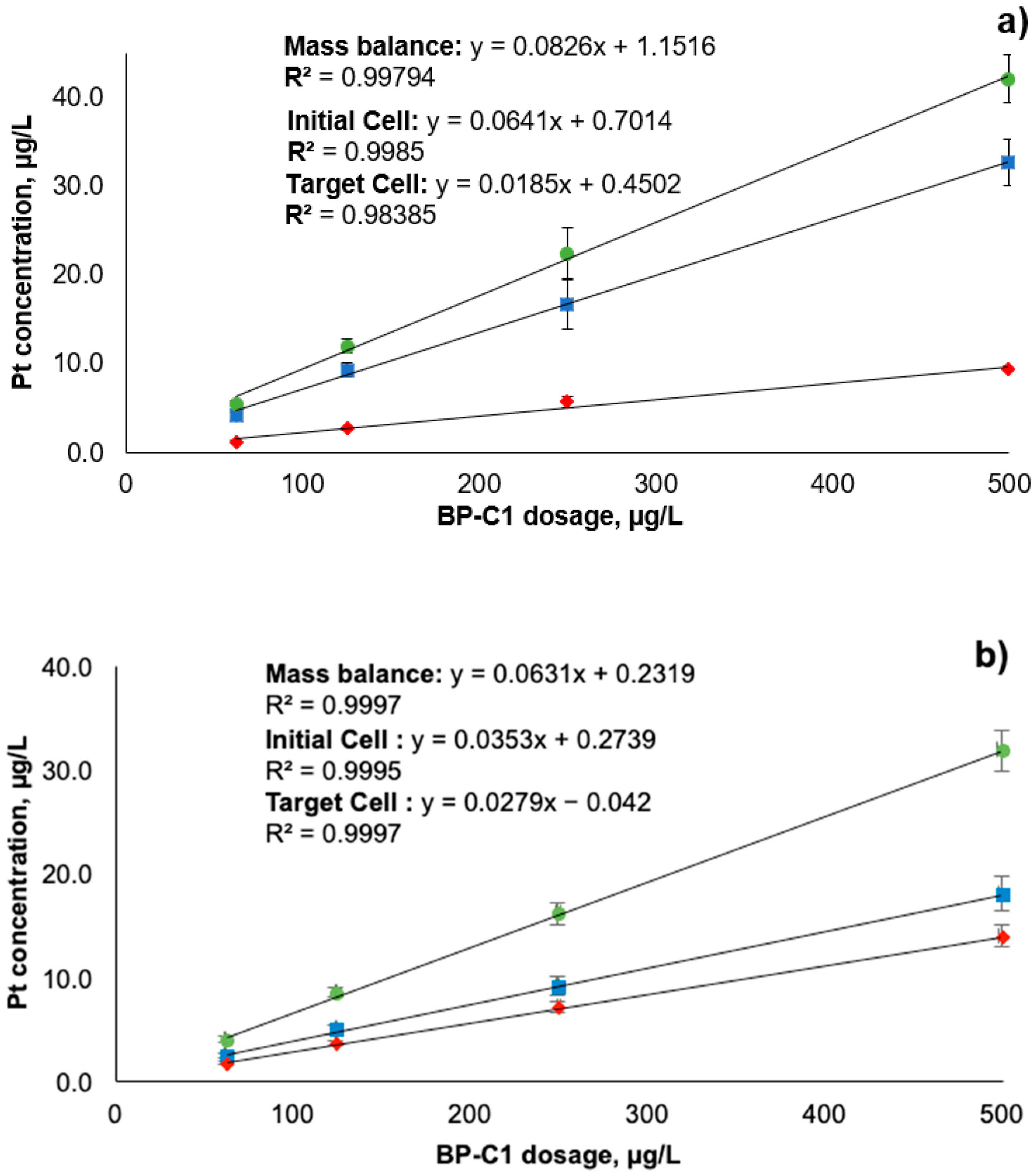
) stand for initial cell after the establishment of equilibrium; red diamonds ( Applsci 11 11008 i002) stand for target cell after crossing the dialysis membrane; green circles ( Applsci 11 11008 i003) stand for total Pt in both cells (mass balance). Error bars represent ± standard deviation (n = 6).
Applsci 11 11008 g002
Figure 3. Ratio of 50% plasma to control buffer for the dialysed fraction of platinum (Pt), indicating plasma protein binding. Plasma and control buffer were dialysed against PBS with pH 7.2 for 4 h at 37 °C across the 8 kDa cut-off vertical membrane. Pt was quantified by ICP-MS after microwave digestion. Dashed line represents linear trend. Error bars represent ± standard deviation (n = 6).
Applsci 11 11008 g003
Table 1. Platinum (Pt) concentration and retention in the cells after dialysis equilibrium was established. Fifty percent plasma and control PBS dialysed against PBS with pH 7.2 for 4 h at 37 °C across the 8 kDa cut-off vertical membrane. Pt was quantified by ICP-MS after microwave digestion. All dosage levels were tested in six parallel measurements (n = 6). Fifty percent plasma samples were compared to the control buffer for the same dosage level.
Sample NameAverage Pt
Concentration, µg/L
RSD, %Relative
Retention%
SD of
Relative
Retention%
p-Value *
50% plasma + 500 µg/L BP-C1 retained fraction32.78.177.66.30.004
50% plasma + 500 µg/L BP-C1 dialysis fraction9.43.4
Control buffer + 500 µg/L BP-C1 retained fraction17.99.056.45.1
Control buffer + 500 µg/L BP-C1 dialysis fraction13.87.2
50% plasma + 250 µg/L BP-C1 retained fraction16.71.374.41.0<0.001
50% plasma + 250 µg/L BP-C1 dialysis fraction5.810.6
Control buffer + 250 µg/L BP-C1 retained fraction9.09.556.35.4
Control buffer + 250 µg/L BP-C1 dialysis fraction7.06.9
50% plasma + 125 µg/L BP-C1 retained fraction9.48.677.86.70.002
50% plasma + 125 µg/L BP-C1 dialysis fraction2.76.6
Control buffer + 125 µg/L BP-C1 retained fraction4.97.958.34.6
Control buffer + 125 µg/L BP-C1 dialysis fraction3.54.5
50% plasma + 62.5 µg/L BP-C1 retained fraction4.211.176.18.40.008
50% plasma + 62.5 µg/L BP-C1 dialysis fraction1.38.9
Control buffer + 62.5 µg/L BP-C1 retained fraction2.39.159.35.4
Control buffer + 62.5 µg/L BP-C1 dialysis fraction1.65.7
* Paired t-test, normality was checked using Shapiro–Wilk test (p < 0.05); SD—standard deviation; RSD—relative standard deviation.
Publisher’s Note: MDPI stays neutral with regard to jurisdictional claims in published maps and institutional affiliations.

