Abstract
Chronic kidney disease (CKD) is a dilapidating problem that causes great global burden since the current therapeutic options are mostly ineffective. Early diagnosis and efficient treatment are essential to prevent the progression of CKD. Nanoparticles as technological drivers of innovation have emerged in biomedical studies for different theranostic applications. Several nanoparticles have been developed, which can be labeled with targeting moieties for both drug delivery and/or imaging systems and are investigated to detect different pathological conditions. The focus of this review is to demonstrate the current research and clinical applications for nanoparticles in the diagnosis of CKD and other renal diseases that will probably result in CKD.
1. Introduction
A general classification of kidney diseases could be its division into acute kidney injury (AKI) and chronic kidney diseases (CKD). AKI usually occurs when the serum creatinine level exceeds 0.3 mg/dL within 48 h or the urine volume is below 0.5 mL/k/h for 6 h, or serum creatinine surges up to more than 1.5 times baseline. Urinary obstruction, hypovolemia, and drug overdose are considered the main causes of AKI. The persistent abnormality (more than 3 months) in the structure and function of the kidney is defined as CKD and is commonly connected with primary glomerular diseases, diabetes, and hypertension. There are different definitions, causes, and therapeutical strategies. The global prevalence of CKD is more than 10%, mostly affecting the elderly [1]. Several adverse outcomes and clinical complications as well as individual and social-economic burdens affect CKD patients [2]. Therefore, the prevention and early diagnosis of CKD is an emerging public health issue. Whether the renal disease is localized within the glomeruli, tubules, vessels, interstitium, and/or cysts, the pathological classification of the disease must be determined. In a few groups of CKD patients, a renal biopsy might be required [3]. CKD is tightly connected with the elevated risk of vascular and early cardiovascular-related death. The current guideline categorizes CKD into five stages according to the glomerular filtration rate (GFR) level. However, the conventional techniques of GFR measuring are time-consuming, inconvenient, and might be inaccurate. Although radiolabeled approaches can result in accurate GFR evaluation, they are less practical [4]. Stage 1 CKD is considered as renal damage, but the GFR is normal (90) or increased. A mild plunge in GFR (60–89) is observed at stage 2. The GFR at stages 3 and 4 drops to 30–59 and 15–29, respectively. Finally, in stage 5, renal failure occurs and the GFR is below 15. All GFRs are demonstrated in mL/min/1.73 m2 [4].
A calculation of the estimated GFR (eGFR) from serum creatinine according to a formula in relation to the characteristics of the patients has become an available and facilitated method of monitoring kidney damage. The Chronic Kidney Disease Epidemiology Collaboration (CKD-EPI) equation now represents the finest way to precisely estimate GFR [4,5].
Nanoparticles (NPs) are valuable matrix structures that can encapsulate proteins, drugs, imaging agents, and/or their combinations in a single nano-sized construction [6,7,8]. Nanotechnology is a rapidly progressing field with wide applications [9,10]. Several medical applications have benefited from using NPs to deliver theranostic agents. In particular, NPs can overcome the physical and biological limitations of conventional therapies and thus provide new opportunities against the stability and solubility problems of many compounds. Rapid progress in nanotechnology through the development of several nanomaterials as diagnostic or therapeutic agents holds promise in treating several diseases [11,12]. Sophisticated targeting tactics and multifunctionality are considered as unique properties of nanomedicine products. Nanoparticles as drug/gene delivery vehicles and contrast agents in imaging have been extensively used [13]. In this context, a wide range of organic, inorganic, polymeric, lipid, and glycan compounds have been successfully applied [14]. A broad body of evidence supports the capability of NPs in the diagnosis and treatment of CKD [15]. NPs have been applied in the measurement of renal morphology and function and targeted delivery of different therapeutics. However, few studies have been conducted on using NPs in CKD [16].
Novel techniques have been developed to generate electrostatic charges and thus turn NPs into hydrophobic structures, increasing sight-specific delivery. Additionally, different ligands can be conjugated to the surface of NPs to increase targeted therapy; this is seen in antibodies and peptides to reach specific cell receptors. The current mini-review highlights the effect of produced NPs in the detection of CKD.
2. Application of NPs in the Diagnosis of CKD
An early prediction of the onset and progression of renal disease is crucial. In this context, specific biomarkers have been considered as valuable tools for diagnosis. Successful diagnosis could be followed by an effective therapeutical intervention to prevent CKD and diminish other possible complications such as hypertension, infection, anemia, and heart failure. The application of the conventional diagnostic technique is currently hindered by several limitations such as inconvenience and lack of sensitivity. Thus, NPs might be crucial in the early detection of CKD since they can provide high sensitivity. Table 1 represents the characteristic features and targets of different NPs used so far in CKD treatment.

Table 1.
Features of NPs for CKD targeting.
The breath assessment of volatile organic compounds has been previously used to detect CKD [16]. Carbon-nanotube-based sensors and breath sample analysis have been utilized as non-invasive tools in diagnosing end-stage renal disease (ESRD) in vivo [17]. Accordingly, gold NP-based sensors have been used to detect CKD in breath samples. This sensor can accurately distinguish between the exhaled breath of CKD patients and healthy individuals. Moreover, the sensor has shown a capability to diagnose early and late-stage CKD [18].
Magnetically assisted hemodialysis (MAHD) has been posed as an effective approach in ESRD management. In this context, biocompatible ferromagnetic NPs bind to targeted binding substances such as proteins, antibodies, and immunoglobulins with a high affinity for toxins in serum. The iron constituents of NPs can be magnetically manipulated and then target different body tissues. In this system, before the hemodialysis NP infusion is performed, a sufficient time for binding the toxic substance to NP is allowed. In vitro studies have shown the adsorbance and clearance of homocysteine [19]. Moreover, a single circulation of MAHD leaves only an immeasurably small magnetic signal, which is equivalent to 8 circulations of hemodialysis.
2.1. Detection of Kidney Injury Biomarkers
A dipstick contains a thin plastic stick and chemical-based strips, which can detect the abnormalities in urine. The presence or increased levels of distinct substances in urine result in changes of the stick color [27]. The incidence and progression of CKD are related to the increment of albuminuria as an important risk factor [28,29]. Only a concentration over 30 mg/dL of urinary albumin be detected via routine urinalysis dipstick. Specific dipsticks or specific antibody techniques are required to detect microalbuminuria [30]. Nonetheless, these strategies are inconvenient and insensitive. Surface-enhanced Raman scattering (SERS) with silver/copper/gold NP surfaces has been applied in this context by employing laser energy [31]. Easy sample processing steps, high sensitivity, rapid analysis, and the availability of portables apparatus are the advantages of the SERS method [32]. The application of silver NPs has significantly increased the Raman signal [31]. It has been reported that the SERS spectroscopy method reveals a strong connection between the reference and predicted albumin concentration, proposing that this technique is appropriate for the determination of absolute albuminuria. SERS as a sensitive tool has been able to detect extremely low levels (3 μg/mL) of albumin [33]. Currently, the available commercial devices have provided point-of-care monitoring of microalbuminuria [32]. A recent investigation developed a disposable electrochemical immunosensors for a point-of-care detection of microalbuminuria in which gold NPs were used on the electrodes via dielectrophoresis. This sensor contained a polystyrene NP core with HAS antibodies that were covalently conjugated to silver nanoshells. The developed nanosensor exhibited high conductivity, biocompatibility, and sensitivity which can be used in the early detection and monitoring of CKD in point-of-care settings [34] (Figure 1).
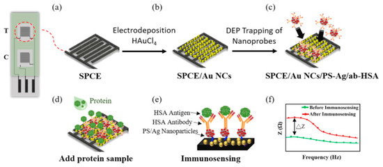
Figure 1.
Schematic illustration of systematic protocol for immunosensor fabrication and operation. Screen-printed carbon electrode (SPCE) interdigitated on a flexible plastic substrate (a). SPCE have modified with gold nanoparticles (SPCE/Au NCs) (b). Polystyrene nanoparticle core with silver nanoshells (PS-Ag) covalently conjugated to Human Serum Albumin (HSA) antibodies and these nanoprobes are trapped on the electrode surface (PS/Ag/ab-HSA nanoprobes) (c). In order to perform immunosensing, HAS protein was dropped onto the sensing areas (d). PS-Ag nanoparticles improved surface areas and incresed the number of immobilized antibodies (e). Immunosensing results with normalized impedance response, good linearity and repeatability dependent on albumin concentration (f). (Reprinted with permission from Ref. [34] Copyright © 2018 Elsevier B.V.).
Serum and urine creatinine are considered important biomarkers of CKD [35]. Peptides are among the suitable detectors of creatinine due to their simple synthesis and modification properties and low molecular weight [36,37]. Phage display technology has been utilized to fabricate peptides that have a specific affinity to creatinine [38]. In this approach, gold NPs, due to their distance-dependent optical features, have been used in combination with peptide probes to detect creatinine. The synergistic effect between these NPs and peptides has shown high selectivity and specificity for detecting creatinine in human urine [39].
Other promising biomarkers such as N-acetyl-β-d-glucosaminidase (NAG), kidney injury molecule-1 (KIM-1), and cystatin C (CysC) have been introduced as efficient tools for CKD prediction [40]. Mounting evidence demonstrated that NPs could significantly enhance the response of immunosensors in recognition of these biomarkers [41,42]. Serum CysC was detected by a disposable amperometric immunosensor through a sandwich-type assay in which gold NPs were used to generate the augmented response with high sensitivity [43]. In another study, a new “light-switch” molecule of the Ru (II) complex ([Ru(dcbpy)2dppz]2+-DPEA) with self-enhanced electrochemiluminescence (ECL) specifications was developed [42]. The combination of this ECL with DNA nanotechnology has been used as an effective signal amplifier in NAG detection high sensitivity excellent linear range (0.1 pg/mL to 1 ng/mL) [42]. In a similar report, KIM-1 was detected via an ECL biosensor in which Pt NPs were used to enhance the electron transfer efficiency [41]. Therefore, nanotechnological advances can pave the way to yield an effective and sensitive detection method for CKD, which in turn will facilitate earlier intervention. MicroRNAs (miRNAs) as short single-stranded RNAs, regulate the expression of genes that are paired with regions within the 3’-untranslated region of target mRNAs [44,45]. The current research highlights the role of small non-coding RNAs in the pathophysiology of renal diseases since the potential role of miRNAs as renal disease biomarkers are gaining increasing attention [46,47]. However, the standardization of measuring methods is required in order to validate the efficiency of these biomarkers. Moreover, selecting the appropriate tissue or biofluid is essential to achieve ideal results in clinical settings. The expression levels of miRNAs involved in kidney disease might represent novel treatment approaches [48].
The bioavailability and activity of insulin-like growth factor (IGF) are mediated by a number of superfamily members of homologous proteins referred to as insulin-like growth factor binding proteins (IGFBPs). Given the pivotal role of these proteins, IGFBP 7 has been reported as a possible independent biomarker of AKI with high specificity and sensitivity [49]. Similarly, IGFBP 2 has been connected with the kidney through the elevation of reactive oxygen species-related apoptosis in the renal tubular epithelium [50], hinting to the potential role of this molecule as a kidney damage biomarker [51,52]. Therefore, detecting IGFBPs with novel and rapid techniques might shed new light to the early treatment of renal diseases.
Arendowski and coworkers analyzed the blood serum of renal cell carcinoma (RCC) patients via laser desorption/ionization mass spectrometry on a gold nanoparticle-enhanced target (AuNPET) approach. This study led to the identification of potential biomarkers of RCC such as creatinine, dihydrouracil, melatonin, palmitoyl glucuronide, tyrosine, and glutamine. The applied nanotechnology-based MS detection method was successful in identifying the metabolic biomarkers of RCC [53].
2.2. Kidney Imaging Method Using Fluorescence Nanomaterials
GFR is a direct indicator of renal function. However, its detection is inconvenient and raises the risk of Iohexol (contrast agents) related-AKI. As mentioned, the CKD-EPI formula is used to calculate GFR. The absorption and secretion of creatinine by tubular cells, which in turn decrease the effectiveness of CKD detection, could be referred to as an important drawback. The application of different fluorescent NPs such as quantum dots, silica, and gold NP offer several advantages in comparison with the conventional method of FGR evaluation. They are entirely filtered by glomeruli, neither induce cytotoxicity nor interfere within body metabolism, are easy to produce, and are cost-effective [54]. Besides, their absorption and emission wavelengths are in the visible (and mainly in the near-infrared) range [55].
Semiconductor nanocrystals (quantum dots) are among the available commercial fluorescent nanomaterials with extensive applications in biology [56]. Moreover, near-infrared, sensitive, and inexpensive fluorescent imaging has widespread applications in detecting several disorders, including cancer [57]. However, the diagnosis of renal insufficiency and staging via noninvasive fluorescent methods are currently in the preclinical phases [57]. The kidney function has been determined by fluorescent imaging using renal-clearable near-infrared-emitting glutathione-coated gold NPs (GS-AuNPs) as a contrast agent. This method could effectively and noninvasively detect kidney dysfunction, disease stages, and show adaptive functioning in animals with unilateral obstructive nephropathy (UUO). The identification of disease stages was in correlation with the outputs of pathological examinations [58]. Traditional markers of kidney dysfunction (Blood urea nitrogen (BUN) and creatinine) are incapable of determining UUO renal dysfunction. GS-AuNPs induced no structural changes in normal mice kidneys and exhibited a very low concentration in the background tissues [59]. The emission wavelength was approximately 800 nm and thus was visible [60]. Additionally, GS-AuNPs have complete renal removal since their hydrodynamic diameter is lower than the renal filtration threshold [61]. Currently, different renal-clearable NPs such as gold, copper, silica, iron oxide NPs, palladium nanosheets, and quantum dots are available [62,63]. Shirai et al. used silica NPs with fluorescent anti-CD 11b to detect fibrosis and inflammation in a UUO model in vivo [64]. Imaging-guided photodynamic therapy (PDT) with renal clearable ultra-small size NPs has also been a focus of researchers. Siwawannapong et al. functionalized Pyropheophorbide-a (Pa), a deep red photosensitizer with polyethylene glycol (PEG), to yield Pa-PEG as ultra-small nanodots which exhibited red-shifted absorbance and efficient singlet oxygen formation upon light irradiation. These Pa-PEG nanodots revealed a significant PDT effect in vitro by attacking many cancer cells. Moreover, Pa-PEG nanodots were used in fluorescence and photoacoustic (PA) imaging methods to determine the optimal period for PDT therapy upon intravenous administration in vivo. An effective accumulation at the tumor site and obtaining the maximum concentration eight hours after injection were noted as the superiorities of this method. Furthermore, the high PA and fluorescent signal from the kidney tissues demonstrated their renal excretion [65] (Figure 2).
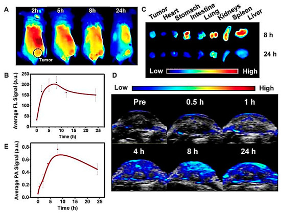
Figure 2.
The fluorescence and photoacoustic imaging of Pa-PEG nanodots. (A) Fluorescence images. (B) Fluorescence signal examination of the tumor. (C) Ex vivo fluorescence images of main organs and tumors. (D) Photoacoustic images of tumor post injecting Pa-PEG nanodots (E) Photoacoustic intensities of localized Pa-PEG nanodots [65].
2.3. Magnetic Resonance Imaging (MRI) Coupled with Nanomaterials
A great percentage of old patients and patients undergoing angiography experience CKD [66]. The use of gadolinium-based or iodinated contrast agents can exacerbate the disease, or even be fatal [67]. Magnetic NP imaging is a safe strategy that applies superparamagnetic iron-oxide NPs, which are processed and preserved in the hepatic iron reserve of the body and do not influence the kidneys [68].
The gold standard technique for diagnosing and evaluating renal diseases is currently performed via biopsy since it can only show a connection between the diagnosis of kidney diseases and related pathological alterations. Nonetheless, an invasive method increases the risk of hematoma [69]. Moreover, mismatches between the samples and overall kidney tissue might occur [70]. Therefore, the application of nanotechnology in MRI might diminish the restrictions and be a good substitute for biopsy [71]. In this context, iron oxide NPs are considered as promising surrogates of gadolinium-based MRI contrast agents [72]. These NPs can be utilized not only to assess kidney functions but also to act as cellular imaging apparatuses [73]. The T2-weighted MRI signals have been decreased by using iron oxide particles since these particles can be absorbed by inflammatory cells [74]. The effects of iron oxide injection on the signal intensity of kidneys were assessed in different nephropathic models in rats. The signal intensity loss was observed in the overall components of kidneys in the obstructive nephropathy model nephrotoxic nephritis rate showed a decreased intensity in the cortical signal. This proposes the promising role of iron oxide NPs in detecting different types of renal lesions [75]. Human studies have also shown similar results [76]. Intravenous injection of iron oxide was used to identify and characterize macrophages in infiltrated normal native and transplanted kidneys. A significant signal decrease was shown in the cortical macrophage infiltration; however, patients with ischemic acute tubular necrosis declined the medulla [76]. Since the infiltration of macrophages is a substantial feature of inflammation, a fall in the signal can represent inflammation-related injury. Moreover, different nephropathies could be differentially diagnosed by assessing the intensity and the area of the MRI signal. Nonetheless, it remains to be clarified if the repeated use of iron oxide NPs induced cytotoxicity, although empirical pharmacokinetic studies and safety assessment have been satisfactory so far [77].
Hydrophilic MNO NPs with PEG (PEG-MNO NPs) through a polyol-like technique have been developed in a one-pot method. The produced NPs showed a low r2/r1 ratio and high relaxivity. In addition, an acidic environment, glutathione, or exposure to the stimulated body fluid increase the stability of these particles. It has been reported that an amino-modified AS1411 aptamer conjugation with PEG-MNO NPAs can visibly visualize renal carcinoma cells with MRI during in vitro studies which exhibit a more prolonged retention time in the tumor site in vivo in comparison with PEG-MNO NPs [78] (Figure 3).
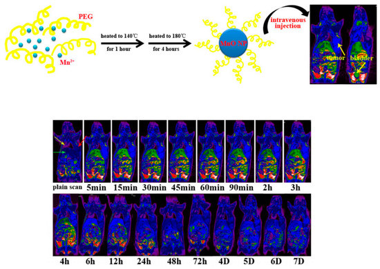
Figure 3.
AS1411-PEG-MnO nanoprobe and MR T1 images of animals with renal carcinoma tumors. (Reprinted with permission from Ref. [78]. Copyright © 2017 Elsevier B.V.).
3. Conclusions and Perspectives
Nanotechnology is a potential tool for the early detection and monitoring of CKD that can provide a prompt therapeutic strategy. Nanotechnology-assisted diagnosis approaches have successfully detected different CKD biomarkers such as CysC, NAG, and KIM-1. Moreover, NPs can enhance the potency of imaging techniques in assessing kidney dysfunction stages and the possible presence of fibrosis and inflammation. The optimization of different features of NPs such as size, shape, surface ligand, and charge can improve their application. Although the production of novel NPs is rapidly increasing in empirical studies, a gradual introduction into the clinic and market is being observed since the stability, renal targeting, cytotoxicity, and biodistribution of these materials must be strictly tested. However, advances in nanotechnology and its application in nephrology might bring great hope for the early treatment of renal diseases, particularly CKD.
Author Contributions
Supervision, E.A. and E.M.; conceptualization, A.E., S.M.D., M.A., S.D., S.M. (Shakar Mammadova), M.V., S.M. (Sevil Mehraliyeva), R.K. and A.N.; writing—original draft preparation, A.E., S.M.D., E.A., S.M. (Shakar Mammadova), A.N., R.K. and S.M. (Sevil Mehraliyeva); writing—review and editing, E.A., E.M., M.V., S.M. (Shakar Mammadova), A.E., R.K., S.M. (Sevil Mehraliyeva) and A.N.; visualization, A.E., M.A., S.D. and S.M.D. All authors have read and agreed to the published version of the manuscript.
Funding
This research received no external funding.
Institutional Review Board Statement
Not applicable.
Informed Consent Statement
Not applicable.
Data Availability Statement
Not applicable.
Acknowledgments
Ebrahim Mostafavi would like to acknowledge the support from the National Institute of Biomedical Imaging and Bioengineering (5T32EB009035).
Conflicts of Interest
The authors declare no conflict of interest.
References
- Luo, S.; Grams, M.E. Epidemiology research to foster improvement in chronic kidney disease care. Kidney Int. 2020, 97, 477–486. [Google Scholar] [CrossRef]
- Saran, R.; Robinson, B.; Abbott, K.C.; Agodoa, L.Y.; Albertus, P.; Ayanian, J.; Balkrishnan, R.; Bragg-Gresham, J.; Cao, J.; Chen, J.L. US renal data system 2016 annual data report: Epidemiology of kidney disease in the United States. Am. J. Kidney Dis. 2017, 69, A7–A8. [Google Scholar] [CrossRef]
- Levin, A.; Bilous, R.; Coresh, J. Chapter 1: Definition and classification of CKD. Kidney Int. Suppl. 2013, 3, 19–62. [Google Scholar]
- Mula-Abed, W.-A.S.; Al Rasadi, K.; Al-Riyami, D. Estimated glomerular filtration rate (eGFR): A serum creatinine-based test for the detection of chronic kidney disease and its impact on clinical practice. Oman Med. J. 2012, 27, 108. [Google Scholar] [CrossRef] [PubMed]
- Silveiro, S.P.; Araújo, G.N.; Ferreira, M.N.; Souza, F.D.; Yamaguchi, H.M.; Camargo, E.G.J.D.C. Chronic Kidney Disease Epidemiology Collaboration (CKD-EPI) equation pronouncedly underestimates glomerular filtration rate in type 2 diabetes. Diabetes Care 2011, 34, 2353–2355. [Google Scholar] [CrossRef] [PubMed]
- Adibkia, K.; Yaqoubi, S.; Dizaj, S.M. Pharmaceutical and Medical Applications of Nanofibers. In Novel Approaches for Drug Delivery; IGI Global: Hershey, Pennsylvania, 2017; pp. 338–363. [Google Scholar]
- Dizaj, S.; Adibkia, K. A Short Overview on the Nanoparticle-based smart Drug Delivery Systems. J. Pharm. Pharm. 2015, 2, 1–2. [Google Scholar]
- Turksoy, V.A. NANOSOLUTIONS project; a safety classification model for engineered nanomaterials. J. Adv. Chem. Pharm. Mater. (JACPM) 2020, 3, 260–262. [Google Scholar]
- Dizaj, S.M. Can nanotechnology present new strategies to overcome COVID-19? J. Adv. Chem. Pharm. Mater. (JACPM) 2020, 3, 258–259. [Google Scholar]
- Kavetskyy, T. The recent reports on ion implantation technique to prepare nanoporous materials. J. Adv. Chem. Pharm. Mater. (JACPM) 2020, 3, 235–237. [Google Scholar]
- Nagraik, R.; Sharma, A.; Kumar, D.; Mukherjee, S.; Sen, F.; Kumar, A.P.J.S.I. Amalgamation of biosensors and nanotechnology in disease diagnosis: Mini-review. Sen. Int. 2021, 2, 100089. [Google Scholar] [CrossRef]
- Ahmadian, E.; Dizaj, S.M.; Sharifi, S.; Shahi, S.; Khalilov, R.; Eftekhari, A.; Hasanzadeh, M. The potential of nanomaterials in theranostics of oral squamous cell carcinoma: Recent progress. Trac Trends Anal. Chem. 2019, 116, 167–176. [Google Scholar] [CrossRef]
- Liu, Y.; Miyoshi, H.; Nakamura, M. Nanomedicine for drug delivery and imaging: A promising avenue for cancer therapy and diagnosis using targeted functional nanoparticles. Int. J. Cancer 2007, 120, 2527–2537. [Google Scholar] [CrossRef]
- Maleki Dizaj, S.; Sharifi, S.; Ahmadian, E.; Eftekhari, A.; Adibkia, K.; Lotfipour, F. An update on calcium carbonate nanoparticles as cancer drug/gene delivery system. Expert Opin Drug Deliv. 2019, 16, 331–345. [Google Scholar] [CrossRef]
- Ma, Y.; Cai, F.; Li, Y.; Chen, J.; Han, F.; Lin, W. A review of the application of nanoparticles in the diagnosis and treatment of chronic kidney disease. Bioact. Mater. 2020, 5, 732–743. [Google Scholar] [CrossRef]
- Brede, C.; Labhasetwar, V. Applications of nanoparticles in the detection and treatment of kidney diseases. Adv. Chronic Kidney Dis. 2013, 20, 454–465. [Google Scholar] [CrossRef]
- Haick, H.; Hakim, M.; Patrascu, M.; Levenberg, C.; Shehada, N.; Nakhoul, F.; Abassi, Z. Sniffing chronic renal failure in rat model by an array of random networks of single-walled carbon nanotubes. ACS Nano 2009, 3, 1258–1266. [Google Scholar] [CrossRef]
- Marom, O.; Nakhoul, F.; Tisch, U.; Shiban, A.; Abassi, Z.; Haick, H.J.N. Gold nanoparticle sensors for detecting chronic kidney disease and disease progression. Nanomedicine 2012, 7, 639–650. [Google Scholar] [CrossRef] [PubMed]
- Stamopoulos, D.; Benaki, D.; Bouziotis, P.; Zirogiannis, P.J.N. In vitro utilization of ferromagnetic nanoparticles in hemodialysis therapy. Nanotechnology 2007, 18, 495102. [Google Scholar] [CrossRef] [PubMed]
- Choi, C.H.J.; Zuckerman, J.E.; Webster, P.; Davis, M.E. Targeting kidney mesangium by nanoparticles of defined size. Proc. Natl. Acad. Sci. USA 2011, 108, 6656–6661. [Google Scholar] [CrossRef] [PubMed]
- Serkova, N.J.; Renner, B.; Larsen, B.A.; Stoldt, C.R.; Hasebroock, K.M.; Bradshaw-Pierce, E.L.; Holers, V.M.; Thurman, J.M. Renal inflammation: Targeted iron oxide nanoparticles for molecular MR imaging in mice. Radiology 2010, 255, 517–526. [Google Scholar] [CrossRef]
- Hultman, K.L.; Raffo, A.J.; Grzenda, A.L.; Harris, P.E.; Brown, T.R.; O’Brien, S. Magnetic resonance imaging of major histocompatibility class II expression in the renal medulla using immunotargeted superparamagnetic iron oxide nanoparticles. Acs Nano 2008, 2, 477–484. [Google Scholar] [CrossRef] [PubMed]
- Radović, N.; Čuzić, S.; Knotek, M. Effect of unilateral ureteral obstruction and anti-angiotensin II treatment on renal tubule and interstitial cell apoptosis in rats. Croat. Med. J. 2008, 49, 600–607. [Google Scholar] [CrossRef] [PubMed]
- Akers, W.J.; Zhang, Z.; Berezin, M.; Ye, Y.; Agee, A.; Guo, K.; Fuhrhop, R.W.; Wickline, S.A.; Lanza, G.M.; Achilefu, S. Targeting of ανβ3-integrins expressed on tumor tissue and neovasculature using fluorescent small molecules and nanoparticles. Nanomedicine 2010, 5, 715–726. [Google Scholar] [CrossRef] [PubMed]
- Simón-Yarza, T.; Tamayo, E.; Benavides, C.; Lana, H.; Formiga, F.R.; Grama, C.N.; Ortiz-de-Solorzano, C.; Kumar, M.R.; Prosper, F.; Blanco-Prieto, M.J. Functional benefits of PLGA particulates carrying VEGF and CoQ10 in an animal of myocardial ischemia. Int. J. Pharm. 2013, 454, 784–790. [Google Scholar] [CrossRef] [PubMed][Green Version]
- Jo, S.-K.; Hu, X.; Kobayashi, H.; Lizak, M.; Miyaji, T.; Koretsky, A.; Star, R.A. Detection of inflammation following renal ischemia by magnetic resonance imaging. Kidney Int. 2003, 64, 43–51. [Google Scholar] [CrossRef]
- Carel, R.; Silverberg, D.; Kaminsky, R.; Aviram, A. Routine urinalysis (dipstick) findings in mass screening of healthy adults. Clin. Chem. 1987, 33, 2106–2108. [Google Scholar] [CrossRef]
- Eftekhari, A.; Ahmadian, E.; Khalilov, R. The main biomarkers for drug-induced nephrotoxicity; a sight on clinical studies. J. Adv. Chem. Pharm. Mater. (JACPM) 2020, 3, 244–247. [Google Scholar]
- Reichel, H.; Zee, J.; Tu, C.; Young, E.; Pisoni, R.L.; Stengel, B.; Duttlinger, J.; Lonnemann, G.; Robinson, B.M.; Pecoits-Filho, R. Chronic kidney disease progression and mortality risk profiles in Germany: Results from the Chronic Kidney Disease Outcomes and Practice Patterns Study. Nephrol. Dial. Transplant. 2020, 35, 803–810. [Google Scholar] [CrossRef]
- Keane, W.F.; Eknoyan, G. Proteinuria, albuminuria, risk, assessment, detection, elimination (PARADE): A position paper of the National Kidney Foundation. Am. J. Kidney Dis. 1999, 33, 1004–1010. [Google Scholar] [CrossRef]
- Mosier-Boss, P.A. Review of SERS substrates for chemical sensing. Nanomaterials 2017, 7, 142. [Google Scholar] [CrossRef] [PubMed]
- McNay, G.; Eustace, D.; Smith, W.E.; Faulds, K.; Graham, D. Surface-enhanced Raman scattering (SERS) and surface-enhanced resonance Raman scattering (SERRS): A review of applications. Appl. Spectrosc. 2011, 65, 825–837. [Google Scholar] [CrossRef] [PubMed]
- Stefancu, A.; Moisoiu, V.; Bocsa, C.; Bálint, Z.; Cosma, D.-T.; Veresiu, I.A.; Chiş, V.; Leopold, N.; Elec, F. SERS-based quantification of albuminuria in the normal-to-mildly increased range. Analyst 2018, 143, 5372–5379. [Google Scholar] [CrossRef] [PubMed]
- Shaikh, M.O.; Zhu, P.-Y.; Wang, C.-C.; Du, Y.-C.; Chuang, C.-H. Electrochemical immunosensor utilizing electrodeposited Au nanocrystals and dielectrophoretically trapped PS/Ag/ab-HSA nanoprobes for detection of microalbuminuria at point of care. Biosens. Bioelectron. 2019, 126, 572–580. [Google Scholar] [CrossRef]
- Peralta, C.A.; Shlipak, M.G.; Judd, S.; Cushman, M.; McClellan, W.; Zakai, N.A.; Safford, M.M.; Zhang, X.; Muntner, P.; Warnock, D.J.J. Detection of chronic kidney disease with creatinine, cystatin C, and urine albumin-to-creatinine ratio and association with progression to end-stage renal disease and mortality. Jama 2011, 305, 1545–1552. [Google Scholar] [CrossRef] [PubMed]
- Fani, M.; Maecke, H.; Okarvi, S.J.T. Radiolabeled peptides: Valuable tools for the detection and treatment of cancer. Theranostics 2012, 2, 481. [Google Scholar] [CrossRef] [PubMed]
- Shapira, S.; Fokra, A.; Arber, N.; Kraus, S. Peptides for diagnosis and treatment of colorectal cancer. Curr. Med. Chem. 2014, 21, 2410–2416. [Google Scholar] [CrossRef]
- Landon, L.A.; Deutscher, S. Combinatorial discovery of tumor targeting peptides using phage display. J. Cell. Biochem. 2003, 90, 509–517. [Google Scholar] [CrossRef]
- Feng, S.; Shi, R.; Xu, P.; Bhamore, J.R.; Bal, J.; Baek, S.H.; Park, C.Y.; Park, J.P.; Park, T. Colorimetric detection of creatinine using its specific binding peptides and gold nanoparticles. New J. Chem. 2020, 44, 15828–15835. [Google Scholar] [CrossRef]
- Alter, M.L.; Kretschmer, A.; Von Websky, K.; Tsuprykov, G.; Reichetzeder, C.; Simon, A.; Stasch, J.-P.; Hocher, B. Early urinary and plasma biomarkers for experimental diabetic nephropathy. Clin. Lab. 2012, 58, 659. [Google Scholar]
- Yang, H.; Wang, H.; Xiong, C.; Chai, Y.; Yuan, R. Highly sensitive electrochemiluminescence immunosensor based on ABEI/H2O2 system with PFO dots as enhancer for detection of kidney injury molecule-1. Biosens. Bioelectron. 2018, 116, 16–22. [Google Scholar] [CrossRef]
- Wang, H.; Yuan, Y.; Zhuo, Y.; Chai, Y.; Yuan, R. Sensitive electrochemiluminescence immunosensor for detection of N-acetyl-β-d-glucosaminidase based on a “light-switch” molecule combined with DNA dendrimer. Anal. Chem. 2016, 88, 5797–5803. [Google Scholar] [CrossRef]
- Lopes, P.; Costa-Rama, E.; Beirão, I.; Nouws, H.P.; Santos-Silva, A.; Delerue-Matos, C. Disposable electrochemical immunosensor for analysis of cystatin C, a CKD biomarker. Talanta 2019, 201, 211–216. [Google Scholar] [CrossRef]
- Nelson, P.T.; Wang, W.X.; Rajeev, B. MicroRNAs (miRNAs) in neurodegenerative diseases. Brain Pathol. 2008, 18, 130–138. [Google Scholar] [CrossRef] [PubMed]
- Esquela-Kerscher, A.; Slack, F. Oncomirs—microRNAs with a role in cancer. Nat. Rev. Cancer 2006, 6, 259–269. [Google Scholar] [CrossRef] [PubMed]
- Liu, Z.; Wang, Y.; Shu, S.; Cai, J.; Tang, C.; Dong, Z. Non-coding RNAs in kidney injury and repair. Am. J. Physiol.-Cell Physiol. 2019, 317, C177–C188. [Google Scholar] [CrossRef] [PubMed]
- Ignarski, M.; Islam, R.; Müller, R.U. Long non-coding RNAs in kidney disease. Int. J. Mol. Sci. 2019, 20, 3276. [Google Scholar] [CrossRef] [PubMed]
- Metzinger-Le Meuth, V.; Fourdinier, O.; Charnaux, N.; Massy, Z.A.; Metzinger, L. The expanding roles of microRNAs in kidney pathophysiology. Nephrol. Dial. Transplant. 2019, 34, 7–15. [Google Scholar] [CrossRef] [PubMed]
- Su, Y.; Gong, Z.; Wu, Y.; Tian, Y.; Liao, X. Diagnostic value of urine tissue inhibitor of metalloproteinase-2 and insulin-like growth factor-binding protein 7 for acute kidney injury: A meta-analysis. PLoS ONE 2017, 12, e0170214. [Google Scholar] [CrossRef]
- Shin, M.; Kang, H.S.; Park, J.-H.; Bae, J.-H.; Song, D.-K.; Im, S.-S. Recent insights into insulin-like growth factor binding protein 2 transcriptional regulation. Endocrinol. Metab. 2017, 32, 11–17. [Google Scholar] [CrossRef]
- Wang, X.; Zhang, Y.; Chang, Y.; Duan, D.; Sun, Z.; Guo, X. Elevation of IGFBP2 contributes to mycotoxin T-2-induced chondrocyte injury and metabolism. Biochem. Biophys. Res. Commun. 2016, 478, 385–391. [Google Scholar] [CrossRef]
- Li, H.-L.; Yan, Z.; Ke, Z.-P.; Tian, X.-F.; Zhong, L.-L.; Lin, Y.-T.; Xu, Y.; Zheng, D.-H. IGFBP2 is a potential biomarker in acute kidney injury (AKI) and resveratrol-loaded nanoparticles prevent AKI. Oncotarget 2018, 9, 36551–36560. [Google Scholar] [CrossRef] [PubMed][Green Version]
- Arendowski, A.; Ossoliński, K.; Nizioł, J.; Ruman, T. Gold nanostructures-assisted laser desorption/ionization mass spectrometry for kidney cancer blood serum biomarker screening. Int. J. Mass Spectrom. 2020, 456, 116396. [Google Scholar] [CrossRef]
- Takeda, M.; Khamdang, S.; Narikawa, S.; Kimura, H.; Kobayashi, Y.; Yamamoto, T.; Cha, S.H.; Sekine, T.; Endou, H. Human organic anion transporters and human organic cation transporters mediate renal antiviral transport. J. Pharmacol. Exp. Ther. 2002, 300, 918–924. [Google Scholar] [CrossRef] [PubMed]
- Huang, J.; Gretz, N. Light-Emitting Agents for Noninvasive Assessment of Kidney Function. ChemistryOpen 2017, 6, 456. [Google Scholar] [CrossRef]
- Michalet, X.; Pinaud, F.F.; Bentolila, L.A.; Tsay, J.M.; Doose, S.; Li, J.J.; Sundaresan, G.; Wu, A.; Gambhir, S.; Weiss, S. Quantum dots for live cells, in vivo imaging, and diagnostics. Science 2005, 307, 538–544. [Google Scholar] [CrossRef]
- Eftekhari, A.; Hasanzadeh, M.; Sharifi, S.; Dizaj, S.M.; Khalilov, R.; Ahmadian, E. Bioassay of saliva proteins: The best alternative for conventional methods in non-invasive diagnosis of cancer. Int. J. Biol. 2019, 124, 1246–1255. [Google Scholar] [CrossRef]
- Penna, F.J.; Chow, J.S.; Minnillo, B.J.; Passerotti, C.C.; Barnewolt, C.E.; Treves, S.T.; Fahey, F.H.; Dunning, P.S.; Freilich, D.A.; Retik, A.B.; et al. Identifying ureteropelvic junction obstruction by fluorescence imaging: A comparative study of imaging modalities to assess renal function and degree of obstruction in a mouse model. J. Urol. 2011, 185, 2405–2413. [Google Scholar] [CrossRef]
- Yu, M.; Liu, J.; Ning, X.; Zheng, J. High-contrast Noninvasive Imaging of Kidney Clearance Kinetics Enabled by Renal Clearable Nanofluorophores. Angew. Chem. 2015, 54, 15434–15438. [Google Scholar] [CrossRef]
- Liu, J.; Yu, M.; Zhou, C.; Yang, S.; Ning, X.; Zheng, J. Passive tumor targeting of renal-clearable luminescent gold nanoparticles: Long tumor retention and fast normal tissue clearance. J. Am. Chem. Soc. 2013, 135, 4978–4981. [Google Scholar] [CrossRef]
- Yu, M.; Zheng, J. Clearance pathways and tumor targeting of imaging nanoparticles. ACS Nano 2015, 9, 6655–6674. [Google Scholar] [CrossRef] [PubMed]
- Yang, S.; Sun, S.; Zhou, C.; Hao, G.; Liu, J.; Ramezani, S.; Yu, M.; Sun, X.; Zheng, J. Renal clearance and degradation of glutathione-coated copper nanoparticles. Bioconjug. Chem. 2015, 26, 511–519. [Google Scholar] [CrossRef] [PubMed]
- Huang, X.; Zhang, F.; Zhu, L.; Choi, K.Y.; Guo, N.; Guo, J.; Tackett, K.; Anilkumar, P.; Liu, G.; Quan, Q. Effect of injection routes on the biodistribution, clearance, and tumor uptake of carbon dots. ACS Nano 2013, 7, 5684–5693. [Google Scholar] [CrossRef]
- Shirai, T.; Kohara, H.; Tabata, Y. Inflammation imaging by silica nanoparticles with antibodies orientedly immobilized. J. Drug Target. 2012, 20, 535–543. [Google Scholar] [CrossRef] [PubMed]
- Siwawannapong, K.; Zhang, R.; Lei, H.; Jin, Q.; Tang, W.; Dong, Z.; Lai, R.-Y.; Liu, Z.; Kamkaew, A.; Cheng, L. Ultra-small Pyropheophorbide-a Nanodots for Near-infrared Fluorescence/Photoacoustic Imaging-guided Photodynamic Therapy. Theranostics 2020, 10, 62–73. [Google Scholar] [CrossRef] [PubMed]
- Ix, J.H.; Mercado, N.; Shlipak, M.G.; Lemos, P.A.; Boersma, E.; Lindeboom, W.; O’Neill, W.W.; Wijns, W.; Serruys, P.W.J. Association of chronic kidney disease with clinical outcomes after coronary revascularization: The Arterial Revascularization Therapies Study (ARTS). Am. Heart J. 2005, 149, 512–519. [Google Scholar] [CrossRef]
- Neuwelt, E.A.; Hamilton, B.E.; Varallyay, C.G.; Rooney, W.R.; Edelman, R.D.; Jacobs, P.M.; Watnick, S.G. Ultrasmall superparamagnetic iron oxides (USPIOs): A future alternative magnetic resonance (MR) contrast agent for patients at risk for nephrogenic systemic fibrosis (NSF)? Kidney Int. 2009, 75, 465–474. [Google Scholar] [CrossRef]
- Lu, M.; Cohen, M.H.; Rieves, D.; Pazdur, R.J. FDA report: Ferumoxytol for intravenous iron therapy in adult patients with chronic kidney disease. Am. J. Hematol. 2010, 85, 315–319. [Google Scholar] [CrossRef]
- Jayasinghe, K.; Stark, Z.; Kerr, P.G.; Gaff, C.; Martyn, M.; Whitlam, J.; Creighton, B.; Donaldson, E.; Hunter, M.; Jarmolowicz, A. Clinical impact of genomic testing in patients with suspected monogenic kidney disease. Genet. Med. 2020, 23, 183–191. [Google Scholar] [CrossRef]
- Walker, P.D. The renal biopsy. Arch. Pathol. Lab. Med. 2009, 133, 181–188. [Google Scholar] [CrossRef]
- Khosroshahi, H.T.; Abedi, B.; Daneshvar, S.; Sarbaz, Y.; Shakeri Bavil, A. Future of the renal biopsy: Time to change the conventional modality using nanotechnology. Int. J. Biomed. Imaging 2017, 2017, 6141734. [Google Scholar] [CrossRef] [PubMed]
- Stabi, K.L.; Bendz, L.M. Ferumoxytol use as an intravenous contrast agent for magnetic resonance angiography. Ann. Pharmacother. 2011, 45, 1571–1575. [Google Scholar] [CrossRef]
- Corot, C.; Robert, P.; Idée, J.-M.; Port, M. Recent advances in iron oxide nanocrystal technology for medical imaging. Adv. Drug Deliv. Rev. 2006, 58, 1471–1504. [Google Scholar] [CrossRef] [PubMed]
- Egger, C.; Cannet, C.; Gérard, C.; Debon, C.; Stohler, N.; Dunbar, A.; Tigani, B.; Li, J.; Beckmann, N. Adriamycin-induced nephropathy in rats: Functional and cellular effects characterized by MRI. J. Magn. Reson. Imaging 2015, 41, 829–840. [Google Scholar] [CrossRef]
- Hauger, O.; Delalande, C.; Deminière, C.; Fouqueray, B.; Ohayon, C.l.; Garcia, S.; Trillaud, H.; Combe, C.; Grenier, N. Nephrotoxic nephritis and obstructive nephropathy: Evaluation with MR imaging enhanced with ultrasmall superparamagnetic iron oxide—Preliminary findings in a rat model. Radiology 2000, 217, 819–826. [Google Scholar] [CrossRef] [PubMed]
- Hauger, O.; Grenier, N.; Deminère, C.; Lasseur, C.; Delmas, Y.; Merville, P.; Combe, C. USPIO-enhanced MR imaging of macrophage infiltration in native and transplanted kidneys: Initial results in humans. Eur. Radiol. 2007, 17, 2898–2907. [Google Scholar] [CrossRef]
- Bourrinet, P.; Bengele, H.H.; Bonnemain, B.; Dencausse, A.; Idee, J.-M.; Jacobs, P.M.; Lewis, J.M. Preclinical safety and pharmacokinetic profile of ferumoxtran-10, an ultrasmall superparamagnetic iron oxide magnetic resonance contrast agent. Investig. Radiol. 2006, 41, 313–324. [Google Scholar] [CrossRef]
- Li, J.; Wu, C.; Hou, P.; Zhang, M.; Xu, K. One-pot preparation of hydrophilic manganese oxide nanoparticles as T1 nano-contrast agent for molecular magnetic resonance imaging of renal carcinoma in vitro and in vivo. Biosens. Bioelectron. 2018, 102, 1–8. [Google Scholar] [CrossRef] [PubMed]
Publisher’s Note: MDPI stays neutral with regard to jurisdictional claims in published maps and institutional affiliations. |
© 2021 by the authors. Licensee MDPI, Basel, Switzerland. This article is an open access article distributed under the terms and conditions of the Creative Commons Attribution (CC BY) license (https://creativecommons.org/licenses/by/4.0/).