Review

Biomedical Effects of the Phytonutrients Turmeric, Garlic, Cinnamon, Graviola, and Oregano: A Comprehensive Review

by
Yamixa Delgado
1,*,
Céline Cassé
2,
Yancy Ferrer-Acosta
3,
Ivette J. Suárez-Arroyo
4,
José Rodríguez-Zayas
1,
Anamaris Torres
5,
Zally Torres-Martínez
6,
Daraishka Pérez
1,
Michael J. González
7,
Ricardo A. Velázquez-Aponte
2,
Josué Andino
1,
Clarissa Correa-Rodríguez
1,
Jean C. Franco
1,
Wandaliz Milán
1,
Gabriela Rosario
1,
Eddian Velázquez
1,
Jaisy Vega
1,
Janmary Colón
8 and
Christopher Batista
9
1
Biochemistry & Pharmacology Department, San Juan Bautista School of Medicine, Urb Turabo Gardens PR-172, Caguas, PR 00726, USA
2
Chemistry Department, University of Puerto Rico-Mayagüez Campus, PR-108, Mayagüez, PR 00682, USA
3
Neuroscience Department, School of Medicine, Universidad Central del Caribe, Ave. Laurel 100, Sta. Juanita, Bayamón, PR 00960, USA
4
Biochemistry Department, School of Medicine, Universidad Central del Caribe, Ave. Laurel 100, Sta. Juanita, Bayamón, PR 00960, USA
5
Biology Department, University of Puerto Rico-Rio Piedras Campus, Av. Universidad Ste. 1401, San Juan, PR 00925, USA
6
Chemistry Department, University of Puerto Rico-Rio Piedras Campus, Av. Universidad Ste. 1401, San Juan, PR 00925, USA
7
School of Public Health, University of Puerto Rico-Medical Sciences Campus, Paseo Dr. José Celso Barbosa, San Juan, PR 00921, USA
8
School of Natural Sciences & Technology, Universidad Ana G. Méndez-Gurabo Campus, Carr. # 189 Km 3.3, Gurabo, PR 00778, USA
9
Biology Department, University of Puerto Rico-Bayamón Campus, Industrial Minillas 170, 63, PR-174, Bayamón, PR 00959, USA
*
Author to whom correspondence should be addressed.
Appl. Sci. 2021, 11(18), 8477; https://doi.org/10.3390/app11188477
Submission received: 12 June 2021 / Revised: 23 August 2021 / Accepted: 1 September 2021 / Published: 13 September 2021
(This article belongs to the Special Issue Bioactive Molecules in Food)

Abstract

:

Featured Application

This review is significant to the researchers using plant-derived products as therapeutics. It evaluates and summarizes the most recent studies published in the last 20 years and combines the latest botanical description, pharmacological, and biomedical effects of several popular phytonutrients as turmeric, garlic, cinnamon, graviola, and oregano, and their active phytochemicals. Furthermore, this review provides the audience with fast, summarized information of >250 scientific articles on some of the most utilized phytonutrients worldwide, of which much tertiary literature is found without the support of robust, reproducible scientific data.

Abstract

Phytonutrients are plant foods that contain many natural bioactive compounds, called phytochemicals, which show specific biological activities. These phytonutrients and their phytochemicals may play an important role in health care maintaining normal organism functions (as preventives) and fighting against diseases (as therapeutics). Phytonutrients’ components are the primary metabolites (i.e., proteins, carbohydrates, and lipids) and phytochemicals or secondary metabolites (i.e., phenolics, alkaloids, organosulfides, and terpenes). For years, several phytonutrients and their phytochemicals have demonstrated specific pharmacological and therapeutic effects in human health such as anticancer, antioxidant, antiviral, anti-inflammatory, antibacterial, antifungal, and immune response. This review summarizes the effects of the most studied or the most popular phytonutrients (i.e., turmeric, garlic, cinnamon, graviola, and oregano) and any reported contraindications. This article also presents the calculated physicochemical properties of the main phytochemicals in the selected phytonutrients using Lipinski’s, Veber’s, and Ghose’s rules. Based on our revisions for this article, all these phytonutrients have consistently shown great potential as preventives and therapeutics on many diseases in vitro, in vivo, and clinical studies.

Graphical Abstract

1. Introduction

For centuries, plants have been considered a significant source of medicinal nutrients and compounds. Historical findings have reported the use of plants by our ancestors to treat numerous diseases [1,2,3]. Consequently, it has been a quest for many individuals to search for herbal supplements and natural therapies to attend to their healthcare needs, prevent diseases, and support their nutrition. Plants produce a large variety of metabolites. Primary metabolites (i.e., innate proteins, lipids, and carbohydrates) are directly involved in their intrinsic metabolic pathways in processes such as normal growth, development, and reproduction. In contrast, secondary metabolites, also known as phytochemicals, confer a selective advantage to the plants, despite not being involved in their main metabolic pathways [4]. These phytochemicals are classified into four main chemical groups: phenolics, alkaloids, organosulfides, and terpenes [5]. Phenolics are the biggest group subdivided into seven groups: curcuminoid, stilbenes, tannins, flavonoids, phenolics acids, lignans, and coumarins [6]. Figure 1 shows a summary of the phytonutrients’ metabolite composition.
Based on this definition of secondary metabolites, phytonutrients can be considered as whole-plant extracts containing one or more phytochemicals. Since one of the functions of such secondary metabolites is to protect the plant organism from pests and diseases, it is not surprising that many of them show activity against human ailments. The scientific literature contains strong evidence supporting healthy diets rich in phytonutrients correlated with the prevention of chronic diseases, preventive medicine being one of the most important types of health care, if not the most [7,8,9,10,11,12]. However, the ingestion of plant-derived foods, also known as “superfoods,” or phytonutrients, in order to take advantage of its therapeutic properties is well under debate. This debate is mainly due to the heterogeneity in the properties of these plant extracts (phytonutrients) compared to the results in their isolated bioactive compounds (phytochemicals).
When we consume superfoods, the first feature altering their biological effect is during the food preparation, possibly inducing chemical decomposition and thermal denaturation of most metabolites, including phytochemicals [13,14]. Secondly, the different physiological barriers in the digestion process through the gastrointestinal tract determine all nutrients’ absorption, bioavailability, and delivery [15]. Multiple research groups have demonstrated the activity of the plant extract being higher when compared to the pure isolated phytochemical when orally administrated [16]. This difference is largely due to the low bioavailability and low absorption of these isolated natural compounds, which is explained by their poor solubility [16,17,18]. To overcome this pharmacokinetic problem and study the therapeutic potential of the pure phytochemicals, it is recommended to use other administration routes or develop improved delivery systems [19,20]. However, some researchers have found that synergistic interactions between the mixture of primary and secondary metabolites in phytonutrients create a natural behavior of micellar nanoparticles [21,22]. These results expose one of the least investigated properties of plant extracts.
The development of these intrinsic micelles in the extract significantly increases the successful delivery and high absorption of the phytochemical molecules [23]. On the other hand, if the phytochemical concentration in the herbal extract is extremely low, its bioactivity would be underestimated. Furthermore, the metabolites concentration, composition, and quality from batch to batch in these extracts are considerably heterogeneous [24]. These inconsistencies make extracts challenging to fulfill the high homogeneous consistency and the reproducibility required to study their therapeutic activity analytically, even when people claim their medicinal effect [5]. Thus, for the development of new drugs, isolated active phytochemicals are preferred over crude extracts. Fortunately, basic and clinical research studies of pure phytochemicals have continued for decades and have given important therapeutic outcomes. Because of these results, almost half of the drugs available in the market are naturally derived compounds [25], showing the pertinence to our review.
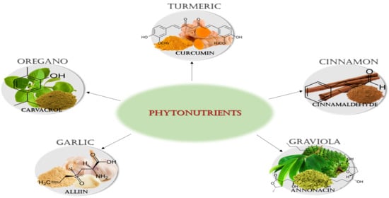
This comprehensive review summarizes the most recent studies published in the last 20 years and combines the latest botanical description, pharmacological, and biomedical effects of several popular phytonutrients as turmeric, garlic, cinnamon, graviola, and oregano, and their active phytochemicals. We emphasized the biomedical areas of the anticancer, antioxidant, antiviral, anti-inflammatory, antibacterial, antifungal, and immune response presented by the mentioned phytonutrients. Besides, special attention is given to potential contraindications found while consuming these phytonutrients/phytochemicals alone or in combination with conventional medicine. After all, the notion of phytonutrients impacting the health status of individuals, in a preventive or therapeutic way, remains an attractive topic for the public, particularly regarding food with tangible health benefits to their diets.

2. Phytonutrients

In the last 20 years, researchers’ interest in natural products has grown in search of alternatives for disease prevention and therapies. In this review, we looked for the health benefits of the selected phytonutrients demonstrated by scientific studies. Furthermore, we constructed Table 1 to show the results of the theoretical calculations of the physicochemical properties or “drug-likeness” relevant for gastrointestinal tract absorption of the main phytochemicals in five phytonutrients: turmeric, garlic, cinnamon, graviola, and oregano.

2.1. Turmeric

2.1.1. Botanical Description

Turmeric, also known as Curcuma longa, is a rhizomatous herbaceous perennial plant that belongs to the Zingiberaceae family (ginger family). This plant is highly branched with long aromatic leaves arranged in two rows. Turmeric flowers have colors ranging from white, green, yellowish, and purple-red [31]. Curcuma plants are wildly cultivated in Southeast Asia and the Indian region, where various parts are used mainly for herbal medicinal applications, dietary supplements, and cuisine purposes [32,33]. An essential part of turmeric used as a spice and herbal supplement is the rhizome, which is adjacent to the plant’s roots. Turmeric powder has a pungent taste and distinctive yellow/orange color due to pigments and curcuminoids phytochemicals in the rhizome [34]. Furthermore, primary metabolites (e.g., proteins and fats) and phytochemicals concentration dictate other physical properties and the color intensity of the turmeric powder, depending on factors such as the type of soil, crop fertilizers, and pH [35].

2.1.2. Phytochemicals

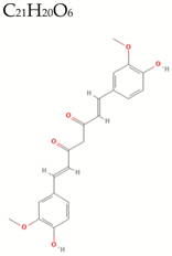
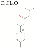
Turmeric’s therapeutic properties may include a wide variety of conditions found in the literature, where most of them come from the bioactive compounds in its rhizome. For years, different research groups have shown that turmeric is extraordinarily rich in valuable phytochemicals with pharmacological properties including polyphenols (e.g., curcuminoids), terpenes (e.g., ar-, α- and β-turmerone, α-zingiber, and β-sesquiphellandrene), flavonoids, coumarins, saponins, tannins, and steroids [36,37,38]. The principal curcuminoids are curcumin and its derivatives demethoxycurcumin and bisdemethoxycurcumin [32,39,40]. Curcumin is considered the major bioactive phytochemicals from turmeric and is around 5% of the rhizome. Other bioactive compounds found in essential turmeric oils are aromatic-tumerones, α-santalene, and aromatic curcumene [41,42]. The biomedical uses of curcumin are limited by its short half-life, low stability, and limited bioavailability [43]. However, there are different strategies under investigation to overcome these limitations, such as using natural enhancers and developing delivery systems to encapsulate the curcumin [44,45]. Various studies have demonstrated that primary and secondary metabolites in turmeric extracts may enhance the bioavailability of curcumin in vivo [43,46]. Some other phytochemicals in combination with curcumin have shown synergistic effects increasing its bioavailability, e.g., quercetin, genistein, terpineol, epigallocatechin-3-gallate, and resveratrol [47,48].

2.1.3. Biomedical effects

Anticancer

Turmeric extracts and isolated curcumin have been extensively studied for cancer applications. Since 1985, turmeric extracts have demonstrated potent cytotoxic activity against cancer in vitro and in vivo studies [49]. Then, it also entered clinical studies for the treatment of cancer [50]. Curcumin has been shown to diminish tumor growth effectively, prevent tumor formation, angiogenesis, migration, and invasion by modulating several cell signaling pathways related to adhesion molecules, cell survival proteins, growth factors, transcription factors, cytokines, kinases, and receptors [51]. Different studies demonstrated that curcumin downregulates cyclin D1, cyclin E, and MDM2, and upregulates p21, p27, and p53 [52]. Due to the low bioavailability of pure curcumin, some researchers prefer to continue studies using turmeric extracts, co-administration with other phytochemicals, or the development of drug delivery systems. For example, Li et al. reported that turmeric extracts (200 mg/kg) induced in vivo tumor growth inhibition and anti-metastatic effects using colorectal CT26, HT29, and HCT116 cancer cells [53]. Furthermore, in combination with the phytochemical quercetin, it reveals a synergistic effect against lung, skin, colorectal, and breast cancer cells [54]. In addition, Almutairi et al. designed a model that encapsulated curcumin in a chitosan polymer nanoparticle (115 nm) to increase its anticancer activity. This curcumin–chitosan nanoparticle showed a sensitive release in a more acidic pH environment, such as in cancer cells [55]. Moreover, several studies using curcumin as an anticancer agent include possible mechanisms of action [56,57,58,59,60].

Antioxidant

Curcumin is an extremely potent antioxidant by inhibiting the formation of reactive oxygen species [61]. In an in vitro study, Ak and Gülçin demonstrated the potent radical scavenging activity of curcumin by inhibiting >95% of lipid peroxidation [62]. Yuliani et al. investigated the antioxidant and neuroprotective effects of curcuminoids on neurons from Sprague–Dawley rats as a potential treatment for dementia. Turmeric extract (200 mg/kg) prevents spatial memory deficits, and its effects were comparable to the standard dementia medicine, citicoline [63]. In addition, Hossen et al. demonstrated the antioxidant properties and protective effects to hepatic organs in orally supplemented rats through a combination of curcumin (62%), flavonoids (37%), and ascorbic acid (10%). The possible mechanism of action was through antioxidant enzyme upregulation and lipid peroxidation inhibition, providing protective effects [64].

Antimicrobial

  • Antiviral
Several studies have demonstrated that the turmeric plant and the isolated phytochemical curcumin exhibited activity against a wide variety of viruses due to its potential to interfere with different cellular signaling pathways, inhibiting virus proliferation and viral expression [65]. The list of viruses that turmeric demonstrated activity are influenza A, dengue, viral hemorrhagic septicemia, human immunodeficiency, herpes simplex, Enterovirus 71, Zika, chikungunya, vesicular stomatitis, human respiratory syncytial, and others [66]. In general, curcumin strongly inhibits virus proliferation and expression. An in vitro study focused on the structure–activity relationship demonstrated that double bonds in the central carbon chain enhanced the curcumin activity against type A influenza virus by its interaction with the receptor-binding region [67]. On the other hand, in another study, researchers claimed that the hydroxyl groups and phenyl rings of curcumin are responsible for the antiviral effect against the herpes simplex virus [68]. Curcumin showed an excellent inhibitory effect in the micromolar range against transmissible gastroenteritis virus in cells in a dose-, temperature- and time-dependent manner [69]. In a very recent systematic review, Kunnumakkara et al. explained the potential of curcumin and other spices against SARS-COV-2 due to their anti-inflammatory properties to inhibit the cytokine storm [70]. Interestingly, curcumin has demonstrated antiviral activity against the SARS-CoV-2 by disrupting the binding of the spike protein to the ACE2 receptor and preventing the virus from entering cells. This group also found that curcumin positively regulates the action of the antioxidant molecule NRF2 while negatively regulating the master inflammatory molecule HMGB1 [71]. These findings suggest that turmeric and its main phytochemical curcumin could not only be a potential treatment but also a prevention alternative for viral infections.
  • Antibacterial
There are also reports showing the antibacterial activity of turmeric [37]. Bangun et al. developed an alginate-based drug delivery system of turmeric extract and tested its activity against Staphylococcus aureus (Gram-positive) and Escherichia coli (Gram-negative). The results showed that this turmeric drug delivery system affected both strains. However, there was more prominent growth inhibition on the Gram-positive bacteria than on the Gram-negative [72]. Another study performed by Czernicka, and colleagues elucidated the antimicrobial potential of turmeric extract against several Gram-positive strains (one strain of Staphylococcus epidermidis and two strains of Bacillus subtilis), revealing that the different fractions of this extract can inhibit bacterial growth [37]. In the same way, Shakeri et al. confirmed that Gram-positive bacteria are more sensitive to curcumin than Gram-negative bacteria due to their abundant hydrophilic lipopolysaccharide’s outer membrane [73].
  • Antifungal
Another significant effect of turmeric is its antifungal activity. Chen et al. showed that turmeric extracts have potent antifungal activity against 20 pathogenic fungi (e.g., Fusarium verticillioides, Curvularia pallescens, Colletotrichum falcatum, Aspergillus niger, Aspergillus terreus, Fusarium oxysporum, Fusarium moniliforme, Fusarium graminearum, Phoma wasabiae, Alternaria alternate, Botrytis cinerea, Chaetomium olivaceum, Penicillium pallidum, Mycogone perniciosa, and Verticillium dahlia) by disrupting the synthesis of the main components of the fungal cell wall and interfering the protein synthesis. From this study, phytochemicals in turmeric have better antifungal activity working in combination than individual compounds [74]. Murugesh and colleagues elucidated that turmeric extracts exhibit a potent anticandidal effect against Candida albicans on in vitro studies [75]. In a randomized clinical trial, researchers demonstrated that the topical administration of curcumin 5% ointment could significantly reduce knee pain in osteoarthritis patients [76]. This finding suggests considering turmeric topical use as a low-cost alternative with lesser side effects considering its antifungal capacity.

Anti-Inflammatory

Turmeric also exhibited potential to treat chronic pain and joint inflammation [77]. In a study using turmeric extracts combined with Allium hookeri extracts, researchers determined that this co-treatment restored the altered skin membrane and inhibited white blood cells and monocyte proliferation in inflamed skin models [78]. Bethapudi et al. demonstrated that oral administration of turmeric extract containing 57% of the bioactive turmerosaccharides significantly reduced pain and inflammation effects on an animal model (mimicking human osteoarthritis). This turmeric extract revealed a similar analgesic effect to tramadol on osteoarthritis pain [79]. In a recent study, Nicoliche et al. summarized the following curcumin’s mechanisms of action against the inflammatory process: inhibition of NF-ΚB (nuclear factor kappa B), MMP-1, 3, 8, 9, and 13 (matrix metalloproteinases), nitric oxide synthase, MAPK (mitogen-activated protein kinase), MCP (monocyte chemoattractant protein), STAT (signal transduction and activation transcription), PI3K (phosphoinositide 3-kinase), lipo-oxygenase, JAK (Janus kinase), and COX-2 (cyclo-oxygenase-2), MIP (migration inhibitory protein); also inhibition on the expression of interleukin-1, -2, -6, -8, -12 and -1β, and TNF-α (tumor necrosis factor-α); significantly improve collagen repair [80]. The study also postulated that curcumin upregulates the peroxisome proliferator-activated receptor-γ (PPAR-γ) [81].

Immunomodulatory

As previously described here, turmeric has antioxidant, antimicrobial, and anti-inflammatory properties leading to improved immune response. In in vivo experiments to study graft-versus-host disease (induced after bone marrow transplantation), mice were pretreated with curcumin (100 µg/mouse). These curcumin-pretreated mice showed an increase in CD4+ and CD8+ cells before the transplant, preventing the disease [82]. Jian et al. studied the effects of curcumin as a dietary supplement in the male Hu sheep model, reporting changes in blood metabolites, antioxidant capacity, testicular development, and immune response. After four months of dietary supplementation, the sheep improved their reproductive system performance [83]. In vivo and clinical studies indicate that curcumin can positively affect several immune cells (i.e., T lymphocyte subsets, macrophages, dendritic cells, B lymphocytes, and natural killer cells), which diminishes the severity of different autoimmune diseases [84]. Additional studies found promising results in patients with several pro-inflammatory illnesses (i.e., cardiovascular disease, renal diseases, arthritis, Crohn’s disease, ulcerative colitis, irritable bowel disease, pancreatitis, peptic ulcer, gastric ulcer, oral lichen planus, vitiligo, psoriasis, acute coronary syndrome, atherosclerosis, diabetes, lupus, acquired immunodeficiency syndrome, β-thalassemia, biliary dyskinesia, and Dejerine-Sottas disease) [85]. Most recently, a study showed that curcumin supports immunomodulatory responses by inhibiting the cell-mediated response of inflammatory cytokines and, thus, mitigating progression to pneumonia and acute respiratory distress syndrome (ARDS) after SARS-CoV-2 infection [71].

2.1.4. Contraindications

Despite the extensive evidence that reveals the beneficial effect of Curcuma longa extract, there might be several side effects and contraindications associated with its use. Previous studies reported that turmeric extract could increase bile secretion, triggering biliary colic and predisposing patients to have gallstones [86]. In addition, a high dose of turmeric supplementation in a 38-year-old man was related to inducing atrioventricular block, which disappeared once the supplementation was discontinued [87]. We must emphasize that this patient took 20–30 pills of curcumin supplement, 75 mg each, twice per day, when the physician’s recommendation was to take only ten capsules per day, with at least four times being the recommended dosage. Furthermore, turmeric supplementation may increase the risk of bleeding in combination with anticoagulant drugs [88]. Moreover, turmeric extracts decreased insulin resistance in diabetic patients due to their hypoglycemic effect [89]. Due to curcumin’s iron chelating property, it is not recommended to patients with iron deficiency [90].

2.2. Garlic

2.2.1. Botanical Description

Central Asia is considered the home of garlic (Allium sativum), a member of the Amaryllidaceae family, even though it has been farmed for a long time worldwide. Garlic is a perennial plant that produces edible bulbs from a tall stem of 25–70 cm and can be grown in mild climates [91]. Garlic bulbs are composed of 10–20 cloves, and those who have flowers are hermaphrodites (some varieties do not produce flowers) [92]. Its leaves and cloves have been used as a spice, food additive, and in traditional medicine for a long time [93]. Garlic has two major subspecies: hardneck (produces flower stalks and results in a bulb circle of 6–11 cloves) and softneck (produces no flowers, and the bulb circle can result in 24 cloves [94,95]. Garlic’s cultivars are divided into eight subtypes (rocambole, marble purple, purple stripes, porcelain, glazed purple stripe, Asiatic, creole, and turban) for hardneck and into two subtypes (artichoke and silverskin) for softneck [95]. Alliums, such as garlic, produce a pungent odor when crushed. Interest in the potential benefits of this plant originates in antiquity (up to 5000 years ago). It is one of the earliest documented examples of plants used for health maintenance and treatment of disease [96].

2.2.2. Phytochemicals

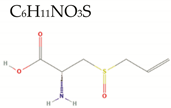
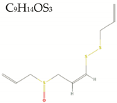
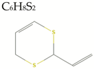
Garlic has various phytoconstituents, including alkaloids, saponins, flavonoids, tannins, phenolics, terpenoids, and organosulfides [97]. In addition, garlic is considered a good source of vitamins and minerals, including vitamin B1, B6, C, manganese, copper, phosphorus, selenium, and calcium [98]. Garlic’s main phytochemicals are organosulfides (sulfur-containing compounds), including allicin, alliin, ajoenes (E-ajoene and Z-ajoene), sulfides (diallyl sulfide, diallyl disulfide, diallyl trisulfide), 2-Vinyl-4H-1,3-dithiin, and allyl methyl sulfide [99]. These organosulfides are produced in garlic cloves [97]. Allicin is the primary bioactive phytochemicals present in the aqueous extract of garlic and is also responsible for the characteristic odor of garlic [94]. Thus, enzyme alliinase converts allicin to alliin when the garlic cloves are sliced/crushed [100,101]. For this reason, several studies have shown that crushed fresh garlic can deliver most of its active phytochemical [99,102,103]. As allicin is chemically unstable, it rearranges into the stable phytochemical ajoene (E- and Z-) [104]. Allyl sulfides are most often found in garlic oil, and vinyl-4H-1,3-dithiin is mainly found in stir-fried garlic and garlic oil [105,106].

2.2.3. Biomedical effects

Anticancer

Interestingly, phytochemicals such as garlic-derived allicin have been combined with commonly used anticancer drugs to enhance the therapeutic effect of current treatments. For example, an experiment performed by Țigu et al. showed that a combination of the anticancer drug, 5-fluorouracil with allicin, hindered colorectal (DLD-1) and lung cancer (SK-MES-1) cell migration and proliferation in vitro [107]. Petrovic et al. studied the effectiveness of intraperitoneal injections of ethanolic homemade garlic extract against an aggressive breast cancer tumor in BalB/c mice. The results showed that, after 28 days of treatment, cancer growth was delayed by 30% compared with untreated mice [108]. In another study, Tanaka et. al, led a randomized double-blinded study on 51 patients with colorectal adenomas that utilized high-aged garlic extract (2.4 mL/day) and low-aged garlic extract (0.16 mL/day) for 12 months. At least one adenoma decreased by 50% (>6 months of uptake) in the high-aged garlic extract group, while there was no decrease in the low-aged garlic extract group [109]. Finally, a recent meta-analysis of epidemiological articles using a total of 11 clinical trials and 12,558 cases concluded that garlic intake could reduce the risk of colorectal cancer [110], coinciding with previous studies [111], while another previous meta-analysis limited to men showed no correlation [112]. These studies show that broader investigations with increased sample size are necessary to clarify the result discrepancies from several epidemiological studies.

Antioxidant

Garlic’s phytochemicals also promote an antioxidant effect. The antioxidant properties of garlic might be associated with two of its main phytochemicals, alliin, and allicin. Bhatt and Patel et al. prepared 900 mg of cooked versus raw garlic and incubated these samples with gastric enzymes. These results showed that cooked garlic lost 90% of phenolic content, leading to less antioxidant activity due to heat (evaporation of active compound) than raw garlic [113]. Lei et al. demonstrated that the scavenging activity of black fermented garlic ethanolic extract is concentration-dependent. This study also showed that this garlic extract increased the mean longevity of flies (Drosophila melanogaster) compared to controls [114]. In a more translational scenario, a randomized, double-blind clinical trial on seventy women with rheumatoid arthritis evaluated the effects of garlic in pain mitigation. Patients received 1000 mg of garlic for a total of 8 weeks. Results showed that pain after activities decreased in the garlic group compared to the placebo. This effect from garlic was attributed to a decrease in oxidative stress, a common feature in this disease [115]. However, there are mixed results in the literature about the oxidative stress reduction mechanism.

Antimicrobial

  • Antiviral
Several studies have shown the antiviral effect of garlic. Pre-clinical studies elucidated that garlic and its organosulfides phytochemicals have great activity against several human and animal viruses by inhibiting viral RNA polymerase, reverse transcriptase, and downregulation of the extracellular-signal-regulated kinase/mitogen-activated protein kinase signaling pathway [116]. The variety of viruses attacked by garlic are adenovirus [117]; SARS-CoV-1 [118]; dengue [119]; herpes simplex [120] influenza A, B, and H1N1 [121,122]; hepatitis [123]; HIV [124]; and rotavirus [125]. Furthermore, in a very recent study, essential garlic oil was found to be acting on the angiotensin-converting enzyme 2 (ACE2) and largely on the main protease of SARS-CoV-2 (PDB6LU7). This activity is crucial to diminish the impact of the host receptor of SARS-CoV-2, and this study proposes that garlic oil active compounds can be used as a COVID-19 preventive treatment [126].
  • Antibacterial
The antibacterial effect of garlic was analyzed in vitro using fresh garlic juice in agar plates against E. coli, P.mirabilis, K.pneumoniae, S.aereus, and P.aeruginosa. The results showed a dose-dependent inhibition in all bacterial strains exposed to a garlic concentration higher than 10% [127]. In another study, two different aqueous garlic extracts (from Allium sativum and Allium tuberosum) were tested in rats infected with one penicillin-sensitive (ATCC 25923) and one methicillin-resistant (ATCC 33592) S. aureus. The two species of garlic were administered orally at 100 and 400 mg/kg) every 6 hours for 24 hrs. Results showed that both garlic extracts could reduce the infection but not against the resistant strain [128]. Several in vitro studies demonstrated the antibacterial effect of fresh garlic extract on E. coli, Klebsiella pneumoniae, Proteus mirabilis, P. aeruginosa, and S. aureus [127], and also against multidrug-resistant E. coli, P. aeruginosa, K. pneumoniae, Serratia marcescens, and methicillin-resistant S. aureus [129]. In a clinical trial that involved 15 patients with Helicobacter pylori, the results showed that the urease breath test to detect H. pylori was lower in patients who took 3 g of garlic cloves twice a day, demonstrating its antimicrobial effect [130].
  • Antifungal
Various studies have discussed the antifungal effect of garlic. Li et al. showed that garlic oil had an inhibitory effect against Candida albicans at a concentration of 0.35 μg/mL [131]. Aala et al. performed an experiment that evaluated the structural characteristic of Trichophyton rubrum in response to garlic and allicin aqueous extracts. The results showed that the allicin extract was more effective in impeding the growth of fungal cells by changing fungi morphology [132]. Another in vitro study indicated that 0.125 and 0.0313 % of garlic oil had a strong antifungal activity by penetrating hyphae cells and destroying their organelles against Penicillium funiculosum [133].

Anti-Inflammatory

The anti-inflammatory effect of garlic was studied by several research groups. Overall, the studies agreed on the antioxidant and anti-inflammatory properties of garlic. However, the results for the mechanisms activated/inhibited by the phytonutrients themselves and their phytochemicals are diverse. We understand that this could be possible due to different garlic preparations and also by the “double-edged sword” of nitric oxide. For example, in an in vitro study, Lee and coworkers showed garlic’s anti-inflammatory activity at μM concentrations. They demonstrated that garlic’s organosulfides Z- and E- ajoene and analogs inhibited nitric oxide/prostaglandins and nitric oxide synthase/cyclooxygenase, the phosphorylation of p38 mitogen-activated protein kinases, and, also, the expression of the following pro-inflammatory cytokines: tumor necrosis factor-α, interleukin-1β, and −6 in a lipopolysaccharide-induced macrophage cell line [134]. In a double-blind clinical trial study, anti-inflammatory effects in 40 peritoneal dialysis patients were investigated by administering a garlic extract twice daily for 8 weeks. The results demonstrated that garlic diminished inflammatory markers in end-stage renal disease patients, specifically interleukin-6, C-reactive protein, and erythrocyte sedimentation rate in the treated group [135]. On the other hand, a previous in vivo study concluded that garlic inhibits platelet aggregation by activating nitric oxide (NO) synthase and the production of NO [136].

Immunomodulatory

As previously described here, garlic induces multiple different functions, including antioxidant, anti-microbial, and anti-inflammatory properties leading to an improvement in the immune response. The immune response induced by the garlic phytochemical allicin was studied in female BALB/c mice. Results showed that allicin treatment reduced parasitaemias and enhanced pro-inflammatory mediators during malaria infection in a dose-dependent manner [137]. In addition, Bruck et al. studied the immune response of allicin in induced liver damage BALB/c male mouse. Results showed that allicin-treated mice showed decreased levels of the pro-inflammatory tumor necrosis factor-α, aminotransferases, and improved hepatic necroinflammation [138]. A randomized, double-blind clinical trial studied the immune and inflammatory effects of 3.6-g aged garlic extract administered daily in 51 obese adults for 6 weeks. Results showed that patients who took the extract supplementation had less pro-inflammatory cytokines, such as interleukin-6 and tumor necrosis factor-α [139]. In a separate study, the immune effect of aged garlic extract supplementation was analyzed in a randomized, double-blind trial with 120 healthy participant adults to examine the proliferation of immune cells and the severity of symptoms during cold and flu season. Results showed that the garlic extract induced increased levels of NK cells and γ/δ-T cells, and reduced the severity of symptoms, days, and incidence [140]. This immune response of garlic is due to the scavenging of oxidizing agents, thereby preventing the formation of pro-inflammatory messengers, such as COX and LOX. In addition, one of the main mechanisms observed is through immunomodulation of inflammatory cytokines and direct stimulation of immune cells [141].

2.2.4. Contraindications

There is limited data about the safety of garlic supplements [142]. Hoshino et al. administered 40 mg of different garlic preparations to adult dogs, and his results showed significant damage caused to gastric mucosa by raw garlic powder and gastric redness caused by boiled garlic powder. Interestingly, no adverse effect was caused by the ingestion of raw garlic extract [143]. In 2014, the first case of pneumonia caused by fermented black garlic was discovered in a 77-year-old female patient who came into the hospital with shortness of breath and cough after taking black garlic. The patient showed health improvement when she stopped taking black garlic [144]. In addition, the first case of drug-induced liver injury by the mild periportal cholestatic reaction was reported in a 43-year-old patient who suffered from hepatopulmonary syndrome following a liver transplant by taking a high dose of Allium sativum as treatment. The patient’s liver enzymes returned to normal after discontinuation of the treatment [142]. According to the National Institutes of Health, garlic supplements may increase the risk of bleeding. As we mentioned before, garlic displays strong antioxidant properties [115] that could lead to the inhibition of platelet adhesion and aggregation [145]. Through these mechanisms, garlic intake might increase the risk of bleeding when combined with other anticoagulants. However, this property would help patients with cardiovascular diseases by the strong garlic antihypertensive action.

2.3. Cinnamon

2.3.1. Botanical Description

Cinnamon, appreciated for centuries for its peculiar flavor and aroma, is the dried inner bark of Cinnamomum verum (syn. C. zeylanicum Blume), an evergreen tree native of Sri Lanka and India. This C. verum is also commonly called “true” cinnamon or Ceylon cinnamon. The Cinnamomum genus, which the cinnamons are part of, belongs to the laurel family (Lauraceae), and it includes about 250 evergreen aromatic trees and shrubs [146]. Most of the spice sold as cinnamon in the United States, however, comes from another cinnamon species, Cinnamonum cassia, also called Chinese cinnamon, because of its geographical origin in the mountains of China [147]. The botanical features of C. verum are summarized as trees (up to 50 ft) with long lance-shaped leaves, small yellow flowers organized in a cluster, and ovoid-shaped fruits. The botanical features of C. cassia are summarized as trees (up to 65 ft) with thin lance-shaped leaves, white flowers, axial inflorescences, and globose drupe fruits [148].

2.3.2. Phytochemicals

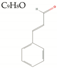
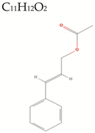
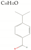
Qualitative phytochemical screening of a methanolic extract from the bark of C. verum showed the presence of all four categories of secondary metabolites. It has also been shown that the phytoprofiles of the cinnamon extracts depend on the botanical part of the tree used for extraction. At the same time, essential oils from the C. verum bark mainly contain cinnamaldehyde and linalool, the flower and fruit extracts are enriched in (E)-cinnamyl acetate, and eugenol is the main compound of leaf extracts [149,150]. The bark of the cinnamon tree has also been reported to contain coumarin, a benzenoid lactone. C. cassia that is particularly rich in coumarin (3462.0 mg/kg in C. cassia vs. 12.3 to 143.0 mg/kg for C. verum) [151]. The solvent and temperature should also be carefully selected according to the molecule one wishes to extract; for example, water is a better solvent for extracting the phenols from C. verum than polar organic solvents at 200 °C [152]. For Klejdus et al., however, the factor for efficient extraction mainly depends on the state of the destruction of the cinnamon cell structures during the extraction protocol [153].

2.3.3. Biomedical effects

Anticancer

In vitro and in vivo studies by Yang et al. showed that the essential oil of cinnamon extracted from the bark of C. cassia significantly inhibits the growth of head and neck cancer cells and tumors in mice. The antitumor activity was believed to be mediated by the trans-cinnamaldehyde acting as a competitive inhibitor of the epidermal growth factor receptor (EGFR). This kinase is often mutated and overexpressed in many tumors and regulates key cancer metabolic pathways, such as proliferation, apoptosis, angiogenesis, and tumor invasiveness [154]. Similarly, Koppikar et al. reported that aqueous bark extract from C. cassia inhibits the growth of cervical carcinoma cells in a dose-dependent manner (IC50 = 80 μg/mL) by apoptosis and loss of mitochondrial membrane potential. The treated cells exhibited reduced migration potential by the downregulation matrix metallopeptidase 2 (MMP-2) and the EGFR [155]. Furthermore, Perng et al. demonstrated that C. verum component 2-methoxy-cinnamaldehyde had an antiproliferative effect on human hepatic adenocarcinoma both in vitro (IC50 = 25.72 μM for 48 h) and in vivo (10–20 mg/kg/d administration of 2-methoxy-cinnamaldehyde). The targeted metabolisms determined by this group were similar to the previous studies (i.e., mitochondrial apoptotic pathway) due to the activation of caspase-3 and -9, a sub-G1 phase cell-cycle arrest, and the downregulation of nuclear factor- Κβ (NF-Kβ) [156].

Antioxidant

A study on the peripheral blood mononuclear cells of rheumatoid arthritis patients showed that cinnamaldehyde and eugenol significantly reduced the levels of pro-inflammatory cytokines tumor necrosis factor-α (TNF-α) and interleukin-6. Additionally, these patients showed enhanced activity of superoxide dismutase, glutathione peroxidase, and catalase enzymes, suggesting an antioxidant effect [157]. In the same way, Davaatseren et al. demonstrated that trans-cinnamaldehyde diminishes the production of nitric oxide and reactive oxygen species in macrophages [158]. Furthermore, cinnamon capsules were orally administered for 12 weeks in a small controlled clinical trial to women with polycystic ovary syndrome. This study demonstrated that cinnamon improved the antioxidant status and lipid profile of these patients by decreasing serum levels of malondialdehyde (derived from lipid peroxidation), total cholesterol, triacylglycerol, and increasing high-density lipoproteins [159].

Antimicrobial

  • Antiviral
In vitro studies concluded that essential oil extracts from the leaves of C. verum extract had an antiviral effect in cells infected with influenza type A (H1N1) [160]. Similarly, a study by Moshaverinia and colleagues suggests that a hydroalcoholic extract of C. verum at 1 mg/mL significantly reduces the viral titer of the human herpes simplex virus type 1 -infected cells [161]. Furthermore, in silico studies by Kulkarni et al. suggest that cinnamaldehyde possesses a strong affinity to the S1 receptor binding domain of the spike (S) glycoprotein of the severe acute respiratory syndrome coronavirus 2 (SARS-CoV-2). Cinnamaldehyde could therefore be an efficient pharmacological agent to inhibit the entry of the virus into the host cells [162].
  • Antibacterial
Ahmed et al. showed that aqueous, methanolic, and acetone extracts from C. verum bark exerted significant antibacterial effects on S. aureus, P. aeruginosa, and E. coli. The inhibitory effect of the extracts was believed to be mediated by cinnamaldehyde [163]. Furthermore, in an in vivo study conducted on aquatic pathogens in zebrafish, Faikoh et al. concluded significant antimicrobial effects of liposome-encapsulated cinnamaldehyde in fish infected by A. hydrophilia, V. vulnificus, S. agalactiae, V. parahaemolyticus, and V. alginolyticus. The antimicrobial activity of cinnamaldehyde was associated with a decrease in the expression of pro-inflammatory interleukins, i.e., -1β, -6, -15, and an increase in anti-inflammatory interleukin-10 [164].
  • Antifungal
In a 2019 study, Kowalska et al. demonstrated the antifungal properties of 1% (v/w) aqueous C. verum bark after a 6-day treatment against Botrytis cinerea, the mycelium responsible for the grey mold disease in tomato plants [165]. Furthermore, cinnamon seems to inhibit the growth of the microorganisms of the Candida family, which are responsible for most of the fungal diseases in humans. In a clinical trial study, Wang et al. showed that an oil extract from C. verum significantly inhibited the growth of three species of C. albicans (minimum inhibitory concentration (MIC) = 0.064 mg/mL), C. tropicalis (MIC = 0.129 mg/mL), and C. krusei (MIC = 0.129 mg/mL) [166]. Additionally, a study conducted on guinea pigs suggests that topical treatments with methanolic extracts of C. verum inhibit the growth of M. canis and T. mentagrophytes, two fungi involved in skin infections in animals and humans [167].

Anti-Inflammatory

A study conducted in an in vitro human skin model for chronic inflammation and fibrosis suggests that a cinnamon concentration of 0.0012% (v:v) significantly inhibits the expression of genes involved in the inflammation and immune DNA damage responses [168]. The authors attributed the effect to cinnamaldehyde and cinnamyl acetate, the two main chemical compounds present in the extract. Likewise, Gunawardena et al. have demonstrated that C. verum and C. cassia extracts inhibited the release of pro-inflammatory nitric oxide molecule and tumor necrosis factor protein in activated macrophages. From these results, the ethanolic extract from C. verum showed more activity than the aqueous extract (IC50 = 36.4 and 122 μg/mL, respectively). The phytochemicals with more potent anti-inflammatory effects were E-cinnamaldehyde and o-methoxycinnamaldehyde [169]. Furthermore, in an in vivo study, 4.5 mL/kg of the ethanolic cinnamon extract was orally administered to a mouse model for colitis. The treated mice exhibited significantly enhanced resorption of their colon fibrotic tissues and reduction in the fibrotic score associated with a decrease in the expression of extracellular matrix proteinases [170].

Immunomodulatory

As previously described, cinnamon has antioxidant, antimicrobial, and anti-inflammatory properties leading to improved immune response. Several studies have concluded that the phytochemicals present in cinnamon extracts inhibit the immune response associated with allergies. Mast cells, key effectors in allergic diseases, are considered promising therapeutic targets. Hagenlocher et al. have shown that cinnamon extracts decrease the release and expression of pro-inflammatory mast cell mediators such as β-hexosaminidase; cytokines CXCL8; and chemokine ligand 2, 3, and 4. From this study, the anti-allergic properties are believed to be mediated by cinnamaldehyde [171]. Similar results have been found in human and murine models for allergic inflammation. Cinnamon extracts significantly inhibited the allergen-specific T-cell proliferation as well as TH1 and TH2 cytokine production [172]. Recent studies have also shown the possibility of cinnamon application in COVID-19 symptoms reduction and as preventive treatment through immune system strengthening [173]. However, further research should focus on the safety and route of cinnamon administration to maximize the therapeutic effects.

2.3.4. Contraindications

While cinnamon possesses a large spectrum of medicinal properties, its regular consumption can also lead to adverse health effects. Due to its cellulose fiber composition, which does not dissolve or biodegrade in the lungs, cinnamon inhalation or its dry consumption can trigger a hypersensitive airway and irritate mucous membranes in the lungs [174]. Due to the apoptotic effect of the cinnamon component cinnamaldehyde on B- and T-cells, the consumption of cinnamon is contraindicated in patients under an immunotherapy treatment [175]. The consumption of cinnamon supplements should be avoided during pregnancy since cinnamon can lead to uterine contractions, miscarriage, or premature labor [176]. Importantly, studies conducted both in vitro and in vivo suggest that the toxic compound coumarin, found abundantly in C. cassia, and less in ceylon cinnamon (~250 times less), is a potential carcinogen to individuals with mutations of the cytochrome P450 2A6 [177].

2.4. Graviola

2.4.1. Botanical Description

A member of the Annonaceae (Custard-apple family), Annona muricata, commonly known as soursop, graviola, paw-paw, or “guanabana”, is a tree native to Central America and West Indies that is abundant at altitudes lower than 900 m above sea level. It is cultivated in tropical and subtropical climates in countries such as Angola, Brazil, Colombia, Costa Rica, Puerto Rico, India, and Venezuela [178]. The graviola tree is mainly appreciated for its edible fruit. Still, its parts (leaves, fruit, bark, root, etc.) have been commonly used in traditional pharmacopeia in the form of macerations, decoction, or as a topical medication [179,180]. While the graviola tree can grow in a large variety of soils, it prefers deep soils with good oxygenation [178]. Botanically speaking, its leaves are large and obovate to elliptically shaped, are green on top, and paler under the top with short petioles and a pungent smell. The tree produces yellow-greenish flowers and lags about two years in producing heart-shaped fruits. It usually bears fruits yearly from that point on (12–24 per year) and can produce up to 50 fruits from its fifth year [181].

2.4.2. Phytochemicals

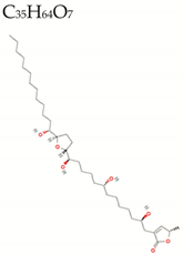
More than two hundred (>200) bioactive compounds have been isolated from the leaves, seeds, root, bark, fruit, and fruit peel of the graviola tree [180]. Most frequently identified are alkaloids, phenolics, and terpenoids [182,183]. Acetogenins are considered the main bioactive compound in the Annonaceae family, with over 120 acetogenins identified from the root, leaves, stems, fruit pulp, and the seed of the family members [184,185]. Acetogenins are a particular class of secondary metabolites that could be considered part of the phenolics integrating polyketides and polyethers found exclusively in the plants of the Annonaceae family [186]. The structure of acetogenins is composed of a long carbon chain (35–38 carbons) as a fatty acid derivative. Graviola leaves contain key medically relevant polyphenolics compounds, including quercetin, rutin, and gallic acid [187,188,189]. The leaves of graviola also contain close to eighty (80) essential oils, including bioactive sesquiterpenes, and compounds such as potassium; calcium; zinc, phosphorus; magnesium; carbohydrates; vitamin A, B, and C; phytosterol; and calcium oxalate [190,191].

2.4.3. Biomedical Effects

Anticancer

Graviola anticancer activity has been extensively studied, and the cytotoxicity of graviola has been reported for several cancer types e.g., breast, colorectal, skin, head and neck, lung, liver, pancreatic, prostate cancer, and leukemia [178,192,193,194]. Most of the antiproliferative properties of the extracts are suggested to be mediated by the graviola acetogenins. The acetogenins exert an inhibitory activity on the NADPH mitochondrial complex 1, a component of the energy transport chain, which is crucial to the synthesis of high quantities of ATP in cancer cells [193,195,196]. Acetogenins have also been shown to target several critical cancer metabolic pathways by inhibiting the Na+/K+ ATPase pump and the hypoxic and glycolytic pathways, inducing apoptosis and cell cycle arrest [196,197,198].

Antioxidant

Studies conducted in vitro and in vivo suggest that graviola contains antioxidant compounds that act as free-radical scavengers and increase the activity of the antioxidant enzymes superoxide dismutase and catalase and downregulate the function of mitochondrial NADPH oxidase complex I [199,200,201]. The leaf and the fruit pulp of graviola are the parts of the tree with the highest antioxidant properties [182]. The antioxidant activity of graviola is believed to be mediated by the following phenolic phytochemicals: quercetin, gallic acid, and graviola leaf polysaccharides [202,203].

Antimicrobial

  • Antiviral
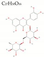
It has been suggested that the phytochemicals polyphenolics in graviola exert some antiviral activity against RNA and DNA viruses [189,204]. A study by Wahab et al. showed that pretreating monkey kidney epithelial cells with a graviola leaf extract 24 h prior to infecting them with the dengue virus serotype 2 inhibited the virus replication. The treatment also increased the survival of the dengue-infected cells [205]. A recent clinical study conducted by Le Donne et al. investigated the antiviral properties of graviola on human papillomavirus (HPV)-infected patients who were supplemented with ellagic acid and graviola extract twice a day for six months. Results showed a 74% HPV clearance in treated patients compared to the 25% clearance for the placebo group [206]. Furthermore, recent in silico studies suggest that rutin, a phytonutrient abundant in graviola, could act as strong ligands and inhibit the function of proteins of the SARS-CoV and SARS-CoV-2 virus, thus suggesting potential therapeutic benefits against the COVID-19 infection [207,208].
  • Antibacterial
Graviola leaf extracts have been shown to exert in vitro antibacterial activity against oral pathogenic strains such as S. mutants, S. mitis, P. gingivalis, P. intermedia, P. intermedia, and C. albicans [209,210]. An in vivo study conducted in albino rats demonstrated the efficiency of graviola unripe fruit extracts to inhibit the growth of S. typhi [211]. Furthermore, aqueous leaf extract and fruit-skin ethanolic graviola extracts showed a strong antibacterial effect against K. pneumoniae, S. aureus, and P. aeruginosa bacteria, i.e., the pathogens responsible for respiratory infections in the human immunodeficiency virus (HIV/AIDS) patients [212].
  • Antifungal
We did not find studies testing the antifungal activity of any graviola extract. However, we found studies evaluating this property in some of graviola’s phytochemicals. In 2017, a research group found that gallic acid has in vitro antifungal activity against dermatophyte strains (between 43.75 and 83.33 μg/mL), and Candida strains (C. albicans MIC = 12.5 μg/mL, and Trichophyton rubrum MIC = 43.75 μg/mL) by inhibiting the ergosterol synthesis. They also confirmed this activity after in vivo studies administrating 80 mg/kg d of gallic acid [213]. In another study, researchers found that quercetin induces apoptosis in Candida albicans through mitochondrial dysfunction by increasing intracellular magnesium [214].

Anti-Inflammatory

The anti-inflammatory properties of graviola have been extensively studied in vitro and in vivo [215]. Cercato et al. reported that a topical application of a graviola leaf extract (0.3, 1, or, 3 mg/ear) significantly reduced ear edema and myeloperoxidase activity in Swiss mice with 12-O-tetradecanoylphorbol-13-acetate (TPA)-induced ear inflammation. The authors were also able to show that the anti-inflammatory effect of the extract was associated with a reduction in the total amount of hydroperoxides and with modulation of catalase antioxidant activity [216]. While studying the anti-inflammatory response in lipopolysaccharide (LPS)-stimulated murine macrophage cell line RAW264.7 treated with graviola ethanolic leaf extracts, Laksmitawati et al. reported a downregulation in pro-inflammatory protein markers, such as tumor necrosis factor-alpha (TNF-α), interleukin-1β, interleukin-6, in the treated macrophages cells compared to untreated controls [217]. Furthermore, graviola aqueous extract suppresses nitric oxide production [218]. Similarly, an in vivo study conducted in rodents by Ishola et al. showed that the administration of a lyophilized graviola fruit extract inhibits the activity of the pro-inflammatory biomarkers cyclooxygenase (COX)-1 and COX-2 in a dose-dependent manner [219].

Immunomodulatory

As previously described, the specific bioactive constituents responsible for the major antioxidant, anti-inflammatory, and antimicrobial properties of graviola include different classes of annonaceous acetogenins (metabolites and products of the polyketide pathway), alkaloids, flavonoids, and sterols. Several studies have reported that graviola possesses immunomodulatory properties. For example, a study conducted in rodents by Umayra et al. shows that administration of an ethanolic graviola leaf extract boosts the immunological response through the activation of phagocytic cells [220]. Furthermore, an immune-enhancing activity of graviola leaf extracts has been observed in RAW 264.7 macrophage cells in vitro, a phenomenon which is believed to be mediated by the activation of the mitogen-activated protein kinase (MAPK) pathways [221].

2.4.4. Contraindications

Studies from patients of the French West Indies, whose diet is rich in graviola, showed the development of a type of Parkinsonism resistant to the common anti-parkinsonism drug to treat tremors, levodopa [192]. This observation must lead scientists to further study if the acetogenins and alkaloids present in the graviola fruit could be toxic to brain cells, specifically dopaminergic neurons, which are the main cells affected in Parkinson’s disease. In general, patients with neurological ailments should avoid the consumption of graviola supplements. Given that graviola is already widely used in traditional medicine, it could potentially be used against many health conditions if properly tested in further clinical studies. In conclusion, the benefits and side-effects of graviola should be carefully evaluated on a case-by-case basis.

2.5. Oregano

2.5.1. Botanical Description

The term oregano refers to a group of several plant genera, including Thymbra, Thymus, Coridothymus, Satureja, and Origanum, containing a high amount of the phytochemical carvacrol in their essential oils. The genus Origanum consists of 43 species. Origanum vulgare (O. vulgare), commonly named “oregano”, is the name of the aromatic plant used as a condiment herb in Mediterranean cuisine [222,223,224]. O. vulgare size is usually 20–80 cm; its 1–4 cm leaves are dark green, with 2-mm bell-shaped calyx purple flowers arranged in erect spikes [225,226,227]. Like other aromatic plants, the oregano plant produces essential oils as secondary metabolites in response to various infectious agents, UV light, and even oxidative stress. Oregano essential oils (OEOs) are usually extracted from the plant leaves and flowering tops. OEOs are famous for their medicinal value and are traditionally used in Turkey to cure diseases such as cough, chronic cold, wounds, gastrointestinal disorders, and skin problems in humans and domestic animals [228].

2.5.2. Phytochemicals

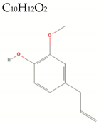
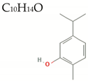
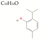
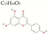
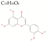
The main bioactive compounds present in the OEOs are the aromatic oxygenated monoterpene thymol (5-methyl-2-(1-methylethyl) phenol) and its constitutive isomer carvacrol (5-isopropyl-2-methylphenol, 2-p-cymenol). The ratio of thymol/carvacrol varies according to the oregano plant’s geographical location [229]. Both compounds are lipophilic, volatile, highly soluble in ethanol, and possess low densities [228,230,231,232]. Other bioactive oregano phytochemicals include o-cymene (2-Isopropyltoluene), apigenin (4′,5,7-trihydroxyflavone), and luteolin (7,3′,4′,5-tetrahydroxyflavone) [233,234]. Due to their low general toxicities, the two main chemicals of O. vulgare, thymol and carvacrol, have been approved as food additives by the Food and Drug Administration (FDA) [235].

2.5.3. Biomedical Effects

Anticancer

The antiproliferative/anticancer properties of oregano have been documented in vitro and animal models for cancers. A recent study by Spyridopoulou et al. showed that OEO exerts dose-dependent cytotoxicity against breast cancer (MCF-7), colon cancer cells (HT-29), melanoma (A375), and hepatocellular carcinoma (HepG2) cells, with respective IC50 values of 0.35, 0.35, 8.90, and 10.0 mg/mL. The authors also showed that the treatment of HT-29 cells with 50 mg/mL of OEO correlated with an attenuated migration and an induced apoptosis-related morphological change in HT-29 cells. Furthermore, the oral administration of OEO for 13 days (0.370 g/kg b.w/day) proved to inhibit the growth of CT26 colon tumors in vivo in BALB/c mice [236]. Another study by Coccimiglio reports that an ethanolic leaf extract of O. vulgare promotes the death of A549 human lung carcinoma in a dose-dependent manner (IC50 = 14.0 μg/mL) [237]. The antiproliferative properties of oregano are believed to be mediated by thymol and carvacrol, which possess antioxidant characteristics while being non-mutagenic to cells [237,238,239]. The anticancer properties of thymol were evidenced in in vitro and in vivo models for colorectal cancers [240,241]. One astonishing property of carvacrol is its potential to specifically target cancer cells while being less toxic to normal cells [242]. Furthermore, carvacrol seems to exert a modulatory effect on the toxicity of cisplatin in vitro, a property that could be exploited for reducing the side-effects associated with classical cisplatin-based antitumor treatments [239].

Antioxidant

An in vitro study by Gavaric et al. showed that OEO possessed a robust antioxidant activity (IC50 = 0.2 µg/mL). While thymol and carvacrol were the components accounting for the antioxidant properties of oregano, the antioxidant activities of the two compounds were much inferior to the one observed for the whole extract with (IC50 = 70–80 mg/mL for thymol and carvacrol). The authors concluded that thymol, carvacrol, and other extract phytocompounds acted in synergy to promote the scavenging of free radicals [243]. According to a study conducted on the human colon carcinoma intestinal Caco-2 cell line, thymol, carvacrol, and their mixture seem to exhibit double-edged anti- or pro-oxidant effects, depending on the concentration at which they are administered (pro-oxidants at sub-cytotoxic concentrations vs. antioxidants at higher concentrations) [244].

Antimicrobial

  • Antiviral
An in vitro study conducted on simian Vero cell line CCL-81 showed that thymol, carvacrol, and p-cymene (all major components of oregano oils) possess antiviral properties against the human herpes simplex virus type 1 with respective IC50 values of 0.002%, 0.037%, and >0.1%. The antiviral properties of the three compounds are believed to be correlated to their ability to interfere with the viral membrane fusion mechanism during the adsorption phase of the virus [245]. Furthermore, an in vitro study by Sánchez and Aznar have reported a dose-dependent titer inhibition of the feline calicivirus and the murine norovirus by thymol, in the 1–2% (v:v) range concentrations [246].
  • Antibacterial
Thymol and carvacrol have been shown to exert antibacterial activities against Gram-positive and Gram-negative bacteria [247]. In studies using thymol concentrations ranging from 26.5–52.9 mg/cm2 showed potent inhibitory activity against the S. aureus, B. subtilis, E. coli, and Salmonella enteritidis [248]. Studies performed by Du et al. showed the following results: strong antibacterial activity of the OEOs, thymol, and carvacrol against E. coli, C. perfringens, and Salmonella strains. They also performed in vivo studies in 448 male broiler chicks by oral gavage using OEO. They found that OEO alleviated intestinal lesions and decreased E. coli populations [249]. In another study, oregano oil showed great antibacterial activity against the following multidrug-resistant bacteria: three Acinetobacter baumannii, three Pseudomonas aeruginosa, and four methicillin-resistant Staphylococcus aureus with inhibitory concentrations ranging from 0.08–0.64 mg/mL [250]. Another in vitro study showed that the use of OEO and carvacrol could curve Group A streptococci erythromycin-resistant bacterial infections [251].
  • Antifungal
The in vitro antifungal properties of OEO, thymol, and carvacrol in the 40–350 mg/mL ranges have been reported in several studies against plant pathogenic fungi Colletotrichum acutatum and Botryodiplodia theobromae [252]; Penicillium digitatum and Penicillium italicum [253]; food-relevant fungi Cladosporium spp. and Aspergillus spp. [254]; longan pathogens, Lasiodiplodia spp., Phomopsis spp., Pestalotiopsis spp. and Geotrichum candidum [255]; and against Fusarium verticillioides and Rhizopus stolonifera [256]. Furthermore, an in vivo study conducted in Caenorhabditis elegans suggests that thymol possesses antifungal activity against Candida albicans, the most prevalent cause of fungal infections in humans [257].

Anti-Inflammatory

OEOs possess a strong anti-inflammatory activity, a property that is proposed to be mediated by its main active compounds: thymol and carvacrol. The impact of the OEOs on 14 protein biomarkers was closely related to the inflammatory response. The results show dose-dependent inhibition of the expression of all the proinflammatory and remodeling biomarkers studied: monocyte chemoattractant protein 1 (MCP-1), vascular cell adhesion molecule 1 (VCAM-1), intracellular cell adhesion molecule 1 (ICAM-1), interferon gamma-induced protein 10 (IP-10), interferon-inducible T-cell alpha chemoattractant (I-TAC), monokine induced by gamma interferon, collagen I, collagen III, epidermal growth factor receptor (EGFR), matrix metalloproteinase 1 (MMP-1), plasminogen activator inhibitor 1 (PAI-1), tissue inhibitor of metalloproteinase (TIMP) 1 and 2, and macrophage colony-stimulating factor (M-CSF) [258]. The anti-inflammatory activity of thymol was also reported in vivo in BALB/c mice affected by LPS-induced endometritis [259].

Immunomodulatory

Recent investigations cited in previous sections have demonstrated that oregano has potent antioxidant, antimicrobial, and anti-inflammatory properties, leading to an improved immune response. Oregano’s immunomodulatory activity can be attributed to thymol by its ability to modify the secretion of cytokines, probably through the regulation of NF-κB, but also through the MAPK signaling pathway, or through their ability to affect the cellular expression of iNOS and the secretion of prostaglandins [260]. De Santis et al. studied the immunomodulatory effects of several 50% (v/v) hydroalcoholic O. vulgare extracts on human-derived dendritic cells type-1 and type-2 macrophages infected with M. bovis Bacille Calmette–Guérin. The authors showed that the hydroalcoholic extract stimulated the anti-mycobacterial innate immunity and limited the inflammatory response in all the tested cell types [261]. On the contrary, Gholijani et al. showed that intraperitoneal injections of 80 mg/kg of thymol or carvacrol in BALB/c mice trigger an immunosuppressive response, a property that could be exploited for treating autoimmune diseases [262].

2.5.4. Contraindications

As detailed in this review, O. vulgare offers a wide range of medicinal benefits. In addition, Schönknecht et al. concluded that including primrose and thymol in combination with conventional therapy could alleviate cough and dyspnea in upper respiratory tract infections [263]. However, in a study of several decades ago, thymol and carvacrol have been shown to induce dose-dependent structural chromosomal aberrations in Rattus norvegicus, when consumed at doses over 40 mg/kg, despite being non-toxic at low to moderate doses [264]. Although all the studies mentioned here cited oregano, more robust studies are needed to have a profound evaluation of its efficacy.

3. Discussion

Phytochemicals are vital cofactors with powerful effects on the body, helping it regain functionality. As shown in this review, even though phytochemicals may have different mechanisms of action and different levels of effectiveness in the body, there are overlapping aspects such as antioxidant, anti-inflammatory, and corrective metabolic effects that produce various positive physiological impacts favoring the healthy state. The physiologic modulation induced by these phytonutrients and their phytochemicals produces functional changes that support repair mechanisms necessary to achieve the homeostasis or balance known as health.
The physicochemical properties calculated for the main phytochemicals in the phytonutrients studied in this review are based on the combination of Lipinski’s, Ghose’s, and Veber’s rules (L-Ro5, GF, VR), described as an approximation for the pharmacokinetics of a molecule in the body [30]. Thus, a molecule whose structure falls out of the range of these rules is predicted to have poor absorption or permeation through the gastrointestinal system and low systemic bioavailability.
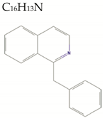
From the evaluation of 25 phytochemicals through the mentioned parameters (Table 1), 23 of them fulfill the requirements of L-Ro5 (HBD ≤ 5, HBA ≤ 10, MW ≤ 500, logP ≤ 5) and VR (RB ≤ 10, PSA ≤ 140), while 2 (annonacin/acetogenin and rutin from graviola) violated more than one parameter. Per GF, the compounds should meet the following: MW (160–480), logP (−0.4–5.6), A (40–130), and TNA (20–70). Accordingly, 13 phytochemicals (curcumin, demethoxycurcumin, bisdemethoxycurcumin, and α-turmerone from turmeric; alliin, allicin, and z-ajoene from garlic; (E)-cinnamyl acetate and eugenol from cinnamon; benzylisoquinoline, coumaric acid, and caffeic acid from graviola; and apigenin and luteolin from oregano) comply with Ghose’s rules.
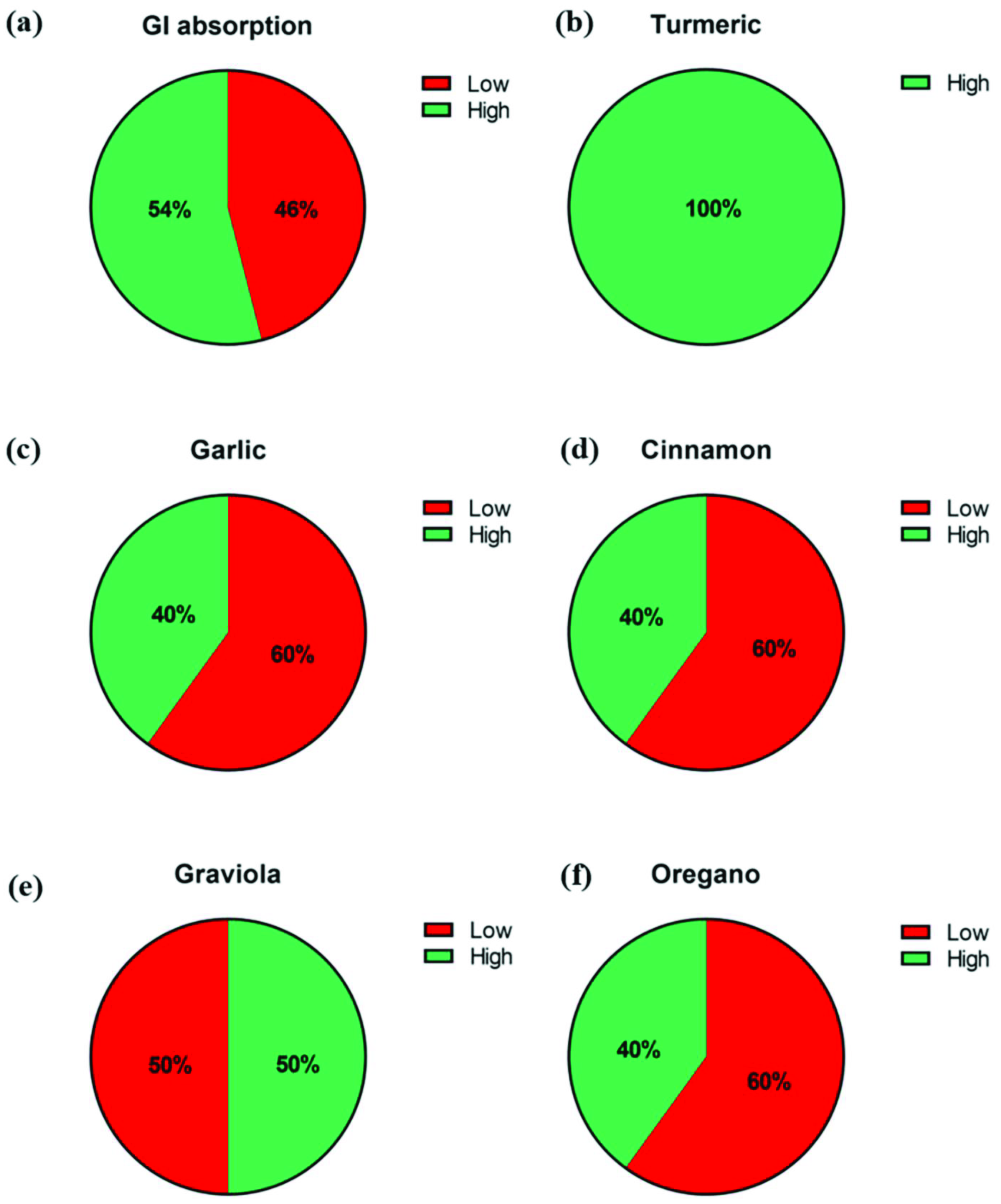
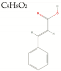
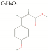
Considering GI tract absorption (Figure 2a), 54% of all phytochemicals studied in this review (curcumin, demethoxycurcumin, bisdemethoxycurcumin, α-turmerone, alliin, z-ajoene, (E)-cinnamyl acetate, eugenol, coumaric acid, caffeic acid, apigenin, and luteolin) met all rules and, thus, have a higher probability of being highly absorbed. Based on L-Ro5, GF, and VR, all described turmeric’s phytochemicals belong to highly absorbed compounds (100%) compared to garlic, cinnamon, oregano (40%) (Figure 2b,c,d,f); and graviola (50%) (Figure 2e).
Graviola’s phytochemicals, annonacin/acetogenin, and rutin violate most of the “drug-likeness” rules. For example, annonacin/acetogenin complies with only 50% L-Ro5 and VR and violates 100% of the GF. For rutin, the compliance for L-Ro5 was 25%, GF was 25%, and VR was 50%. Thus, it is predicted that annonacin/acetogenin and rutin have the lowest probability of absorption in the GI.
Other researchers have proposed that the lipophilicity, considering the ionizable groups at pH 7.4 (LogD), is much more important for physiological absorption or permeation [265]. Thus, compounds that fall below 1 and above 5 for LogD are less likely to be absorbed. Based on this, alliin from garlic; protocatechuic acid from cinnamon; and annonacin/acetogenin, cinnamic acid, coumaric acid, caffeic acid, and rutin from graviola fall out this LogD range.
However, the predictions of these rules are also based on molecules passively transported into the cells. This means that L-Ro5, GF, and VR do not take into consideration actively transported substrates by biological transporters (e.g., cellular receptors or channels) [266]. On the contrary, we understand that many therapeutic compounds are actively transported in the organism, especially plant-based compounds. Due to this, other studies have shown that most of the violators of these rules are natural products [267].

4. Conclusions

All the phytonutrients mentioned in this review article, when used properly, have demonstrated a large variety of health benefits. Yet, a medical evaluation is needed before making any decision on utilizing phytonutrients and phytochemicals regularly or in combination with another pharmacological treatment. Although the biomedical properties of turmeric, garlic, cinnamon, graviola, and oregano in vitro and in vivo have shown mostly positive effects, there is a critical need for well-designed studies in humans to gain a better understanding of their physiological activities and underlying mechanisms in the human body.

Author Contributions

Coordination, Y.D.; conceptualization, Y.D.; formal analysis, Y.D. and I.J.S.-A.; theoretical calculations, Y.D., A.T., D.P., E.V. and J.V.; writing—original draft preparation, Y.D., J.A., C.C.-R., J.C.F., W.M., G.R., J.C.; A.T. and C.B.; writing—review and editing, Y.D., C.C., Y.F.-A., Z.T.-M., R.A.V.-A., J.R.-Z. and M.J.G.; supervision, Y.D. All authors have read and agreed to the published version of the manuscript.

Funding

This study was funded by the Career Development Grant (Y.D., Y.F.-A.) from Sloan Scholars Mentoring Network, Biomedical Fellowships from Fundación Intellectus (Z.T.-M., J.V. and Y.D.), and PR INBRE P20 GM103475-17 (I.J.S.-A.).

Institutional Review Board Statement

Not applicable.

Informed Consent Statement

Not applicable.

Data Availability Statement

Not applicable.

Acknowledgments

The authors thank Estela Estapé (Director of the SJBSM Research Center) for her outstanding dedication and service as a senior advisor in the writing process of this review.

Conflicts of Interest

The authors declare no conflict of interest. The funders had no role in the design of the study; in the collection, analyses, or interpretation of data; in the writing of the manuscript, or in the decision to publish the results.

References

  1. Doosti, F.; Dashti, S.; Tabatabai, S.M.; Hosseinzadeh, H. Traditional Chinese and Indian medicine in the treatment of opioid-dependence: A review. Avicenna J. Phytomed. 2013, 3, 205–215. [Google Scholar] [PubMed]
  2. Pan, S.Y.; Litscher, G.; Gao, S.H.; Zhou, S.F.; Yu, Z.L.; Chen, H.Q.; Zhang, S.F.; Tang, M.K.; Sun, J.N.; Ko, K.M. Historical perspective of traditional indigenous medical practices: The current renaissance and conservation of herbal resources. Evid. Based Complement Altern. Med. 2014, 2014, 525340. [Google Scholar] [CrossRef]
  3. Senti, S.; Habicht, M.E.; Rayo, E.; Eppenberger, P.E.; Rühli, F.J.; Galassi, F.M. Egyptian canopic jars at the crossroad of medi-cine and archaeology: Overview of 100 years of research and future scientific expectations. Pathobiology 2018, 85, 267–275. [Google Scholar] [CrossRef] [Green Version]
  4. Molyneux, R.J.; Lee, S.T.; Gardner, D.R.; Panter, K.E.; James, L.F. Phytochemicals: The good, the bad and the ugly? Phytochemistry 2007, 68, 2973–2985. [Google Scholar] [CrossRef] [PubMed]
  5. Koche, D.; Shirsat, R.; Kawale, M. An overview of major classes of phytochemicals: Their types and role in disease prevention. Hislopia J. 2016, 9, 1–11. [Google Scholar]
  6. Giada, M.D.L.R. Food Phenolic Compounds: Main Classes, Sources and Their Antioxidant Power. Oxidative Stress Chronic Degener. Dis. A Role Antioxid. 2013. [Google Scholar] [CrossRef] [Green Version]
  7. Boeing, H.; Bechthold, A.; Bub, A.; Ellinger, S.; Haller, D.; Kroke, A.; Leschik-Bonnet, E.; Müller, M.J.; Oberritter, H.; Schulze, M.B.; et al. Critical review: Vegetables and fruit in the prevention of chronic diseases. Eur. J. Nutr. 2012, 51, 637–663. [Google Scholar] [CrossRef] [Green Version]
  8. De Lima, R.M.T.; Dos Reis, A.C.; de Menezes, A.P.M.; Santos, J.V.O.; Filho, J.; Ferreira, J.R.O.; de Alencar, M.V.O.B.; da Mata, A.M.O.F.; Khan, I.N.; Islam, A.; et al. Protective and therapeutic potential of ginger (zingiber officinale) extract and [6]-gingerol in cancer: A comprehensive review. Phytother. Res. 2018, 32, 1885–1907. [Google Scholar] [CrossRef]
  9. Kibe, M.N.; Konyole, S.O.; Oloo, M.O.; Ochieng, N.G.; Kathure, D. The role of phytochemicals in prevention and control of chronic diseases. Int. J. Curr. Res. 2017, 9, 62540–62543. [Google Scholar]
  10. Napolitano, G.; Fasciolo, G.; Di Meo, S.; Venditti, P. Vitamin e supplementation and mitochondria in experimental and fun-ctional hyperthyroidism: A mini-review. Nutrients 2019, 11, 2900. [Google Scholar] [CrossRef] [Green Version]
  11. Reddavide, R.; Rotolo, O.; Caruso, M.G.; Stasi, E.; Notarnicola, M.; Miraglia, C.; Nouvenne, A.; Meschi, T.; Angelis, G.L.D.; Di Mario, F.; et al. The role of diet in the prevention and treatment of Inflammatory Bowel Diseases. Acta Biomed. 2018, 89, 60–75. [Google Scholar] [CrossRef]
  12. Salas-Salvadó, J.; Becerra-Tomás, N.; Papandreou, C.; Bulló, M. Dietary Patterns Emphasizing the Consumption of Plant Foods in the Management of Type 2 Diabetes: A Narrative Review. Adv. Nutr. 2019, 10, S320–S331. [Google Scholar] [CrossRef] [Green Version]
  13. Kourouma, V.; Mu, T.-H.; Zhang, M.; Sun, H.-N. Effects of cooking process on carotenoids and antioxidant activity of or ange-fleshed sweet potato. LWT 2019, 104, 134–141. [Google Scholar] [CrossRef]
  14. Palermo, M.; Pellegrini, N.; Fogliano, V. The effect of cooking on the phytochemical content of vegetables. J. Sci. Food Agric. 2014, 94, 1057–1070. [Google Scholar] [CrossRef] [PubMed]
  15. Chen, F.; Wen, Q.; Jiang, J.; Li, H.L.; Tan, Y.F.; Li, Y.H.; Zeng, N.K. Could the gut microbiota reconcile the oral bioavailability conundrum of traditional herbs? J. Ethnopharmacol. 2016, 179, 253–264. [Google Scholar] [CrossRef]
  16. Zimmermann, G.R.; Lehár, J.; Keith, C.T. Multi-target therapeutics: When the whole is greater than the sum of the parts. Drug Discov. Today 2007, 12, 34–42. [Google Scholar] [CrossRef] [PubMed]
  17. Xu, W.; Wen, M.; Yu, J.; Zhang, Q.; Polyakov, N.E.; Dushkin, A.V.; Su, W. Mechanochemical preparation of kaempferol intermolecular complexes for enhancing the solubility and bioavailability. Drug Dev. Ind. Pharm. 2018, 44, 1924–1932. [Google Scholar] [CrossRef]
  18. Mouhid, L.; Corzo-Martínez, M.; Torres, C.; Vázquez, L.; Reglero, G.; Fornari, T.; de Ramírez Molina, A. Improving in vivo efficacy of bioactive molecules: An overview of potentially antitumor phytochemicals and currently available lipid-based de-livery systems. J. Oncol. 2017, 2017, 7351976. [Google Scholar] [CrossRef] [PubMed] [Green Version]
  19. McClements, D.J. Advances in nanoparticle and microparticle delivery systems for increasing the dispersibility, stability, and bioactivity of phytochemicals. Biotechnol. Adv. 2020, 38, 107287. [Google Scholar] [CrossRef] [PubMed]
  20. Subramanian, A.P.; Jaganathan, S.K.; Manikandan, A.; Pandiaraj, K.N.; Gomathi, N.; Supriyanto, E. Recent trends in nano-based drug delivery systems for efficient delivery of phytochemicals in chemotherapy. RSC Adv. 2016, 6, 48294–48314. [Google Scholar] [CrossRef]
  21. G Peixoto, M.P.; Treter, J.; de Resende, P.E.; da Silveira, N.P.; Ortega, G.G.; Lawrence, M.J.; Dreiss, C.A. Wormlike micellar aggregates of saponins from ilex paraguariensis a. St. Hil. (mate): A characterisation by cryo-tem, rheology, light scattering and small-angle neutron scattering. J. Pharm. Sci. 2011, 100, 536–546. [Google Scholar] [CrossRef]
  22. Kregiel, D.; Berlowska, J.; Witonska, I.; Antolak, H.; Proestos, C.; Babic, M.; Babic, L.; Zhang, B. Saponin-based, biological-active surfactants from plants. Appl. Charact. Surfactants 2017, 183, 184–205. [Google Scholar]
  23. Zhao, Q.; Luan, X.; Zheng, M.; Tian, X.-H.; Zhao, J.; Zhang, W.-D.; Ma, B.-L. Synergistic mechanisms of constituents in herbal extracts during intestinal absorption: Focus on natural occurring nanoparticles. Pharmaceutics 2020, 12, 128. [Google Scholar] [CrossRef] [PubMed] [Green Version]
  24. Khan, I.A.; Smillie, T. Implementing a “Quality by Design” Approach to Assure the Safety and Integrity of Botanical Dietary Supplements. J. Nat. Prod. 2012, 75, 1665–1673. [Google Scholar] [CrossRef] [PubMed]
  25. Newman, D.J.; Cragg, G.M. Natural products as sources of new drugs over the nearly four decades from 01/1981 to 09/. J. Nat. Prod. 2020, 83, 770–803. [Google Scholar] [CrossRef] [PubMed]
  26. Available online: https://pubchem.ncbi.nlm.nih.gov (accessed on 31 May 2021).
  27. ChemSpider | Search and share chemistry. 2021. Available online: http://www.chemspider.com/ (accessed on 4 March 2021).
  28. PhysChem, ADME/Tox Calculations | ACD/Labs Percepta Software. 2021. Available online: https://www.acdlabs.com/products/percepta/index.php (accessed on 9 March 2021).
  29. US EPA. EPI Suite™-Estimation Program Interface | US EPA. 2021. Available online: https://www.epa.gov/tsca-screening-tools/epi-suitetm-estimation-program-interface (accessed on 12 March 2021).
  30. Jablonsky, M.; Haz, A.; Burčová, Z.; Kreps, F.; Jablonsky, J. Pharmacokinetic properties of biomass-extracted substances iso-lated by green solvents. Bioresources 2019, 14, 6294–6303. [Google Scholar]
  31. Leong-Škorničková, J.; Šída, O.; Wijesundara, S.; Marhold, K. On the identity of turmeric: The typification of Curcuma longa L. (Zingiberaceae). Bot. J. Linn. Soc. 2008, 157, 37–46. [Google Scholar] [CrossRef]
  32. Chatzinasiou, L.; Booker, A.; MacLennan, E.; Mackonochie, M.; Heinrich, M. Turmeric (Curcuma longa L.) products: What quality differences exist? J. Herb. Med. 2019, 17-18, 100281. [Google Scholar] [CrossRef]
  33. Rao, P.S.; Ramanjaneyulu, Y.S.; Prisk, V.R.; Schurgers, L.J. A Combination of Tamarindus indica seeds and Curcuma longa Rhizome Extracts Improves Knee Joint Function and Alleviates Pain in Non-Arthritic Adults Following Physical Activity. Int. J. Med. Sci. 2019, 16, 845–853. [Google Scholar] [CrossRef] [Green Version]
  34. Hossain, M.A.; Ishimine, Y. Growth, yield and quality of turmeric (curcuma longa l.) cultivated on dark-red soil, gray soil and red soil in okinawa, japan. Plant Prod. Sci. 2005, 8, 482–486. [Google Scholar] [CrossRef]
  35. Dhakal, S.; Schmidt, W.F.; Kim, M.; Tang, X.; Peng, Y.; Chao, K. Detection of additives and chemical contaminants in turmeric powder using FT-IR spectroscopy. Foods 2019, 8, 143. [Google Scholar] [CrossRef] [PubMed] [Green Version]
  36. Amalraj, A.; Pius, A.; Gopi, S.; Gopi, S. Biological activities of curcuminoids, other biomolecules from turmeric and their de-rivatives—A review. J. Tradit. Complement. Med. 2016, 7, 205–233. [Google Scholar] [CrossRef] [Green Version]
  37. Czernicka, L.; Grzegorczyk, A.; Marzec, Z.; Antosiewicz, B.; Malm, A.; Kukula-Koch, W. Antimicrobial Potential of Single Metabolites of Curcuma longa Assessed in the Total Extract by Thin-Layer Chromatography-Based Bioautography and Image Analysis. Int. J. Mol. Sci. 2019, 20, 898. [Google Scholar] [CrossRef] [Green Version]
  38. Irshad, S.; Muazzam, A.; Shahid, Z.; Dalrymple, M.B. Curcuma longa (Turmeric): An auspicious spice for antibacterial, phytochemical and antioxidant activities. Pak. J. Pharm. Sci. 2018, 31, 2689–2696. [Google Scholar]
  39. Choi, Y.; Ban, I.; Lee, H.; Baik, M.-Y.; Kim, W. Puffing as a Novel Process to Enhance the Antioxidant and Anti-Inflammatory Properties of Curcuma longa L. (Turmeric). Antioxidants 2019, 8, 506. [Google Scholar] [CrossRef] [PubMed] [Green Version]
  40. Rodríguez Castaño, P.; Parween, S.; Pandey, A.V. Bioactivity of curcumin on the cytochrome p450 enzymes of the steroido-genic pathway. Int. J. Mol. Sci. 2019, 20, 4606. [Google Scholar] [CrossRef] [PubMed] [Green Version]
  41. Toden, S.; Theiss, A.; Wang, X.; Goel, A. Essential turmeric oils enhance anti-inflammatory efficacy of curcumin in dextran sulfate sodium-induced colitis. Sci. Rep. 2017, 7, 1–12. [Google Scholar] [CrossRef] [PubMed] [Green Version]
  42. Yu, Y.; Shen, Q.; Lai, Y.; Park, S.Y.; Ou, X.; Lin, D.; Jin, M.; Zhang, W. Anti-inflammatory Effects of Curcumin in Microglial Cells. Front. Pharmacol. 2018, 9, 386. [Google Scholar] [CrossRef] [Green Version]
  43. Anand, P.; Kunnumakkara, A.B.; Newman, R.A.; Aggarwal, B.B. Bioavailability of Curcumin: Problems and Promises. Mol. Pharm. 2007, 4, 807–818. [Google Scholar] [CrossRef]
  44. Chen, Y.; Lu, Y.; Lee, R.J.; Xiang, G. Nano Encapsulated Curcumin: And Its Potential for Biomedical Applications. Int. J. Nanomed. 2020, ume 15, 3099–3120. [Google Scholar] [CrossRef]
  45. Agarwal, D.K.; Agarwal, S. Current Trends in Drug Delivery System of Curcumin and its Therapeutic Applications. Mini Rev. Med. Chem. 2020, 20, 1190–1232. [Google Scholar] [CrossRef]
  46. Kim, J.H.; Gupta, S.C.; Park, B.; Yadav, V.R.; Aggarwal, B.B. Turmeric (curcuma longa) inhibits inflammatory nuclear fac-tor (nf)-κb and nf-κb-regulated gene products and induces death receptors leading to suppressed proliferation, induced chemosensitization, and suppressed osteoclastogenesis. Mol. Nutr. Food Res. 2012, 56, 454–465. [Google Scholar] [CrossRef] [Green Version]
  47. Rizeq, B.; Gupta, I.; Ilesanmi, J.; Alsafran, M.; Rahman, M.; Ouhtit, A. The Power of Phytochemicals Combination in Cancer Chemoprevention. J. Cancer 2020, 11, 4521–4533. [Google Scholar] [CrossRef]
  48. Siviero, A.; Gallo, E.; Maggini, V.; Gori, L.; Mugelli, A.; Firenzuoli, F.; Vannacci, A. Curcumin, a golden spice with a low bio-availability. J. Herb. Med. 2015, 5, 57–70. [Google Scholar] [CrossRef]
  49. Kuttan, R.; Bhanumathy, P.; Nirmala, K.; George, M. Potential anticancer activity of turmeric (Curcuma longa). Cancer Lett. 1985, 29, 197–202. [Google Scholar] [CrossRef]
  50. Aggarwal, B.B.; Kumar, A.; Bharti, A.C. Anticancer potential of curcumin: Preclinical and clinical studies. Anticancer Res. 2003, 23, 363–398. [Google Scholar] [PubMed]
  51. Kunnumakkara, A.B.; Bordoloi, D.; Harsha, C.; Banik, K.; Gupta, S.C.; Aggarwal, B.B. Curcumin mediates anticancer effects by modulating multiple cell signaling pathways. Clin. Sci. 2017, 131, 1781–1799. [Google Scholar] [CrossRef] [PubMed]
  52. Goel, A.; Kunnumakkara, A.B.; Aggarwal, B.B. Curcumin as “curecumin”: From kitchen to clinic. Biochem. Pharm. 2008, 75, 787–809. [Google Scholar] [CrossRef] [Green Version]
  53. Li, M.; Yue, G.G.-L.; Tsui, S.K.-W.; Fung, K.-P.; Lau, C.B.-S. Turmeric extract, with absorbable curcumin, has potent anti-metastatic effect in vitro and in vivo. Phytomedicine 2018, 46, 131–141. [Google Scholar] [CrossRef] [PubMed]
  54. Srivastava, N.S.; Srivastava, R.A.K. Curcumin and quercetin synergistically inhibit cancer cell proliferation in multiple cancer cells and modulate Wnt/β-catenin signaling and apoptotic pathways in A375 cells. Phytomedicine 2019, 52, 117–128. [Google Scholar] [CrossRef]
  55. Almutairi, F.M.; el Rabey, H.A.; Tayel, A.A.; Alalawy, A.I.; Al-Duais, M.A.; Sakran, M.I.; Zidan, N.S. Augmented anti-cancer activity of curcumin loaded fungal chitosan nanoparticles. Int. J. Biol. Macromol. 2020, 155, 861–867. [Google Scholar] [CrossRef]
  56. Kasi, P.D.; Tamilselvam, R.; Skalicka-Woźniak, K.; Nabavi, S.M.; Daglia, M.; Bishayee, A.; Pazoki-Toroudi, H. Molecular targets of curcumin for cancer therapy: An updated review. Tumor Biol. 2016, 37, 13017–13028. [Google Scholar] [CrossRef]
  57. Liu, Y.; Sun, H.; Makabel, B.; Cui, Q.; Li, J.; Su, C.; Ashby, C.R., Jr.; Chen, Z.; Zhang, J. The targeting of non-coding RNAs by curcumin: Facts and hopes for cancer therapy (Review). Oncol. Rep. 2019, 42, 20–34. [Google Scholar] [CrossRef]
  58. Song, X.; Zhang, M.; Dai, E.; Luo, Y. Molecular targets of curcumin in breast cancer (Review). Mol. Med. Rep. 2019, 19, 23–29. [Google Scholar] [CrossRef] [Green Version]
  59. Wang, H.; Zhang, K.; Liu, J.; Yang, J.; Tian, Y.; Yang, C.; Li, Y.; Shao, M.; Su, W.; Song, N. Curcumin regulates cancer pro-gression: Focus on ncrnas and molecular signaling pathways. Front. Oncol. 2021, 11, 660712. [Google Scholar] [CrossRef]
  60. Wang, M.; Jiang, S.; Zhou, L.; Yu, F.; Ding, H.; Li, P.; Zhou, M.; Wang, K. Potential Mechanisms of Action of Curcumin for Cancer Prevention: Focus on Cellular Signaling Pathways and miRNAs. Int. J. Biol. Sci. 2019, 15, 1200–1214. [Google Scholar] [CrossRef] [PubMed] [Green Version]
  61. Tanvir, E.M.; Hossen, M.S.; Hossain, M.F.; Afroz, R.; Gan, S.H.; Khalil, M.I.; Karim, N. Antioxidant properties of popular turmeric (curcuma longa) varieties from bangladesh. J. Food Qual. 2017, 8471785. [Google Scholar] [CrossRef] [Green Version]
  62. Ak, T.; Gülçin, I. Antioxidant and radical scavenging properties of curcumin. Chem. Interact. 2008, 174, 27–37. [Google Scholar] [CrossRef]
  63. Yuliani, S.; Mustofa; Partadiredja, G. Turmeric (Curcuma longa L.) extract may prevent the deterioration of spatial memory and the deficit of estimated total number of hippocampal pyramidal cells of trimethyltin-exposed rats. Drug Chem. Toxicol. 2017, 41, 62–71. [Google Scholar] [CrossRef] [PubMed]
  64. Hossen, S.; Tanvir, E.M.; Prince, M.B.; Paul, S.; Saha, M.; Ali, Y.; Gan, S.H.; Khalil, I.; Karim, N. Protective mechanism of turmeric (Curcuma longa) on carbofuran-induced hematological and hepatic toxicities in a rat model. Pharm. Biol. 2017, 55, 1937–1945. [Google Scholar] [CrossRef] [PubMed] [Green Version]
  65. Praditya, D.; Kirchhoff, L.; Brüning, J.; Rachmawati, H.; Steinmann, J.; Steinmann, E. Anti-infective Properties of the Golden Spice Curcumin. Front. Microbiol. 2019, 10, 912. [Google Scholar] [CrossRef] [Green Version]
  66. Jennings, M.; Parks, R. Curcumin as an Antiviral Agent. Viruses 2020, 12, 1242. [Google Scholar] [CrossRef] [PubMed]
  67. Ou, J.L.; Mizushina, Y.; Wang, S.Y.; Chuang, D.Y.; Nadar, M.; Hsu, W.L. Structure-activity relationship analysis of curcumin analogues on anti-influenza virus activity. FEBS J. 2013, 280, 5829–5840. [Google Scholar] [CrossRef] [PubMed] [Green Version]
  68. Treml, J.; Gazdová, M.; Šmejkal, K.; Šudomová, M.; Kubatka, P.; Hassan, S.T.S. Natural Products-Derived Chemicals: Breaking Barriers to Novel Anti-HSV Drug Development. Viruses 2020, 12, 154. [Google Scholar] [CrossRef] [PubMed] [Green Version]
  69. Li, Y.; Wang, J.; Liu, Y.; Luo, X.; Lei, W.; Xie, L. Antiviral and virucidal effects of curcumin on transmissible gastroenteritis virus in vitro. J. Gen. Virol. 2020, 101, 1079–1084. [Google Scholar] [CrossRef]
  70. Kunnumakkara, A.B.; Rana, V.; Parama, D.; Banik, K.; Girisa, S.; Henamayee, S.; Thakur, K.K.; Dutta, U.; Garodia, P.; Gupta, S.C.; et al. COVID-19, cytokines, inflammation, and spices: How are they related? Life Sci. 2021, 119201. [Google Scholar] [CrossRef] [PubMed]
  71. Thimmulappa, R.K.; Mudnakudu-Nagaraju, K.K.; Shivamallu, C.; Subramaniam, K.J.T.; Radhakrishanan, A.; Bhojraj, S.; Kuppusamy, G. Antivarial and immunomodulatory activity of curcumin: A case for prophylatic therapy for COVID-19. Heliyon 2021, 7, e06350. [Google Scholar] [CrossRef] [PubMed]
  72. Bangun, H.; Arianto, A.; Bangun, Y.S.; Nainggolan, M. Antibacterial Activity of Mucoadhesive Gastroretentive Drug Delivery System of Alginate Beads Containing Turmeric Extract-PVP Solid Dispersion. Open Access Maced. J. Med. Sci. 2019, 7, 3868–3873. [Google Scholar] [CrossRef] [Green Version]
  73. Shakeri, A.; Panahi, Y.; Johnston, T.P.; Sahebkar, A. Biological properties of metal complexes of curcumin. BioFactors 2019, 45, 304–317. [Google Scholar] [CrossRef]
  74. Chen, C.; Long, L.; Zhang, F.; Chen, Q.; Chen, C.; Yu, X.; Liu, Q.; Bao, J.; Long, Z. Antifungal activity, main active compo-nents and mechanism of curcuma longa extract against fusarium graminearum. PLoS ONE 2018, 13, e0194284. [Google Scholar]
  75. Murugesh, J.; Annigeri, R.G.; Mangala, G.K.; Mythily, P.H.; Chandrakala, J. Evaluation of the antifungal efficacy of differ-ent concentrations of curcuma longa on candida albicans: An in vitro study. JOMFP 2019, 23, 305. [Google Scholar]
  76. Jamali, N.; Adib-Hajbaghery, M.; Soleimani, A. The effect of curcumin ointment on knee pain in older adults with osteoar-thritis: A randomized placebo trial. BMC Complement. Med. Ther. 2020, 20, 305. [Google Scholar] [CrossRef] [PubMed]
  77. Jurenka, J.S. Anti-inflammatory properties of curcumin, a major constituent of Curcuma longa: A review of preclinical and clinical research. Altern. Med. Rev. J. Clin. Ther. 2009, 14, 141–153. [Google Scholar]
  78. Lee, S.-Y.; Cho, S.-S.; Li, Y.; Bae, C.-S.; Park, K.M.; Park, D.-H. Anti-inflammatory effect of curcuma longa and allium hook-eri co-treatment via nf-κb and cox-2 pathways. Sci. Rep. 2020, 10, 5718. [Google Scholar] [CrossRef]
  79. Velusami, C.C.; Bethapudi, B.; Murugan, S.; Illuri, R.; Mundkinajeddu, D. Bioactive turmerosaccharides from Curcuma longa Extract (NR-INF-02): Potential ameliorating effect on osteoarthritis pain. Pharmacogn. Mag. 2017, 13, 623–S627. [Google Scholar] [CrossRef]
  80. Nicoliche, T.; Maldonado, D.C.; Faber, J.; Silva, M.C.P. Evaluation of the articular cartilage in the knees of rats with induced arthritis treated with curcumin. PLoS ONE 2020, 15, e0230228. [Google Scholar] [CrossRef]
  81. Jacob, A.; Wu, R.; Zhou, M.; Wang, P. Mechanism of the Anti-inflammatory Effect of Curcumin: PPAR-γActivation. PPAR Res. 2007, 2007, 1–5. [Google Scholar] [CrossRef] [PubMed] [Green Version]
  82. Park, M.J.; Moon, S.J.; Lee, S.H.; Yang, E.J.; Min, J.K.; Cho, S.G.; Yang, C.W.; Park, S.H.; Kim, H.Y.; Cho, M.L. Curcu-min attenuates acute graft-versus-host disease severity via in vivo regulations on th1, th17 and regulatory t cells. PLoS ONE 2013, 8, e67171. [Google Scholar]
  83. Jiang, Z.; Wan, Y.; Li, P.; Xue, Y.; Cui, W.; Chen, Q.; Chen, J.; Wang, F.; Mao, D. Effect of Curcumin Supplement in Summer Diet on Blood Metabolites, Antioxidant Status, Immune Response, and Testicular Gene Expression in Hu Sheep. Animals 2019, 9, 720. [Google Scholar] [CrossRef] [PubMed] [Green Version]
  84. Abdollahi, E.; Momtazi, A.A.; Johnston, T.P.; Sahebkar, A. Therapeutic effects of curcumin in inflammatory and im-mune-mediated diseases: A nature-made jack-of-all-trades? J. Cell Physiol. 2018, 233, 830–848. [Google Scholar] [CrossRef] [PubMed]
  85. Gupta, S.C.; Patchva, S.; Aggarwal, B.B. Therapeutic Roles of Curcumin: Lessons Learned from Clinical Trials. AAPS J. 2013, 15, 195–218. [Google Scholar] [CrossRef] [Green Version]
  86. Asher, G.N.; Spelman, K. Clinical utility of curcumin extract. Altern. Ther. Health Med. 2013, 19, 20–22. [Google Scholar] [PubMed]
  87. Lee, S.-W.; Nah, S.-S.; Byon, J.-S.; Ko, H.J.; Park, S.-H.; Lee, S.-J.; Shin, W.-Y.; Jin, D.-K. Transient complete atrioventricular block associated with curcumin intake. Int. J. Cardiol. 2011, 150, e50–e52. [Google Scholar] [CrossRef] [PubMed]
  88. Heck, A.M.; DeWitt, B.A.; Lukes, A.L. Potential interactions between alternative therapies and warfarin. Am. J. Health Pharm. 2000, 57, 1221–1227. [Google Scholar] [CrossRef]
  89. Pivari, F.; Mingione, A.; Brasacchio, C.; Soldati, L. Curcumin and Type 2 Diabetes Mellitus: Prevention and Treatment. Nutrients 2019, 11, 1837. [Google Scholar] [CrossRef] [Green Version]
  90. Jiao, Y.; Wilkinson, J.; Di, X.; Wang, W.; Hatcher, H.; Kock, N.D.; D’Agostino, R., Jr.; Knovich, M.A.; Torti, F.M.; Torti, S.V. Curcumin, a cancer chemopreventive and chemotherapeutic agent, is a biologically active iron chelator. Blood 2009, 113, 462–469. [Google Scholar] [CrossRef] [Green Version]
  91. Great Plains Herbaria-Allium Sativum. 2021. Available online: https://ngpherbaria.org/portal/taxa/index.php?taxon=71501&cl=VPlants (accessed on 2 April 2021).
  92. Wisconsin Horticulture. Garlic, Allium Sativum. 2021. Available online: https://hort.extension.wisc.edu/articles/garlic-allium-sativum/ (accessed on 8 April 2021).
  93. Mikaili, P.; Maadirad, S.; Moloudizargari, M.; Aghajanshakeri, S.; Sarahroodi, S. Therapeutic Uses and Pharmacological Properties of Garlic, Shallot, and Their Biologically Active Compounds. Iran. J. Basic Med. Sci 2013, 16, 1031–1048. [Google Scholar] [PubMed]
  94. Bayan, L.; Koulivand, P.H.; Gorji, A. Garlic: A review of potential therapeutic effects. Avicenna J. Phytomed. 2014, 4, 1–14. [Google Scholar] [PubMed]
  95. Bratt, D.A. Garlic and Other Alliums. The Lore and the Science. R. Soc. Chem. 2010, 49, 7162. [Google Scholar] [CrossRef]
  96. Rivlin, R.S. Historical Perspective on the Use of Garlic. J. Nutr. 2001, 131, 951S–954S. [Google Scholar] [CrossRef] [PubMed] [Green Version]
  97. Divya, B.J.; Suman, B.; Venkataswamy, M.; Thyagaraju, K. A STUDY ON PHYTOCHEMICALS, FUNCTIONAL GROUPS AND MINERAL COMPOSITION OF ALLIUM SATIVUM (GARLIC) CLOVES. Int. J. Curr. Pharm. Res. 2017, 9, 42. [Google Scholar] [CrossRef] [Green Version]
  98. Saif, S.; Hanif, M.A.; Rehman, R.; Riaz, M. Garlic. In Medicinal Plants of South Asia; Hanif, M.A., Nawaz, H., Khan, M.M., Byrne, H.J., Eds.; Elsevier: New York, NY, USA, 2020; Chapter 23; pp. 301–315. [Google Scholar]
  99. El-Saber Batiha, G.; Magdy Beshbishy, A.; G Wasef, L.; Elewa, Y.H.A.; A Al-Sagan, A.; Abd El-Hack, M.E.; Taha, A.E.; M Abd-Elhakim, Y.; Prasad Devkota, H. Chemical constituents and pharmacological activities of garlic (allium sativum l.): A review. Nutrients 2020, 12, 872. [Google Scholar] [CrossRef] [Green Version]
  100. Shang, A.; Cao, S.-Y.; Xu, X.-Y.; Gan, R.-Y.; Tang, G.-Y.; Corke, H.; Mavumengwana, V.; Li, H.-B. Bioactive Compounds and Biological Functions of Garlic (Allium sativum L.). Foods 2019, 8, 246. [Google Scholar] [CrossRef] [Green Version]
  101. Borlinghaus, J.; Albrecht, F.; Gruhlke, M.C.H.; Nwachukwu, I.D.; Slusarenko, A.J. Allicin: Chemistry and biological prop-erties. Molecules 2014, 19, 12591–12618. [Google Scholar] [CrossRef] [Green Version]
  102. Amagase, H.; Petesch, B.L.; Matsuura, H.; Kasuga, S.; Itakura, Y. Intake of Garlic and Its Bioactive Components. J. Nutr. 2001, 131, 955S–962S. [Google Scholar] [CrossRef]
  103. Lawson, L.D.; Hunsaker, S.M. Allicin Bioavailability and Bioequivalence from Garlic Supplements and Garlic Foods. Nutrients 2018, 10, 812. [Google Scholar] [CrossRef] [Green Version]
  104. Kaschula, C.H.; Hunter, R.; Cotton, J.; Tuveri, R.; Ngarande, E.; Dzobo, K.; Schäfer, G.; Siyo, V.; Lang, D.; Kusza, D.A.; et al. The garlic compound ajoene targets protein folding in the endoplasmic reticulum of cancer cells. Mol. Carcinog. 2016, 55, 1213–1228. [Google Scholar] [CrossRef] [PubMed]
  105. Palazzolo, C.T.; De Paola, M.; Quesada, I.; Camargo, A.; Castro, C. 2-Vinyl-4H-1,3-Dithiin, a Bioavailable Compound from Garlic, Inhibits Vascular Smooth Muscle Cells Proliferation and Migration by Reducing Oxidative Stress. Plant Foods Hum. Nutr. 2020, 75, 355–361. [Google Scholar] [CrossRef] [PubMed]
  106. Wang, H.; Yang, J.-H.; Hsieh, S.-C.; Sheen, L.-Y. Allyl Sulfides Inhibit Cell Growth of Skin Cancer Cells through Induction of DNA Damage Mediated G2/M Arrest and Apoptosis. J. Agric. Food Chem. 2010, 58, 7096–7103. [Google Scholar] [CrossRef] [PubMed]
  107. Țigu, A.B.; Toma, V.-A.; Moț, A.C.; Jurj, A.; Moldovan, C.S.; Fischer-Fodor, E.; Berindan-Neagoe, I.; Pârvu, M. The syner-gistic antitumor effect of 5-fluorouracil combined with allicin against lung and colorectal carcinoma cells. Molecules 2020, 25, 1947. [Google Scholar] [CrossRef] [PubMed]
  108. Petrovic, V.; Nepal, A.; Olaisen, C.; Bachke, S.; Hira, J.; Søgaard, C.K.; Røst, L.M.; Misund, K.; Andreassen, T.; Melø, T.M.; et al. Anti-cancer potential of homemade fresh garlic extract is related to increased endo-plasmic reticulum stress. Nutrients 2018, 10, 450. [Google Scholar] [CrossRef] [Green Version]
  109. Tanaka, S.; Haruma, K.; Kunihiro, M.; Nagata, S.; Kitadai, Y.; Manabe, N.; Sumii, M.; Yoshihara, M.; Kajiyama, G.; Chayama, K. Effects of aged garlic extract (AGE) on colorectal adenomas: A double-blinded study. Hiroshima J. Med. Sci. 2004, 53, 39–45. [Google Scholar]
  110. Zhou, X.; Qian, H.; Zhang, D.; Zeng, L. Garlic intake and the risk of colorectal cancer: A meta-analysis. Medicine 2020, 99, e18575. [Google Scholar] [CrossRef]
  111. Fleischauer, A.T.; Poole, C.; Arab, L. Garlic consumption and cancer prevention: Meta-analyses of colorectal and stomach cancers. Am. J. Clin. Nutr. 2000, 72, 1047–1052. [Google Scholar] [CrossRef] [Green Version]
  112. Chiavarini, M.; Minelli, L.; Fabiani, R. Garlic consumption and colorectal cancer risk in man: A systematic review and me-ta-analysis. Public. Health Nutr. 2016, 19, 308–317. [Google Scholar] [CrossRef] [PubMed]
  113. Bhatt, A.; Patel, V. Antioxidant activity of garlic using conventional extraction and in vitro gastrointestinal digestion. Free. Radic. Antioxid. 2013, 3, 30–34. [Google Scholar] [CrossRef] [Green Version]
  114. Lei, M.; Xu, M.; Zhang, Z.; Zhang, M.; Gao, Y. The Analysis of Saccharide in Black Garlic and its Antioxidant Activity. Adv. J. Food Sci. Technol. 2014, 6, 755–760. [Google Scholar] [CrossRef]
  115. Moosavian, S.P.; Paknahad, Z.; Habibagahi, Z. A randomized, double-blind, placebo-controlled clinical trial, evaluating the garlic supplement effects on some serum biomarkers of oxidative stress, and quality of life in women with rheumatoid arthritis. Int. J. Clin. Pr. 2020, 74, e13498. [Google Scholar] [CrossRef] [PubMed]
  116. Rouf, R.; Uddin, S.J.; Sarker, D.K.; Islam, M.T.; Ali, E.S.; Shilpi, J.A.; Nahar, L.; Tiralongo, E.; Sarker, S.D. Antiviral po-tential of garlic (allium sativum) and its organosulfur compounds: A systematic update of pre-clinical and clinical data. Trends Food Sci. Technol. 2020, 104, 219–234. [Google Scholar] [CrossRef] [PubMed]
  117. Khanal, S.; Ghimire, P.; Dhamoon, A.S. The Repertoire of Adenovirus in Human Disease: The Innocuous to the Deadly. Biomed 2018, 6, 30. [Google Scholar] [CrossRef] [Green Version]
  118. Mehrbod, P.; Aini, I.; Amini, E.; Eslami, M.; Torabi, A.; Bande, F.; Kheiri, M.T. Assessment of direct immunofluorescence assay in detection of antiviral effect of garlic extract on influenza virus. Afr. J. Microbiol. Res. 2013, 7, 2608–2612. [Google Scholar] [CrossRef]
  119. Alejandria, M.M. Dengue haemorrhagic fever or dengue shock syndrome in children. BMJ Clin. Evid. 2015, 2015, 0917. [Google Scholar]
  120. Straface, G.; Selmin, A.; Zanardo, V.; De Santis, M.; Ercoli, A.; Scambia, G. Herpes Simplex Virus Infection in Pregnancy. Infect. Dis. Obstet. Gynecol. 2012, 2012, 1–6. [Google Scholar] [CrossRef]
  121. Chavan, R.D.; Shinde, P.; Girkar, K.; Madage, R.; Chowdhary, A. Assessment of anti-influenza activity and hemagglutina-tion inhibition of plumbago indica and allium sativum extracts. Pharm. Res. 2016, 8, 105. [Google Scholar]
  122. Klenk, H.-D.; Matrosovich, M.; Stech, J. Chapter 6-Avian influenza: Molecular mechanisms of pathogenesis and host range. In Animal Viruses: Molecular Biology, 1st ed.; Mettenleiter, T.C., Sobrino, F., Eds.; Caister Academic Press: Poole, UK, 2008; pp. 253–301. [Google Scholar]
  123. Matheny, S.C.; Kingery, J.E. Hepatitis A. Am. Fam. Physician 2012, 86, 1027–1034. [Google Scholar]
  124. Wang, L.; Jiao, H.; Zhao, J.; Wang, X.; Sun, S.; Lin, H. Allicin alleviates reticuloendotheliosis virus-induced immunosuppres-sion via erk/mitogen-activated protein kinase pathway in specific pathogen-free chickens. Front. Immunol. 2017, 8, 1856. [Google Scholar] [CrossRef] [PubMed] [Green Version]
  125. Vlasova, A.N.; Amimo, J.O.; Saif, L.J. Porcine Rotaviruses: Epidemiology, Immune Responses and Control Strategies. Viruses 2017, 9, 48. [Google Scholar] [CrossRef] [PubMed]
  126. Thuy, B.; My, T.; Hai, N.; Hieu, L.; Hoa, T.; Thi Phuong Loan, H.; Triet, N.; Anh, T.; Quy, P.; Tat, P.; et al. Investigation into SARS-CoV-2 resistance of compounds in garlic essential oil. ACS Omega 2020, 5, 8312–8320. [Google Scholar] [CrossRef]
  127. Trivedi, N.A.; Yadav, S.; Bhatt, J.D. Antimicrobial activity of fresh garlic juice: An in vitro study. AYU 2015, 36, 203–207. [Google Scholar] [CrossRef] [Green Version]
  128. Venâncio, P.C.; Figueroba, S.R.; Nani, B.D.; Ferreira, L.; Muniz, B.V.; Fiol, F.D.S.D.; Sartoratto, A.; Rosa, E.; Groppo, F.C. Antimicrobial Activity of Two Garlic Species (Allium Sativum and A. Tuberosum) Against Staphylococci Infection. In Vivo Study in Rats. Adv. Pharm. Bull. 2017, 7, 115–121. [Google Scholar] [CrossRef]
  129. Farrag, H.A.; Hosny, A.E.-D.M.; Hawas, A.M.; Hagras, S.A.; Helmy, O.M. Potential efficacy of garlic lock therapy in combating biofilm and catheter-associated infections; experimental studies on an animal model with focus on toxicological aspects. Saudi Pharm. J. 2019, 27, 830–840. [Google Scholar] [CrossRef] [PubMed]
  130. Zardast, M.; Namakin, K.; Kaho, J.E.; Hashemi, S.S. Assessment of antibacterial effect of garlic in patients infected with Helicobacter pylori using urease breath test. Avicenna J. Phytomed. 2016, 6, 495–501. [Google Scholar] [PubMed]
  131. Li, W.-R.; Shi, Q.-S.; Dai, H.-Q.; Liang, Q.; Xie, X.-B.; Huang, X.-M.; Zhao, G.-Z.; Zhang, L.-X. Antifungal activity, kinetics and molecular mechanism of action of garlic oil against Candida albicans. Sci. Rep. 2016, 6, 22805. [Google Scholar] [CrossRef]
  132. Aala, F.; Yusuf, U.K.; Nulit, R.; Rezaie, S. Inhibitory effect of allicin and garlic extracts on growth of cultured hyphae. Iran. J. Basic Med. Sci 2014, 17, 150–154. [Google Scholar]
  133. Li, W.R.; Shi, Q.S.; Liang, Q.; Huang, X.M.; Chen, Y.B. Antifungal effect and mechanism of garlic oil on penicillium funic-ulosum. Appl. Microbiol. Biotechnol. 2014, 98, 8337–8346. [Google Scholar] [CrossRef]
  134. Lee, D.Y.; Li, H.; Lim, H.J.; Lee, H.J.; Jeon, R.; Ryu, J.-H. Anti-Inflammatory Activity of Sulfur-Containing Compounds from Garlic. J. Med. Food 2012, 15, 992–999. [Google Scholar] [CrossRef] [Green Version]
  135. Zare, E.; Alirezaei, A.; Bakhtiyari, M.; Mansouri, A. Evaluating the effect of garlic extract on serum inflammatory markers of peritoneal dialysis patients: A randomized double-blind clinical trial study. BMC Nephrol. 2019, 20, 26. [Google Scholar] [CrossRef]
  136. Rahman, K.; Lowe, G.M. Garlic and Cardiovascular Disease: A Critical Review. J. Nutr. 2006, 136, 736S–740S. [Google Scholar] [CrossRef]
  137. Feng, Y.; Zhu, X.; Wang, Q.; Jiang, Y.; Shang, H.; Cui, L.; Cao, Y. Allicin enhances host pro-inflammatory immune responses and protects against acute murine malaria infection. Malar. J. 2012, 11, 268. [Google Scholar] [CrossRef] [Green Version]
  138. Bruck, R.; Aeed, H.; Brazovsky, E.; Noor, T.; Hershkoviz, R. Allicin, the active component of garlic, prevents immune-mediated, concanavalin A-induced hepatic injury in mice. Liver Int. 2005, 25, 613–621. [Google Scholar] [CrossRef] [PubMed]
  139. Xu, C.; Mathews, A.E.; Rodrigues, C.; Eudy, B.J.; Rowe, C.A.; O’Donoughue, A.; Percival, S.S. Aged garlic extract supple-mentation modifies inflammation and immunity of adults with obesity: A randomized, double-blind, placebo-controlled clin-ical trial. Clin. Nutr. ESPEN 2018, 24, 148–155. [Google Scholar] [CrossRef] [Green Version]
  140. Nantz, M.P.; Rowe, C.A.; Muller, C.E.; Creasy, R.A.; Stanilka, J.M.; Percival, S.S. Supplementation with aged garlic ex-tract improves both nk and γδ-t cell function and reduces the severity of cold and flu symptoms: A randomized, dou-ble-blind, placebo-controlled nutrition intervention. Clin. Nutr. 2012, 31, 337–344. [Google Scholar] [CrossRef]
  141. Schafer, G.; Kaschula, C.H. The immunomodulation and anti-inflammatory effects of garlic oregano sulfur compounds in cancer chemoprevention. Anticancer Agents Med. Chem. 2014, 14, 233–240. [Google Scholar] [CrossRef]
  142. Shaikh, S.A.; Tischer, S.; Choi, E.K.; Fontana, R.J. Good for the lung but bad for the liver? Garlic-induced hepatotoxicity following liver transplantation. J. Clin. Pharm. Ther. 2017, 42, 646–648. [Google Scholar] [CrossRef] [PubMed]
  143. Hoshino, T.; Kashimoto, N.; Kasuga, S. Effects of garlic preparations on the gastrointestinal mucosa. J. Nutr. 2001, 131, 1109S–1113S. [Google Scholar] [CrossRef]
  144. Suzuki, Y.; Saito, J.; Misa, K.; Fukuhara, N.; Fukuhara, A.; Munakata, M. A case of black garlic-induced pneumonia as an adverse reaction. Allergol. Int. 2016, 65, 353–355. [Google Scholar] [CrossRef] [PubMed] [Green Version]
  145. Dubey, H.; Singh, A.; Patole, A.M.; Tenpe, C.R. Antihypertensive effect of allicin in dexamethasone-induced hypertensive rats. Integr. Med. Res. 2017, 6, 60–65. [Google Scholar] [CrossRef]
  146. Barceloux, D.G. Cinnamon (cinnamomum species). Dis. Mon. 2009, 55, 327–335. [Google Scholar] [CrossRef]
  147. Thomas, J.; Kuruvilla, K. Cinnamon. In Handbook of Herbs and Spices; Elsevier: Amsterdam, The Netherlands, 2012; pp. 182–196. [Google Scholar]
  148. Ramazani, E.; Yazdfazeli, M.; Emami, S.A.; Mohtashami, L.; Javadi, B.; Asili, J.; Tayarani-Najaran, Z. Protective effects of Cinnamomum verum, Cinnamomum cassia and cinnamaldehyde against 6-OHDA-induced apoptosis in PC12 cells. Mol. Biol. Rep. 2020, 47, 2437–2445. [Google Scholar] [CrossRef] [PubMed]
  149. Behbahani, B.A.; Falah, F.; Arab, F.L.; Vasiee, M.; Yazdi, F.T. Chemical Composition and Antioxidant, Antimicrobial, and Antiproliferative Activities of Cinnamomum zeylanicum Bark Essential Oil. Evid. Based Complement. Altern. Med. 2020, 2020, 1–8. [Google Scholar] [CrossRef] [PubMed]
  150. Rao, P.V.; Gan, S.H. Cinnamon: A Multifaceted Medicinal Plant. Evid. Based Complement. Altern. Med. 2014, 2014, 1–12. [Google Scholar] [CrossRef] [PubMed] [Green Version]
  151. Ananthakrishnan, R.; Chandra, P.; Kumar, B.; Rameshkumar, K.B. Quantification of coumarin and related phenolics in cinnamon samples from south india using UHPLC-ESI-QqQLIT-MS/MS. Int. J. Food Prop. 2018, 21, 50–57. [Google Scholar] [CrossRef] [Green Version]
  152. Khuwijitjaru, P.; Sayputikasikorn, N.; Samuhasaneetoo, S.; Penroj, P.; Siriwongwilaichat, P.; Adachi, S. Subcritical water extraction of flavoring and phenolic compounds from cinnamon bark (Cinnamomum zeylanicum). J. Oleo Sci. 2012, 61, 349–355. [Google Scholar] [CrossRef] [PubMed] [Green Version]
  153. Klejdus, B.; Kováčik, J. Quantification of phenols in cinnamon: A special focus on “total phenols” and phenolic acids including desi-orbitrap MS detection. Ind. Crops Prod. 2016, 83, 774–780. [Google Scholar] [CrossRef]
  154. Yang, X.-Q.; Zheng, H.; Ye, Q.; Li, R.-Y.; Chen, Y. Essential oil of Cinnamon exerts anti-cancer activity against head and neck squamous cell carcinoma via attenuating epidermal growth factor receptor-tyrosine kinase. J. BUON 2016, 20, 1518–1525. [Google Scholar]
  155. Koppikar, S.J.; Choudhari, A.S.; Suryavanshi, S.A.; Kumari, S.; Chattopadhyay, S.; Kaul-Ghanekar, R. Aqueous Cinnamon Extract (ACE-c) from the bark of Cinnamomum cassia causes apoptosis in human cervical cancer cell line (SiHa) through loss of mitochondrial membrane potential. BMC Cancer 2010, 10, 210. [Google Scholar] [CrossRef] [Green Version]
  156. Cherng, J.-M.; Perng, D.-S.; Tsai, Y.-H.; Cherng, J.; Wang, J.-S.; Chou, K.-S.; Shih, C.-W. Discovery of a novel anticancer agent with both anti-topoisomerase I and II activities in hepatocellular carcinoma SK-Hep-1 cells in vitro and in vivo: Cinnamomum verum component 2-methoxycinnamaldehyde. Drug Des. Dev. Ther. 2016, 10, 141–153. [Google Scholar] [CrossRef] [Green Version]
  157. Mateen, S.; Rehman, T.; Shahzad, S.; Naeem, S.S.; Faizy, A.F.; Khan, A.Q.; Khan, M.S.; Husain, F.M.; Moin, S. Anti-oxidant and anti-inflammatory effects of cinnamaldehyde and eugenol on mononuclear cells of rheumatoid arthritis patients. Eur. J. Pharmacol. 2019, 852, 14–24. [Google Scholar] [CrossRef]
  158. Davaatseren, M.; Jo, Y.-J.; Hong, G.-P.; Hur, H.J.; Park, S.; Choi, M.-J. Studies on the Anti-Oxidative Function of trans-Cinnamaldehyde-Included β-Cyclodextrin Complex. Molecules 2017, 22, 1868. [Google Scholar] [CrossRef] [Green Version]
  159. Borzoei, A.; Rafraf, M.; Niromanesh, S.; Farzadi, L.; Narimani, F.; Doostan, F. Effects of cinnamon supplementation on anti-oxidant status and serum lipids in women with polycystic ovary syndrome. J. Tradit. Complement. Med. 2018, 8, 128–133. [Google Scholar] [CrossRef] [PubMed]
  160. Setzer, W. Essential oils as complementary and alternative medicines for the treatment of influenza. Am. J. Essent. Oil Nat. Prod. 2016, 4, 16–22. [Google Scholar]
  161. Lavaee, F.; Moshaverinia, M.; Rastegarfar, M.; Moattari, A. Evaluation of the effect of hydro alcoholic extract of cinnamon on herpes simplex virus-1. Dent. Res. J. 2020, 17, 114. [Google Scholar] [CrossRef]
  162. Kulkarni, S.A.; Nagarajan, S.K.; Ramesh, V.; Palaniyandi, V.; Selvam, S.P.; Madhavan, T. Computational evaluation of major components from plant essential oils as potent inhibitors of SARS-CoV-2 spike protein. J. Mol. Struct. 2020, 1221, 128823. [Google Scholar] [CrossRef] [PubMed]
  163. Ahmed, H.M.; Ramadhani, A.M.; Erwa, I.Y.; Ishag, O.A.O.; Saeed, M.B. Phytochemical screening, chemical composition and antimicrobial activity of cinnamon verum bark. Int. Res. J. Pure Appl. Chem. 2020, 36–43. [Google Scholar] [CrossRef]
  164. Faikoh, E.N.; Hong, Y.-H.; Hu, S.-Y. Liposome-encapsulated cinnamaldehyde enhances zebrafish (Danio rerio) immunity and survival when challenged with Vibrio vulnificus and Streptococcus agalactiae. Fish. Shellfish. Immunol. 2014, 38, 15–24. [Google Scholar] [CrossRef]
  165. Kowalska, J.; Tyburski, J.; Krzymińska, J.; Jakubowska, M. Cinnamon powder: An in vitro and in vivo evaluation of anti-fungal and plant growth promoting activity. Eur. J. Plant. Pathol. 2020, 156, 237–243. [Google Scholar] [CrossRef] [Green Version]
  166. Wang, G.S.; Deng, J.H.; Ma, Y.H.; Shi, M.; Li, B. Mechanisms, clinically curative effects, and antifungal activities of cinnamon oil and pogostemon oil complex against three species of candida. J. Tradit. Chin. Med. 2012, 32, 19–24. [Google Scholar] [CrossRef] [Green Version]
  167. Ayatollahi Mousavi, S.A.; Kazemi, A. In vitro and in vivo antidermatophytic activities of some iranian medicinal plants. Med. Mycol. 2015, 53, 852–859. [Google Scholar] [CrossRef] [Green Version]
  168. Han, X.; Parker, T.L. Antiinflammatory Activity of Cinnamon (Cinnamomum zeylanicum) Bark Essential Oil in a Human Skin Disease Model. Phytother. Res. 2017, 31, 1034–1038. [Google Scholar] [CrossRef] [Green Version]
  169. Gunawardena, D.; Karunaweera, N.; Lee, S.; van Der Kooy, F.; Harman, D.; Raju, R.; Bennett, L.; Gyengesi, E.; Sucher, N.; Münch, G. Anti-inflammatory activity of cinnamon (C. zeylanicum and C. cassia) extracts—Identification of E-cinnamaldehyde and o-methoxy cinnamaldehyde as the most potent bioactive compounds. Food Funct. 2015, 6, 910–919. [Google Scholar] [CrossRef]
  170. Hagenlocher, Y.; Satzinger, S.; Civelek, M.; Feilhauer, K.; Köninger, J.; Bischoff, S.C.; Lorentz, A. Cinnamon reduces in-flammatory response in intestinal fibroblasts in vitro and in colitis in vivo leading to decreased fibrosis. Mol. Nutr. Food Res. 2017, 61, 1601085. [Google Scholar] [CrossRef]
  171. Hagenlocher, Y.; Kiessling, K.; Schäffer, M.; Bischoff, S.C.; Lorentz, A. Cinnamaldehyde is the main mediator of cinnamon extract in mast cell inhibition. Eur. J. Nutr. 2014, 54, 1297–1309. [Google Scholar] [CrossRef]
  172. Ose, R.; Tu, J.; Schink, A.; Maxeiner, J.; Schuster, P.; Lucas, K.; Saloga, J.; Bellinghausen, I. Cinnamon extract inhibits aller-gen-specific immune responses in human and murine allergy models. Clin. Exp. Allergy 2020, 50, 41–50. [Google Scholar] [CrossRef]
  173. Lucas, K.; Fröhlich-Nowoisky, J.; Oppitz, N.; Ackermann, M. Cinnamon and Hop Extracts as Potential Immunomodulators for Severe COVID-19 Cases. Front. Plant Sci. 2021, 12, 589783. [Google Scholar] [CrossRef]
  174. Grant-Alfieri, A.; Schaechter, J.; Lipshultz, S.E. Ingesting and Aspirating Dry Cinnamon by Children and Adolescents: The “Cinnamon Challenge”. Pediatrics 2013, 131, 833–835. [Google Scholar] [CrossRef] [PubMed] [Green Version]
  175. Roth-Walter, F.; Moskovskich, A.; Gomez-Casado, C.; Diaz-Perales, A.; Oida, K.; Singer, J.; Kinaciyan, T.; Fuchs, H.C.; Jensen-Jarolim, E. Immune Suppressive Effect of Cinnamaldehyde Due to Inhibition of Proliferation and Induction of Apoptosis in Immune Cells: Implications in Cancer. PLoS ONE 2014, 9, e108402. [Google Scholar] [CrossRef]
  176. Ernst, E. Herbal medicinal products during pregnancy: Are they safe? BJOG Int. J. Obstet. Gynaecol. 2002, 109, 227–235. [Google Scholar] [CrossRef]
  177. Hsieh, C.J.; Sun, M.; Osborne, G.; Ricker, K.; Tsai, F.C.; Li, K.; Tomar, R.; Phuong, J.; Schmitz, R.; Sandy, M.S. Cancer hazard identification integrating human variability: The case of coumarin. Int. J. Toxicol. 2019, 38, 501–552. [Google Scholar] [CrossRef] [PubMed]
  178. Rethinam, P.; Sundararaj, P. Annona muricata l., soursop (graviola)-nature’s gift to mankind with amazing medicinal benefits. Int. J. Innov. Horticult. 2016, 5, 73–80. [Google Scholar]
  179. Chatrou, L.W.; Pirie, M.; Erkens, R.; Couvreur, T.; Neubig, K.M.; Abbott, J.R.; Mols, J.B.; Maas, J.W.; Saunders, R.; Chase, M.W. A new subfamilial and tribal classification of the pantropical flowering plant family Annonaceae informed by molecular phylogenetics. Bot. J. Linn. Soc. 2012, 169, 5–40. [Google Scholar] [CrossRef] [Green Version]
  180. Coria-Téllez, A.V.; Montalvo-Gónzalez, E.; Yahia, E.M.; Obledo-Vázquez, E.N. Annona muricata: A comprehensive review on its traditional medicinal uses, phytochemicals, pharmacological activities, mechanisms of action and toxicity. Arab. J. Chem. 2018, 11, 662–691. [Google Scholar] [CrossRef] [Green Version]
  181. Annona muricata Sour Sop PFAF Plant Database. 2021. Available online: https://pfaf.org/user/Plant.aspx?LatinName=Annona+muricata (accessed on 7 July 2021).
  182. Agu, K.C.; Okolie, P.N. Proximate composition, phytochemical analysis, and in vitro antioxidant potentials of extracts of Annona muricata(Soursop). Food Sci. Nutr. 2017, 5, 1029–1036. [Google Scholar] [CrossRef] [PubMed] [Green Version]
  183. Viraraghavan, S.S.N. Phytochemical screening of hydroalcohol fruit extract of annona muricata. Int. J. Adv. Res. Dev. 2018, 3, 681–685. [Google Scholar]
  184. Anaya Esparza, L.M.; Montalvo-González, E. Bioactive compounds of soursop (annona muricata l.) fruit. In Bioactive Com-Pounds in Underutilized Fruits and Nuts; Murthy, H.N., Bapat, V.A., Eds.; Springer International Publishing: Cham, Switzerland, 2020; pp. 175–189. [Google Scholar]
  185. Rady, I.; Bloch, M.; Chamcheu, R.-C.N.; Mbeumi, S.B.; Anwar, R.; Mohamed, H.; Babatunde, A.S.; Kuiate, J.-R.; Noubissi, F.K.; El Sayed, K.A.; et al. Anticancer Properties of Graviola (Annona muricata): A Comprehensive Mechanistic Review. Oxidative Med. Cell. Longev. 2018, 2018, 1–39. [Google Scholar] [CrossRef] [PubMed] [Green Version]
  186. Simpson, D.; Amos, S. Other plant metabolites. In Pharmacognos; Badal, S., Delgoda, R., Eds.; Academic Press: Boston, MA, USA, 2017; Chapter 12; pp. 267–280. [Google Scholar]
  187. Jan, A.T.; Kamli, M.R.; Murtaza, I.; Singh, J.B.; Ali, A.; Haq, Q. Dietary Flavonoid Quercetin and Associated Health Benefits—An Overview. Food Rev. Int. 2010, 26, 302–317. [Google Scholar] [CrossRef]
  188. Kahkeshani, N.; Farzaei, F.; Fotouhi, M.; Alavi, S.S.; Bahramsoltani, R.; Naseri, R.; Momtaz, S.; Abbasabadi, Z.; Rahimi, R.; Farzaei, M.H.; et al. Pharmacological effects of gallic acid in health and disease: A mechanistic review. Iran. J. Basic Med. Sci. 2019, 22, 225–237. [Google Scholar] [CrossRef]
  189. Seo, D.J.; Choi, C. Viral disease and use of polyphenolic compounds. In Polyphenols: Prevention and Treatment of Human Disease, 2nd ed.; Watson, R.R., Preedy, V.R., Zibadi, S., Eds.; Academic Press: Cambridge, MA, USA, 2018; Chapter 25; pp. 301–312. [Google Scholar]
  190. Enweani, I.; Obroku, J.; Enahoro, T.; Omoifo, C. The biochemical analysis of soursop (annona muricata l.) and sweetsop (a.Squamosa l.) and their potential use as oral rehydration therapy. J. Food Agric. Environ. 2004, 22, 39–43. [Google Scholar]
  191. Gyesi, J.N.; Opoku, R.; Borquaye, L.S. Chemical Composition, Total Phenolic Content, and Antioxidant Activities of the Essential Oils of the Leaves and Fruit Pulp of Annona muricata L. (Soursop) from Ghana. Biochem. Res. Int. 2019, 2019, 1–9. [Google Scholar] [CrossRef] [Green Version]
  192. Lannuzel, A.; Michel, P.P.; Caparros-Lefebvre, D.; Abaul, J.; Hocquemiller, R.; Ruberg, M. Toxicity of Annonaceae for do-paminergic neurons: Potential role in atypical parkinsonism in Guadeloupe. Mov. Disord. 2002, 17, 84–90. [Google Scholar] [CrossRef]
  193. Deep, G.; Kumar, R.; Jain, A.K.; Dhar, D.; Panigrahi, G.K.; Hussain, A.; Agarwal, C.; El-Elimat, T.; Sica, V.P.; Oberlies, N.H.; et al. Graviola inhibits hypoxia-induced NADPH oxidase activity in prostate cancer cells reducing their prolifera-tion and clonogenicity. Sci. Rep. 2016, 6, 23135. [Google Scholar] [CrossRef] [Green Version]
  194. Qazi, A.K.; A Siddiqui, J.; Jahan, R.; Chaudhary, S.; A Walker, L.; Sayed, Z.; Jones, D.T.; Batra, S.K.; A Macha, M. Emerging therapeutic potential of graviola and its constituents in cancers. Carcinogenesis 2018, 39, 522–533. [Google Scholar] [CrossRef] [PubMed]
  195. McLaughlin, J.L. Paw Paw and Cancer: Annonaceous Acetogenins from Discovery to Commercial Products. J. Nat. Prod. 2008, 71, 1311–1321. [Google Scholar] [CrossRef] [PubMed]
  196. Torres, M.P.; Rachagani, S.; Purohit, V.; Pandey, P.; Joshi, S.; Moore, E.D.; Johansson, S.L.; Singh, P.K.; Ganti, A.K.; Batra, S.K. Graviola: A novel promising natural-derived drug that inhibits tumorigenicity and metastasis of pancreatic cancer cells in vitro and in vivo through altering cell metabolism. Cancer Lett. 2012, 323, 29–40. [Google Scholar] [CrossRef] [PubMed] [Green Version]
  197. Chung, I.; Yap, C.V.; Subramaniam, K.S.; Khor, S.W. Annonacin exerts antitumor activity through induction of apoptosis and extracellular signal-regulated kinase inhibition. Pharmacogn. Res. 2017, 9, 378–383. [Google Scholar] [CrossRef] [PubMed]
  198. Yiallouris, A.; Patrikios, I.; Johnson, E.O.; Sereti, E.; Dimas, K.; de Ford, C.; Fedosova, N.U.; Graier, W.F.; Sokratous, K.; Kyriakou, K.; et al. Annonacin promotes selective cancer cell death via nka-dependent and serca-dependent pathways. Cell Death Dis. 2018, 9, 764. [Google Scholar] [CrossRef] [PubMed]
  199. Byun, E.-B.; Song, H.-Y.; Kim, W.S. Polysaccharides from Annona muricata leaves protect normal human epidermal keratinocytes and mice skin from radiation-induced injuries. Radiat. Phys. Chem. 2020, 170, 108672. [Google Scholar] [CrossRef]
  200. George, V.C.; Kumar, D.; Rajkumar, V.; Suresh, P.; Kumar, R.A. Quantitative assessment of the relative antineoplastic potential of the n-butanolic leaf extract of annona muricata linn. In normal and immortalized human cell lines. Asian Pac. J. Cancer Prev. 2012, 13, 699–704. [Google Scholar] [CrossRef]
  201. Zamudio-Cuevas, Y.; Díaz-Sobac, R.; Vázquez-Luna, A.; Landa-Solís, C.; Cruz-Ramos, M.; Santamaría-Olmedo, M.; Martínez-Flores, K.; Fuentes-Gómez, A.J.; López-Reyes, A. The antioxidant activity of soursop decreases the expression of a member of the NAPDH oxidase family. Food Funct. 2014, 5, 303–309. [Google Scholar] [CrossRef]
  202. Baskar, R.; Rajeswari, V.; Sathishkumar, T. In vitro antioxidant studies in leaves of Annona species. Indian J. Exp. Boil. 2007, 45, 480–485. [Google Scholar]
  203. Biba, V.; Amily, A.; Sangeetha, S.; Remani, P. Anticancer, antioxidant and antimicrobial activity of annonaceae family. World J. Pharm. Pharm. Sci. 2014, 3, 1595–1604. [Google Scholar]
  204. Padma, P.; Pramod, N.P.; Thyagarajan, S.P.; Khosa, R.L. Effect of the extract of annona muricata and petunia nyctagini-flora on herpes simplex virus. J. Ethnopharmacol. 1998, 61, 81–83. [Google Scholar] [CrossRef]
  205. Wahab, N.Z.A.; Ibrahim, N.; Kamarudin, M.K.A.; Lananan, F.; Juahir, H.; Ghazali, A.; Ireana Yusra, A.F. Cytotoxicity and antiviral activity of annona muricata aqueous leaves extract against dengue virus type. J. Fundam. Appl. Sci. 2018, 10, 580–589. [Google Scholar]
  206. Le Donne, M.; Lentini, M.; Alibrandi, A.; Salimbeni, V.; Giuffre’, G.; Mazzeo, F.; Triolo, O.; D’Anna, R. Antiviral activity of Ellagic acid and Annona Muricata in cervical HPV related pre-neoplastic lesions: A randomized trial. J. Funct. Foods 2017, 35, 549–554. [Google Scholar] [CrossRef]
  207. Elmi, A.; Sayem, S.A.-J.; Ahmed, M.; Abdoul-Latif, F. NATURAL COMPOUNDS FROM DJIBOUTIAN MEDICINAL PLANTS AS INHIBITORS OF COVID-19 BY IN SILICO INVESTIGATIONS. Int. J. Curr. Pharm. Res. 2020, 52–57. [Google Scholar] [CrossRef]
  208. Jianfeng, Y.; Shengxi, S.; Bin, L.; Zhihao, W.; Yi-Zhou, J.; Yunqing, L.; Feng, C.; Bing, L. Emergency antiviral drug discovery during a pandemic-a case study on the application of natural compounds to treat covid-19. ChemiRxiv 2020. [Google Scholar] [CrossRef]
  209. Pai, B.M.; Rajesh, G.; Shenoy, R.; Rao, A. Anti-microbial efficacy of soursop leaf extract (annona muricata) on oral patho-gens: An in-vitro study. JCDR 2016, 10, ZC01–ZC04. [Google Scholar]
  210. Rodriguez-Perez, J.L.; A Millones-Gomez, P. Antibacterial Effect of Annona muricata L. Leaves on Streptococcus mutans ATCC 25175 Strains. J. Clin. Diagn. Res. 2019, 13. [Google Scholar] [CrossRef]
  211. Dada, E.O.; Faleye, O.S. In-vitro and in-vivo Activities of the Ethanol Pulp Extract of Annona muricata (Linn) Fruit in Albino Rats Infected with Salmonella typhi. J. Adv. Med. Pharm. Sci. 2016, 5, 1–12. [Google Scholar] [CrossRef]
  212. Iyanda-Joel, W.O.; Omonigbehin, E.A.; Iweala, E.E.J.; Chinedu, S.N. Antibacterial studies on fruit-skin and leaf extracts of Annona muricata in Ota, Nigeria. IOP Conf. Ser. Earth Environ. Sci. 2019, 331, 012029. [Google Scholar] [CrossRef] [Green Version]
  213. Li, Z.-J.; Liu, M.; Dawuti, G.; Dou, Q.; Ma, Y.; Liu, H.-G.; Aibai, S. Antifungal Activity of Gallic Acid In Vitro and In Vivo. Phytother. Res. 2017, 31, 1039–1045. [Google Scholar] [CrossRef]
  214. Kwun, M.S.; Lee, D.G. Quercetin-induced yeast apoptosis through mitochondrial dysfunction under the accumulation of magnesium in Candida albicans. Fungal Biol. 2020, 124, 83–90. [Google Scholar] [CrossRef] [PubMed]
  215. Wahab, S.M.A.; Jantan, I.; Haque, A.; Arshad, L. Exploring the Leaves of Annona muricata L. as a Source of Potential Anti-inflammatory and Anticancer Agents. Front. Pharmacol. 2018, 9, 661. [Google Scholar] [CrossRef] [PubMed]
  216. Cercato, L.M.; Araújo, J.M.D.; Oliveira, A.S.; Melo, A.J.O.; Lima, B.S.; dos Santos, E.W.P.; Dos S Neto, A.G.; de Albu-querque-Júnior, R.L.C.; Duarte, M.C.; Araujo, A.A.S. Reduced cutaneous inflammation associated with antioxidant action after topical application of the aqueous extract of annona muri-cata leaves. Inflammopharmacology 2021, 29, 307–315. [Google Scholar] [CrossRef]
  217. Laksmitawati, D.R.; Prasanti, A.P.; Larasinta, N.; Syauta, G.A.; Hilda, R.; Ramadaniati, H.U.; Widyastuti, A.; Karami, N.; Afni, M.; Rihibiha, D.D.; et al. Anti-Inflammatory Potential of Gandarusa (Gendarussa vulgaris Nees) and Soursoup (Annona muricata L) Extracts in LPS Stimulated-Macrophage Cell (RAW264.7). J. Nat. Remedies 2016, 16, 73–81. [Google Scholar] [CrossRef] [Green Version]
  218. Choi, M.; Kang, Y.-R.; Zu, H.D.; Lim, I.-S.; Jung, S.K.; Chang, Y.H. Effects of Time on Phenolics and in vitro Bioactivity in Autoclave Extraction of Graviola (Annona muricata) Leaf. Biotechnol. Bioprocess. Eng. 2020, 25, 9–15. [Google Scholar] [CrossRef]
  219. Ishola, I.O.; Awodele, O.; Olusayero, A.M.; Ochieng, C.O. Mechanisms of Analgesic and Anti-Inflammatory Properties of Annona muricata Linn. (Annonaceae) Fruit Extract in Rodents. J. Med. Food 2014, 17, 1375–1382. [Google Scholar] [CrossRef] [Green Version]
  220. Umayra, A.N.; Qasim, M.J.; Al-Yasari, A.K.S.; Dakheel, M.H. Immunological Role of Annona muricata (Dietary Supplement of gravula) Against Cypermethrin’s Toxic Effects in Female Rats. Ann. Trop. Med. Public Health 2020, 23, 103–109. [Google Scholar] [CrossRef]
  221. Kim, G.-T.; Tran, N.K.S.; Choi, E.-H.; Song, Y.-J.; Song, J.-H.; Shim, S.-M.; Park, T.-S. Immunomodulatory efficacy of stand-ardized annona muricata (graviola) leaf extract via activation of mitogen-activated protein kinase pathways in raw 264.7 macrophages. Evid. Based Complementary Altern. Med. 2016, 2016, 2905127. [Google Scholar] [CrossRef] [PubMed] [Green Version]
  222. Baser, K.H.C. Biological and Pharmacological Activities of Carvacrol and Carvacrol Bearing Essential Oils. Curr. Pharm. Des. 2008, 14, 3106–3119. [Google Scholar] [CrossRef]
  223. Leyva-López, N.; Gutiérrez-Grijalva, E.P.; Vazquez-Olivo, G.; Heredia, J.B. Essential oils of oregano: Biological activity be-yond their antimicrobial properties. Molecules 2017, 22, 989. [Google Scholar] [CrossRef] [Green Version]
  224. Nostro, A.; Papalia, T. Antimicrobial Activity of Carvacrol: Current Progress and Future Prospectives. Recent Pat. Anti-Infect. Drug Discov. 2012, 7, 28–35. [Google Scholar] [CrossRef]
  225. Grbović, F.; Stanković, M.S.; Ćurčić, M.; Đorđević, N.; Šeklić, D.; Topuzović, M.; Marković, S. In vitro cytotoxic activity of origanum vulgare l. On hct-116 and mda-mb-231 cell lines. Plants 2013, 2, 371–378. [Google Scholar]
  226. Grevsen, K.; Fretté, X.; Christensen, L.P. Content and composition of volatile terpenes, flavonoids and phenolic acids in greek oregano (origanum vulgare l. Ssp hirtum) at different development stages during cultivation in cool temperate climate. Eur. J. Horticult. Sci. 2009, 74, 193–203. [Google Scholar]
  227. Oregano | Diseases and Pests, Description, Uses, Propagation. 2021. Available online: https://plantvillage.psu.edu/topics/oregano/infos/diseases_and_pests_description_uses_propagation (accessed on 20 May 2021).
  228. Gutiérrez-Grijalva, E.P.; Picos-Salas, M.A.; Leyva-López, N.; Criollo-Mendoza, M.S.; Vazquez-Olivo, G.; Heredia, J.B. Fla-vonoids and phenolic acids from oregano: Occurrence, biological activity and health benefits. Plants 2017, 7, 2. [Google Scholar] [CrossRef] [PubMed] [Green Version]
  229. Elpiniki, S.; Alexandra, D.S.; Nicholaos, G.D. Ecology, cultivation and utilization of the aromatic greek oregano (origanum vulgare l.): A review. Not. Bot. Horti Agrobot. Cluj-Napoca 2019, 47, 545–552. [Google Scholar]
  230. Guarda, A.; Rubilar, J.F.; Miltz, J.; Galotto, M.J. The antimicrobial activity of microencapsulated thymol and carvacrol. Int. J. Food Microbiol. 2011, 146, 144–150. [Google Scholar] [CrossRef] [PubMed]
  231. Sharifi-Rad, M.; Varoni, E.M.; Iriti, M.; Martorell, M.; Setzer, W.N.; Maria, D.M.C.; Salehi, B.; Soltani-Nejad, A.; Rajabi, S.; Tajbakhsh, M.; et al. Carvacrol and human health: A comprehensive review. Phytother. Res. 2018, 32, 1675–1687. [Google Scholar] [CrossRef] [PubMed]
  232. Zhu, P.; Chen, Y.; Fang, J.; Wang, Z.; Xie, C.; Hou, B.; Chen, W.; Xu, F. Solubility and solution thermodynamics of thymol in six pure organic solvents. J. Chem. Thermodyn. 2016, 92, 198–206. [Google Scholar] [CrossRef]
  233. Ruiz Reyes, E.; Moreira Castro, J. Secondary metabolites in medicinal plants to heal for gastrointestinal problems. A review of Ecuadorian ancestral medicine. Rev. Bases Cienc. 2017, 2, 1–16. [Google Scholar]
  234. Pozzatti, P.; Scheid, L.A.; Spader, T.B.; Atayde, M.L.; Santurio, J.M.; Alves, S.H. In vitro activity of essential oils extracted from plants used as spices against fluconazole-resistant and fluconazole-susceptible Candida spp. Can. J. Microbiol. 2008, 54, 950–956. [Google Scholar] [CrossRef]
  235. Food and Drug Administration Code of Federal Regulations. 2021. Available online: https://www.accessdata.fda.gov/scripts/cdrh/cfdocs/cfcfr/CFRSearch.cfm?fr=172.515&SearchTerm=thymol (accessed on 28 March 2021).
  236. Spyridopoulou, K.; Fitsiou, E.; Bouloukosta, E.; Tiptiri-Kourpeti, A.; Vamvakias, M.; Oreopoulou, A.; Papavassilopoulou, E.; Pappa, A.; Chlichlia, K. Extraction, Chemical Composition, and Anticancer Potential of Origanum onites L. Essential Oil. Molecules 2019, 24, 2612. [Google Scholar] [CrossRef] [PubMed] [Green Version]
  237. Coccimiglio, J.; Alipour, M.; Jiang, Z.-H.; Gottardo, C.; Suntres, Z. Antioxidant, Antibacterial, and Cytotoxic Activities of the Ethanolic Origanum vulgare Extract and Its Major Constituents. Oxidative Med. Cell. Longev. 2016, 2016, 1–8. [Google Scholar] [CrossRef] [PubMed] [Green Version]
  238. Llana-Ruiz-Cabello, M.; Gutiérrez-Praena, D.; Pichardo, S.; Moreno, F.J.; Bermúdez, J.M.; Aucejo, S.; Cameán, A.M. Cyto-toxicity and morphological effects induced by carvacrol and thymol on the human cell line caco-2. Food. Chem. Toxicol. 2014, 64, 281–290. [Google Scholar] [CrossRef]
  239. Potočnjak, I.; Gobin, I.; Domitrović, R. Carvacrol induces cytotoxicity in human cervical cancer cells but causes cisplatin re-sistance: Involvement of mek-erk activation. Phytother. Res. 2018, 32, 1090–1097. [Google Scholar] [CrossRef]
  240. Zeng, Q.; Che, Y.; Zhang, Y.; Chen, M.; Guo, Q.; Zhang, W.J.D.D. Thymol isolated from thymus vulgaris l. Inhibits colorectal cancer cell growth and metastasis by suppressing the wnt/β-catenin pathway. Drug Des. Dev. Ther. 2020, 14, 2535. [Google Scholar] [CrossRef]
  241. Khan, I.; Bahuguna, A.; Kumar, P.; Bajpai, V.K.; Kang, S.C. In vitro and in vivo antitumor potential of carvacrol nanoemulsion against human lung adenocarcinoma a549 cells via mitochondrial mediated apoptosis. Sci. Rep. 2018, 8, 144. [Google Scholar] [CrossRef] [Green Version]
  242. Günes-Bayir, A.; Kocyigit, A.; Güler, E.M.; Bilgin, M.G.; Ergün, İ.S.; Dadak, A. Effects of carvacrol on human fibroblast (ws-1) and gastric adenocarcinoma (ags) cells in vitro and on wistar rats in vivo. Mol. Cell. Biochem. 2018, 448, 237–249. [Google Scholar] [CrossRef]
  243. Gavaric, N.; Mozina, S.S.; Kladar, N.; Bozin, B. Chemical profile, antioxidant and antibacterial activity of thyme and oregano essential oils, thymol and carvacrol and their possible synergism. J. Essent. Oil Bear Plants 2015, 18, 1013–1021. [Google Scholar] [CrossRef]
  244. Llana-Ruiz-Cabello, M.; Gutiérrez-Praena, D.; Puerto, M.; Pichardo, S.; Jos, Á.; Cameán, A.M. In vitro pro-oxidant/antioxidant role of carvacrol, thymol and their mixture in the intestinal Caco-2 cell line. Toxicol. Vitr. 2015, 29, 647–656. [Google Scholar] [CrossRef]
  245. Sharifi-Rad, J.; Salehi, B.; Schnitzler, P.; Ayatollahi, S.A.; Kobarfard, F.; Fathi, M.; Eisazadeh, M.; Sharifi-Rad, M. Susceptibil-ity of herpes simplex virus type 1 to monoterpenes thymol, carvacrol, p-cymene and essential oils of sinapis arvensis l., lallemantia royleana benth. and pulicaria vulgaris gaertn. Cell Mol. Biol. 2017, 63, 42–47. [Google Scholar] [CrossRef]
  246. Sánchez, G.; Aznar, R. Evaluation of Natural Compounds of Plant Origin for Inactivation of Enteric Viruses. Food Environ. Virol. 2015, 7, 183–187. [Google Scholar] [CrossRef]
  247. Zarrini, G.; Delgosha, Z.B.; Moghaddam, K.M.; Shahverdi, A.R. Post-antibacterial effect of thymol. Pharm. Biol. 2010, 48, 633–636. [Google Scholar] [CrossRef]
  248. Gniewosz, M.; Synowiec, A. Antibacterial activity of pullulan films containing thymol. Flavour Fragr. J. 2011, 26, 389–395. [Google Scholar] [CrossRef]
  249. Du, E.; Gan, L.; Li, Z.; Wang, W.; Liu, D.; Guo, Y. In vitro antibacterial activity of thymol and carvacrol and their effects on broiler chickens challenged with Clostridium perfringens. J. Anim. Sci. Biotechnol. 2015, 6, 1–12. [Google Scholar] [CrossRef] [Green Version]
  250. Lu, M.; Dai, T.; Murray, C.K.; Wu, M.X. Bactericidal Property of Oregano Oil against Multidrug-Resistant Clinical Isolates. Front. Microbiol. 2018, 9, 2329. [Google Scholar] [CrossRef] [PubMed] [Green Version]
  251. Magi, G.; Marini, E.; Facinelli, B. Antimicrobial activity of essential oils and carvacrol, and synergy of carvacrol and erythro-mycin, against clinical, erythromycin-resistant group a streptococci. Front. Microbiol. 2015, 6, 165. [Google Scholar] [CrossRef] [PubMed] [Green Version]
  252. Numpaque, M.A.; Oviedo, L.A.; Gil, J.H.; García, C.M.; Durango, D.L. Thymol and carvacrol: Biotransformation and antifungal activity against the plant pathogenic fungi colletotrichum acutatum and botryodiplodia theobromae. Trop. Plant. Pathol. 2011, 36, 3–13. [Google Scholar] [CrossRef] [Green Version]
  253. Pérez-Alfonso, C.O.; Martínez-Romero, D.; Zapata, P.J.; Serrano, M.; Valero, D.; Castillo, S. The effects of essential oils car-vacrol and thymol on growth of penicillium digitatum and p. Italicum involved in lemon decay. Int. J. Food Microbiol. 2012, 158, 101–106. [Google Scholar] [CrossRef] [PubMed]
  254. Abbaszadeh, S.; Sharifzadeh, A.; Shokri, H.; Khosravi, A.R.; Abbaszadeh, A. Antifungal efficacy of thymol, carvacrol, eugenol and menthol as alternative agents to control the growth of food-relevant fungi. J. Mycol. Med. 2014, 24, e51–e56. [Google Scholar] [CrossRef] [PubMed]
  255. Suwanamornlert, P.; Sangchote, S.; Chinsirikul, W.; Sane, A.; Chonhenchob, V. Antifungal activity of plant-derived compounds and their synergism against major postharvest pathogens of longan fruit in vitro. Int. J. Food Microbiol. 2018, 271, 8–14. [Google Scholar] [CrossRef]
  256. Ochoa-Velasco, C.E.; Navarro-Cruz, A.R.; Vera-López, O.; Palou, E.; Avila-Sosa, R. Growth modeling to control (in vitro) Fusarium verticillioides and Rhizopus stolonifer with thymol and carvacrol. Revista Argentina de Microbiología 2018, 50, 70–74. [Google Scholar] [CrossRef]
  257. Shu, C.; Sun, L.; Zhang, W. Thymol has antifungal activity against candida albicans during infection and maintains the innate immune response required for function of the p38 mapk signaling pathway in caenorhabditis elegans. Immunol. Res. 2016, 64, 1013–1024. [Google Scholar] [CrossRef] [PubMed]
  258. Han, X.; Parker, T.L. Anti-inflammatory, tissue remodeling, immunomodulatory, and anticancer activities of oregano (origanum vulgare) essential oil in a human skin disease model. Biochim. Open. 2017, 4, 73–77. [Google Scholar] [CrossRef] [PubMed]
  259. Wu, H.; Jiang, K.; Yin, N.; Ma, X.; Zhao, G.; Qiu, C.; Deng, G. Thymol mitigates lipopolysaccharide-induced endometritis by regulating the tlr4- and ros-mediated nf-κb signaling pathways. Oncotarget 2017, 8, 20042–20055. [Google Scholar] [CrossRef] [PubMed]
  260. Liang, D.; Li, F.; Fu, Y.; Cao, Y.; Song, X.; Wang, T.; Wang, W.; Guo, M.; Zhou, E.; Li, D.; et al. Thymol inhibits LPS-stimulated inflammatory response via down-regulation of NF-kB and MAPK signaling pathways in mouse mammary epithelial cells. Inflammation 2014, 37, 214–222. [Google Scholar] [CrossRef]
  261. de Santis, F.; Poerio, N.; Gismondi, A.; Nanni, V.; di Marco, G.; Nisini, R.; Thaller, M.C.; Canini, A.; Fraziano, M. Hydroal-coholic extract from origanum vulgare induces a combined anti-mycobacterial and anti-inflammatory response in innate immune cells. PLoS ONE 2019, 14, e0213150. [Google Scholar] [CrossRef] [Green Version]
  262. Gholijani, N.; Amirghofran, Z. Effects of thymol and carvacrol on T-helper cell subset cytokines and their main transcription factors in ovalbumin-immunized mice. J. Immunotoxicol. 2016, 13, 729–737. [Google Scholar] [CrossRef] [PubMed] [Green Version]
  263. Schönknecht, K.; Krauss, H.; Jambor, J.; Fal, A. Treatment of cough in respiratory tract infections-the effect of combining the natural active compounds with thymol. Wiadomości Lekarskie 2017, 69, 791–798. [Google Scholar]
  264. Ris, M.M.; Deitrich, R.A.; Von Wartburg, J.-P. Inhibition of aldehyde reductase isoenzymes in human and rat brain. Biochem. Pharmacol. 1975, 24, 1865–1869. [Google Scholar] [CrossRef]
  265. Tinworth, C.P.; Young, R.J. Facts, Patterns, and Principles in Drug Discovery: Appraising the Rule of 5 with Measured Physicochemical Data. J. Med. Chem. 2020, 63, 10091–10108. [Google Scholar] [CrossRef]
  266. Pollastri, M. Overview on the Rule of Five. Curr. Protoc. Pharmacol. 2010, 49, 9.12.1–9.12.8. [Google Scholar] [CrossRef] [PubMed]
  267. Benet, L.Z.; Hosey, C.M.; Ursu, O.; Oprea, T. BDDCS, the Rule of 5 and drugability. Adv. Drug Deliv. Rev. 2016, 101, 89–98. [Google Scholar] [CrossRef] [PubMed] [Green Version]
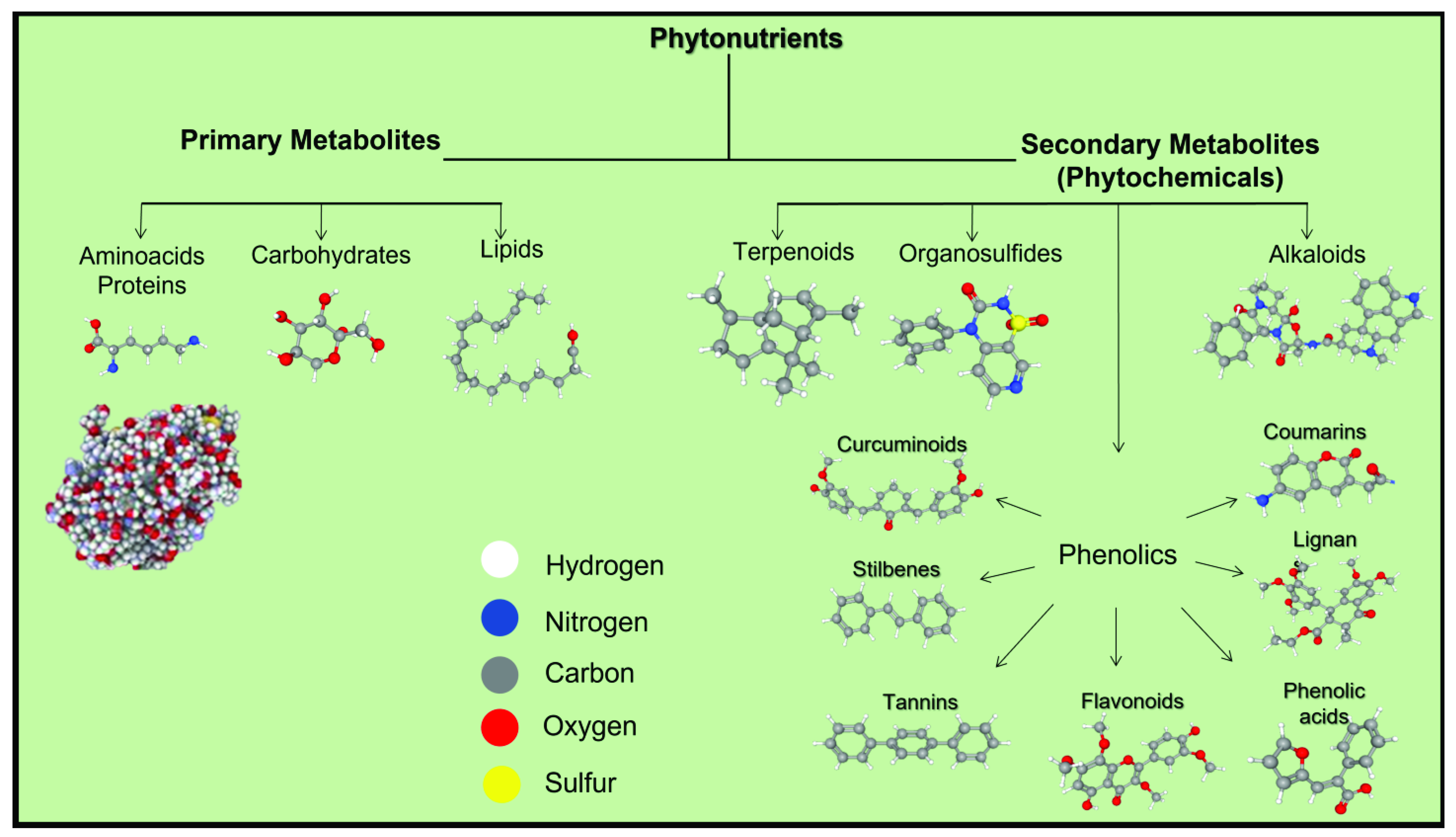
Figure 1. Phytonutrients’ composition. Plants produce primary metabolites (i.e., innate proteins, lipids, and carbohydrates) for their normal metabolic functioning and secondary metabolites (i.e., phytochemicals), primarily to protect them from predators. These phytochemicals are classified into four main chemical groups: phenolics, alkaloids, organosulfides, and terpenes. Phenolics are the biggest group subdivided into seven subgroups: curcuminoid, stilbenes, tannins, flavonoids, phenolics acids, lignans, and coumarins. 3D structures were visualized using PubChem [26] ball-and-stick model.
Applsci 11 08477 g001
Figure 2. GI absorption for described phytonutrients. (a) Percent (%) of phytochemicals of all selected phytonutrients with high or low probability for GI absorption. Percent (%) of (b) turmeric, (c) garlic, (d) cinnamon, (e) graviola, and (f) oregano phytochemicals with high or low probability for GI absorption.
Applsci 11 08477 g002
Table 1. Theoretical calculations of the physicochemical properties for the main phytochemicals of the selected phytonutrients.
Phytochemical Compound NameEmpirical Formula/StructureMW (Da)HBA/HBD/RBLog PLog DA (cm3)PSA (Å2)GI Absorption/L-RO5, GF, and VR Violations
Turmeric
Curcumin Applsci 11 08477 i001368.46/3/82.92.610693.1High/0
Demethoxycurcumin Applsci 11 08477 i002338.35/2/73.22.6 9783.8High/0
Bisdemethoxycurcumin Applsci 11 08477 i003308.34/2/63.42.8 9174.6High/0
α-Turmerone Applsci 11 08477 i004218.31/0/44.44.16917High/0
Garlic
Alliin Applsci 11 08477 i005177.24/3/5−0.5−3.34499.6High/0
Negative LogD
Allicin Applsci 11 08477 i006162.3 1/0/51.2 1.44661.6Low/
TNA < 20
Diallylsulfide Applsci 11 08477 i007114.2 0/0/42.6 2.93725.3Low/
TNA < 20
MW < 160
A < 40
Z-Ajoene Applsci 11 08477 i008234.4 1/0/83.1 2.8 6886.9High/0
2-Vinyl-4H-1,3-dithiin Applsci 11 08477 i009144.30/0/12.2 2.7 4550.6Low/
TNA < 20
MW < 160
Cinnamon
(E)-Trans-Cinnamaldehyde Applsci 11 08477 i010132.2 1/0/22.1 1.8 4217.1Low/
TNA < 20
MW < 160
(E)-Cinnamyl Acetate Applsci 11 08477 i011176.2 2/0/42.6 2.6 5326.3High/0
Eugenol Applsci 11 08477 i012164.22/1/32.22.54929.5High/0
Cuminaldehyde Applsci 11 08477 i013148.21/0/23.03.14717.1Low/
MW < 160
Protocatechuic Acid Applsci 11 08477 i014154.14/3/11.2−1.93777.8Low/TNA < 20
MW < 160
A < 40
Negative LogD
Graviola
Benzylisoquinoline Applsci 11 08477 i015219.31/0/24.04.37212.9High/0
Annonacin/Acetogenin Applsci 11 08477 i016596.97/4/266.47.3169116Low/
TNA > 70
MW > 500
RB > 10
LogP > 5.6
A > 130
High LogD
Cinnamic Acid Applsci 11 08477 i017148.22/1/22.4 −0.7 4437.3Low/
TNA < 20
MW < 160
Negative LogD
Coumaric Acid Applsci 11 08477 i018164.23/2/22.4 −1.4 4657.5High/0
Negative LogD
Caffeic Acid Applsci 11 08477 i019180.24/3/21.4−1.74877.8High/0
Negative LogD
Rutin Applsci 11 08477 i020610.516/10/61.8−1.8138266Low/
TNA > 70
MW > 500
HBA > 10
HBD > 5
A > 130
PSA > 140
Negative LogD
Oregano
Carvacrol Applsci 11 08477 i021150.21/1/13.33.14720.2Low/
MW < 160
Thymol Applsci 11 08477 i022150.21/1/13.33.1 4720.2Low/
MW < 160
O-Cymene Applsci 11 08477 i023134.20/0/14.04.1 450Low/
MW < 160
Apigenin Applsci 11 08477 i024270.25/3/12.11.37087High/0
Luteolin Applsci 11 08477 i025286.26/4/12.41.172107High/0
MW: molecular weight; LogP: lipophilicity; LogD: lipophilicity considering ionizable groups at pH 7.4; A: molar refractivity; HBD: hydrogen bond donors, HBA: hydrogen bond acceptors; RB: rotatable bonds; PSA: polar surface area; TNA: total number of atoms; L-Ro5: Lipinski’s Rule of 5; GF: Ghose Filter; VR: Veber’s Rules; Predicted data of Empirical formula, Structure, MW (Da), H-bond Acceptor/Donor, Log P, Log D, and A were generated using PubChem [26], ChemSpider [27], ACD/Labs Percepta Platform-PhysChem Module [28] and US Environmental Protection Agency’s EPISuite™ [29]; Favorable properties or “drug-likeness” for GI tract absorption are predicted by the combination of L-RO5, GF, and VR: MW (160–500 Da); HBD ≤ 5; HBA ≤ 10; A (40–130); LogP (−0.4–5.6); RB ≤ 10: PSA < 140; TNA (20–70) [30].
Publisher’s Note: MDPI stays neutral with regard to jurisdictional claims in published maps and institutional affiliations.