Review

Co-Adjuvant Nanoparticles for Radiotherapy Treatments of Oncological Diseases

1
Department of Materials Science, University of Milano-Bicocca, via R. Cozzi 55, I-20125 Milano, Italy
2
Institute of Physics of the Czech Academy of Sciences (FZU), Cukrovarnická 10/112, 162 00 Prague, Czech Republic
*
Author to whom correspondence should be addressed.
Appl. Sci. 2021, 11(15), 7073; https://doi.org/10.3390/app11157073
Submission received: 21 June 2021 / Revised: 27 July 2021 / Accepted: 28 July 2021 / Published: 30 July 2021
(This article belongs to the Special Issue Applications of Medical Physics)

Abstract

:
Nanomedicine is emerging as promising approach for the implementation of oncological methods. In this review, we describe the most recent methods exploiting heavy nanoparticles and hybrid nanomaterials aiming at improving the traditional X-rays-based treatments. High-Z nanoparticles are proposed as radiosensitizers due to their ability to stop the ionizing radiation and to increase the locally delivered therapeutic dose. Other nanoparticles working as catalysts can generate reactive oxygen species upon X-rays exposure. Thanks to their high toxicity and reactivity, these species promote DNA cancer cells damage and apoptosis. Hybrid nanoparticles, composed by scintillators coupled to organic molecules, are suitable in X-rays activated photodynamic therapy. This work highlights the roles played by the diverse nanoparticles, upon ionizing radiation irradiation, according to their physico-chemical properties, surface functionalization, and targeting strategies. The description of nanoparticle qualities demanded by the oncological nanomedicine is presented in relation to the processes occurring in biological medium when X-ray radiation interacts with heavy nanoparticles, including the scintillation mechanisms, the stopping power amplification, and the disputed modeling of the effective deposit of energy within nanomaterials. The comprehension of these issues in nanomedicine drives the strategies of nanoparticles engineering and paves the way for the development of advanced medical therapies.

1. Introduction

Today, cancer is still one of the greatest global causes of death. In 2018, there were 18.1 million new cases and 9.5 million cancer-related deaths worldwide. By 2040, the number of new cancer cases per year is expected to rise to 29.5 million with an estimation of cancer-related deaths close to 16.4 million [1,2]. Currently, regular screening and surveillance programs required for early diagnosis/intervention are the best ways to improve the outcome and survival. The diagnostic tools in use in healthcare rely on imaging methods, like optical imaging, radiography, magnetic resonance imaging (MRI), and computed tomography (CT), as well as nuclear imaging techniques [3,4]. More recently image-guided cancer treatments and molecular diagnostics have enabled to subcategorize each cancer type allowing efficient intervention with targeted therapeutics [5,6]. Over the last years, ceaseless efforts have been put by the scientific and medical communities in the cancer research with the aim of finding new diagnosis alternatives for the fabrication of simpler, broad use, and cost-effective imaging tools and scanners [7,8,9]. The most important goal is to surpass the typical drawbacks of conventional diagnostic procedures, which are, for instance, related to the choice among the available contrast agents presenting low accuracy and specificity, short half-life, low radiodensity, and rapid clearance [10,11]. These disadvantages result in the need of massive and prolonged exposure to high levels of electromagnetic radiation of the patient for the collection of images with satisfactory resolution [12,13]. Conventional cancer treatments are chemotherapy, radiotherapy, and surgery. Despite, in the past years, these consolidate procedures have enabled an active fight against cancer ensuring long-term survival and quality of life for several malignancies in oncological patients, their broad use has been always associated to diverse drawbacks and side effects. One of the main problems of chemotherapy is the low specificity of chemotherapeutic drugs for cancer cells. Chemotherapy suffers from non-specific biodistribution and poor tumor accumulation of the therapeutic drugs, resulting in high toxicity, damages of the healthy cells, as well as systemic side effects [14,15]. Surgery can represent the only potentially curative therapy of high-risk patients. However, the high tumors recurrence rate associated with surgical resection as single oncological protocol is responsible for a significant morbidity and failure percentage in many malignancies [16]. Lastly, the radiation therapy is limited by the maximum cumulative radiation dose allowed without incurring significant injuries to the adjacent tissues or organs [17,18]. More recently, small molecules-based therapies and immuno-oncology have been demonstrated to be effective owing to some recent successes in the clinic [19,20]. Besides the monotherapies, the combination of the established and accessible therapeutic protocols with immunotherapy has been validated to be efficient in cancer patients [21,22]; however, the ability of the molecular agents used in immunological therapies to reduce immune suppression, or to enhance activation of cytotoxic processes in the tumor environment is associated to a severe toxicity, whose side effects results under debate [23,24].
Novel and targeted cancer therapeutic techniques have been recently developed to guarantee effective, efficient, affordable, and less invasive cancer cares to patients [25]. The search of developing tailored treatments for specific oncological demands has found its productive ground in the branch of nanomedicine; in order to visualize the timetable of the scientific efforts boosting the research on nanomedicine, we the proposed, for instance, works and reviews published in the recent twenty years [26,27,28,29,30,31,32]. The European Medicines Agency (EMA) defines nanomedicine as including all applications of nanotechnology and nanoparticles for tumor targeting, imaging, and therapy [27,33]. Nanoparticles (NPs) definition refers to those materials as large as 1- to 100-nm made from both inorganic and organic materials. NPs can be administered systemically (via injection inhalation, and oral intake) or locally (via intratumoral delivery and implantable devices such as implants loaded with NPs or anticancer agents) [34,35,36,37]. Organic based NPs used in biomedical applications include liposomes [38], polymer constructs [39], and micelles [40]. Inorganic NPs comprise mostly metal, semiconductor, and insulating systems (see [41] and within references to have an overview of the available NPs). Within the past few decades, inorganic NPs have been proposed in the biomedical fields for the implementation of the cancer therapies (chemotherapy, drug delivery [42,43], and hyperthermia [44,45], as well as radiotherapy, photodynamic therapy and X-ray activated photodynamic therapy [17,46]), or for the use as diagnostic agents for imaging techniques [12], as well as for the applications as theranostic tools for oncology and other fatal diseases [47]. The attractiveness of these NPs relies on the versatility and tunability of the physico-chemical qualities, which depend on their composition, morphology, size, and high surface-to-volume ratio. Specifically, NPs modifiable features, like shape, size, and surface charge [28], are key aspects for surface accommodation of functional groups or hydrophobic molecules. The functionalization of NPs surfaces results in enhanced solubility and biocompatibility, in improved stability and retention time in bodily fluids [48,49], and in the ability of the nanosystems to cross physiological barriers for their use in vivo [50]. Moreover, the engineering of the NPs surfaces by targeting ligands enables the specificity of their diagnostic and therapeutic activity towards the malignancy and illness sites in the patients’ body and the release of the antitumoral drugs in a stable and controlled manner [51,52]. The adjustment of the physico-chemical properties of NPs by facile doping strategies as well as by the engineering of their size, crystallinity, and surface chemistry would also affect their luminescence behavior. Fluorescent NPs, such as lanthanide doped inorganic nanoparticles [53,54,55], quantum dots [56], silica based nanosystems [57,58], as well as metal clusters and nanoparticles [59,60] have been proposed for in vitro and in vivo bioimaging, sensing, drug delivery, and light-triggered oncological therapies. The tuning of the luminescence is a crucial point to satisfy the performance requirements demanded to the nanomaterials for their exploitation in medicine [53,61,62]. Even though many NPs are composed by purely organic and inorganic materials, the development of organic/inorganic hybrid materials is eagerly hunted; hybrid NPs in form of core-shell structures, metal organic frameworks (MOFs), or of nanocomposites where hybrids NPs are dispersed into a polymeric matrix, enable to create a new class of functionalized theranostic NPs that possess unique advantages, exploiting synergistically the feature of their single components [63].
The world of nanomedicine and nanotheranostic is wide and covers many frontiers of medicine. The use of NPs of diverse composition can represent an alternative to the traditional oncology and, in parallel, a way to implement the established therapies and diagnostic tools. In this review, we focus our attention on the utilization of NPs in the X-rays based oncological treatments, with efforts in describing the multiplicity of the roles that the diverse NPs can play according to their compositions and surface functionalization from the radiosensitization in radiotherapy (RT), to the triggered activation of cancer killing agents in the photodynamic therapies (PDT) and in the X-ray activated photodynamic therapy (X-PDT). NPs suitable for X-rays can be divided in a simple way into passive and active nanoparticles. Passive nanomaterials include dense and high atomic number (Z) NPs (for instance, metal containing nanoparticles), able to efficiently stop the ionizing X-ray radiation enhancing the energy deposition within the tissue and thus increase the RT effects at lower doses. A group of active nanoparticles, such as inorganic photo (-and radio) catalysts, possess lower Z than the passive NPs but can sensitize the RT effects by generating an excess of reactive oxygen species (ROS) in cells upon X-rays and by causing ROS-dependent functional disorder/cell death/apoptosis. Another class of active NPs comprises inorganic/organic multicomponent systems allowing a dual effect because of the interaction with the ionizing beam: firstly, an improved response to the external stimuli due to the inorganic dense part; and then the triggered activation of cancer killing agents, such as singlet oxygen sensitizer, nearby the malignancy.
Specifically, in this review we present especially the use of inorganic NPs and hybrids that can efficiently interact with the ionizing irradiation in the RT. The use of organic NPs will be only briefly pictured for topic completeness, regarding especially the possibility to employ light molecular photosensitizers, like porphyrin molecules, with X-ray radiation. The exploitation of biomolecules and drugs in chemo-radiotherapy able, for instance, to increase the cells susceptibility to radiation [64] or inhibit the DNA repair after the treatment [65] are not included in this review.

2. Radiotherapy Mechanism

Radiotherapy is one of the most effective and widely used cancer therapeutic modalities. About 50% of cancer patients are treated with radiotherapy for curative and palliative purposes both as single therapeutic modality and in combination with other treatment methods [66,67]. Although RT cannot be applied to all types of cancer, it is established as a principal option for treatment of many malignancies, such as breast carcinoma, lung carcinoma, melanoma, gastrointestinal cancers, head and neck cancers, gynecological cancers, hematologic malignancies, prostate or cervix tumors, central nervous system neoplasms and thyroid carcinomas, as well as for an effective reduction of pain in the of case bone and brain metastases [68,69].
RT exploits ionizing beams—X-rays or radiation emitted by radionuclides—to stop the rapid proliferation of cancer cells and to achieve the therapeutic goals [70,71]. For the description of the RT mechanism, this review focuses mainly on RT applied by an external X-ray source. As summarized in Figure 1, the interaction of the high energy radiation within tissues induces the killing of tumor cells directly by the damage of the DNA and of the molecular structure leading to the termination of cell division and proliferation, and even to cell necrosis or apoptosis; otherwise, cancer cells destruction can arise indirectly from the formation of cytotoxic ROS upon interaction with the cellular aqueous environment [72,73]. A detailed description of biochemical reactions occurring in cellular environment upon the interaction with the ionizing radiation is depicted by Clement and co-worker in Ref. [65]. Briefly, in cellular environment, water radiolysis by ionizing radiation results in the formation of several species, mainly: e−aq (hydrated electrons), HO• (hydroxyl radicals), H• (hydrogen radicals), H2 (radiolytic hydrogen), H2O2 (hydrogen peroxide), and HO2 (hydroperoxyl radical). Afterwards, these products can react and create further ROS, such as O2 (superoxide), organic radicals (R•), hydroperoxides (ROOH), and singlet oxygen (1O2). Under physiological conditions cells generate a certain amount of ROS, whose concentration is regulated by the antioxidant system to maintain cellular homeostasis. Perturbing the cellular balance by radiation-induced ROS creation leads to oxidative stress that can trigger DNA damage and other cell death mechanisms, for instance by necrosis, apoptosis, autophagy, mutation, and senescence.
Whatever the cancer cells killing mechanism, i.e., by physically direct ionization or using free radicals by water ionization, the goal of radiotherapy is to deliver the maximum dose to the target tumor tissue while safeguarding the surrounding normal tissue. Clinical radiotherapy uses high-energy external X-ray beam, from 6 MeV up to about 20 MeV, from linear accelerators. In typical launched protocols, patients are treated through fractionated regime with dose of 1.8–2 Gy over the course of 4–8 weeks to limit toxicity to normal tissues [74] but in advanced treatment planning the delivered dose to cancer can reach values close to 15–20 Gy [75]. Interactions of such X-rays with biological tissue can occur by (1) photoelectric effect, (2) Compton effect, and (3) pair production, according to the energy of the external X-ray source. In clinical RT the X-rays beams of few tens of MV are widely used for deep seated tumors, while low energy beams (up to 200 kV) have very few applications due to the lower penetration depth (<5 mm) [76]; at these working energies the Compton and the photoelectric effects are dominant (Figure 2), and the deposit of their energy occurs through a cascade of secondary photoelectrons and Auger electrons, stimulating all the radiobiological effects above depicted.
The radiobiological effects and efficacy in producing a permanent DNA damage in cells are related to the type and the energy of interacting radiation by the linear energy transfer (LET) physical factor i.e., energy deposited per unit distance along the ionization track. LET is higher for heavy ions than for photons (for example a 250 keV X-ray photon have an average LET of 2 keV/μm, whereas alpha particles LET is of hundreds of keV/μm) [78]. The main difference between high LET radiation and low LET radiation is due to a diverse induction of DNA damage that is the primary target required for cell killing: it has been proven that high LET radiation causes more extensive and less repairable cellular damage by very dense events of energy deposition along the track, without penetrating deeply into the human tissues; in turn low LET X-rays deposit energy in tissue in a highly dispersed manner and are thus able to reach deeper tissue penetrations [79]. In RT, the secondary products of the first ionizing event, especially of the photoelectric effect, include the Auger electrons. High LET makes the Auger electrons highly toxic to the cells inducing damage in macromolecular targets of a cancer cell and in the cell membrane, as detailed reviewed in [80] by A. Ku and co-authors, who display the use of Auger electrons in radiation dosimetry and for cancer treatments, such as targeted radiotherapy.
From the above picture, it is evidenced that the effective result of RT and the estimation of the dose deposited in the malignancy environment depend on the interactions in tissue involving a given particle and secondary particles. The modeling of the energy deposition by the ionizing radiation and the following radiobiological consequences are of fundamental importance towards the goal of surpassing the common drawback afflicting RT, like the lack of tissue selectivity, hindering the discrimination between the tumor and the healthy cells, and the need of high energy primary beam enabling the penetration of the radiation in deep tumors and the release of an amount of energy adequate to start the biochemical ROS production to destroy the proliferation of the malignant cells. The most advanced strategies exploit the use of NPs to enhance a localized therapeutic effect of RT, in order to reduce the local dose to be delivered and the toxicity towards normal surrounding tissue, and thus to improve the quality of life of the patience during and after the radiological treatments.

3. Nanoparticles in Radiotherapy

In the following sections, we aim at describing the most used and promising NPs that can be exploited in RT to overcome its limitations and to combine the common RT procedures with novel challenging strategies. Among these strategies, this review emphasizes the X-PDT. Nanomaterials are classified into passive (mostly inorganic containing heavy elements) and active (inorganic nanocatalysts and nanohybrids) NPs. For clarity, a summary of the properties and the application area of the NPs presented in the manuscript is provided at the end of this section, in Table 1. It is worth to notice that the boundaries of the NPs classes are smooth. In particular, the categorization of the passive and single component inorganic active nanosystems is rather shaky. We have arbitrary chosen to consider as passive NPs all the dense and high Z systems that enable to enhance the energy depositions in the cancer environment; in turn, only nanometric catalysts with lower Z are sorted as active NPs enabling generation of ROS thanks to the high reactivity of their surface upon irradiation. Nevertheless, we are aware that (as discussed in Section 2) ionizing radiation, by interacting with passive heavy NPs, produces secondary charges that contribute at generating ROS localized in the neighborhoods of the ionizing event.

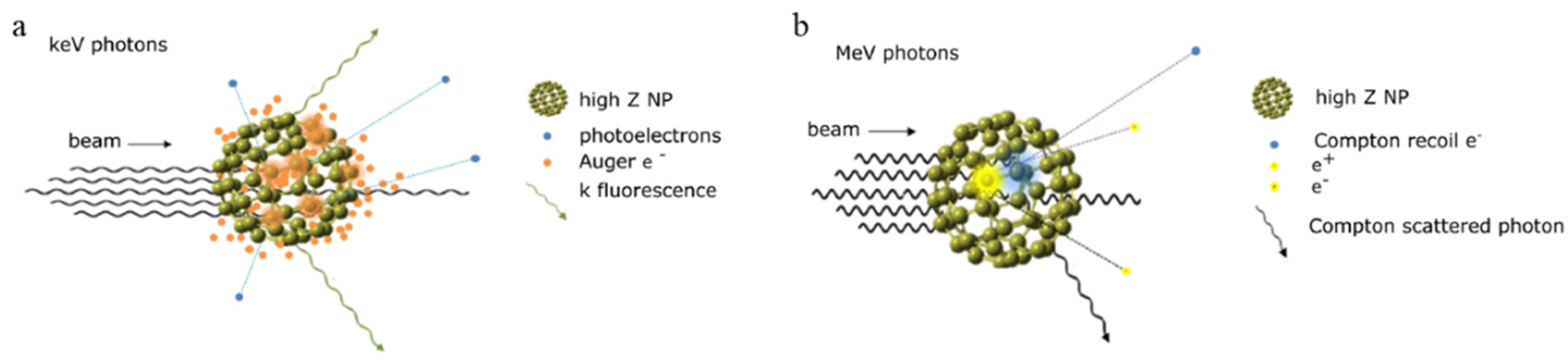
3.1. Passive Nanoparticles as Enhancers of Energy Deposition in RT

The scientific and medical communities strive to find X-ray based therapeutic solutions with the aim to improve the survival rate and life quality in oncological patients subjected to RT by reducing the illness recurrence, which is related mostly to the massive doses required for stopping the cancer cells division but delivered also towards the surrounding healthy tissue. High Z and dense inorganic nanomaterials represent the key to reduce RT parameters, such as external beam energy, delivered dose, and exposition time, while boosting all the toxic and fatal effects of the radiation [74,76]. The idea to exploit high Z materials comes from simple considerations. Indeed, the presence of heavy NPs in a regime of low energy ionizing radiation (<60 keV) maximizes the X-ray beams attenuation by photoelectric effects, whose cross section σ depends on (Z/E) n, with n ≅ 3–4 [77]. This dependency reveals that high Z materials turn out to be supportive in RT by enhancing the deposit of energy in the tumor and in the proximity of the malignant cells (thus, the dose delivered during radiotherapy) [81].
Since the early 1970s, the ability of diverse materials, like water/tissue equivalent materials, alloys of metals, and heavy metals, at stopping the ionizing radiation have been tested for applications in radiotherapy [82]. Later, it has been discovered that metal implants, composed by aluminum, tin, stainless steel, titanium, and lead, can alter the delivered doses in radiotherapy in patients affected by neck and mandibular cancers, due to the difference in Z value between the biological tissues and the metals [83,84]. In the last few decades, the scientific research has looked at the capacity of nanosized heavy metal particles in promoting the dose delivery in cancer tissue as a chance to improve the RT procedure and its therapeutic outcomes, due to the correlation between the enhanced energy dose and the increased cancer cell destruction [85].
Over the past few years, NP sensitizers have been proposed in combination with radiotherapy in order to improve tumor response and control the disease growth. For instance, there has been lot of interest in metallic—gold, gadolinium, silver, bismuth, and platinum—and bimetallic based NPs, as well as in nanometric metal oxides [86,87]. Thanks to the high atomic number, good biocompatibility, and relatively strong photoelectric absorption coefficient, gold nanoparticles (AuNPs, Z = 79) are largely studied for diagnostic and therapeutic applications in cancer therapy [88]. Especially, Au nanoparticles have been under investigation as possible agents for amplification of radiation dose in tumors, in the so called “gold nanoparticle-assisted radiation therapy”. From the first decades of 2000’s, AuNPs’ action has been predicted to increase the fraction of the X-rays energy deposited close to gold NPs because of increased photoelectric interactions, thereby enhancing the local radiation dose also according to their shape, functionalization, concentration, and intracellular distribution [89]. In vivo studies performed by Hainfeld et al. [90] and Herold et al. [91] by injecting Au NPs and microspheres in tumor-bearing mice, have reported the occurrence of biologically effective dose enhancement by using kilovoltage photon beams; while the use of isotopes producing low-energy photons has been foreseen for significantly improving the clinical potential of interstitial brachytherapy. More recent works assess the Au actions considering the increment of dose together with the corollary activation of ROS within malignant cells [92] (see for example Figure 3 reproduced from Ref. [92]). On-going research promotes the optimization of nanoparticles targeting and functionalization to reduce the total doses to be delivered, and to combine ionizing radiation with other therapeutic modalities [93,94,95,96]
A valid candidate as sensitizers of RT is Gadolinium (Gd, Z = 64). In addition to its well-known application as contrast agent in MRI, recently the additional role of gadolinium nanostructures in RT has been modeled by computational analysis. Modeling results reveal the capability of Gd NPs at increasing the final delivered dose to the tumors site and the suitability in brachytherapy applications [97,98]. Moreover, studies on gadolinium-NPs, namely AGuIX, confirm the occurrence of sensitization under ionizing radiation in vitro assay on HeLa cells, revealing the attractiveness of gadolinium-based NPs as alternative to the widely used gold NPs for improving the efficacy of radiation cancer treatments [99].The use of platinum (Pt, Z = 78) NPs (PtNPs) as promising radiation sensitizers in radiotherapy cancer treatment has come into light in a small number of works with respect to the Au and Gd NPs. For instance, in 2010, it has been examined the case of platinum nanoparticles upon irradiation by fast carbon ions (LET = 13.4 keV μm−1). Their use enables the strong enhancement of the biological efficiency of ionizing radiations and the amplification of tumor cells DNA lethal damage (single strand breaks, and mostly double strand breaks) induced by irradiation; moreover, formation of radicals in PtNP proximity has been proved to further contribute to the DNA strand breaks [100]. More recently, porous Pt NPs have opened the way for novel therapeutic strategies. Indeed, while promoting radiation induced cell cycle arrest by an effective energy deposition within the cancer cells, porous Pt systems enable also to generate oxygens (O2) from hydrogen peroxides (H2O2). The final outcomes of exploiting porous Pt NPs are the formation of DNA breaks with the generation of ROS and, at the same time, the hampering of the establishment of hypoxia condition in the tumor, being one of the major causes of cancer radioresistance [101].
Metal oxides nanoparticles are considered an alternative to full metallic systems. Hafnium oxide (hafnia, HfO2) has been considered in RT as sensitizer to increase energy dose deposit within cancer cells, owing to its high atomic number, electron density, and chemical stability. Hafnium (Z = 72) oxide nanoparticles (NBTXR3) have been designed in terms of size, surface charge, and shape, to concentrate in tumor cells to achieve intracellular high-energy dose deposit with reduced health hazards. Monte Carlo simulation considering a high-energy source of a few MeV predicts an enhancement of local energy deposit, within and close to the tumors where NPs are located, almost 9 times larger with respect to RT alone. In vitro and in vivo studies on the HT1080 fibrosarcoma cell line with NBTXR3 nanoparticles with high-energy sources confirm the results of the simulation, denoting a higher radio-induced cancer cell destruction rate in presence of high Z NPs [102]. Another recently discovered effect of NBTXR3 marks their ability to impact on the activation of the antitumor immune-response, open the way for their use in radio-immunotherapy [103]. Other interesting high Z materials enabling strong absorption of X-rays, are tantalum pentoxide (Ta2O5) NPs and bismuth oxide nanoparticles—Bi2Se3, Bi2S3, and Bi2O3, the latter showing a multiplicity of characteristics for multimodal imaging, radiosensitization, as well as in radioprotection [81].

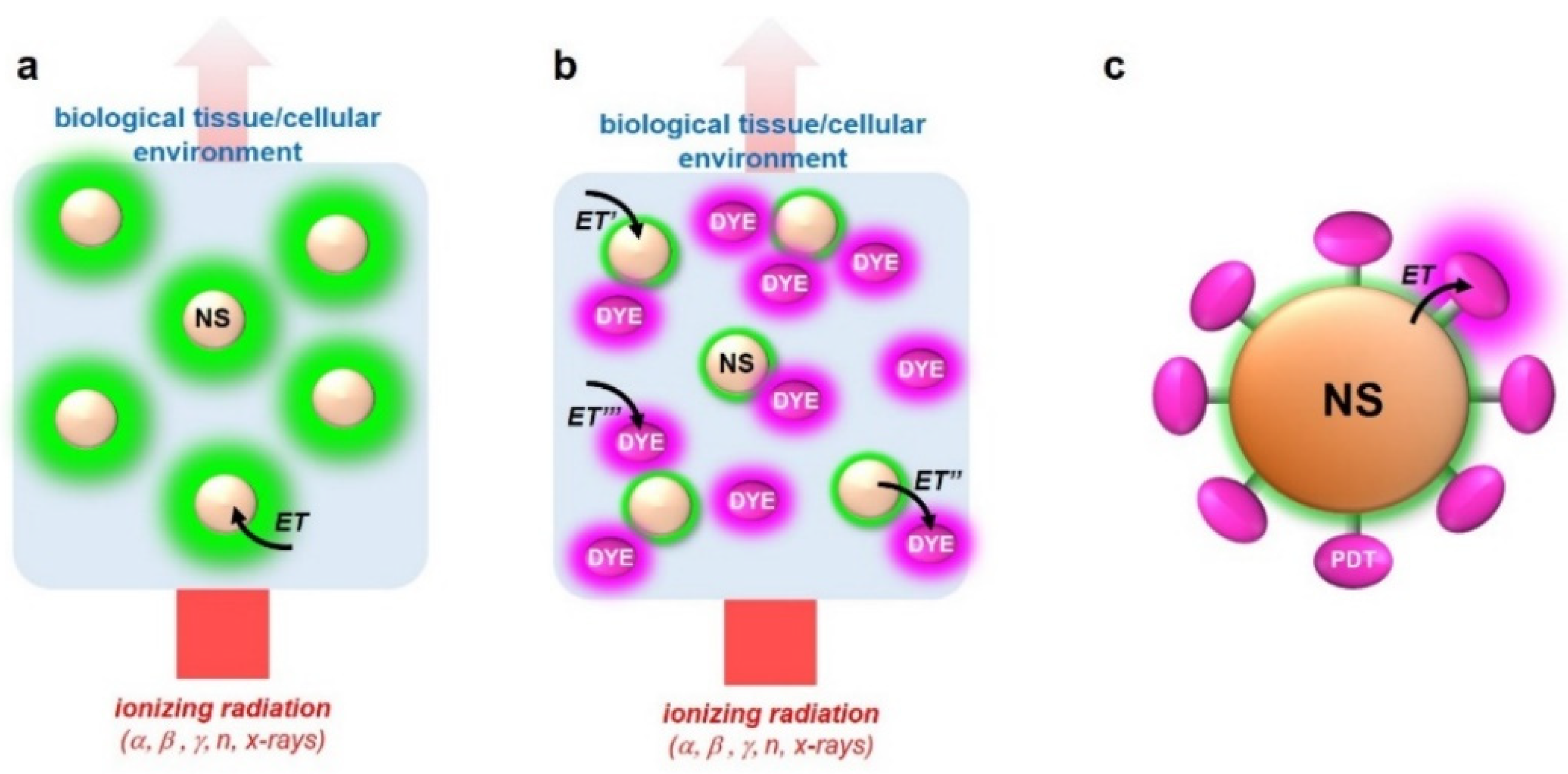
3.2. Active Nanoparticles and Catalysts Inducing ROS Generation

As extensively explained above, the augmented photoelectric effect cross section in presence of NPs with heavy Z enables the creations of secondary charges, mostly Auger electrons, and the productions of direct and indirect DNA cancer cells damages. However, as reviewed by Guerreiro et al. in Ref. [104], there are many evidences that also metal oxide NPs with lower atomic number (made up by Si, Al, Ti, Zn, Fe, Ce and Mn for instance) are responsible of radiosensitization phenomena within tumor cells. This output is imputed to the chemical and catalytic activities of the NPs surfaces contributing to the overall generation of radiation-induced radicals. From a general point of view, nanocatalysts are materials able to produce ROS under high energy light (photocatalysts) and ionizing radiation (radiocatalysts) to decompose unwanted chemical and biological agents. Despite the attempt to treat the radiocatalysis as similar to the well-known photocatalysis [65], it is worth to notice that the use of ionizing radiation guarantees diverse advantages, such as the increased rate of a catalytic reaction, the acceleration of degradation of pollutants, the overcoming of limited penetration of UV/Visible (Vis) light in solutions and biological tissue, as well as the possibility to use a wider range of catalyst also with wider band gap (Eg) and greater redox capabilities [105,106,107]. If we consider the case of (nano) titanium oxide, TiO2, as representative of photo-/radiocatalysis in semiconductors, catalysis phenomenon can be summarized in the following steps: (1) excitation by high energy ionizing radiation or light source with energy larger than the Eg of the material; (2) promotion of an electron (e) from the valence band to the conduction band, and the concomitant generation of a hole (h+) in the valence band; in an efficient nanocatalyst, where recombination of the generated e/h+ is restrained, the last step (3) is the diffusion of e/h+ charges towards the materials surface to rapidly (in sub-nanoseconds time range) initiate redox reactions among reactants adsorbed at the nanocatalyst surface, and to start the formation of ROS. In presence of aqueous environment, electrons reduce molecular oxygen forming superoxide radical anion; hydroxyl radicals and hydrogen peroxide molecules are induced by the holes oxidizing water molecules and hydroxide ions. In TiO2, it has been also proven the occurrence of singlet oxygen creation (1O2) after oxidation of superoxide radical ion [108,109,110]. The reactive species, especially hydroxyl radicals, can be used to decompose a variety of pollutants in environmental applications [111]. More important for the goal of this review is the effective damage that the generated ROS can produce on DNA of cancer cells and biomolecules, thus contributing to the biological damage induced during radiotherapy, as well as to the lethal prolonged metabolic oxidative stress after irradiation [112]. Due to the large active surface area, it has been established the importance of morphological features (e.g., size, composition, specific surface ligands, surface charges) on the amount of ROS generated by NPs [113]. Numerous examples in the literature report chemistry strategies to fix up the NP surface properties also through functionalization/coating procedures [114,115].
In this review, we aim at providing straightforward examples illustrating the importance to conduct further research on lower Z radiocatalysts, whose effect in reducing cancer growth in RT is as effective as the one of high Z and dense NPs, thanks to the boosted ROS productions. We consider two well-known radio-photocatalyst NPs, specifically titanium and zinc oxide (TiO2 and ZnO, respectively), which have been approved by Food and Drug Administration (FDA) [116]. In nanomedicine, the ability of ZnO and TiO2 in producing ROS by themselves under UV/Vis excitation has been exploited for the photodynamic therapy [117]. Indeed, PDT is considered as a clinically deployed efficient and non-invasive alternative to the surgery and to the current oncological therapies. PDT is based on the cytotoxic effects originating when biocompatible photosensitizers (PSs) are photoexcited, producing ROS and singlet oxygen moieties that induce the cell death through oxidative damage of cellular membranes [118]. PSs can include organic dyes and aromatic hydrocarbons, porphyrins and related photosensitizers, transition metal complexes, and semiconductors [119]. Unfortunately, the PSs approved for routine PDT treatment require UV/Vis light to be activated. In this spectral region, the human tissue transparency is low, thus making PDT ineffective for tumors seated at depths larger than 1 cm [120]. Interestingly, TiO2 and ZnO have been proved to create ROS also under ionizing radiation and to be suitable for X-ray based oncological treatments. For instance, a work of Youkhana, Esho Qasho, et al. reports the radiosensitization phenomenon in presence of anatase TiO2 NPs under X-ray beam with energy set from keV to MeV [121]. Tests on cell viability assays have been performed on phantoms and in vitro to determine cells survival curves and the dose enhancement percentage. The obtained results evidence the occurrence of sensitization driven by two different mechanisms: in the keV range, the photoelectric effect coupled to the difference in the atomic numbers of the biological tissue and the TiO2 NPs are responsible of the dose enhancement; when MeV energy is used, the cross section of the photoelectric effect decreases while the active catalytic surfaces of the NPs start to play the major role in the generation of cell killing ROS, leading to the final therapeutic sensitization of RT. The possibility to take advantage of the two mechanisms, i.e., photoelectric and Z-dependent radiosensitization, together with the generation of ROS capable of destroying tumor cells by nanocatalysts, is at the base of the development of novel composite materials, like Au nanoparticles supported on TiO2 (Au@TiO2), enabling the simultaneous and synergetic establishment of the two processes, which lead to superior performances over the single elements [122].
Nanoparticles of ZnO are attractive materials for biomedical applications due to noticeably low toxicity. ZnO NPs have been widely explored for anticancer, antidiabetic, antibacterial, antifungal, and anti-inflammatory activities, as well as for drug delivery applications. Especially, ZnO NPs anticancer action relies on their ability to trigger ROS generation and apoptosis mechanisms under external excitations. Recently, the X-ray radiation-induced therapeutic behavior of ZnO NPs coated by a silica layer (ZnO/SiO2) has been studied on prostate adenocarcinoma cell lines. The potentiality of ZnO/SiO2 nanoparticles to increase radiation-induced killing of the cells has been validated by comparing cells survival after RT with and without NPs. Results display a 2-fold augmented killing rate in presence of ZnO based NPs; the findings show the active response to ionizing radiation of ZnO/SiO2 by means of the radiocatalytic mechanisms occurring at its surface. The proven catalytic reactions induced by high energy radiation make ZnO/SiO2 nanoparticles suitable for biomedical applications [123]. The combined use of ZnO with chemotherapeutics represents a step forward in the use of ZnO as anticancer. The loading of the anticancer drug, like doxorubicin as in ref [124], into the ZnO nanoparticles, results in an augmented cytotoxicity effects of the cancer therapy, due to the simultaneous combination of the anticancer action of the drug with the lethal ROS production by ZnO. Current research on nanocatalysts for RT focuses on other oxides, like superparamagnetic iron oxide nanoparticles (SPIONs) that, although the relatively low atomic number of iron (Fe, Z = 26), are used in combination with kV and MV X-rays beams and allow to enhance the impact of X-rays in tumor cells thanks to the production of ROS from catalysis reaction at the oxides surface [125]. Also, studies on cerium oxide NPs (CONPs) have demonstrated their capacity to enhance ROS production during RT in human pancreatic cancer cells and, at the same time, to play a protective role from toxic side effects of RT in normal tissues [126].

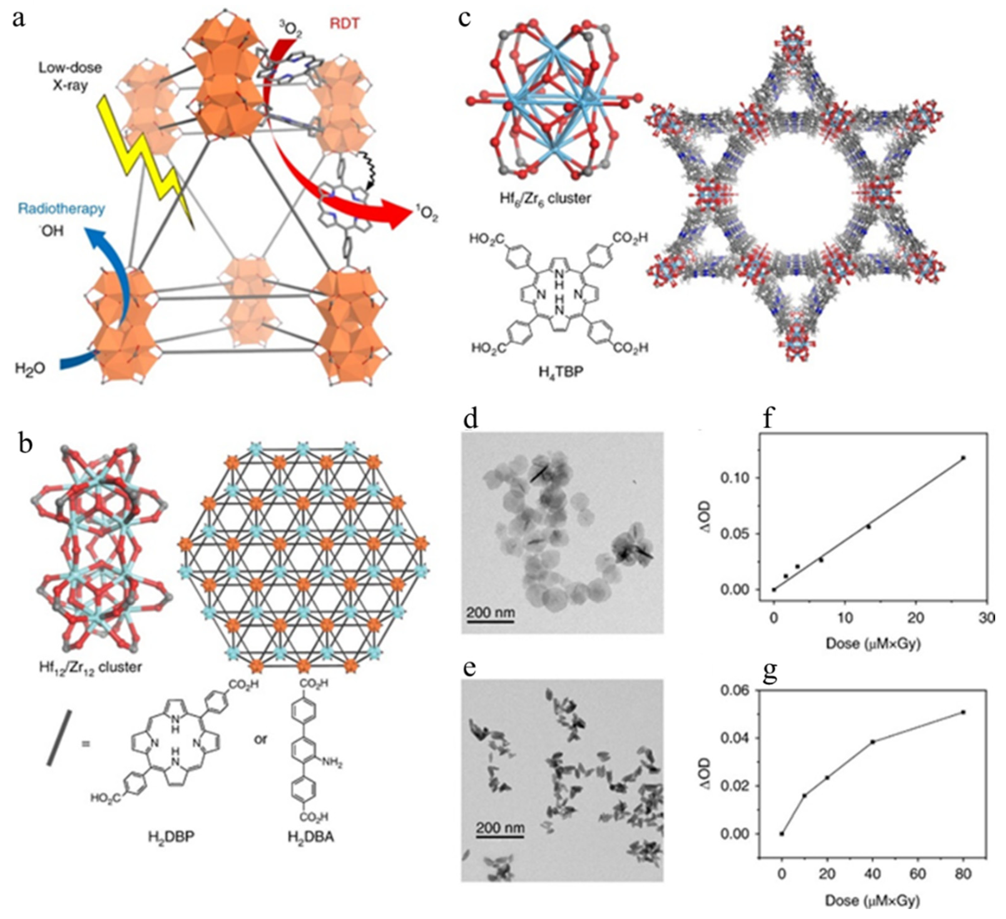
3.3. Multicomponent Nanoscintillators for X-PDT

The attempt of finding oncological cures alternative to surgery, chemotherapy and radiotherapy relies on the use of selective and non-invasive approaches with reduced side effects. In the previous paragraph, we mentioned at the advent of PDT as FDA approved oncological treatment, which has been proposed as alternative to the RT for the treatment of primary tumors of skin, esophagus and lung. Far from being an exhaustive explanation, in brief, PDT principle is based on photoexcitation of a PS (among which we can mention noble metal complexes, metal organic frameworks, metal oxides or carbon-based nanostructures, as well as polymeric PS and small organic moieties [127]) to locally generate ROS species and cause cancer cells death. After interaction with light, the PS is excited from ground state (S0) to unstable singlet excited states. In particular, the first excited state (S1) may undergo intersystem crossing (ISC) to form a more stable excited triplet state (T1) with longer lifetime (≈µs). Triplet state of PS can produce ROS by interacting with water and molecular oxygen. The first interaction generates free radicals (type I), while the second one produces singlet oxygen moieties (type II). However, the most common PSs can be excited by UV/Vis light; in this wavelengths range light is reflected and scattered by the human tissues, rendering the PDT ineffective for deep-tissue treatment [128]. Moreover, PDT painful side effects can appear immediately after treatment as well as after months. In the literature, many research studies have been reviewing in detail the PDT processes, the advantages and limitations, together with the PSs guideline, for instance see refs [129,130,131,132,133]. One possible solution relies on the so-called second-generation photosensitizers showing higher molar extinction coefficient in the near-infrared region where biological tissues present a transparency window [134,135]. Thanks to their high penetrability, X-rays offer another solution for adapting the PDT principle to deep tumors. From the first decades of the last century extensive studies have displayed the activation of the PS under ionizing radiation [136,137]. Recently, an extensive review by Larue et al. has offered an overview on the efficacy of X-rays in enhancing the toxic activity of some PSs, like hematoporphyrin derivatives, protoporphyrins, or metalloporphyrins They have listed the available photosensitizers used as radiosensitizers or as precursors of PDT effects in combination with radiation therapy [138].
A step forward in the progress of alternative therapies is provided by the so-called X-PDT. The strategy counts on the employment of scintillating nanoparticles (nanoscintillator; NS), able to convert the ionizing radiations into ultraviolet or visible light through a three stage scintillation process consisting in-conversion, transport, and luminescence [139,140]. NS can be embedded in a biological surrounding, where both the absorption of X-rays by the NS and the deposition of energy by the exciting beam (or the following secondary particles) in its proximity can occur. Afterwards, NS can emit fluorescence (Figure 4a), alternatively, when NS is embedded in biological environment with other elements (organic dyes and therapeutics, for example), the energy can be transferred to the secondary moieties through multiple radiative emission/reabsorption process and/or through efficient energy transfer mechanisms (ET). The interaction can occur between the high-energy beam and the aqueous environment with the consequent excitation of the NS and then the activation of organics nearby (ET′); or the NS can directly absorb the X-rays and transfer the energy to the dye (ET″). Lastly, also the organic molecules can interact with the primary beam by themselves (ET‴) (Figure 4b). In X-PDT, high Z and dense NS are designed to be grafted by a PS therapeutic agent (Figure 4c). In order to employ energy transfer from the NS to the PS for triggering the ROS and 1O2 production, the PS absorption spectrum should be resonant with the NS emission. Potentially, the exploitation of X-rays and heavy NS fulfills all the constraints for RT applications and for inducing radiosensitization (Section 3.1); at the same time, the use of PS, mostly 1O2 producing PS, being attested to be more efficient in the killing of malignant biological tissues and cells [127], guarantees an additional cytotoxicity towards the cancer cells destruction, mimic the action of the traditional PDT. The outcome of the RT- and PDT coupling remains unclear: besides the concept that the final result is just a sum of the effects from the two individual therapies, it has been demonstrated that X-PDT can produce important synergetic effects resulting in an improved efficiency in killing the tumor cells and in the overall reduction of the total dose to the patient [18].
Many inorganic nanomaterials like oxides, fluorides, silica-based nanostructures and semiconductor nanocrystals have been combined with organic photosensitizers towards X-PDT applications [141,142,143,144,145,146,147,148]. We can consider, for instance the case of silica-based nanotubes coupled to PS, displaying noteworthy therapeutic effects thanks to the synergy between RT and PDT [149]. Previous studies have proved the low toxicity of stoichiometric chrysotile [Mg3(Si2O5)(OH)4] nanotubes featuring also the ability to pass the blood brain barrier, potentially important in cancer brain treatments [58]. Thanks to their density, chrysotile nanotubes have been proven to respond to X-ray irradiation by emitting light in the blue wavelength range, suitable to excite meso-tetra(4-sulfonatophenyl) porphyrin (H2TPPS4−) as PS. Thus, the surface of chrysotile scintillating nanotubes has been functionalized by an ionic self-assembly strategy with this efficient PS, able to produce singlet oxygen toxic species upon photoexcitation (Figure 5a). The study of luminescence behavior under X-rays (RL) and light (PL) excitations discloses the occurrence of a fast energy transfer (in ps range) from the chrysotile to the PS, together with the sensitization of singlet oxygen production upon ionizing radiation (Figure 5b). As expected, also bare nanotubes enable a sensitization of the 1O2 under ionizing radiation, due to the enhancement in energy deposition in presence of elements with higher Z than the surrounding biological medium. A further stabilization of the functionalized nanotubes has been obtained by covering their surface with polymeric shell. This ultimate composition boosts the ability of the NS to enhance the production of singlet oxygen in an aqueous environment by ET mechanism to PS under X-ray irradiation. Tests in vitro—mimic the radiotherapy dose depositions—show that stabilized chrysotile nanosystems functionalized with porphyrins enable to sensitize the singlet oxygen species at the lowest doses thanks to the contribution of a high-density material, and the effective toxic action of PSs activated by the nanoscintillators (Figure 5c,d). This opens the possibility of effectively reducing the high-energy radiation exposure of patients during oncological therapies.
Metal-organic frameworks containing a heavy metal, such as Zr or Hf, have shown attractive scintillating performances; especially, MOF architecture can include high energy radiation interacting dense nodes linked through photosensitizer molecules for therapeutic and diagnosis applications [150,151,152]. A variation of the organic component allows for structural modifications useable to tailor the MOF properties quite easily, thus enabling the fabrication of molecular-size MOFs—based scintillators where organic dyes or therapeutics can be used as ligands interconnecting high Z NS. In this architecture, the proximity of NS and ligands enables the prompt activation of the ligands by energy transfer, after the interaction of ionizing radiation with the scintillators. The adaptability of these nanometric architectures has been exploited by Lu et al. in [152] to create cutting edge Hf based scintillating MOF (nMOF) for simultaneous X-PDT and immunotherapy (Figure 6). The synergetic combination of radiotherapy and immunotherapy represents a step forward towards the extension of the applications of radiotherapy for both local and systemic tumor rejection, by positively reducing the high-dose related RT toxicity.
In this work, hybrid nMOFs have been designed by linking Hf clusters and porphyrin-based photosensitizer ligands. The nMOFs exhibit the suitability to activate X-PDT mechanisms and the ability to stop the ionizing radiation and to trigger efficiently the 1O2 species creation by PS (Figure 6f,g). In vitro experiments display that hybrid nMOF can induce cytotoxicity and DNA double-strand breaks for doses < 1 Gy, i.e., lower than the doses typically delivered in X-rays based oncological therapies. Cancer killing action by nMOF has been demonstrated in vivo by injection in subcutaneous tumor models of radioresistant head and neck squamous cell carcinoma (SQ20B), glioblastoma (U87M G) and prostate cancer (PC-3) xenografts, and into colorectal cancer (CT26) tumor-bearing mice. All tested cancer models disclose a rapid tumor regression at extremely low X-ray doses with minimal toxicity. Being a porous system, nMOF channels and pores have been loaded also with small-molecule IDO inhibitor (IDOi), which are able to reverse immunosuppression and control tumor growth, for the activation of immunotherapy modality. The synergy between IDO inhibition and X-PDT upon low-dose X-ray irradiation leads to the local regression and eradication of the tumors in in vivo models. Moreover, the blockade of inhibitory checkpoints by IDOi has been evaluated to reduce other systemic tumors placed far from the irradiation points. A comparison with no porous Hf based systems (NBTXR3; Nanobiotix), commonly used as sensitizer in RT, reveals the efficacy of the synergetic treatments with respect to the individual therapeutic modalities. The use of nanosystems with tunable properties, such as scintillating MOFs, enables to design materials with multiple therapeutic actions that can be simultaneously activated by X-rays at low doses resulting in a synergism among the treatments.
Table 1. Table summarizing the properties of the diverse nanoparticles, explored in the manuscript, together with the proposed application and the expected effects in RT, the type of radiations (with the final delivered dose), and the stage of the investigation (in vitro or in vivo studies).
Type of NPSizeSurface FunctionalizationType of RadiationMaximun DoseApplicationType of StudiesRef.
Gold NPs15 nmCapped with citrateX-rays (50 kVp);
Carbon ions (165 MeV/u);
4 GyPassive Radio-sensitizationIn vitro[92]
Gadolinium NPssub-5 nmCoated with polysiloxane shellX-rays (220 kVp);
gamma rays (6 MV)
8 GyPassive Radio-sensitizationIn vitro[99]
Porous platinum NPs116 nmConjugated with PEGX-rays (250 kVp)10 GyPassive Radio-sensitizationIn vivo[101]
Hafnium oxide NPs50 nmCoated with a biocompatible agentGamma rays (1.25 MeV and 0.38 MeV)4 GyPassive Radio-sensitizationIn vitro and in vivo[102]
Anatase titanium oxide NPs30 nmFunctionalized with amine or PEGX-rays (80 kV and 6 MV)8 GyActive ROS generationPhantoms and in vitro[121]
Zinc oxide NPs8–100 nmCoated with silica shellX-rays (200 kVp)10 GyActive ROS generationIn vitro[123]
Cerium oxide NPs5–8 nmNoneX-rays (160 kV)5 GyActive ROS generationIn vitro[126]
Chrysotile NTs20 × 60 nmFunctionalized with PEO and porphyrinX-rays (20 kV)12 GyX-PDTIn vitro[149]
DBP Hf nMOF72 nmNoneX-rays (225 kVp)1 GyX-PDTTumor models[152]

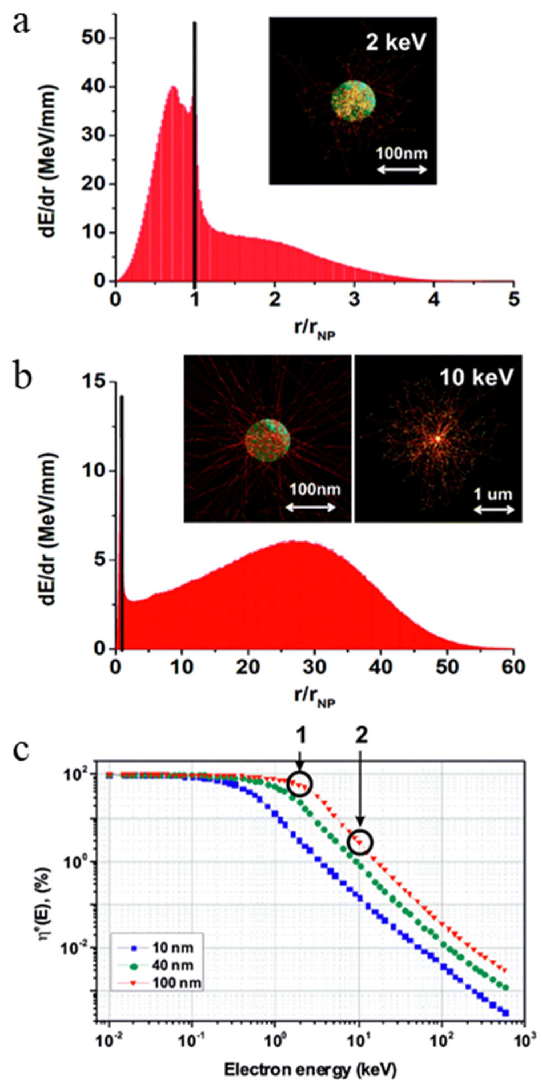
4. The Effect of the Energy Release vs. X-PDT Efficacy

The perspective of discovering novel materials mostly shaped in form of organic and inorganic nanoparticle or hybrids demands a deeper comprehension of the processes occurring within the material after the interaction with the ionizing radiation. Indeed, the reduced material dimension is critical in presence of high energy ionizing radiation, as the migration distance of secondary charges generated in the NPs along the track from the point of the ionizing event (relaxation mechanisms) are significantly larger than the common nanometric radius of the NPs. Therefore, a fraction of energy escapes from the NPs, limiting the efficiency of activation of the RT and X-PDT mechanisms [18,153].
The effective energy deposition in RT, together with the activation of the scintillating NPs and the trigger of the PS therapeutic effects in X-PDT, are key open question in nanosystems. In nanomedicine, the debate can regard the comprehension of the increase of X-rays absorption cross section, the effective energy release within the nanomaterials embedded in the biological surrounding, and the radiation dose-enhancement in the vicinity of the malignancy due to the presence of high-Z elements. The main goal of exploiting the passive NPs or single component active NPs is to increase the RT effects at the lowest doses in the disease site, and to localize the radiation energy deposition and RT damages only in the cancer site, while preserving from the radiation lethal toxicity the healthy tissue and organ in the patient [154,155]. As discussed in Section 5, this point can be addressed by creating tumor-targeting NPs, such as NPs functionalized by folic acid, able to accumulate only in the cancer tissue and thus to locally increase the absorbed dose [93,156]. Moreover, the question on the X-PDT processes in hybrids is under debate: here, besides the mechanism due to the presence of high-Z nanoscintillator, the effect of the photo-toxicity of the PS grafted on their surface should be considered, together with the possible interaction of the high energy beam directly with the PS. If on the one hand, different publications in the literature have demonstrated the occurrence of a synergetic effect given by the simultaneous application of the RT and the PDT in presence of PS grafted on NS [18,152,157], on the other hand the X-PDT phenomenology lacks unique explanation—i.e., whether the dominant therapeutic effect is due to the presence of high Z elements allowing for an enhancement of radiation dose deposition close to the injured tissues, or to the establishment of a tradeoff between the RT and PDT enabling simultaneously the reduction of the total dose to the patient and the induction of additional cell death mechanisms by toxic singlet oxygen moieties.
In this context, Monte Carlo simulations are powerful tools enabling to properly connect the clinical beam simulations to the phenomena expected to occur at the NP level. Many Monte Carlo codes have been developed [158,159] to explore the dose enhancement in RT, especially with X-ray beams, electron beams, and proton beams; each of these simulations helps to quantify the spatial energy distribution resulting from the interaction between the X-ray photon at diverse energies and the NP/NS, which finally drives the sensitization efficiency in RT and the activation of molecular singlet oxygen sensitizers in PDT and X-PDT. In addition, these computational studies allow to model the release of secondary particles after the first ionization event in relation to the nanomaterials physical, chemical, and structural properties. For instance, interesting computational studies have explored the use of passive high Z agents, like iodine, gadolinium, and gold, to treat human brain tumors in synchrotron stereotactic radiotherapy (SSRT) with irradiation by monochromatic X-rays from a synchrotron source, tuned at an optimal energy [160]. Especially, Edouard et al. in [161] have developed theoretical dosimetric investigation, by using a Monte Carlo code developed for neutrons, photons, and electrons transport (MCNPX) on an idealized phantom representing a human head bearing a brain tumor volume. Simulations of irradiation have been conducted for diverse beams: monochromatic X-ray beams from a synchrotron source (30–120 keV); polychromatic X-ray beams have been used to simulate irradiations from a conventional X-ray tube (80, 120, and 180 kVp), and from a linear accelerator (6 MV). The results on the SSRT dose enhancement have been compared for the diverse ionizing radiation energy ranges and in the presence of iodine elements. The enthusiastic data proves the major RT enhancement in presence of high Z agents for the medium energy beam by monochromatic source at 80 keV, despite the lower penetration of the keV X-rays with respect to the conventionally used higher energy X-rays. At keV energies, the maximum dose can be delivered to the tumor while preserving skull and healthy tissues from excessive radiations exposure. In particular, the simulation on X-ray penetration and dose deposition performed by Edouard and co-workers reveals that by using 6 MV irradiation, the doses delivered to the skull are similar to the value obtained for 80 keV from monochromatic source but, at the same time, the doses delivered to the healthy brain are hazardously higher if compared to low energy monochromatic and polychromatic beams. Moreover, the dose enhancement for 6 MV irradiation is almost negligible. This may be imputed to the different type of X-rays interaction with the biological medium, ruled by Compton and photoelectric effects working in the MeV and keV range, respectively. This work highlights the importance of computational modeling in order to find an optimized treatment in presence of radiosensitizers, able to significantly reduce the dose to the healthy tissue, hopefully avoiding the implication of megavoltage irradiation.
In another work, Bulin et al. in [162] have quantified, by Monte Carlo GEANT4 program, the amount of energy deposited in diverse scintillators such as gadolinium oxides (Gd2O3) nanoparticles, by varying the concentration of the NS in an aqueous medium, the NS dimension, and the energy of the first high energy beam, as well as the position of the first interaction event. In this work, the first estimation of the energy transfer from the primary interaction of an ionizing radiation beam with a sphere of water representing a volume of tumor, loaded with nanoscintillators, has been conducted by means of the mass energy absorption coefficient. The results reveal that the maximum energy transfer enhancement occurs for energies around 40–60 keV, confirming the importance of exploiting beams of ionizing radiation in the kiloelectronvolt energy range in presence of high-Z elements. However, Bulin and co-workers have highlighted that in such a composite compound, made by a large volume of water loaded with a small concentration of NS, the interaction between the first photon and the matter can occur either in the NS or in water. Especially, when the interaction occurs in the NS a fraction of the expected transferred energy can escape from the nanoparticle, typically due to the larger migration distance of the secondary particles generated after the first interaction with respect to the NS dimensions, and the deposited energy is shared between the water and the NS. In this context, the Monte Carlo GEANT4 calculation has helped to determine the spatial distribution of deposited energy in the volume surrounding the nanoparticle where the primary interaction occurs and to evaluate the fraction of energy loss during the energy relaxation and charge migration processes in the NS. The new parameter η has been introduced, which quantifies the percentage of energy that is deposited in the NS itself, as a function of both NS and external source parameters. The outcome of the computational investigation performed for three different NS diameters reveals that even for the nanoparticles of 100 nm of diameter, the deposited energy decreases very quickly when the incident beam energy increases, resulting in a large amount of energy loss outside the NS and deposited in the surrounding medium (see Figure 7). From this consideration, the estimation of η is crucial especially in the case of hybrid nanomaterials for X-PDT, where after the activation of the scintillating process in the nanoparticles by the external irradiation beam, the subsequent trigger of the singlet oxygen sensitizers is expected by energy transfer mechanisms (see Figure 4). The correct evaluation of the energy deposition is critical to optimize the singlet oxygen production in presence of NS for therapeutic purposes and to exploit the synergy among RT, radiosensitization and PDT.
The development of further theoretical studies devoted to the modeling of energy deposition in a biological medium and of scintillation phenomena in nanomaterials and hybrids is essential for the exploitation of NP/NS in nanomedicine. Computational simulations coupled with the experimental proofs would provide the guidelines for tuning the nanomaterial composition in order to optimize the interaction with X-rays and the activation of therapeutics or diagnostic agents, and thus to accomplish the different requirements according to the application target.

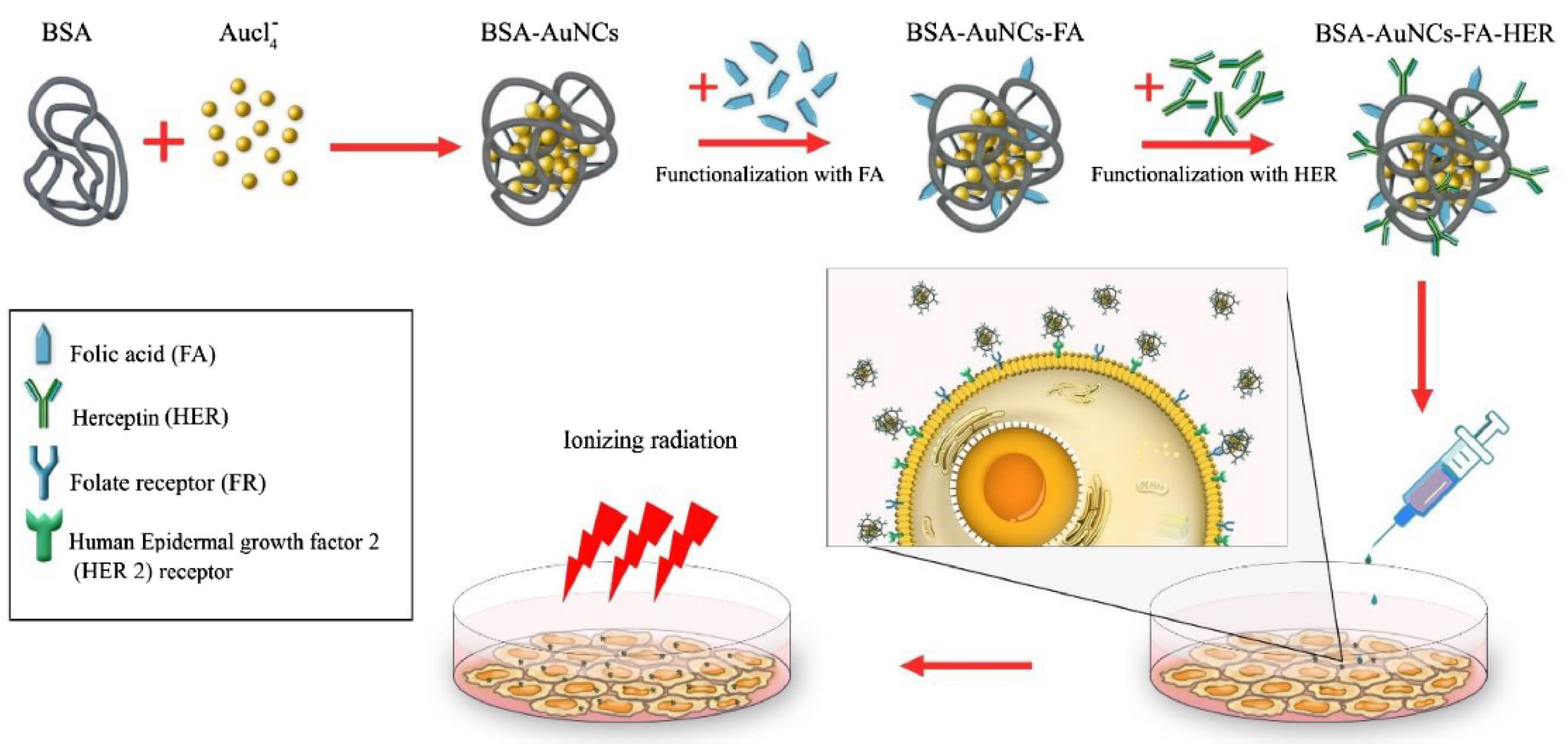
5. Targeting Strategies for Enhancing NPs Sensitizing Effects

Two important features are required to design effective nanotechnologies conceived for X-rays based oncological treatments: one is a selective tumor cell targeting and the other is a rapid clearance from human body after treatment [163,164]. These achievements are highly dependent on the size of the nanomaterials that can affect the efficacy of the treatment reducing its side effects (such as undesirable ROS generation, disruption of cellular compartments, immune reactions, inflammation, destruction of the host homeostasis) [165,166]: for instance, it has been reported that non-targeted heavy nanoparticles with diameter ~50 nm have the highest cellular uptake and are able to increase the efficacy of RT treatment [164,167]. Thanks to their size, nanoparticles tend to accumulate more in tumor tissues with respect to the normal ones due to the enhanced permeability and retention (EPR) effect [168]. This passive targeting exploits the fact that tumors have a leaky vasculature, different pH, and different local temperature, and are devoid of an efficient lymphatic drainage system. Therefore, nanoparticles with suitable physico-chemical characteristics and size can extravasate from leaky tumor vessels and accumulate in the tumors [169]. Passive targeting, however, may induce multi-drug resistance (MDR), a poor drug diffusion, and a-specific accumulation in liver and spleen [163,170]. One of the most promising approaches for improving the retention and accumulation of NPs in tumor cells relies on grafting the NPs surface with cancer cell-specific targeting ligands. Tumor accumulation of actively targeted nanoparticles has been found to be 5 times faster and approximately 2-fold higher than their passive counterparts, within the 60 nm diameter range [164]. Among the ligands used to target cancer cells we can mention antibodies, peptides, and aptamers to recognize and bind overexpressed cancer cell receptors (such as epidermal growth factor receptors), together with folate receptors and transferrin-receptors to enhance the cellular uptake of nanoparticles [171,172,173]. Some monoclonal antibodies have been exploited in strategies combining chemotherapeutic agents or immunotherapy with ionizing radiation; in vitro and in vivo results have proven that antibodies promote and potentiate apoptosis and cytotoxic effects, thus damaging DNA of the cancer cells. Successful medical outcomes have been obtained especially for the treatment of breast cancer [174,175,176]. In this context, the human epidermal receptor-2 (HER-2) is the most widely studied overexpressed targeting receptor for therapeutic applications [177]. The most commonly ligand targeted to HER-2, is the anti-HER-2 mAb [178]. The functionalization of gold nanoclusters (AuNCs) with anti-HER2-mAb and/or folic acid (FA) as a single and dual-targeted radiosensitizers has shown the increasing of cellular internalization and of the RT efficiency (Figure 8) [179].
Despite the fact that just an exceedingly small amount of antibody is needed to target a specific site, the expensive production and immunogenic properties may restrain their applications in clinical trials [180]. An alternative is represented by the aptamers that are attractive for in vivo visualization, as they can couple some diagnostic agents, such as fluorescent or radionuclide labels, bioconjugates and nanoparticles, for the delivery of therapeutic agents and as sensitizers in RT, especially for brain tumors [181,182]. Aptamer functionalized metal oxides and metals containing nanostructures have been proposed for tumor-targeted delivery and efficient PDT treatment [183,184]. ZnO catalyst NPs have been studied for targeted PDT therapy in combination with chemotherapy by loading the NPs with anti-cancer drugs. The results presented in [184] show an enhanced cellular uptake of the NPs and they validate the successful therapeutic cytotoxic effect with a higher rate of death of cancer cells, if compared to the one of single photo—or chemotherapy.
In spite of the significant advancements made in targeting strategies of NPs, potential harmful effects may occur depending on the route of delivery. With the systemic intravenous delivery, only a small fraction of NPs has the possibility of reaching the target site, even in case of targeted NPs. Thus, a large number of NPs could remain in systemic circulation, leading to drug accumulation or systemic toxicity, and affecting the health of patients [37,185]. For this reason, other delivery routes, such as localized delivery via intratumoral injection have been studied. Localized delivery can be advantageous particularly in tumor sites where conventional systemic delivery routes fail, enabling the accumulation of the delivered NP at higher concentrations in the targeted tumor volume. Consequently, the use NPs localized delivery strategies helps to avoid repeated dosing, to decrease the final toxicity, and to increase patient compliance [186]. To improve the beneficial effect of localized delivery strategies, recently NP-loaded or drug-embedded implants have been obtained from biodegradable polymers. NP can degrade into biocompatible compounds that are easily absorbed in the body with virtually no side effects [187,188].
The final evaluation of the anticancer performances of each type of nanoparticles relies on many factors. The radiotherapeutic performance of nanoparticles is affected by their intrinsic parameters, mainly size, shape, surface charge/functionalization, and toxicity, as well as by external factors, such as the targeted type of tumor, the administration methodology, and the irradiation setup [76,189,190]. From this picture coupled to the description of the issue on the energy/dose deposition in heavy or hybrid materials at the nanoscale (Section 3 and Section 4), it is clear that the comprehension of the physical phenomena occurring in NP exploited in X-rays-based oncotherapy, together with deeper studies for the engineering of nanoparticles performances and targeting, are mandatory. The research for a guideline towards the assessment of the relationship between the NPs key physico-chemical parameters and their therapeutic efficacy in RT is an open roundtable. However, it represents a challenging matter for the chance to design NPs with properties and performances that can be appropriate to address all the pending requests made by the clinical advanced therapies, to increase the survivor rate in oncology, and to improve the life quality of oncological patients.

6. Conclusions

The advanced nanomedicine is currently promoting the development of novel nanoparticles able to address the increasing demands for the fight against cancer. This paper proposes an overview of the inorganic and hybrid nanoparticles of interest for X-ray based oncological strategies. The aim is to highlight the characteristics that such nanoparticles should satisfy to be exploitable in oncology to promote the overcoming of the limitations of the traditional RT, with the following reduction of the collateral effects and improvement of the therapeutic efficacy. Indeed, this review points out that the physico-chemical properties of the nanoparticles, together with the choice on surface functionalization and the targeting strategies, affect the key parameters of RT and X-PDT, including energy deposition and total delivered dose in cancer tissues. Heavy nanoparticles or hybrids represent a successful chance in cancer fight as they can both assist the traditional RT as radiosensitizers and boost the therapeutic effects on malignant cells through the synergetic combination of radio and photodynamic therapies in X-PDT. The cutting-edge scientific research looks especially at the development of hybrid systems, whose properties are easily tunable with huge consequences for the therapeutic performances. However, the production of high-performing inorganic nanoparticles and nanohybrids for medical applications involving the use of ionizing radiation must be accompanied by the advancement in the knowledge on scintillation at the nanoscale, in correlation with the physical-chemical features of nanomaterials. In this context, this review shows the importance of the theoretical studies on the ionizing radiation energy deposition in biological medium in the presence of heavy nanoparticles, as well as on the stimulation of anticancer activities in hybrid nanoscintillators coupled to toxic single oxygen agents. These studies aim at guiding the optimization of nanoparticles’ qualities according to the specific medical applications. The future achievement of novel experimental knowledge and theoretical descriptions of the phenomena occurring in heavy and hybrids materials at nanoscale would represent a step beyond in the field of nanomedicine for the achievement of more and more efficient oncological X-rays based therapies.

Author Contributions

Conceptualization, I.V.; Supervision, I.V.; Writing—original draft, I.V., R.C., V.S.; Writing—review and editing, I.V., R.C., V.S., R.C. and V.S. have contributed equally. All authors have read and agreed to the published version of the manuscript.

Funding

This research was funded by Marie Skłodowska-Curie Actions Widening Fellowships (MSCA-WF): NUMBER 101003405—HANSOME.

Institutional Review Board Statement

Not applicable.

Informed Consent Statement

Not applicable.

Data Availability Statement

Not applicable.

Acknowledgments

This work was supported by (MSCA-WF)—HANSOME (101003405).

Conflicts of Interest

The authors declare no conflict of interest.

References

  1. Siegel, R.L.; Miller, K.D.; Jemal, A. Cancer statistics, 2020. CA A Cancer J. Clin. 2020, 70, 7–30. [Google Scholar] [CrossRef] [PubMed]
  2. Pucci, C.; Martinelli, C.; Ciofani, G. Innovative approaches for cancer treatment: Current perspectives and new challenges. Ecancermedicalscience 2019, 13, 961. [Google Scholar] [CrossRef] [PubMed]
  3. Massoud, T.F.; Gambhir, S.S. Molecular imaging in living subjects: Seeing fundamental biological processes in a new light. Genes Dev. 2003, 17, 545–580. [Google Scholar] [CrossRef] [PubMed] [Green Version]
  4. Lusic, H.; Grinstaff, M.W. X-ray-computed tomography contrast agents. Chem. Rev. 2013, 113, 1641–1666. [Google Scholar] [CrossRef] [Green Version]
  5. Vogelstein, B.; Papadopoulos, N.; Velculescu, V.E.; Zhou, S.; Diaz, L.A.; Kinzler, K.W. Cancer genome landscapes. Science 2013, 339, 1546–1558. [Google Scholar] [CrossRef]
  6. Braicu, C.; Buse, M.; Busuioc, C.; Drula, R.; Gulei, D.; Raduly, L.; Rusu, A.; Irimie, A.; Atanasov, A.G.; Slaby, O. A comprehensive review on MAPK: A promising therapeutic target in cancer. Cancers 2019, 11, 1618. [Google Scholar] [CrossRef] [Green Version]
  7. Smith-Bindman, R.; Miglioretti, D.L.; Larson, E.B. Rising use of diagnostic medical imaging in a large integrated health system. Health Aff. 2008, 27, 1491–1502. [Google Scholar] [CrossRef] [Green Version]
  8. Fazel, R.; Krumholz, H.M.; Wang, Y.; Ross, J.S.; Chen, J.; Ting, H.H.; Shah, N.D.; Nasir, K.; Einstein, A.J.; Nallamothu, B.K. Exposure to low-dose ionizing radiation from medical imaging procedures. N. Engl. J. Med. 2009, 361, 849–857. [Google Scholar] [CrossRef] [Green Version]
  9. Wan, J.C.; Massie, C.; Garcia-Corbacho, J.; Mouliere, F.; Brenton, J.D.; Caldas, C.; Pacey, S.; Baird, R.; Rosenfeld, N. Liquid biopsies come of age: Towards implementation of circulating tumour DNA. Nat. Rev. Cancer 2017, 17, 223. [Google Scholar] [CrossRef]
  10. Prasad, R.; Jain, N.; Conde, J.; Srivastava, R. Localized nanotheranostics. Mater. Today Adv. 2020, 8, 100087. [Google Scholar] [CrossRef]
  11. Beckett, K.R.; Moriarity, A.K.; Langer, J.M. Safe use of contrast media: What the radiologist needs to know. Radiographics 2015, 35, 1738–1750. [Google Scholar] [CrossRef] [PubMed]
  12. Wu, X.; Yang, H.; Yang, W.; Chen, X.; Gao, J.; Gong, X.; Wang, H.; Duan, Y.; Wei, D.; Chang, J. Nanoparticle-based diagnostic and therapeutic systems for brain tumors. J. Mater. Chem. B 2019, 7, 4734–4750. [Google Scholar] [CrossRef]
  13. Frullano, L.; Meade, T.J. Multimodal MRI contrast agents. JBIC J. Biol. Inorg. Chem. 2007, 12, 939–949. [Google Scholar] [CrossRef] [PubMed]
  14. Brigger, I.; Dubernet, C.; Couvreur, P. Nanoparticles in cancer therapy and diagnosis. Adv. Drug Deliv. Rev. 2012, 64, 24–36. [Google Scholar] [CrossRef]
  15. Nurgali, K.; Jagoe, R.T.; Abalo, R. Editorial: Adverse Effects of Cancer Chemotherapy: Anything New to Improve Tolerance and Reduce Sequelae? Front. Pharmacol. 2018, 9, 245. [Google Scholar] [CrossRef] [PubMed]
  16. Mahvi, D.A.; Liu, R.; Grinstaff, M.W.; Colson, Y.L.; Raut, C.P. Local cancer recurrence: The realities, challenges, and opportunities for new therapies. CA A Cancer J. Clin. 2018, 68, 488–505. [Google Scholar] [CrossRef] [Green Version]
  17. Lucky, S.S.; Soo, K.C.; Zhang, Y. Nanoparticles in photodynamic therapy. Chem. Rev. 2015, 115, 1990–2042. [Google Scholar] [CrossRef] [PubMed]
  18. Bulin, A.-L.; Broekgaarden, M.; Simeone, D.; Hasan, T. Low dose photodynamic therapy harmonizes with radiation therapy to induce beneficial effects on pancreatic heterocellular spheroids. Oncotarget 2019, 10, 2625–2643. [Google Scholar] [CrossRef] [PubMed] [Green Version]
  19. Weinmann, H. Cancer immunotherapy: Selected targets and small-molecule modulators. ChemMedChem 2016, 11, 450–466. [Google Scholar] [CrossRef]
  20. Finn, O. Immuno-oncology: Understanding the function and dysfunction of the immune system in cancer. Ann. Oncol. 2012, 23, viii6–viii9. [Google Scholar] [CrossRef]
  21. Wang, J.; Shi, M.; Ling, R.; Xia, Y.; Luo, S.; Fu, X.; Xiao, F.; Li, J.; Long, X.; Wang, J. Adjuvant chemotherapy and radiotherapy in triple-negative breast carcinoma: A prospective randomized controlled multi-center trial. Radiother. Oncol. 2011, 100, 200–204. [Google Scholar] [CrossRef]
  22. Ko, H.-J.; Kim, Y.-J.; Kim, Y.-S.; Chang, W.-S.; Ko, S.-Y.; Chang, S.-Y.; Sakaguchi, S.; Kang, C.-Y. A combination of chemoimmunotherapies can efficiently break self-tolerance and induce antitumor immunity in a tolerogenic murine tumor model. Cancer Res. 2007, 67, 7477–7486. [Google Scholar] [CrossRef] [PubMed] [Green Version]
  23. Kerr, W.G.; Chisholm, J.D. The next generation of immunotherapy for cancer: Small molecules could make big waves. J. Immunol. 2019, 202, 11–19. [Google Scholar] [CrossRef] [Green Version]
  24. Huck, B.R.; Kötzner, L.; Urbahns, K. Small molecules drive big improvements in immuno-oncology therapies. Angew. Chem. Int. Ed. 2018, 57, 4412–4428. [Google Scholar] [CrossRef] [Green Version]
  25. ClinicalTrials. Database of Privately and Publicly Funded Clinical Studies Conducted Around the World. Available online: https://clinicaltrials.gov (accessed on 30 July 2021).
  26. Whitesides, G.M. The ‘right’ size in nanobiotechnology. Nat. Biotechnol. 2003, 21, 1161–1165. [Google Scholar] [CrossRef]
  27. Ferrari, M. Cancer nanotechnology: Opportunities and challenges. Nat. Rev. Cancer 2005, 5, 161–171. [Google Scholar] [CrossRef]
  28. Doane, T.L.; Burda, C. The unique role of nanoparticles in nanomedicine: Imaging, drug delivery and therapy. Chem. Soc. Rev. 2012, 41, 2885–2911. [Google Scholar] [CrossRef] [PubMed]
  29. Rizzo, L.Y.; Theek, B.; Storm, G.; Kiessling, F.; Lammers, T. Recent progress in nanomedicine: Therapeutic, diagnostic and theranostic applications. Curr. Opin. Biotechnol. 2013, 24, 1159–1166. [Google Scholar] [CrossRef] [PubMed] [Green Version]
  30. Chen, H.; Zhang, W.; Zhu, G.; Xie, J.; Chen, X. Rethinking cancer nanotheranostics. Nat. Rev. Mater. 2017, 2, 1–18. [Google Scholar] [CrossRef] [PubMed]
  31. Shi, J.; Kantoff, P.W.; Wooster, R.; Farokhzad, O.C. Cancer nanomedicine: Progress, challenges and opportunities. Nat. Rev. Cancer 2017, 17, 20. [Google Scholar] [CrossRef] [PubMed]
  32. Dai Phung, C.; Tran, T.H.; Nguyen, H.T.; Jeong, J.-H.; Yong, C.S.; Kim, J.O. Current developments in nanotechnology for improved cancer treatment, focusing on tumor hypoxia. J. Control. Release 2020, 324, 413–429. [Google Scholar] [CrossRef]
  33. Kim, B.Y.; Rutka, J.T.; Chan, W.C. Nanomedicine. N. Engl. J. Med. 2010, 363, 2434–2443. [Google Scholar] [CrossRef] [Green Version]
  34. Furasova, A.D.; Fakhardo, A.F.; Milichko, V.A.; Tervoort, E.; Niederberger, M.; Vinogradov, V.V. Synthesis of a rare-earth doped hafnia hydrosol: Towards injectable luminescent nanocolloids. Colloids Surf. B Biointerfaces 2017, 154, 21–26. [Google Scholar] [CrossRef]
  35. Hao, Y.; Altundal, Y.; Moreau, M.; Sajo, E.; Kumar, R.; Ngwa, W. Potential for enhancing external beam radiotherapy for lung cancer using high-Z nanoparticles administered via inhalation. Phys. Med. Biol. 2015, 60, 7035. [Google Scholar] [CrossRef] [Green Version]
  36. Dong, Y.; Feng, S.-S. Poly (d, l-lactide-co-glycolide)/montmorillonite nanoparticles for oral delivery of anticancer drugs. Biomaterials 2005, 26, 6068–6076. [Google Scholar] [CrossRef]
  37. Chenthamara, D.; Subramaniam, S.; Ramakrishnan, S.G.; Krishnaswamy, S.; Essa, M.M.; Lin, F.-H.; Qoronfleh, M.W. Therapeutic efficacy of nanoparticles and routes of administration. Biomater. Res. 2019, 23, 1–29. [Google Scholar] [CrossRef] [PubMed]
  38. Bozzuto, G.; Molinari, A. Liposomes as nanomedical devices. Int. J. Nanomed. 2015, 10, 975. [Google Scholar] [CrossRef] [Green Version]
  39. Prabhu, R.H.; Patravale, V.B.; Joshi, M.D. Polymeric nanoparticles for targeted treatment in oncology: Current insights. Int. J. Nanomed. 2015, 10, 1001. [Google Scholar]
  40. Khalid, K.; Tan, X.; Mohd Zaid, H.F.; Tao, Y.; Lye Chew, C.; Chu, D.-T.; Lam, M.K.; Ho, Y.-C.; Lim, J.W.; Chin Wei, L. Advanced in developmental organic and inorganic nanomaterial: A review. Bioengineered 2020, 11, 328–355. [Google Scholar] [CrossRef] [PubMed] [Green Version]
  41. Lohse, S.E.; Murphy, C.J. Applications of colloidal inorganic nanoparticles: From medicine to energy. J. Am. Chem. Soc. 2012, 134, 15607–15620. [Google Scholar] [CrossRef]
  42. Zhao, C.-Y.; Cheng, R.; Yang, Z.; Tian, Z.-M. Nanotechnology for cancer therapy based on chemotherapy. Molecules 2018, 23, 826. [Google Scholar] [CrossRef] [PubMed] [Green Version]
  43. Ghosh, P.; Han, G.; De, M.; Kim, C.K.; Rotello, V.M. Gold nanoparticles in delivery applications. Adv. Drug Deliv. Rev. 2008, 60, 1307–1315. [Google Scholar] [CrossRef]
  44. Jaque, D.; Maestro, L.M.; Del Rosal, B.; Haro-Gonzalez, P.; Benayas, A.; Plaza, J.; Rodriguez, E.M.; Sole, J.G. Nanoparticles for photothermal therapies. Nanoscale 2014, 6, 9494–9530. [Google Scholar] [CrossRef]
  45. Maestro, L.M.; Haro-González, P.; Del Rosal, B.; Ramiro, J.; Caamano, A.; Carrasco, E.; Juarranz, A.; Sanz-Rodríguez, F.; Solé, J.G.; Jaque, D. Heating efficiency of multi-walled carbon nanotubes in the first and second biological windows. Nanoscale 2013, 5, 7882–7889. [Google Scholar] [CrossRef]
  46. Cline, B.; Delahunty, I.; Xie, J. Nanoparticles to mediate X-ray-induced photodynamic therapy and Cherenkov radiation photodynamic therapy. Wiley Interdiscip. Rev. Nanomed. Nanobiotechnol. 2019, 11, e1541. [Google Scholar] [CrossRef]
  47. Wong, X.Y.; Sena-Torralba, A.; Alvarez-Diduk, R.; Muthoosamy, K.; Merkoci, A. Nanomaterials for nanotheranostics: Tuning their properties according to disease needs. ACS Nano 2020, 14, 2585–2627. [Google Scholar] [CrossRef]
  48. Shang, L.; Nienhaus, K.; Nienhaus, G.U. Engineered nanoparticles interacting with cells: Size matters. J. Nanobiotechnol. 2014, 12, 1–11. [Google Scholar] [CrossRef] [PubMed] [Green Version]
  49. Salata, O.V. Applications of nanoparticles in biology and medicine. J. Nanobiotechnol. 2004, 2, 1–6. [Google Scholar] [CrossRef] [Green Version]
  50. Teleanu, D.M.; Chircov, C.; Grumezescu, A.M.; Volceanov, A.; Teleanu, R.I. Blood-brain delivery methods using nanotechnology. Pharmaceutics 2018, 10, 269. [Google Scholar] [CrossRef] [PubMed] [Green Version]
  51. Nam, J.; Won, N.; Bang, J.; Jin, H.; Park, J.; Jung, S.; Jung, S.; Park, Y.; Kim, S. Surface engineering of inorganic nanoparticles for imaging and therapy. Adv. Drug Deliv. Rev. 2013, 65, 622–648. [Google Scholar] [CrossRef]
  52. Sun, T.; Zhang, Y.S.; Pang, B.; Hyun, D.C.; Yang, M.; Xia, Y. Engineered nanoparticles for drug delivery in cancer therapy. Angew. Chem. Int. Ed. 2014, 53, 12320–12364. [Google Scholar] [CrossRef]
  53. Liu, Y.; Tu, D.; Zhu, H.; Chen, X. Lanthanide-doped luminescent nanoprobes: Controlled synthesis, optical spectroscopy, and bioapplications. Chem. Soc. Rev. 2013, 42, 6924–6958. [Google Scholar] [CrossRef]
  54. Labrador-Páez, L.; Pedroni, M.; Speghini, A.; García-Solé, J.; Haro-González, P.; Jaque, D. Reliability of rare-earth-doped infrared luminescent nanothermometers. Nanoscale 2018, 10, 22319–22328. [Google Scholar] [CrossRef]
  55. Villa, I.; Villa, C.; Monguzzi, A.; Babin, V.; Tervoort, E.; Nikl, M.; Niederberger, M.; Torrente, Y.; Vedda, A.; Lauria, A. Demonstration of cellular imaging by using luminescent and anti-cytotoxic europium-doped hafnia nanocrystals. Nanoscale 2018, 10, 7933–7940. [Google Scholar] [CrossRef]
  56. Gao, X.; Cui, Y.; Levenson, R.M.; Chung, L.W.; Nie, S. In vivo cancer targeting and imaging with semiconductor quantum dots. Nat. Biotechnol. 2004, 22, 969–976. [Google Scholar] [CrossRef]
  57. Li, L.; Wang, W.; Tang, J.; Wang, Y.; Liu, J.; Huang, L.; Wang, Y.; Guo, F.; Wang, J.; Shen, W. Classification, synthesis, and application of luminescent silica nanoparticles: A review. Nanoscale Res. Lett. 2019, 14, 1–23. [Google Scholar] [CrossRef]
  58. Villa, C.; Campione, M.; Santiago-González, B.; Alessandrini, F.; Erratico, S.; Zucca, I.; Bruzzone, M.G.; Forzenigo, L.; Malatesta, P.; Mauri, M. Self-assembled pH-sensitive fluoromagnetic nanotubes as archetype system for multimodal imaging of brain cancer. Adv. Funct. Mater. 2018, 28, 1707582. [Google Scholar] [CrossRef]
  59. Santiago-González, B.; Monguzzi, A.; Pinchetti, V.; Casu, A.; Prato, M.; Lorenzi, R.; Campione, M.; Chiodini, N.; Santambrogio, C.; Meinardi, F. “Quantized” Doping of Individual Colloidal Nanocrystals Using Size-Focused Metal Quantum Clusters. ACS Nano 2017, 11, 6233–6242. [Google Scholar] [CrossRef] [PubMed]
  60. Seferos, D.G.D.; Daniel, W.; Massich, M.; Patel, P.; Mirkin, C. Gold nanoparticles for biology and medicine. Angewchem. Int. Ed 2010, 49, 3280–3294. [Google Scholar]
  61. del Rosal, B.; Jia, B.; Jaque, D. Beyond phototherapy: Recent advances in multifunctional fluorescent nanoparticles for light-triggered tumor theranostics. Adv. Funct. Mater. 2018, 28, 1803733. [Google Scholar] [CrossRef]
  62. Yao, J.; Yang, M.; Duan, Y. Chemistry, biology, and medicine of fluorescent nanomaterials and related systems: New insights into biosensing, bioimaging, genomics, diagnostics, and therapy. Chem. Rev. 2014, 114, 6130–6178. [Google Scholar] [CrossRef]
  63. Taylor-Pashow, K.M.; Della Rocca, J.; Huxford, R.C.; Lin, W. Hybrid nanomaterials for biomedical applications. Chem. Commun. 2010, 46, 5832–5849. [Google Scholar] [CrossRef] [PubMed]
  64. Kwatra, D.; Venugopal, A.; Anant, S. Nanoparticles in radiation therapy: A summary of various approaches to enhance radiosensitization in cancer. Transl. Cancer Res. 2013, 2, 330–342. [Google Scholar]
  65. Clement, S.; Campbell, J.M.; Deng, W.; Guller, A.; Nisar, S.; Liu, G.; Wilson, B.C.; Goldys, E.M. Mechanisms for Tuning Engineered Nanomaterials to Enhance Radiation Therapy of Cancer. Adv. Sci. 2020, 7, 2003584. [Google Scholar] [CrossRef] [PubMed]
  66. Chen, H.H.; Kuo, M.T. Improving radiotherapy in cancer treatment: Promises and challenges. Oncotarget 2017, 8, 62742. [Google Scholar] [CrossRef] [PubMed] [Green Version]
  67. Baskar, R.; Lee, K.A.; Yeo, R.; Yeoh, K.-W. Cancer and radiation therapy: Current advances and future directions. Int. J. Med. Sci. 2012, 9, 193. [Google Scholar] [CrossRef] [Green Version]
  68. Delaney, G.; Jacob, S.; Featherstone, C.; Barton, M. The role of radiotherapy in cancer treatment: Estimating optimal utilization from a review of evidence-based clinical guidelines. Cancer: Interdiscip. Int. J. Am. Cancer Soc. 2005, 104, 1129–1137. [Google Scholar] [CrossRef] [PubMed]
  69. Gutt, R.; Dawson, G.; Cheuk, A.V.; Fosmire, H.; Moghanaki, D.; Kelly, M.; Jolly, S. Palliative radiotherapy for the management of metastatic cancer: Bone metastases, spinal cord compression, and brain metastases. Fed. Pract. 2015, 32, 12S. [Google Scholar]
  70. Behr, T.M.; Béhé, M.; Löhr, M.; Sgouros, G.; Angerstein, C.; Wehrmann, E.; Nebendahl, K.; Becker, W. Therapeutic advantages of Auger electron-over β-emitting radiometals or radioiodine when conjugated to internalizing antibodies. Eur. J. Nucl. Med. 2000, 27, 753–765. [Google Scholar] [CrossRef] [PubMed]
  71. Lutz, W.; Winston, K.R.; Maleki, N. A system for stereotactic radiosurgery with a linear accelerator. Int. J. Radiat. Oncol. Biol. Phys. 1988, 14, 373–381. [Google Scholar] [CrossRef]
  72. Hall, E.J.; Giaccia, A.J. Radiobiology for the Radiologist; Lippincott Williams & Wilkins: Philadelphia, PA, USA, 2006; Volume 6. [Google Scholar]
  73. Azzam, E.I.; Jay-Gerin, J.-P.; Pain, D. Ionizing radiation-induced metabolic oxidative stress and prolonged cell injury. Cancer Lett. 2012, 327, 48–60. [Google Scholar] [CrossRef] [Green Version]
  74. Voyant, C.; Julian, D.; Roustit, R.; Biffi, K.; Lantieri, C. Biological effects and equivalent doses in radiotherapy: A software solution. Rep. Pract. Oncol. Radiother. 2014, 19, 47–55. [Google Scholar] [CrossRef] [PubMed] [Green Version]
  75. Moding, E.J.; Kastan, M.B.; Kirsch, D.G. Strategies for optimizing the response of cancer and normal tissues to radiation. Nat. Rev. Drug Discov. 2013, 12, 526–542. [Google Scholar] [CrossRef] [PubMed] [Green Version]
  76. Retif, P.; Pinel, S.; Toussaint, M.; Frochot, C.; Chouikrat, R.; Bastogne, T.; Barberi-Heyob, M. Nanoparticles for Radiation Therapy Enhancement: The Key Parameters. Theranostics 2015, 5, 1030–1044. [Google Scholar] [CrossRef] [Green Version]
  77. Kuncic, Z.; Lacombe, S. Nanoparticle radio-enhancement: Principles, progress and application to cancer treatment. Phys. Med. Biol. 2018, 63, 02TR01. [Google Scholar] [CrossRef] [PubMed]
  78. Knoll, G.F. Radiation Detection and Measurement; John Wiley & Sons: Hoboken, NJ, USA, 2010. [Google Scholar]
  79. Niemantsverdriet, M.; van Goethem, M.-J.; Bron, R.; Hogewerf, W.; Brandenburg, S.; Langendijk, J.A.; van Luijk, P.; Coppes, R.P. High and low LET radiation differentially induce normal tissue damage signals. Int. J. Radiat. Oncol. Biol. Phys. 2012, 83, 1291–1297. [Google Scholar] [CrossRef] [PubMed] [Green Version]
  80. Ku, A.; Facca, V.J.; Cai, Z.; Reilly, R.M. Auger electrons for cancer therapy—A review. EJNMMI Radiopharm. Chem. 2019, 4, 1–36. [Google Scholar] [CrossRef] [PubMed] [Green Version]
  81. Choi, J.; Kim, G.; Cho, S.B.; Im, H.-J. Radiosensitizing high-Z metal nanoparticles for enhanced radiotherapy of glioblastoma multiforme. J. Nanobiotechnol. 2020, 18, 1–23. [Google Scholar] [CrossRef] [PubMed]
  82. Thoraeus, R. Attenuation of Gamma Radiation from 60Co, 137Cs, 192Ir, and 226Ra in Various Materials Used in Radiotherapy. Acta Radiol. Ther. Phys. Biol. 1965, 3, 81–86. [Google Scholar] [CrossRef]
  83. Allal, A.S.; Michel Richter, M.; Russo, M.; Rouzaud, M.; Dulguerov, P.; Kurtz, J.M. Dose variation at bone/titanium interfaces using titanium hollow screw osseointegrating reconstruction plates. Int. J. Radiat. Oncol. Biol. Phys. 1998, 40, 215–219. [Google Scholar] [CrossRef]
  84. Melian, E.; Fatyga, M.; Lam, P.; Steinberg, M.; Reddy, S.P.; Petruzzelli, G.J.; Glasgow, G.P. Effect of metal reconstruction plates on cobalt-60 dose distribution: A predictive formula and clinical implications. Int. J. Radiat. Oncol. Biol. Phys. 1999, 44, 725–730. [Google Scholar] [CrossRef]
  85. Pottier, A.; Borghi, E.; Levy, L. New use of metals as nanosized radioenhancers. Anticancer Res. 2014, 34, 443–453. [Google Scholar] [PubMed]
  86. Ahmed, S.; Rao, A.G.; Sankarshan, B.; Vicas, C.; Namratha, K.; Umesh, T.; Somashekar, R.; Byrappa, K. Evaluation of Gold, Silver and Silver–Gold (bimetallic) nanoparticles as radiosensitizers for radiation therapy in cancer treatment. Cancer Oncol. Res 2016, 4, 42–51. [Google Scholar] [CrossRef]
  87. Liu, Y.; Zhang, P.; Li, F.; Jin, X.; Li, J.; Chen, W.; Li, Q. Metal-based nanoenhancers for future radiotherapy: Radiosensitizing and synergistic effects on tumor cells. Theranostics 2018, 8, 1824. [Google Scholar] [CrossRef]
  88. Singh, P.; Pandit, S.; Mokkapati, V.; Garg, A.; Ravikumar, V.; Mijakovic, I. Gold nanoparticles in diagnostics and therapeutics for human cancer. Int. J. Mol. Sci. 2018, 19, 1979. [Google Scholar] [CrossRef]
  89. Carter, J.D.; Cheng, N.N.; Qu, Y.; Suarez, G.D.; Guo, T. Nanoscale energy deposition by X-ray absorbing nanostructures. J. Phys. Chem. B 2007, 111, 11622–11625. [Google Scholar] [CrossRef] [PubMed]
  90. Hainfeld, J.F.; Dilmanian, F.A.; Slatkin, D.N.; Smilowitz, H.M. Radiotherapy enhancement with gold nanoparticles. J. Pharm. Pharmacol. 2008, 60, 977–985. [Google Scholar] [CrossRef] [PubMed] [Green Version]
  91. Herold, D.M.; Das, I.J.; Stobbe, C.C.; Iyer, R.V.; Chapman, J.D. Gold microspheres: A selective technique for producing biologically effective dose enhancement. Int. J. Radiat. Biol. 2000, 76, 1357–1364. [Google Scholar]
  92. Liu, Y.; Liu, X.; Jin, X.; He, P.; Zheng, X.; Dai, Z.; Ye, F.; Zhao, T.; Chen, W.; Li, Q. The dependence of radiation enhancement effect on the concentration of gold nanoparticles exposed to low-and high-LET radiations. Phys. Med. 2015, 31, 210–218. [Google Scholar] [CrossRef]
  93. Babaei, M.; Ganjalikhani, M. The potential effectiveness of nanoparticles as radio sensitizers for radiotherapy. BioImpacts BI 2014, 4, 15. [Google Scholar]
  94. Cooper, D.R.; Bekah, D.; Nadeau, J.L. Gold nanoparticles and their alternatives for radiation therapy enhancement. Front. Chem. 2014, 2, 86. [Google Scholar] [CrossRef] [Green Version]
  95. Liu, X.; Liu, Y.; Zhang, P.; Jin, X.; Zheng, X.; Ye, F.; Chen, W.; Li, Q. The synergistic radiosensitizing effect of tirapazamine-conjugated gold nanoparticles on human hepatoma HepG2 cells under X-ray irradiation. Int. J. Nanomed. 2016, 11, 3517. [Google Scholar] [CrossRef] [PubMed] [Green Version]
  96. Cui, L.; Her, S.; Borst, G.R.; Bristow, R.G.; Jaffray, D.A.; Allen, C. Radiosensitization by gold nanoparticles: Will they ever make it to the clinic? Radiother. Oncol. 2017, 124, 344–356. [Google Scholar] [CrossRef] [PubMed]
  97. Toossi, M.T.B.; Ghorbani, M.; Mehrpouyan, M.; Akbari, F.; Sabet, L.S.; Meigooni, A.S. A Monte Carlo study on tissue dose enhancement in brachytherapy: A comparison between gadolinium and gold nanoparticles. Australas. Phys. Eng. Sci. Med. 2012, 35, 177–185. [Google Scholar] [CrossRef] [PubMed]
  98. Delorme, R.; Taupin, F.; Flaender, M.; Ravanat, J.L.; Champion, C.; Agelou, M.; Elleaume, H. Comparison of gadolinium nanoparticles and molecular contrast agents for radiation therapy-enhancement. Med. Phys. 2017, 44, 5949–5960. [Google Scholar] [CrossRef] [PubMed]
  99. Luchette, M.; Korideck, H.; Makrigiorgos, M.; Tillement, O.; Berbeco, R. Radiation dose enhancement of gadolinium-based AGuIX nanoparticles on HeLa cells. Nanomed. Nanotechnol. Biol. Med. 2014, 10, 1751–1755. [Google Scholar] [CrossRef] [PubMed]
  100. Porcel, E.; Liehn, S.; Remita, H.; Usami, N.; Kobayashi, K.; Furusawa, Y.; Le Sech, C.; Lacombe, S. Platinum nanoparticles: A promising material for future cancer therapy? Nanotechnology 2010, 21, 085103. [Google Scholar] [CrossRef] [PubMed]
  101. Li, Y.; Yun, K.-H.; Lee, H.; Goh, S.-H.; Suh, Y.-G.; Choi, Y. Porous platinum nanoparticles as a high-Z and oxygen generating nanozyme for enhanced radiotherapy in vivo. Biomaterials 2019, 197, 12–19. [Google Scholar] [CrossRef]
  102. Maggiorella, L.; Barouch, G.; Devaux, C.; Pottier, A.; Deutsch, E.; Bourhis, J.; Borghi, E.; Levy, L. Nanoscale radiotherapy with hafnium oxide nanoparticles. Future Oncol. 2012, 8, 1167–1181. [Google Scholar] [CrossRef] [PubMed]
  103. Zhang, P.; Darmon, A.; Marill, J.; Anesary, N.M.; Paris, S. Radiotherapy-activated hafnium oxide nanoparticles produce abscopal effect in a mouse colorectal cancer model. Int. J. Nanomed. 2020, 15, 3843. [Google Scholar] [CrossRef] [PubMed]
  104. Guerreiro, A.; Chatterton, N.; Crabb, E.M.; Golding, J.P. A comparison of the radiosensitisation ability of 22 different element metal oxide nanoparticles using clinical megavoltage X-rays. Cancer Nanotechnol. 2019, 10, 1–20. [Google Scholar] [CrossRef]
  105. Welsher, K.; Sherlock, S.P.; Dai, H. Deep-tissue anatomical imaging of mice using carbon nanotube fluorophores in the second near-infrared window. Proc. Natl. Acad. Sci. USA 2011, 108, 8943–8948. [Google Scholar] [CrossRef] [Green Version]
  106. Kamkaew, A.; Chen, F.; Zhan, Y.; Majewski, R.L.; Cai, W. Scintillating nanoparticles as energy mediators for enhanced photodynamic therapy. ACS Nano 2016, 10, 3918–3935. [Google Scholar] [CrossRef] [Green Version]
  107. Hoertz, P.G.; Magnus-Aryitey, D.; Gupta, V.; Norton, C.; Doorn, S.; Ennis, T. Photocatalytic and radiocatalytic nanomaterials for the degradation of organicspecies. Radiat. Phys. Chem. 2013, 84, 51–58. [Google Scholar] [CrossRef]
  108. Nosaka, Y.; Daimon, T.; Nosaka, A.Y.; Murakami, Y. Singlet oxygen formation in photocatalytic TiO2 aqueous suspension. Phys. Chem. Chem. Phys. 2004, 6, 2917–2918. [Google Scholar] [CrossRef]
  109. Fujishima, A.; Zhang, X.; Tryk, D.A. TiO2 photocatalysis and related surface phenomena. Surf. Sci. Rep. 2008, 63, 515–582. [Google Scholar] [CrossRef]
  110. Linsebigler, A.L.; Lu, G.; Yates, J.T., Jr. Photocatalysis on TiO2 surfaces: Principles, mechanisms, and selected results. Chem. Rev. 1995, 95, 735–758. [Google Scholar] [CrossRef]
  111. Higgins, M.M.; Banu, A.; Pendleton, S.; Rojas, J. Radiocatalytic performance of oxide-based nanoparticles for targeted therapy and water remediation. Radiat. Phys. Chem. 2020, 173, 108871. [Google Scholar] [CrossRef]
  112. Barcellos-Hoff, M.H.; Park, C.; Wright, E.G. Radiation and the microenvironment–tumorigenesis and therapy. Nat. Rev. Cancer 2005, 5, 867–875. [Google Scholar] [CrossRef] [Green Version]
  113. D’Arienzo, M.; Mostoni, S.; Crapanzano, R.; Cepek, C.; Di Credico, B.; Fasoli, M.; Polizzi, S.; Vedda, A.; Villa, I.; Scotti, R. Insight into the influence of ZnO defectivity on the catalytic generation of environmentally persistent free radicals in ZnO/SiO2 systems. J. Phys. Chem. C 2019, 123, 21651–21661. [Google Scholar] [CrossRef]
  114. Wang, D.; Xie, T.; Li, Y. Nanocrystals: Solution-based synthesis and applications as nanocatalysts. Nano Res. 2009, 2, 30–46. [Google Scholar] [CrossRef] [Green Version]
  115. Yin, H.; Casey, P.S.; McCall, M.J.; Fenech, M. Effects of surface chemistry on cytotoxicity, genotoxicity, and the generation of reactive oxygen species induced by ZnO nanoparticles. Langmuir 2010, 26, 15399–15408. [Google Scholar] [CrossRef]
  116. Azizi-Lalabadi, M.; Ehsani, A.; Divband, B.; Alizadeh-Sani, M. Antimicrobial activity of Titanium dioxide and Zinc oxide nanoparticles supported in 4A zeolite and evaluation the morphological characteristic. Sci. Rep. 2019, 9, 1–10. [Google Scholar] [CrossRef] [Green Version]
  117. Bogdan, J.; Pławińska-Czarnak, J.; Zarzyńska, J. Nanoparticles of titanium and zinc oxides as novel agents in tumor treatment: A review. Nanoscale Res. Lett. 2017, 12, 1–15. [Google Scholar] [CrossRef] [PubMed] [Green Version]
  118. Yang, B.; Chen, Y.; Shi, J. Reactive oxygen species (ROS)-based nanomedicine. Chem. Rev. 2019, 119, 4881–4985. [Google Scholar] [CrossRef] [PubMed]
  119. Abrahamse, H.; Hamblin, M.R. New photosensitizers for photodynamic therapy. Biochem. J. 2016, 473, 347–364. [Google Scholar] [CrossRef] [PubMed] [Green Version]
  120. Zhou, Z.; Song, J.; Nie, L.; Chen, X. Reactive oxygen species generating systems meeting challenges of photodynamic cancer therapy. Chem. Soc. Rev. 2016, 45, 6597–6626. [Google Scholar] [CrossRef] [PubMed] [Green Version]
  121. Youkhana, E.Q.; Feltis, B.; Blencowe, A.; Geso, M. Titanium dioxide nanoparticles as radiosensitisers: An in vitro and phantom-based study. Int. J. Med. Sci. 2017, 14, 602. [Google Scholar] [CrossRef] [Green Version]
  122. Higgins, M.C.M.; Clifford, D.M.; Rojas, J.V. Au@ TiO2 nanocomposites synthesized by X-ray radiolysis as potential radiosensitizers. Appl. Surf. Sci. 2018, 427, 702–710. [Google Scholar] [CrossRef]
  123. Generalov, R.; Kuan, W.B.; Chen, W.; Kristensen, S.; Juzenas, P. Radiosensitizing effect of zinc oxide and silica nanocomposites on cancer cells. Colloids Surf. B Biointerfaces 2015, 129, 79–86. [Google Scholar] [CrossRef]
  124. Sharma, H.; Kumar, K.; Choudhary, C.; Mishra, P.K.; Vaidya, B. Development and characterization of metal oxide nanoparticles for the delivery of anticancer drug. Artif. Cells Nanomed. Biotechnol. 2016, 44, 672–679. [Google Scholar] [CrossRef]
  125. Mahmoudi, M.; Sant, S.; Wang, B.; Laurent, S.; Sen, T. Superparamagnetic iron oxide nanoparticles (SPIONs): Development, surface modification and applications in chemotherapy. Adv. Drug Deliv. Rev. 2011, 63, 24–46. [Google Scholar] [CrossRef] [Green Version]
  126. Wason, M.S.; Colon, J.; Das, S.; Seal, S.; Turkson, J.; Zhao, J.; Baker, C.H. Sensitization of pancreatic cancer cells to radiation by cerium oxide nanoparticle-induced ROS production. Nanomed. Nanotechnol. Biol. Med. 2013, 9, 558–569. [Google Scholar] [CrossRef] [Green Version]
  127. Lan, M.; Zhao, S.; Liu, W.; Lee, C.S.; Zhang, W.; Wang, P. Photosensitizers for photodynamic therapy. Adv. Healthc. Mater. 2019, 8, 1900132. [Google Scholar] [CrossRef] [PubMed]
  128. Mallidi, S.; Anbil, S.; Bulin, A.-L.; Obaid, G.; Ichikawa, M.; Hasan, T. Beyond the barriers of light penetration: Strategies, perspectives and possibilities for photodynamic therapy. Theranostics 2016, 6, 2458. [Google Scholar] [CrossRef] [Green Version]
  129. Brown, S.B.; Brown, E.A.; Walker, I. The present and future role of photodynamic therapy in cancer treatment. Lancet Oncol. 2004, 5, 497–508. [Google Scholar] [CrossRef]
  130. Allison, R.R.; Sibata, C.H. Oncologic photodynamic therapy photosensitizers: A clinical review. Photodiagnosis Photodyn. Ther. 2010, 7, 61–75. [Google Scholar] [CrossRef]
  131. Dolmans, D.E.; Fukumura, D.; Jain, R.K. Photodynamic therapy for cancer. Nat. Rev. Cancer 2003, 3, 380–387. [Google Scholar] [CrossRef]
  132. Park, J.; Lee, Y.-K.; Park, I.-K.; Hwang, S.R. Current Limitations and Recent Progress in Nanomedicine for Clinically Available Photodynamic Therapy. Biomedicines 2021, 9, 85. [Google Scholar] [CrossRef] [PubMed]
  133. Allison, R.R.; Downie, G.H.; Cuenca, R.; Hu, X.-H.; Childs, C.J.; Sibata, C.H. Photosensitizers in clinical PDT. Photodiagnosis Photodyn. Ther. 2004, 1, 27–42. [Google Scholar] [CrossRef]
  134. Kou, J.; Dou, D.; Yang, L. Porphyrin photosensitizers in photodynamic therapy and its applications. Oncotarget 2017, 8, 81591. [Google Scholar] [CrossRef] [PubMed] [Green Version]
  135. DeRosa, M.C.; Crutchley, R.J. Photosensitized singlet oxygen and its applications. Coord. Chem. Rev. 2002, 233, 351–371. [Google Scholar] [CrossRef]
  136. Luksiene, Z.; Kalvelyte, A.; Supino, R. On the combination of photodynamic therapy with ionizing radiation. J. Photochem. Photobiol. B: Biol. 1999, 52, 35–42. [Google Scholar] [CrossRef]
  137. Schwartz, S.; Absolon, K.; Vermund, H. Some relationships of porphyrins, X-rays and tumors. Univ. Minn. Med. Bull 1955, 27, 1–37. [Google Scholar]
  138. Larue, L.; Mihoub, A.B.; Youssef, Z.; Colombeau, L.; Acherar, S.; André, J.-C.; Arnoux, P.; Baros, F.; Vermandel, M.; Frochot, C. Using X-rays in photodynamic therapy: An overview. Photochem. Photobiol. Sci. 2018, 17, 1612–1650. [Google Scholar] [CrossRef] [PubMed]
  139. Vasil’ev, A.N. Microtheory of scintillation in crystalline materials. In Proceedings of the International Conference on Engineering of Scintillation Materials and Radiation Technologies, Minsk, Belarus, 26–30 January 2016; pp. 3–34. [Google Scholar]
  140. Dujardin, C.; Auffray, E.; Bourret-Courchesne, E.; Dorenbos, P.; Lecoq, P.; Nikl, M.; Vasil’ev, A.N.; Yoshikawa, A.; Zhu, R. Needs, Trends, and Advances in Inorganic Scintillators. IEEE Trans. Nucl. Sci. 2018, 65, 1977–1997. [Google Scholar] [CrossRef] [Green Version]
  141. Villa, I.; Moretti, F.; Fasoli, M.; Rossi, A.; Hattendorf, B.; Dujardin, C.; Niederberger, M.; Vedda, A.; Lauria, A. The Bright X-Ray Stimulated Luminescence of HfO2 Nanocrystals Activated by Ti Ions. Adv. Opt. Mater. 2020, 8, 1901348. [Google Scholar] [CrossRef]
  142. Bulin, A.-L.; Truillet, C.; Chouikrat, R.; Lux, F.o.; Frochot, C.l.; Amans, D.; Ledoux, G.; Tillement, O.; Perriat, P.; Barberi-Heyob, M. X-ray-induced singlet oxygen activation with nanoscintillator-coupled porphyrins. J. Phys. Chem. C 2013, 117, 21583–21589. [Google Scholar] [CrossRef]
  143. Procházková, L.; Pelikánová, I.T.; Mihóková, E.; Dědic, R.; Čuba, V. Novel scintillating nanocomposite for X-ray induced photodynamic therapy. Radiat. Meas. 2019, 121, 13–17. [Google Scholar] [CrossRef]
  144. Liu, Y.; Chen, W.; Wang, S.; Joly, A.G. Investigation of water-soluble x-ray luminescence nanoparticles for photodynamic activation. Appl. Phys. Lett. 2008, 92, 043901. [Google Scholar] [CrossRef]
  145. Rossi, F.; Bedogni, E.; Bigi, F.; Rimoldi, T.; Cristofolini, L.; Pinelli, S.; Alinovi, R.; Negri, M.; Dhanabalan, S.C.; Attolini, G.; et al. Porphyrin conjugated SiC/SiOx nanowires for X-ray-excited photodynamic therapy. Sci. Rep. 2015, 5, 7606. [Google Scholar] [CrossRef] [PubMed]
  146. Yang, W.; Read, P.W.; Mi, J.; Baisden, J.M.; Reardon, K.A.; Larner, J.M.; Helmke, B.P.; Sheng, K. Semiconductor nanoparticles as energy mediators for photosensitizer-enhanced radiotherapy. Int. J. Radiat. Oncol. Biol. Phys. 2008, 72, 633–635. [Google Scholar] [CrossRef] [PubMed]
  147. Ren, X.-D.; Hao, X.-Y.; Li, H.-C.; Ke, M.-R.; Zheng, B.-Y.; Huang, J.-D. Progress in the development of nanosensitizers for X-ray-induced photodynamic therapy. Drug Discov. Today 2018, 23, 1791–1800. [Google Scholar] [CrossRef]
  148. Sun, W.; Zhou, Z.; Pratx, G.; Chen, X.; Chen, H. Nanoscintillator-mediated X-ray induced photodynamic therapy for deep-seated tumors: From concept to biomedical applications. Theranostics 2020, 10, 1296. [Google Scholar] [CrossRef] [PubMed]
  149. Villa, I.; Villa, C.; Crapanzano, R.; Secchi, V.; Tawfilas, M.; Trombetta, E.; Porretti, L.; Brambilla, A.; Campione, M.; Torrente, Y. Functionalized Scintillating Nanotubes for Simultaneous Radio-and Photodynamic Therapy of Cancer. ACS Appl. Mater. Interfaces 2021, 13, 12997–13008. [Google Scholar] [CrossRef]
  150. Perego, J.; Villa, I.; Pedrini, A.; Padovani, E.; Crapanzano, R.; Vedda, A.; Dujardin, C.; Bezuidenhout, C.X.; Bracco, S.; Sozzani, P. Composite fast scintillators based on high-Z fluorescent metal–organic framework nanocrystals. Nat. Photonics 2021, 15, 393–400. [Google Scholar] [CrossRef]
  151. Zhang, X.; Wasson, M.C.; Shayan, M.; Berdichevsky, E.K.; Ricardo-Noordberg, J.; Singh, Z.; Papazyan, E.K.; Castro, A.J.; Marino, P.; Ajoyan, Z. A historical perspective on porphyrin-based metal–organic frameworks and their applications. Coord. Chem. Rev. 2020, 429, 213615. [Google Scholar] [CrossRef]
  152. Lu, K.; He, C.; Guo, N.; Chan, C.; Ni, K.; Lan, G.; Tang, H.; Pelizzari, C.; Fu, Y.-X.; Spiotto, M.T. Low-dose X-ray radiotherapy–radiodynamic therapy via nanoscale metal–organic frameworks enhances checkpoint blockade immunotherapy. Nat. Biomed. Eng. 2018, 2, 600–610. [Google Scholar] [CrossRef] [PubMed]
  153. Morgan, N.Y.; Kramer-Marek, G.; Smith, P.D.; Camphausen, K.; Capala, J. Nanoscintillator conjugates as photodynamic therapy-based radiosensitizers: Calculation of required physical parameters. Radiat. Res. 2009, 171, 236–244. [Google Scholar] [CrossRef] [Green Version]
  154. McMahon, S.J.; Hyland, W.B.; Muir, M.F.; Coulter, J.A.; Jain, S.; Butterworth, K.T.; Schettino, G.; Dickson, G.R.; Hounsell, A.R.; O’sullivan, J.M. Biological consequences of nanoscale energy deposition near irradiated heavy atom nanoparticles. Sci. Rep. 2011, 1, 1–10. [Google Scholar] [CrossRef] [Green Version]
  155. Banaee, N. Enhanced dose measurement of zinc oxide nanoparticles by radiochromic polymer dosimeter and Monte Carlo simulation. Rep. Pract. Oncol. Radiother. 2020, 25, 515–520. [Google Scholar] [CrossRef]
  156. Khoshgard, K.; Hashemi, B.; Arbabi, A.; Rasaee, M.J.; Soleimani, M. Radiosensitization effect of folate-conjugated gold nanoparticles on HeLa cancer cells under orthovoltage superficial radiotherapy techniques. Phys. Med. Biol. 2014, 59, 2249–2263. [Google Scholar] [CrossRef] [PubMed]
  157. Bulin, A.L.; Broekgaarden, M.; Chaput, F.; Baisamy, V.; Garrevoet, J.; Busser, B.; Brueckner, D.; Youssef, A.; Ravanat, J.L.; Dujardin, C. Radiation Dose-Enhancement Is a Potent Radiotherapeutic Effect of Rare-Earth Composite Nanoscintillators in Preclinical Models of Glioblastoma. Adv. Sci. 2020, 7, 2001675. [Google Scholar] [CrossRef]
  158. Incerti, S.; Douglass, M.; Penfold, S.; Guatelli, S.; Bezak, E. Review of Geant4-DNA applications for micro and nanoscale simulations. Phys. Med. 2016, 32, 1187–1200. [Google Scholar] [CrossRef] [PubMed] [Green Version]
  159. Sakata, D.; Kyriakou, I.; Okada, S.; Tran, H.N.; Lampe, N.; Guatelli, S.; Bordage, M.C.; Ivanchenko, V.; Murakami, K.; Sasaki, T. Geant4-DNA track-structure simulations for gold nanoparticles: The importance of electron discrete models in nanometer volumes. Med. Phys. 2018, 45, 2230–2242. [Google Scholar] [CrossRef] [Green Version]
  160. Boudou, C.; Balosso, J.; Estève, F.; Elleaume, H. Monte Carlo dosimetry for synchrotron stereotactic radiotherapy of brain tumours. Phys. Med. Biol. 2005, 50, 4841. [Google Scholar] [CrossRef] [PubMed]
  161. Edouard, M.; Broggio, D.; Prezado, Y.; Estève, F.; Elleaume, H.; Adam, J.-F. Treatment plans optimization for contrast-enhanced synchrotron stereotactic radiotherapy. Med. Phys. 2010, 37, 2445–2456. [Google Scholar] [CrossRef] [PubMed]
  162. Bulin, A.-L.; Vasil’Ev, A.; Belsky, A.; Amans, D.; Ledoux, G.; Dujardin, C. Modelling energy deposition in nanoscintillators to predict the efficiency of the X-ray-induced photodynamic effect. Nanoscale 2015, 7, 5744–5751. [Google Scholar] [CrossRef]
  163. De Jong, W.H.; Hagens, W.I.; Krystek, P.; Burger, M.C.; Sips, A.J.; Geertsma, R.E. Particle size-dependent organ distribution of gold nanoparticles after intravenous administration. Biomaterials 2008, 29, 1912–1919. [Google Scholar] [CrossRef]
  164. Sykes, E.A.; Chen, J.; Zheng, G.; Chan, W.C. Investigating the impact of nanoparticle size on active and passive tumor targeting efficiency. ACS Nano 2014, 8, 5696–5706. [Google Scholar] [CrossRef] [PubMed]
  165. Her, S.; Jaffray, D.A.; Allen, C. Gold nanoparticles for applications in cancer radiotherapy: Mechanisms and recent advancements. Adv. Drug Deliv. Rev. 2017, 109, 84–101. [Google Scholar] [CrossRef] [PubMed]
  166. Wolfram, J.; Zhu, M.; Yang, Y.; Shen, J.; Gentile, E.; Paolino, D.; Fresta, M.; Nie, G.; Chen, C.; Shen, H. Safety of nanoparticles in medicine. Curr. Drug Targets 2015, 16, 1671–1681. [Google Scholar] [CrossRef] [PubMed] [Green Version]
  167. Sah, B.; Antosh, M.P. Effect of size on gold nanoparticles in radiation therapy: Uptake and survival effects. J. Nano Med. 2019, 2, 1013. [Google Scholar]
  168. Shi, Y.; Van der Meel, R.; Chen, X.; Lammers, T. The EPR effect and beyond: Strategies to improve tumor targeting and cancer nanomedicine treatment efficacy. Theranostics 2020, 10, 7921. [Google Scholar] [CrossRef] [PubMed]
  169. Martinelli, C.; Pucci, C.; Ciofani, G. Nanostructured carriers as innovative tools for cancer diagnosis and therapy. APL Bioeng. 2019, 3, 011502. [Google Scholar] [CrossRef] [PubMed] [Green Version]
  170. Alasvand, N.; Urbanska, A.; Rahmati, M.; Saeidifar, M.; Gungor-Ozkerim, P.; Sefat, F. Therapeutic nanoparticles for targeted delivery of anticancer drugs. In Multifunctional Systems for Combined Delivery, Biosensing and Diagnostics; Elsevier: Amsterdam, The Netherlands, 2017; pp. 245–259. [Google Scholar]
  171. Montaseri, H.; Kruger, C.A.; Abrahamse, H. Organic nanoparticle based active targeting for photodynamic therapy treatment of breast cancer cells. Oncotarget 2020, 11, 2120. [Google Scholar] [CrossRef]
  172. Mills, J.K.; Needham, D. Targeted drug delivery. Expert Opin. Ther. Pat. 1999, 9, 1499–1513. [Google Scholar] [CrossRef]
  173. Piktel, E.; Niemirowicz, K.; Wątek, M.; Wollny, T.; Deptuła, P.; Bucki, R. Recent insights in nanotechnology-based drugs and formulations designed for effective anti-cancer therapy. J. Nanobiotechnology 2016, 14, 1–23. [Google Scholar] [CrossRef] [Green Version]
  174. Larson, S.M.; Carrasquillo, J.A.; Cheung, N.-K.V.; Press, O.W. Radioimmunotherapy of human tumours. Nat. Rev. Cancer 2015, 15, 347–360. [Google Scholar] [CrossRef] [PubMed]
  175. Bernard-Marty, C.; Lebrun, F.; Awada, A.; Piccart, M.J. Monoclonal antibody-based targeted therapy in breast cancer. Drugs 2006, 66, 1577–1591. [Google Scholar] [CrossRef]
  176. Sutton, D.; Nasongkla, N.; Blanco, E.; Gao, J. Functionalized micellar systems for cancer targeted drug delivery. Pharm. Res. 2007, 24, 1029–1046. [Google Scholar] [CrossRef]
  177. Iqbal, N.; Iqbal, N. Human epidermal growth factor receptor 2 (HER2) in cancers: Overexpression and therapeutic implications. Mol. Biol. Int. 2014, 2014. [Google Scholar] [CrossRef] [PubMed]
  178. Zheng, J.; Ren, W.; Chen, T.; Jin, Y.; Li, A.; Yan, K.; Wu, Y.; Wu, A. Recent advances in superparamagnetic iron oxide based nanoprobes as multifunctional theranostic agents for breast cancer imaging and therapy. Curr. Med. Chem. 2018, 25, 3001–3016. [Google Scholar] [CrossRef] [PubMed]
  179. Samani, R.K.; Tavakoli, M.B.; Maghsoudinia, F.; Motaghi, H.; Hejazi, S.H.; Mehrgardi, M.A. Trastuzumab and folic acid functionalized gold nanoclusters as a dual-targeted radiosensitizer for megavoltage radiation therapy of human breast cancer. Eur. J. Pharm. Sci. 2020, 153, 105487. [Google Scholar] [CrossRef]
  180. Peer, D.; Karp, J.M.; Hong, S.; Farokhzad, O.C.; Margalit, R.; Langer, R. Nanocarriers as an emerging platform for cancer therapy. Nano-Enabled Med. Appl. 2020, 2, 751–760. [Google Scholar]
  181. Zhang, X.; Peng, L.; Liang, Z.; Kou, Z.; Chen, Y.; Shi, G.; Li, X.; Liang, Y.; Wang, F.; Shi, Y. Effects of aptamer to U87-EGFRvIII cells on the proliferation, radiosensitivity, and radiotherapy of glioblastoma cells. Mol. Ther. Nucleic Acids 2018, 10, 438–449. [Google Scholar] [CrossRef] [Green Version]
  182. Delač, M.; Motaln, H.; Ulrich, H.; Lah, T.T. Aptamer for imaging and therapeutic targeting of brain tumor glioblastoma. Cytom. Part A 2015, 87, 806–816. [Google Scholar] [CrossRef] [PubMed] [Green Version]
  183. Zhao, Y.; Wang, J.; Cai, X.; Ding, P.; Lv, H.; Pei, R. Metal–Organic Frameworks with Enhanced Photodynamic Therapy: Synthesis, Erythrocyte Membrane Camouflage, and Aptamer-Targeted Aggregation. ACS Appl. Mater. Interfaces 2020, 12, 23697–23706. [Google Scholar] [CrossRef]
  184. Han, Z.; Wang, X.; Heng, C.; Han, Q.; Cai, S.; Li, J.; Qi, C.; Liang, W.; Yang, R.; Wang, C. Synergistically enhanced photocatalytic and chemotherapeutic effects of aptamer-functionalized ZnO nanoparticles towards cancer cells. Phys. Chem. Chem. Phys. 2015, 17, 21576–21582. [Google Scholar] [CrossRef]
  185. Maiti, S.; Sen, K.K. Introductory chapter: Drug delivery concepts. In Advanced Technology for Delivering Therapeutics; Books on Demand: Norderstedt, Germany, 2017; pp. 1–12. [Google Scholar]
  186. Boateng, F.; Ngwa, W. Delivery of nanoparticle-based radiosensitizers for radiotherapy applications. Int. J. Mol. Sci. 2020, 21, 273. [Google Scholar] [CrossRef] [Green Version]
  187. Boateng, F.; Ngwa, W. Novel bioerodable eluting-spacers for radiotherapy applications with in situ dose painting. Br. J. Radiol. 2019, 92, 20180745. [Google Scholar] [CrossRef] [PubMed]
  188. Sinha, N.; Cifter, G.; Sajo, E.; Kumar, R.; Sridhar, S.; Nguyen, P.L.; Cormack, R.A.; Makrigiorgos, G.M.; Ngwa, W. Brachytherapy application with in situ dose painting administered by gold nanoparticle eluters. Int. J. Radiat. Oncol. Biol. Phys. 2015, 91, 385–392. [Google Scholar] [CrossRef] [PubMed] [Green Version]
  189. Zhang, J.; Tang, H.; Liu, Z.; Chen, B. Effects of major parameters of nanoparticles on their physical and chemical properties and recent application of nanodrug delivery system in targeted chemotherapy. Int. J. Nanomed. 2017, 12, 8483. [Google Scholar] [CrossRef] [PubMed] [Green Version]
  190. DuRoss, A.N.; Neufeld, M.J.; Rana, S.; Thomas, C.R., Jr.; Sun, C. Integrating nanomedicine into clinical radiotherapy regimens. Adv. Drug Deliv. Rev. 2019, 144, 35–56. [Google Scholar] [CrossRef] [PubMed]
Figure 1. The direct and indirect cellular effects of ionizing radiation on macromolecules. Absorption of ionizing radiation by living cells directly disrupts atomic structures, producing chemical and biological changes and indirectly through radiolysis of cellular water and generation of reactive chemical species by stimulation of oxidases and nitric oxide synthases. Ionizing radiation may also disrupt mitochondrial functions significantly contributing to persistent alterations in lipids, proteins, nuclear DNA (nDNA) and mitochondrial DNA (mtDNA). Reproduced from Cancer letters 2012, 327, 48–60 [73]. Copyright Elsevier 2012.
Applsci 11 07073 g001
Figure 2. Schematic illustration of inelastic interactions with a high-Z nanoparticle for: (a) incident keV photons (orange clouds represent photoelectric events); (b) incident MeV photons (blue and yellow clouds represent Compton scatter and pair production events, respectively); Image reproduced with permission from Prof. Z. Kuncic and Dr. Y. Gholami from Phys. Med. Biol. 63 (2018) 02TR01 [77].
Applsci 11 07073 g002
Figure 3. (ac) Radiation enhancement effect of 14 nm citrate-capped gold nanoparticles (AuNPs). (a) AuNPs improve the cell-killing effects of X-rays and fast carbon ions. (b) AuNPs improve the hydroxyl radical production of X-rays (assessed by 3-CCA). (c) AuNPs improve the hydroxyl radical production of carbon ions (assessed by 3-CCA). Reproduced with permission from Ref. [92]. Copyright Elsevier 2015.
Applsci 11 07073 g003
Figure 4. Sketch of the working principle of X-ray activated X-PDT. (a) Inorganic nanoscintillators (NS) can be easily embedded in a biological surrounding. (b) The NS scintillation luminescence can be exploited to excite a secondary photoactive moiety (dye) by energy transfer (ET). (c) Multicomponent hybrid nanosystems can be fabricated by decorating the NS surfaces with specific photoactive functionalities, for example PDT agents, whose action is sensitized by ET from the NS that mainly interacts with the ionizing radiation.
Applsci 11 07073 g004
Figure 5. (a) Sketch of the hybrid nanoscintillator composed based on a chrysotile nanotube (NT) functionalized with and H2TPPS4− porphyrin (Por*) as singlet oxygen photosensitizer and PEO as stabilizer for colloidal dispersion (NT-PEO-Por*). (b) Outline of the photophysical process involved in the sensitization of singlet oxygen (1O2*) production upon irradiation with X-rays. Free electrons and holes generated by interaction with the ionizing radiation in the NT localize at an emissive state that transfers its energy to the photosensitizer molecules on the surface promoting their excited singlet state (S1*). Upon intersystem crossing (ISC), the energy is transferred from the photosensitizer triplet to the dispersed molecular oxygen in triplet ground-state 3O2, which is promoted to its excited singlet state. (c) Evaluation of cell metabolic activity by the MTT test on U-87 cells stained with 20, 40, and 60 μg cm−2 of NT-PEO-Por*. (d) Relative fraction of dead cells estimated by the Trypan blue cell exclusion assay on U-87cells stained with 20 μg cm−2 of NT-PEO-Por* as a function of the nominal dose delivered. (e) Evaluation of cell metabolic activity by the MTT test on U-87 cells stained with 20 μg cm−2 of NT-PEO-Por* as a function of the nominal dose delivered. MTT assays and Trypan blue cell counting were performed in triplicate. Statistical analysis: two-way ANOVA, p < 0.0001 ****. Error bars are the standard deviations of the mean values calculated for five independent experiments. Reproduced with permission from ACS Appl. Mater. Interfaces 2021, 13, 11 [149].
Applsci 11 07073 g005
Figure 6. (a) Schematic illustration of the mechanisms of X-ray-induced RT–RDT by MOF nanocrystals. (b) Structure models of Hf12/Zr12 secondary building units (SBUs), and 5,15-di(p-benzoato)porphyrin-Hf (DBP-Hf), 5,15-di(p-benzoato)porphyrin-Zr (DBP-Zr) and 2,5-di(p-benzoato)aniline- Hf (DBA-Hf) MOFs nanocrystals. (c) Structure models of Hf6/Zr6 SBUs, and TBP-Hf and TBP-Zr nMOFs. (d,e) Transmission electron microscopy images of DBP-Hf (d) and TBP-Hf (e). The nMOF syntheses were repeated more than 20 times in each case and the particle morphologies and sizes were similar. (f,g) 1O2 generation of DBP-Hf (f) and TBP-Hf (g) upon X-ray irradiation (225 kVp, 13 mA), as determined by 4-nitroso-N,N-dimethylaniline assay. For DBP-Hf, the change in optical density (ΔOD) at 439 nm is linearly fitted against the product of MOFs concentration (μM) and X-ray dose (Gy). For DBP-Hf or TBP-Hf samples irradiated with 10 Gy at Hf concentrations of 10 μM, two independent experiments were performed, and similar results were obtained. Reprinted from Nature Biomed. Eng. 2, 600–610 (2018) [152] Copyright Springer Nature 2017.
Applsci 11 07073 g006
Figure 7. (a,b) Deposited energy per unit of normalized radial distance from the center of the NP (diameter of 100 nm of Gd2O3) for (1)—2 keV electrons and (2)—10 keV electrons. Visualization of the spatial distribution of the deposited energy is shown as insets. (c) Percentage of deposited energy per electron generated in a Gd2O3 nanoparticle for three different diameters, as a function of the primary electron energy. Reproduced from Nanoscale 2015, 7, 5744–5751 [162]. Copyright Royal Society of Chemistry 2015.
Applsci 11 07073 g007
Figure 8. Schematic illustration of the functionalization process of bovine serum albumin (BSA) gold nanoclusters (AuNCs) with folic acid and Herceptin and the mechanism of cellular internalization by binding with folate and human epidermal growth factor 2 receptors on the surface of SK-BR3 cells. Reproduced from European Journal of Pharmaceutical Sciences 2020, 153, 105487. [179] Copyright Elsevier 2020.
Applsci 11 07073 g008
Publisher’s Note: MDPI stays neutral with regard to jurisdictional claims in published maps and institutional affiliations.

