Article

Identification of Fungal Community Associated with Deterioration of Optical Observation Instruments of Museums in Northern Vietnam

1
Institute of Biotechnology, Vietnam Academy of Science and Technology, Hanoi 100000, Vietnam
2
Vietnam-Russia Tropical Centre, Hanoi 100000, Vietnam
3
LMI DRISA, Department of Life Sciences, University of Science and Technology of Hanoi, Vietnam Academy of Science and Technology, Hanoi 100000, Vietnam
4
Graduate University of Science and Technology, Vietnam Academy of Science and Technology, Hanoi 100000, Vietnam
5
Department of Biosciences (UGC-SAP-II & DST-FIST-I), Veer Narmad South Gujarat University, Surat 395007, India
6
General Department of Technology, Ministry of Defense, Hanoi 100000, Vietnam
*
Author to whom correspondence should be addressed.
These authors have contributed equally to this work and share the first authorship.
Appl. Sci. 2021, 11(12), 5351; https://doi.org/10.3390/app11125351
Submission received: 5 May 2021 / Revised: 4 June 2021 / Accepted: 5 June 2021 / Published: 9 June 2021

Abstract

:
Fungi are the most harmful microorganisms responsible for the deterioration of nonmetallic materials such as glass, polymers, and composites. To date, biological aspects of glass deterioration have been poorly investigated. The present study aimed to evaluate the diversity of the fungal community colonizing eyepieces of binoculars collected from museums of the northern provinces of Vietnam and the biodeterioration effects on accurate glass reproductions. A total of 40 isolates belonging to 14 genera were identified based on internal transcribed spacer (ITS) sequencing, morphological features, and maximum likelihood analysis. The most abundant fungal genera included Aspergillus (43.8%) and Penicillium (31.3%). Among those detected, Byssochlamys, Curvularia, Phomopsis, Coprinellus, Perenniporia, Talaromyces, Pithomyces, Neopestalotiopsis, Trichoderma, Pleospora, and Humicola were found for the first time. Of the 40 strains tested, 8 strains showed great organic acid production, and the extent of mycelium covered from 33.6 to 46.24%. Specifically, the highest extracellular polymeric substance production was observed in Byssochlamys spectabilis BXMA1-2 (14.96 g/L), Aspergillus niger BXMA5-2 (12.17 g/L), and Aspergillus ochraceopetaliformis BMLC1-2 (9.89 g/L). Glass biodeterioration experiments revealed that the light transmission through the fungal-treated glasses was decreased by 30–42.2% as compared to the nontreated glass. In addition, the main alterations resulted from hyphal fingerprints and spots, leading to apparent damage and biocorrosion.

1. Introduction

For many years, the deterioration and corrosion of glass due to physicochemical processes have been documented [1]. Moreover, the role of microorganisms in glass deterioration has been investigated in recent decades [2,3,4,5]. Many studies have proved that along with the physicochemical attack, the deterioration process is strongly stimulated by microbial contamination. Microorganisms can grow on glass materials and cause damage in situ, such as crack formation, pitting, etching, chipping, leaching, and discoloration [6]. Consequently, the glass will lose its transparency and quality as well as light transmission. Many studies have been conducted mainly focusing on the deterioration of stained-glass samples at historic churches in Germany [7], Spain [8,9], and Brazil [10], but little is known about the biodeterioration of modern glass materials of optical instruments.
Among microorganisms, fungi are very widely dispersed and grow in almost every environment on the planet. Fungi are the most common biological invaders responsible for the biodegradation and biocorrosion of glass materials [3,6,10]. Fungi are well-known producers of organic acids and therefore contribute to the biodegradation processes [4,11]. The presence of a tiny amount of substrates from water vapor in the air is enough to initiate the life of fungi on the surface of the glass. The inorganic composition and physical properties of the glass support fungal growth, and fungi can acquire the elements needed for growth from the glass itself [12].
Moreover, fungi can form spores highly resistant to adverse conditions and easily travel into the air, binding to all materials [13]. The biodegradation of glass caused by fungi often occurs slowly but continuously [6]. Aspergillus, Cladosporium, Trichoderma, Penicillium, Chaetomium, Aureobasidium, Eurotium, Phoma, Scopulariopsis, and Rhizopus are commonly reported on the surface of glass materials [2,6,10]. Experimental evidence showed that significant chemical and morphological changes in the surface layer were observed in a short period after inoculation with fungi [3,4].
The tropical climate, characterized by temperatures above 25 °C and humidity of 80–100%, imposes challenging conditions for preserving materials. Such environmental conditions are perfect for the growth of fungi and, therefore, facilitate the colonization of inorganic materials, including glass surfaces [2,7]. Especially in the rainy season, many of the nutrients come continuously from water vapor in the air, promoting biodeterioration and corrosion activities of fungi [10]. Fungus-mediated alteration of the glass is a slow and multistep process starting with the deposition of the organic material on the glass surface [14]. In suitable environmental conditions, the glass surface facilitates the growth of airborne fungal species. Colonization of fungi initiates biofilm formation, and slowly the mineralization process occurs on the glass surface, leading to glass decay. In many African, South-East Asian, and Latin American regions, high temperature and relative humidity provide perfect growth conditions for fungi attacking glass materials, especially optical instruments [15]. However, the risk and severity of damage to instruments vary widely within these regions. Moreover, a recent study showed that the biodeterioration of optical glass is induced by lubricant used in the operation of optical instruments [16]. So, timely cleaning of the glass surface is essential for the durability and service life of glass and optical instruments.
In this context, the present study aimed to evaluate the co-occurrence and diversity of culturable fungi isolated from eyepieces of binoculars in the northern provinces of Vietnam. Another objective of this work was to assess the biodeterioration of optical glass reproductions by isolated fungi under laboratory conditions. Understanding the fungal community growing on eyepieces of binoculars plays an important role in developing modern glasses resistant to harmful fungi. In addition, this finding will also provide crucial information for effective conservation solutions and prolong the use of optical devices in studied areas.

2. Materials and Methods

2.1. Materials

The binoculars (model 6nu5 8 × 30 M) used frequently for nature and bird watching were collected between June and September 2019 from a total of 3 three different museums located in northern Vietnam, including Muong Cultural Space Museum, Hanoi city; Museum of Biology, Phu Tho province; Thu Museum, Vinh Phuc province. In each museum, five binoculars used for around 4 years were collected. During 4 years of use, the exterior of binoculars was cleaned with ethanol solution every month. After that, five eyepieces contaminated with fungi were removed from five binoculars, placed in sterile plastic bags, and directly transported to the laboratory for fungal isolation.

2.2. Methods

2.2.1. Microscopical Investigations

The surface of fungus-contaminated eyepieces was primarily observed using an OPTIKA stereo microscope at a magnification of 60×. The figures were taken by using a digital camera via OPTIKA Vision Pro software. ImageJ v.1.51 software was used to analyze the figures to assess the level of fungal mycelium covering the glass surface. The colonization of fungi on the surface of eyepieces was then examined under a JEOL 5410 scanning electron microscope (SEM) (Japan). After that, the surface of eyepieces was cleaned twice with ethanol 70% to remove biomass and fungal byproducts (biofilms, biogenic crystals) entirely, and the biodeterioration patterns on the glass surface were analyzed.

2.2.2. Fungal Isolation

For each sample, the surface of eyepieces was swabbed using a sterile tube previously wetted in sterilized water and submerged into 1 mL sterile water containing 0.05% Tween 80, shaking at 200 rpm/min for 30 min. About 100 µL of the supernatant was then spread on a Czapek–Dox agar medium (30 g sucrose, 7.5 g peptone, 1 g K2HPO4, 0.5 g MgSO4·7H2O, 0.5 g KCl, 0.01 g FeSO4, pH 7.0) and incubated at 30 °C for 4 to 5 days. Fungal colonies were streaked to obtain pure strains for the analysis of morphological features. Fungal spore and spore chain were observed under a light microscope.

2.2.3. Identification and Analysis of Fungal Community

Fungal strains were incubated on the Czapek–Dox agar medium at 30 °C for 4 to 5 days, and then colonies were characterized by the morphological method. Subsequently, the pure cultures were used for genomic DNA extraction using Fungi/Yeast DNA Extraction Kit (Norgen, Canada). Molecular identification of fungal strains was carried out based on the analysis of internal transcribed spacer (ITS) region sequences. Specifically, the ITS regions were amplified using primer pairs ITS1F (5′-CTT GGT CAT TTA GAG GAA GTA A-3′) and ITS4 (5′-TCC TCC GCT TAT TGA TAT GC-3′) with PCR conditions described previously [17]. According to the manufacturer’s protocol, the PCR amplicons were purified using the QIAQuick PCR Purification Kit (Qiagen, Germany). Finally, the purified products were sequenced using ABI PRISM 3100-Avant Genetic Analyzer (Applied Biosystems, USA). The obtained sequences were analyzed by using BioEdit v7.2.5, and a similarity search was performed using the BLASTn tool on GenBank (http://www.ncbi.nlm.nih.gov/) for fungal identification. Sequences were aligned using the ClustalX software v. 1.81 [18]. The clustering tree was constructed by maximum likelihood based on the analysis of the ITS sequences using MEGA7. The ITS sequences of fungal strains were deposited to GenBank.

2.2.4. Screening of Fungi for Significant Growth and pH Reduction

All fungal strains were incubated on MT1 medium (2.5 g glucose, 0.75 g (NH4)2SO4, 1.0 g MgSO4·7H2O, 1.0 g NaCl, 0.1 g CaCl2·2H2O, 0.5 mL trace element (1.3 g CuSO4·5H2O, 6.9 g FeSO4·7H2O, 3.5 g MnCl2·4H2O, 7.2 g ZnSO4·7H2O, 0.5 g NiCl2·6H2O), pH 7.2) and MT2 medium (2.0 g glucose, 2.0 g NaNO3, 1.0 g K2HPO4, 0.5 g MgSO4, 0.5 g KCl, 0.1 g FeSO4, 0.1 g CaCO3, pH 6.5) to evaluate their growth and pH reduction [14,19].

2.2.5. Glass Biodeterioration Experiments

The glass biodeterioration experiment was performed according to the guideline of the ISO 9022-11:2015 document (https://www.iso.org/standard/67535.html, accessed on April 2015). Briefly, fungal strains were separately incubated on potato dextrose agar (PDA) medium at 30 °C for two weeks to enable their growth and spore formation. Fungal spores were harvested to prepare a suspension of approximately 106 spores/mL in mineral salts medium (0.7 g KH2PO4, 0.7 g K2HPO4, 1 g NH4NO3, 0.7 g MgSO4·7H2O, 0.005 g NaCl, 0.002 g FeSO4·7H2O, 0.002 g ZnSO4·7H2O, 0.001 g MnSO4·7H2O, pH 7.0) supplemented with 0.05% Tween 80. Then, the spore suspension was spread on the previously sterilized (180 °C for 1 h) surface of the glass (10 cm × 20 cm) and incubated at 30 °C, and relative humidity was adjusted to 90% to stimulate the germination of spores into fungal mycelia. The glass samples used as the negative control were prepared in the same way, but no spores were inoculated. After 28 days of incubation, the coverage of fungal mycelia on the glass surface was evaluated in comparison with the untreated glasses using the ImageJ v.1.51 software [9,20].
In addition, the light transmittance through glass samples was analyzed by a spectrophotometer UV-2550 at wavelengths from 400 to 800 nm, both before and after the cleaning procedure. The light absorbance values at a given wavelength were determined.
Finally, the growth and colonization of fungi on glass surfaces during the biodeterioration experiment were also observed under the SEM JEOL 5410 (Japan). The alteration of the glass surface was evaluated right after the cleaning procedure to assess the corrosion patterns.

2.2.6. Assessment of Exopolysaccharide (EPS) Production

Fungal plugs (1 cm × 1 cm) grown on Czapek–Dox agar medium for 72 h at 28 °C were transferred into a 250 mL flask containing 50 mL of Czapek–Dox broth. The cultures were then incubated on a rotary shaker at 150 rpm for two days at 28 °C. Then, 4 mL of prepared fungal inoculant was transferred into 96 mL of modified minimal Czapek–Dox medium containing 5 g/L of glucose as a sole carbon source for 7 days [21]. The EPS production in the broth culture of each fungus was measured as described by Jaroszuk-Ściseł et al. (2020) [22]. Briefly, crude EPS was collected by addition of 96% ethanol/supernatant (1:1 v/v) and then left for 24 h at 4 °C. The precipitate was harvested by centrifugation at 10,000 rpm for 15 min and freeze-dried immediately to yield a white powder. The powder was weighed, and the concentration in grams per liter of cultivation broth was calculated.

2.2.7. Statistical Analysis

All the data are expressed as mean ± standard deviation (SD) of three replicates. Values with different letters within a column are significantly different according to Fisher’s least significant difference (LCD) test (p < 0.05).

3. Results

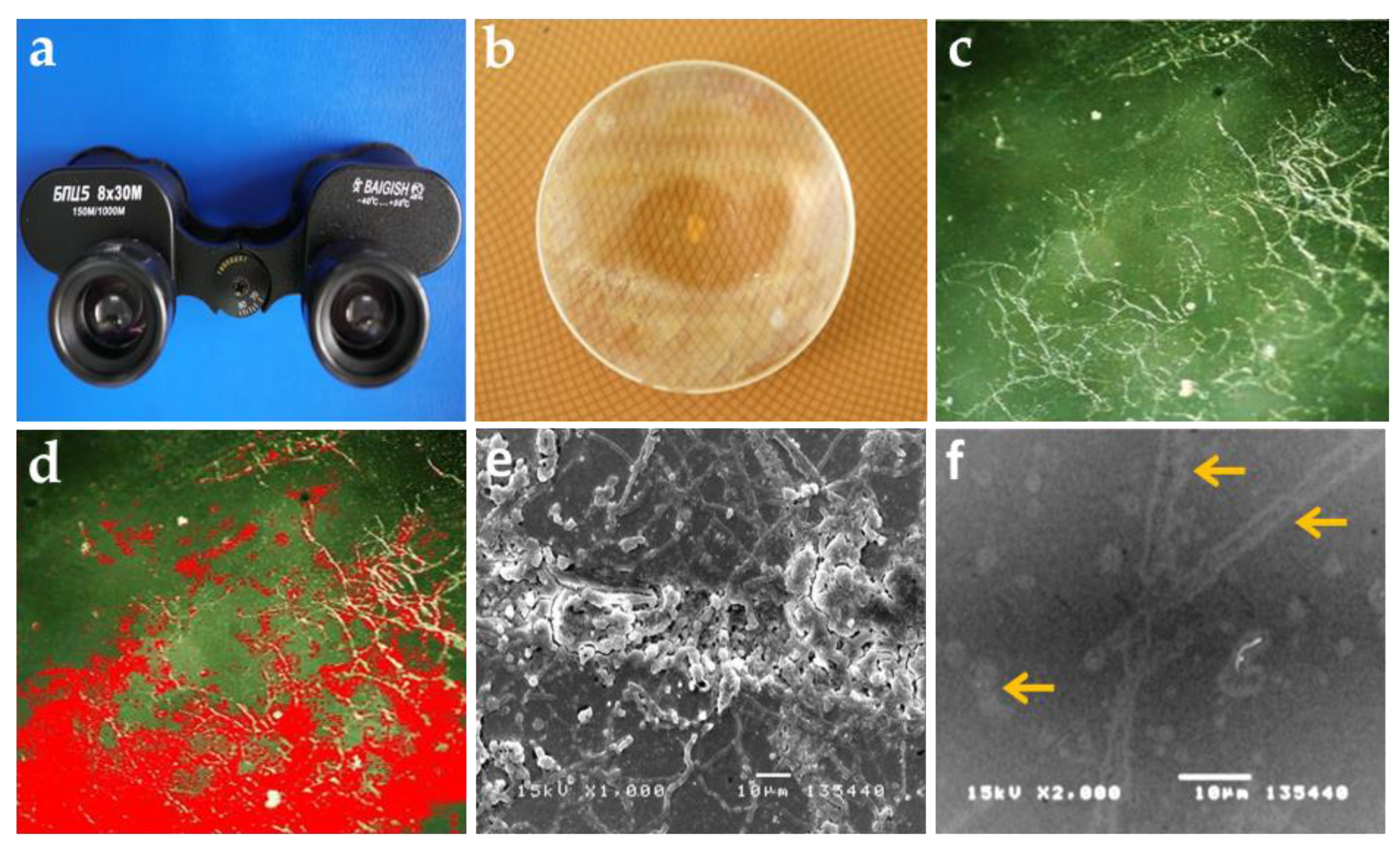
3.1. Visual Examination and Observation of Biodeterioration

The growth of fungi over the inner surface of binocular eyepieces and the extent of glass surface covered by hyphae can be observed in Figure 1a,b. As viewed under the light microscope, the coverage of fungal hyphae was extensive on the surface of 15 glass samples, with the extent of coverage ranging from 21 to 48% (Figure 1c,d), which corresponded to harmful grades 2 and 3 based on the ISO 9022-11 criteria (Table 1). The growth of fungi caused dense colonization on the surface of all glass samples (Figure 1e). After the cleaning procedure, imprints of fungal hyphae, including spots and stains, fingerprints, and etching, that covered the entire surface were detected by using SEM (Figure 1f). In addition, crack formation was not observed on any samples, indicating that the fungi did not bind strongly to the glass surface.

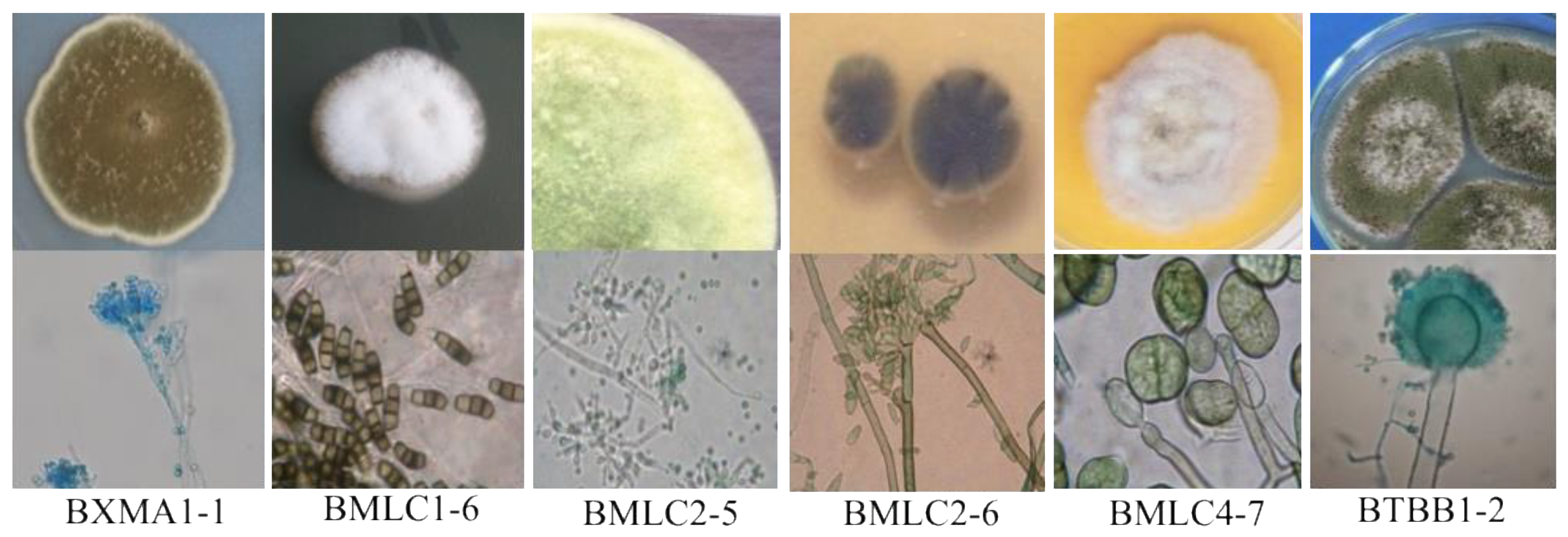
3.2. Distribution and Identification of the Isolated Fungi

A total of 186 fungal colonies were recovered from Muong Cultural Space Museum (n = 49), Museum of Biology (n = 73), and Thu Museum (n = 64) samples. Analysis of morphological and spore features of fungal isolates primarily classified them into 40 distinct clusters (Figure 2). Specifically, the most remarkable diversity and highest abundance of fungal species were found in Thu Museum samples (n = 18), followed by the Muong Cultural Space Museum samples (n = 16) and Museum of Biology samples (n = 6). Notably, at least three different fungal species were found in each glass sample, indicating co-contamination. Thus, the disinfection of fungal-contaminated optical equipment would face a significant challenge.
The molecular identification based on the ITS sequencing revealed high similarities (between 99 and 100%) to known fungal species belonging to 14 genera, including Penicillium, Byssochlamys, Talaromyces, Cladosporium, Phomopsis, Neopestalotiopsis, Trichoderma, Coprinellus, Perenniporia, Curvularia, Pithomyces, Pseudopithomyces, and Aspergillus (Figure S1). Aspergillus (43.8%) and Penicillium (31.3%) were the two predominant fungal genera, followed by species of the genus Curvularia (4%). The comparative sequence analyses combined with the clustering tree analysis further allowed us to assign 40 different fungal species (Figure S1). Fungal strains distributed throughout the tree in multiple single branches demonstrate high genetic diversity in both local and regional scales.

3.3. Evaluation of Growth, pH Changes, and Biodeterioration

To mimic the natural environment and evaluate the pH reduction of fungal isolates, two glucose mineral media were used in this study. All fungal strains secreted organic acids on two mineral salt media in the present study, leading to a significant pH reduction compared to the original pH value for almost all cases (Table 2). In the MT1 medium, a drastic decline in pH was caused by most fungi. Strong growth and pH reduction were observed for Aspergillus fumigatus BMLC1-1, Aspergillus niger BXMA5-2, Cladosporium tenuissimum BMLC2-7, Penicillium decumbens BTBB5-2, and Perenniporia cft tephropora BXMA4-4, suggesting that they might take advantage of other fungal species to develop on glass surfaces.
According to ISO 9022-11, the glass corrosion of each fungal strain was evaluated in the present study. It was seen that fungal species were growing on the surface of all glass samples, albeit to varying degrees. The extent of mycelium covered from 2.26 to 46.24%, compared to the noninoculated glass samples (Table 2). The most vigorous growth was observed for Byssochlamys spectabilis BXMA1-2 (46.2%), followed by Curvularia cft veruculosa BXMA2-2 (41.0%), Penicillium ramusculum BXMA4-2 (36.2%), Aspergillus niger BXMA5-2 (35.7%), Penicillium oxalicum BTBB2-2 (35.6%), Humicola cft insolens BMLC5-5 (35.3%), Aspergillus ochraceopetaliformis BMLC1-2 (33.6%), and Talaromyces cft trachyspermus BTBB1-3 (33.0%), which are highlighted in bold. These fungal species seem to have a good affinity for adhesion on the glass surface by developing mycelium networks in a relatively short time (28 days). Additionally, these strains also exhibited a significant reduction in pH in the two glucose mineral media tested. Based on the growth, pH reduction, and hyphal surface coverage, 8 fungal strains, including B. spectabilis BXMA1-2, C. cft veruculosa BXMA2-2, P. ramusculum BXMA4-2, A. niger BXMA5-2, P. oxalicum BTBB2-2, H. cft insolens BMLC5-5, A. ochraceopetaliformis BMLC1-2, T. cft trachyspermus BTBB1-3, were selected for further study (Table 2).

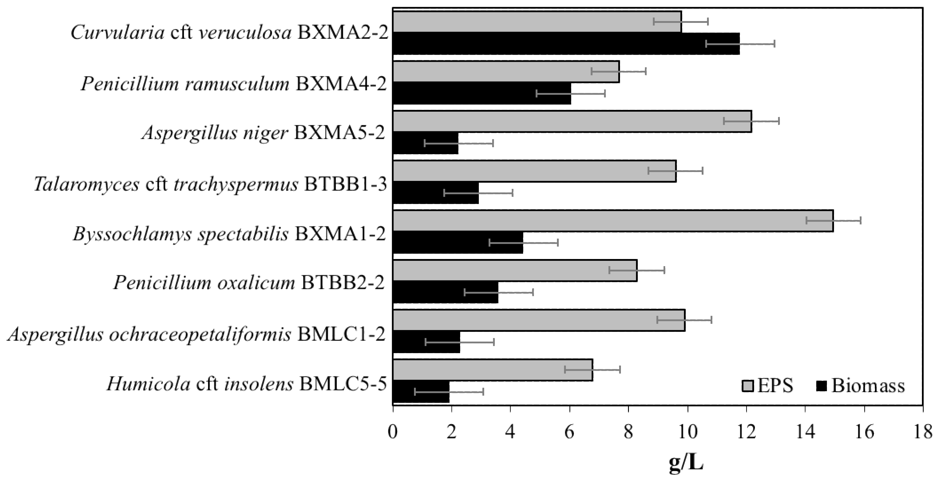
3.4. EPS Production by Fungal Strains

All fungi were selected and evaluated for their EPS production. Among these (Figure 3), the highest EPS production was reported for Byssochlamys spectabilis BXMA1-2. The yield of crude EPS and mycelial growth from the modified Czapek–Dox medium were 14.96 and 4.4 g/L, respectively. Aspergillus niger BXMA5-2 (12.17 g/L) and Aspergillus ochraceopetaliformis BMLC1-2 (9.89 g/L) were also found to produce high levels of EPS. Talaromyces trachyspermus, Curvularia veruculosa, Talaromyces trachyspermus, and Humicola insolens are for the first time reported as EPS producers in this present study. During cultivation, the cells formed pellets with high hairiness, leading to maximum biomass and EPS production.

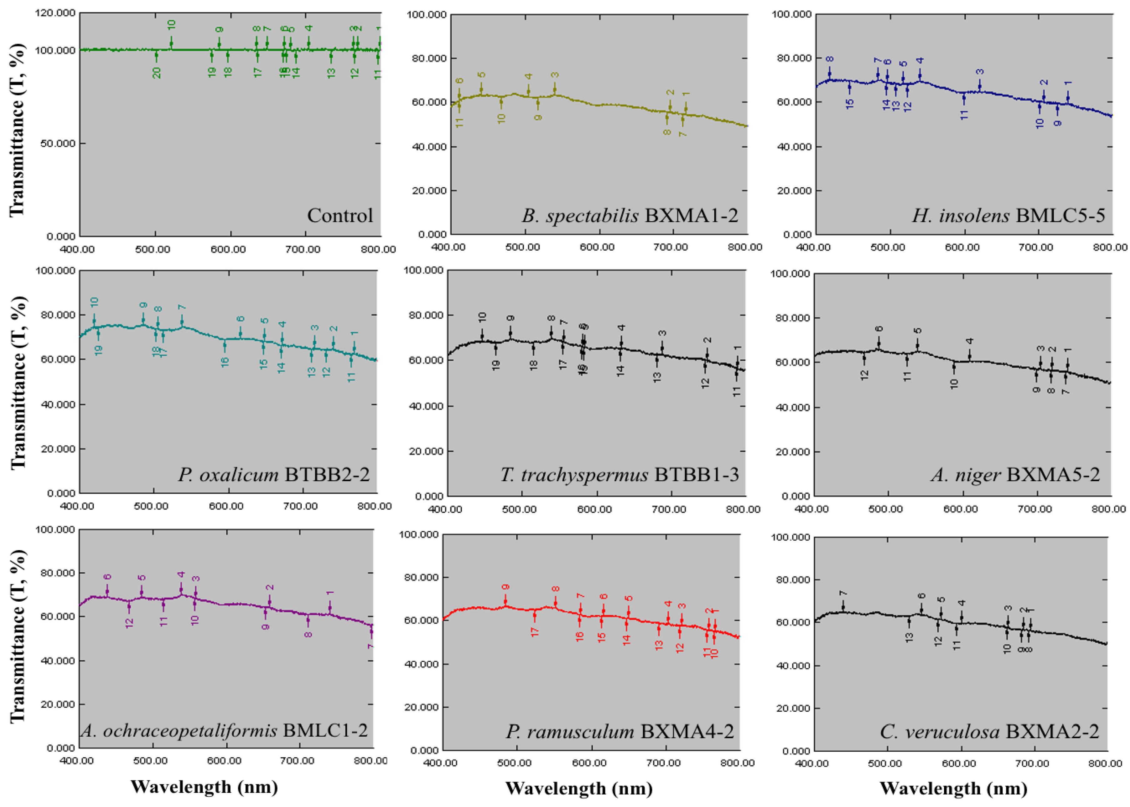
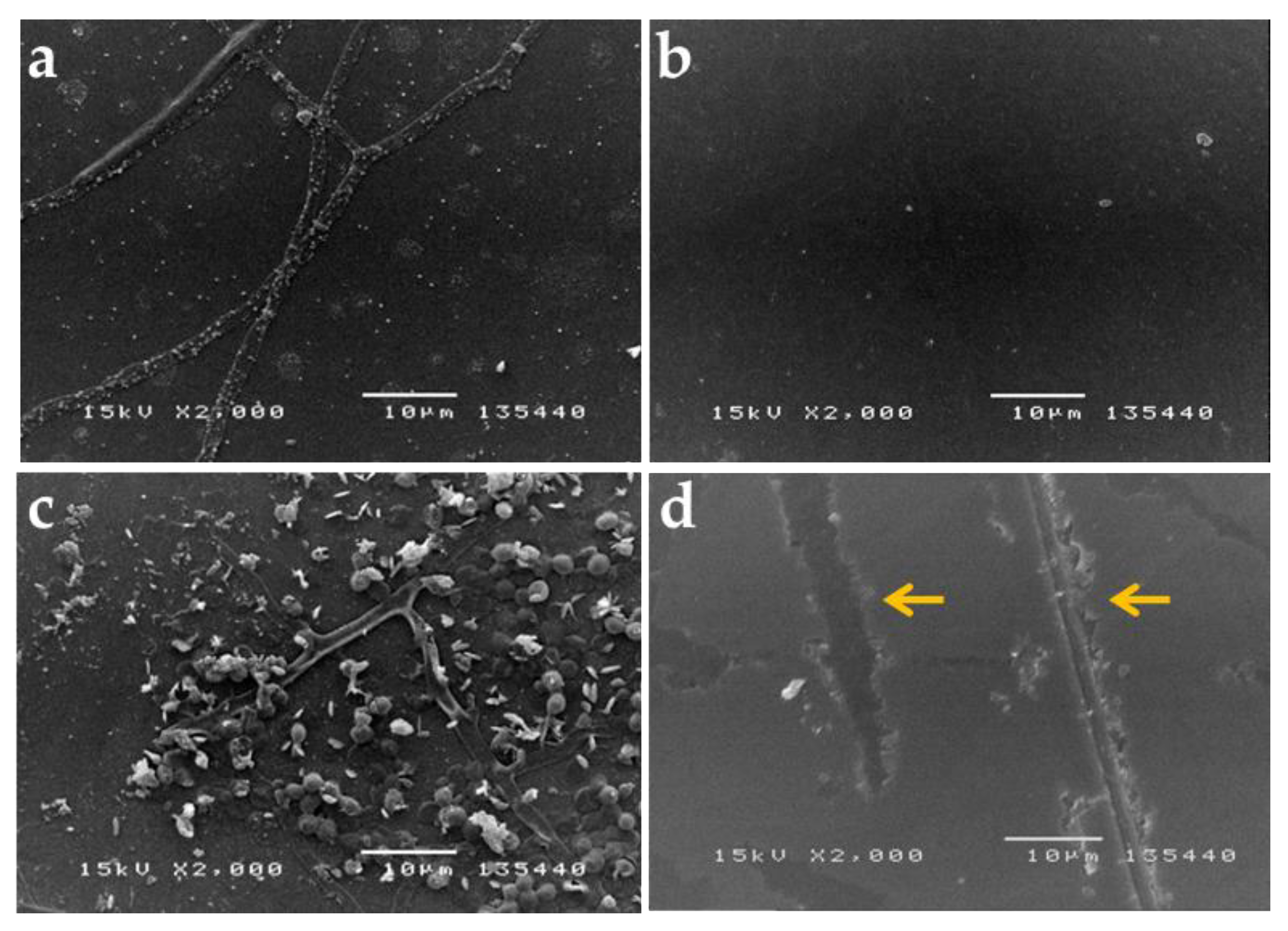
3.5. Biodeterioration of the Glass Reproductions

The biodeteriogenic potential of the selected fungal strains was assessed by measuring light transmission through the glass in the visible light spectrum (wavelengths from 400 to 800 nm). After 28 days of incubation, the light transmission value through the fungal-inoculated glass samples significantly decreased by 30–42.2% compared to the noninoculated ones (Figure 4). The glass treated with Byssochlamys spectabilis BXMA1-2 exhibited the highest reduction in light transmission, followed by Curvularia cft veruculosa BXMA2-2, Aspergillus niger BXMA5-2, and Penicillium ramusculum BXMA4-2.
The glass surfaces showed some little fingerprints after 28 days, attributed to the presence of fungi (Figure 5a,b). This causes corrosion of the glass and loss of quality of the equipment. After the glass surface cleaning procedure, the ratio of light transmission through fungus-treated glasses was reduced by 0.5 to 17.8% compared with nontreated glasses (Table S1). After six months of incubation, the fungal strain produced a well-developed mycelia biofilm on the glass surfaces (Figure 5c). The phenomenon was observed on all fungus-treated glasses. The cleaning procedure showed precise etching and hyphal fingerprints (Figure 5d).

4. Discussion

The fungi could acquire the elements needed for growth from the deteriorating glass without an external supply of mineral nutrients [11]. Fungi have a great ability to metabolize a wide range of carbon sources and grow on every inorganic material easily [23]. Thus, the coinfection of different microorganisms and favorable environmental conditions play a significant role and could affect the biodeterioration and biocorrosion outcomes on the optical instruments.
The main component of glass is SiO2 and other metal oxides, i.e., Na2O, K2O, CaO, Al2O3, B2O, BaO, ZnO, As2O3, and La2O3. Elements such as Mg, Zn, Co, and Cr are also added to change the glass properties, including the magnification coefficient, mechanical strength, refractive index, color, and opacity transparency [24,25]. The glass surface of binoculars is also coated by antireflective layers, including the outer layer consisting of low-refractive-index dielectric materials that are environmentally resistant, such as MgF2, Na3AlF6, CaF2, and LiF2. The inner layer is usually a dielectric with a high refractive index and good adhesion to the glass surface, including ZnO, TiO2, CeF3, and ThO2 [26]. Moreover, a lipid layer is coated around the edges of eyepieces to prevent dirt particles from spreading and sticking to the inner glass surface and the optical system. This lipid layer consists of long-chain hydrocarbons, fatty acids, and some alkaline metals such as Li, Na, K, Ca, Ba, Al, Zn, and Pb, together with dust and other organic contaminants, providing the primary growth medium to initiate microbial growth. Such nutrients are sufficient for airborne fungal spores to germinate and develop into hyphae. Once the hyphal growth occurs on the glass surface, it quickly corrodes the inner surface of eyepieces.
Aspergillus, Cladosporium, and Penicillium are the most prevalent fungi genera commonly found in indoor and outdoor environments, especially glass materials [4,27,28]. Rodrigues et al. identified six fungal genera, among which Penicillium and Cladosporium were the two predominant fungal genera isolated from stained-glass windows in Portugal [4]. In addition, Cladosporium and Phoma were also reported from glass samples collected at two Catalonian churches [9]. Aspergillus, Penicillium, and Alternaria were found to contribute to the formation of pits in optical glass [16]. In the present study, Aspergillus and Penicillium were the two most dominant fungal genera, accounting for 75% of the total identified fungal strains. They are known as primary contaminants that harm historical and cultural heritages and cause glass corrosion [2,9,29]. Because of the ability to produce numerous spores easily dispersed in the air, Aspergillus and Penicillium are primary corrosive agents [30]. Earlier regional studies and reports on the frequent occurrence of these fungi in all areas of the northern provinces of Vietnam suggest the high load of these fungal spores. Therefore, chances of risk are increased for fungal contamination of optical equipment if no preventive measures are taken.
Interestingly, fungal genera Byssochlamys, Curvularia, Phomopsis, Coprinellus, Perenniporia, Talaromyces, Pithomyces, Neopestalotiopsis, Pleospora, and Humicola were identified for the first time as harmful agents on optical instruments. Byssochlamys, Curvularia, Phomopsis, Coprinellus, and Perenniporia were only found in Muong Cultural Space Museum, while Cladosporium, Humicola, Pithomyces, Neopestalotiopsis, Pleospora, and Trichoderma were only found in Thu Museum. In contrast, Talaromyces was only found in the Museum of Biology. The fungal community involved in the biodeterioration process of binocular eyepieces was more diverse than that of historical church window glasses [4].
The identified fungal species might secrete various organic acids, extracellular enzymes, pigments, and EPSs to colonize on glass materials; consequently, biofilms will be developed on the glass surface, accelerating biodegradation and biocorrosion processes [31,32,33]. Fungi are well known for their potential to overproduce and accumulate various organic acids [34,35]. Intrinsic abilities provide the fungi a competitive advantage over other organisms and the ability to proliferate on inorganic materials such as metal and glass [36]. As for the biodeterioration of glass, organic acids produced by fungi dissolve metal oxides and minerals on the glass surface, thus releasing nutrient ions for growth [19]. Organic acid production recorded here was in agreement with previous studies showing that Aspergillus, Penicillium, and Cladosporium are organic acid producers commonly found as the primary contaminants causing biocorrosion of glass materials [1,2,14]. As documented, the most common acids produced by fungi include formic, citric, acetic, oxalic, gluconic, and itaconic acids [34]. The magnitude of corrosion ranges from 0.27 to 0.03 mA cm−2 for 1% organic acids [37] and causes significant etching of glasses. Fungi readily digest the organic materials such as oils from fingerprints and lens coatings and produce strong hydrofluoric acid as a waste product, which decreases the glass surface pH to lower than 3.0, causing permanent etching of the glass [38]. Thus, glass damage is even more potent in vivo because of the condensation and accumulation of a mixture of the above-mentioned organic acids.
Furthermore, organic acids move through the silica network and continue to dissolve metal oxides, leading to carbonate and sulfate salt production due to the absorption of CO2 and SO2 from the atmosphere [39]. Krumbein et al. demonstrated the formation of carbonate, sulfate, and silicate crystals due to fungal acids excreted during the biocorrosion process [11]. Altogether, these agents, in turn, etch and destroy the antireflective layers and the surface of glass permanently, resulting in a decreased degree of transparency and refractive index and a loss of image brightness and sharpness. Moreover, the acidic environment also favors fungal growth and, together with the hot, moist atmosphere conditions, causes physical–chemical corrosion, which speeds up glass deterioration.
In this study, the high level of EPSs recorded for Aspergillus, Byssochlamys, and Penicillium species was consistent with many previous studies [40,41,42]. The EPS composition varies depending on producer species. Aspergillus and Penicillium species produce EPSs composed of glucose, mannose, galactose, galactosamine, and acetate [43,44,45]. Due to excess sugars in EPSs, organic acid production occurs on glass surfaces, leading to glass corrosion. EPS production by the fungi is usually intracellular and secreted outside the cell to form biofilms, leading to permanent colonization of the fungi on the surface of materials [42]. However, the glass biodeterioration ability might differ between fungal species, and the identification of harmful fungi is crucial to protect glass material. Light scattering and absorption by the fungal mycelia on the glass surface could account for the rapid loss of light transmission and image quality. The etching of the glass by excretion of organic acids and other metabolic products results in grooves formed in the glass surface in contact with the hyphae. Furthermore, the fungi remove the glass surface layer and part of the glass matrix, leading to alteration of optical properties [2,20].
The high fungal biomass, colonization, EPS formation, and biofilm development are responsible for the etching and hyphal fingerprints on the glass. Furthermore, EPSs contain polysaccharides, uronic acids, and some enzymes, contributing to the corrosion process [46]. It is important to note that all fungal strains under investigation produce EPSs, contributing to chemical deterioration by acid and chelate production. Therefore, all these results demonstrate that all fungal strains could biodeteriorate glasses and that the degree of damage depended on fungal characteristics. The present study will provide baseline information about the significant fungal species responsible for the biodeterioration of optical instruments. It will be helpful in the development of fungus-resistant optical glass. A more detailed investigation is needed to determine the proper method of protection and preservation of such museum specimens.

5. Conclusions

Fungal biodeterioration of optical equipment represents a serious concern leading to notable research efforts over the last decades. In this study, we found that Aspergillus sp., Penicillium sp., and Cladosporium sp. were major fungi colonizing the binocular eyepieces at museums in northern Vietnam. Moreover, this is the first report that identifies Byssochlamys spectabilis, Curvularia veruculosa, Humicola insolens, and Talaromyces lagena as robust glass-deteriorating fungi. The growth of fungi resulted in dense colonization and biofilm formation on the glass surface, corresponding to the deterioration and corrosion of binocular eyepieces. Organic acid and EPS production were the main factors contributing to the leaching of elements from glass surfaces and severe physical, chemical, and aesthetical modifications. Thus, effective procedures for protecting optical observation equipment can be proposed based on an understanding of the fungal community. The proper cleaning of the optical lens is highly recommended to control fungal contamination as per the manufacturer’s guidelines. However, for prolonged storage in museums, the optical devices must be kept under a controlled condition with unfavorable temperature and humidity for fungi. Finally, to stop the biodeterioration, an understanding of microbial communities is required to optimize biocides against fungi that reduce the biodeterioration of optical glasses.

Supplementary Materials

The following are available online at https://www.mdpi.com/article/10.3390/app11125351/s1, Figure S1: Clustering tree from the maximum likelihood analysis of all fungi isolated from binocular eyepieces using ITS sequences. Cunninghamella elegans CBS 160.28T was selected as the outgroup taxon and 100% bootstrap values indicate the relevant internode. Identified strains are indicated in bold, Table S1: Effect of mycelium growing on glass plates after 28 days of alcohol-purge test as per ISO 9022-11 on light transmission at wavelengths of visible light range from 400 to 800 nm.

Author Contributions

Conceptualization, C.C.N., Q.H.N., and Q.-T.P.; methodology, T.H.N.V., T.T.X.L., T.T.H.D. and T.T.H.L.; investigation, V.D.N. and T.H.N.; writing—original draft preparation, C.C.N. and Q.H.N.; writing—review and editing, P.D., N.T.Q., and Q.-T.P.; visualization, N.T.N.; project administration, C.C.N. and T.H.N. All authors have read and agreed to the published version of the manuscript.

Funding

This research was funded by the Vietnam Academy of Science and Technology, grant number NVCC 08.08/20-20, and Vietnam-Russia Tropical Centre, grant number TCKT2020.

Institutional Review Board Statement

Not applicable.

Informed Consent Statement

Not applicable.

Data Availability Statement

Data sharing is not applicable to this article.

Acknowledgments

The authors would like to acknowledge the support of the VAST Culture Collection of Microorganisms, Institute of Biotechnology, Vietnam Academy of Science and Technology (www.vccm.vast.vn).

Conflicts of Interest

The authors declare no conflict of interest.

References

  1. Schreiner, M. Glass of the past: The degradation and deterioration of medieval glass artifacts. Microchim. Acta 1991, 104, 255–264. [Google Scholar] [CrossRef]
  2. Drewello, R.; Weissmann, R. Microbially influenced corrosion of glass. Appl. Microbiol. Biotechnol. 1997, 47, 337–346. [Google Scholar] [CrossRef]
  3. Gorbushina, A.A.; Palinska, K.A. Biodeteriorative processes on glass: Experimental proof of the role of fungi and cyanobacteria. Aerobiologia 1999, 15, 183–192. [Google Scholar] [CrossRef]
  4. Rodrigues, A.; Gutierrez-Patricio, S.; Miller, A.Z.; Saiz-Jimenez, C.; Wiley, R.; Nunes, D.; Vilarigues, M.; Macedo, M.F. Fungal biodeterioration of stained-glass windows. Int. Biodeter. Biodegr. 2014, 90, 152–160. [Google Scholar] [CrossRef]
  5. Sterflinger, K.; Piñar, G. Microbial deterioration of cultural heritage and works of art-tilting at windmills? Appl. Microbiol. Biotechnol. 2013, 97, 9637–9646. [Google Scholar] [CrossRef] [Green Version]
  6. Pinna, D. Coping with Biological Growth on Stone Heritage Objects: Methods, Products, Applications, and Perspectives; Apple Academic Press: Palm Bay, FL, USA, 2017. [Google Scholar]
  7. Müller, E.; Drewello, U.; Drewello, R.; Weißmann, R.; Wuertz, S. In situ analysis of biofilms on historic window glass using confocal laser scanning microscopy. J. Cult. Herit. 2001, 2, 31–42. [Google Scholar] [CrossRef]
  8. Carmona, N.; Laiz, L.; Gonzalez, J.M.; Garcia-Heras, M.; Villegas, M.A.; Saiz-Jimenez, C. Biodeterioration of historic stained glasses from the Cartuja de Miraflores (Spain). Int. Biodeter. Biodegr. 2006, 58, 155–161. [Google Scholar] [CrossRef]
  9. Piñar, G.; Garcia-Valles, M.; Gimeno-Torrente, D.; Fernandez-Turiel, J.L.; Ettenauer, J.; Sterflinger, K. Microscopic, chemical, and molecular-biological investigation of the decayed medieval stained window glasses of two Catalonian churches. Int. Biodeter. Biodegr. 2013, 84, 388–400. [Google Scholar] [CrossRef] [PubMed]
  10. Corrêa Pinto, A.M.; Palomar, T.; Alves, L.C.; da Silva, S.H.M.; Monteiro, R.C.; Macedo, M.F.; Vilarigues, M.G. Fungal biodeterioration of stained-glass windows in monuments from Belém do Pará (Brazil). Int. Biodeter. Biodegr. 2019, 138, 106–113. [Google Scholar] [CrossRef]
  11. Krumbein, W.E.; Urzì, C.E.; Gehrmann, C. Biocorrosion and biodeterioration of antique and medieval glass. Geomicrobiol. J. 1991, 9, 139–160. [Google Scholar] [CrossRef]
  12. Marvasi, M.; Vedovato, E.; Balsamo, C.; Macherelli, A.; Dei, L.; Mastromei, G.; Perito, B. Bacterial community analysis on the Mediaeval stained glass window “Natività” in the Florence Cathedral. J. Cult. Herit. 2009, 10, 124–133. [Google Scholar] [CrossRef]
  13. Bindschedler, S.; Cailleau, G.; Verrecchia, E. Role of fungi in the biomineralization of calcite. Minerals 2016, 6, 41. [Google Scholar] [CrossRef] [Green Version]
  14. Weaver, J.L.; DePriest, P.T.; Plymale, A.E.; Pearce, C.I.; Arey, B.; Koestler, R.J. Microbial interactions with silicate glasses. NPJ Mater. Degrad. 2021, 5, 11. [Google Scholar] [CrossRef]
  15. Watkins, R.D. Mould in optical instruments. Community Eye Health 2003, 16, 28. [Google Scholar]
  16. Bartosik, M.; Zakowska, Z.; Cedzińska, K.; Rozniakowski, K. Biodeterioration of optical glass induced by lubricants used in optical instruments technology. Pol. J. Microbiol. 2010, 59, 295–300. [Google Scholar] [CrossRef] [PubMed]
  17. Ihrmark, K.; Bödeker, I.T.M.; Cruz-Martinez, K.; Friberg, H.; Kubartova, A.; Schenck, J.; Strid, Y.; Stenlid, J.; Brandström-Durling, M.; Clemmensen, K.E.; et al. New primers to amplify the fungal ITS2 region—evaluation by 454-sequencing of artificial and natural communities. FEMS Microbiol. Ecol. 2012, 82, 666–677. [Google Scholar] [CrossRef] [PubMed]
  18. Thompson, J.D.; Gibson, T.J.; Higgins, D.G. Multiple sequence alignment using ClustalW and ClustalX. Curr. Protoc. Bioinform. 2003, 2.3.1–2.3.22. [Google Scholar] [CrossRef]
  19. Liaud, N.; Giniés, C.; Navarro, D.; Fabre, N.; Crapart, S.; Gimbert, I.H.; Levasseur, A.; Raouche, S.; Sigoillot, J.-C. Exploring fungal biodiversity: Organic acid production by 66 strains of filamentous fungi. Fungal Biol. Biotechnol. 2014, 1, 1. [Google Scholar] [CrossRef] [Green Version]
  20. Garcia-Vallès, M.; Gimeno-Torrente, D.; Martínez-Manent, S.; Fernández-Turiel, J.L. Medieval stained glass in a Mediterranean climate: Typology, weathering and glass decay, and associated biomineralization processes and products. Am. Mineral. 2003, 88, 1996–2006. [Google Scholar] [CrossRef]
  21. Vasil’chenko, L.G.; Khromonygina, V.V.; Karapetyan, K.N.; Vasilenko, O.V.; Rabinovich, M.L. Cellobiose dehydrogenase formation by filamentous fungus Chaetomium sp. INBI 2-26(-). J. Biotechnol. 2005, 119, 44–59. [Google Scholar] [CrossRef]
  22. Jaroszuk-Ściseł, J.; Nowak, A.; Komaniecka, I.; Choma, A.; Jarosz-Wilkołazka, A.; Osińska-Jaroszuk, M.; Tyśkiewicz, R.; Wiater, A.; Rogalski, J. Differences in production, composition, and antioxidant activities of exopolymeric substances (EPS) obtained from cultures of endophytic Fusarium culmorum strains with different effects on cereals. Molecules 2020, 25, 616. [Google Scholar] [CrossRef] [Green Version]
  23. Davis, S.; Chemello, C. Glass: Conservation and Preservation. In Encyclopedia of Global Archaeology; Smith, C., Ed.; Springer: New York, NY, USA, 2014; pp. 3047–3050. [Google Scholar]
  24. Stábile, F.M.; Volzone, C.; Ortiga, J. Thermal evolution of Na2O-K2O-CaO-SiO2-P2O5-Al2O3 glass system, and possible applications as biomedical devices. Procedia Manuf. Sci. 2015, 8, 332–337. [Google Scholar] [CrossRef] [Green Version]
  25. Partyka, J.; Gasek, K.; Pasiut, K.; Gajek, M. Effect of addition of BaO on sintering of glass–ceramic materials from SiO2–Al2O2–Na2O–K2O–CaO/MgO system. J. Therm. Anal. Calorim. 2016, 125, 1095–1103. [Google Scholar] [CrossRef] [Green Version]
  26. Song, H.S.; Yoo, Y.J.; Lee, G.J.; Chang, K.S.; Song, Y.M. Optical design of porous ZnO/TiO2 films for highly transparent glasses with broadband ultraviolet protection. J. Nanomater. 2017, 2017, 2738015. [Google Scholar] [CrossRef] [Green Version]
  27. Johansson, W.; Peralta, A.; Jonson, B.; Anand, S.; Österlund, L.; Karlsson, S. Transparent TiO2 and ZnO thin films on glass for UV protection of PV modules. Front. Mater. Sci. 2019, 6, 259. [Google Scholar] [CrossRef]
  28. Frisvad, J.; Gravesen, S. Penicillium and Aspergillus from Danish homes and working places with indoor air problems: Identification and mycotoxin determination. Health Implic. Fungi Indoor Environ. 1994, 2, 281–290. [Google Scholar]
  29. Guiamet, P.; Borrego, S.; Lavin, P.; Perdomo, I.; de Saravia, S.G. Biofouling and biodeterioration in materials stored at the Historical Archive of the Museum of La Plata, Argentine and at the National Archive of the Republic of Cuba. Colloids Surf. B 2011, 85, 229–234. [Google Scholar] [CrossRef]
  30. Fog Nielsen, K. Mycotoxin production by indoor molds. Fungal Genet. Biol. 2003, 39, 103–117. [Google Scholar] [CrossRef]
  31. Vadillo-Rodríguez, V.; Logan, B.E. Localized attraction correlates with bacterial adhesion to glass and metal oxide substrata. Environ. Sci. Technol. 2006, 40, 2983–2988. [Google Scholar] [CrossRef]
  32. Borrego, S.; Guiamet, P.; Gómez de Saravia, S.; Batistini, P.; Garcia, M.; Lavin, P.; Perdomo, I. The quality of air at archives and the biodeterioration of photographs. Int. Biodeter. Biodegrad. 2010, 64, 139–145. [Google Scholar] [CrossRef]
  33. Trovão, J.; Portugal, A. Current knowledge on the fungal degradation abilities profiled through biodeteriorative plate essays. Appl. Sci. 2021, 11, 4196. [Google Scholar] [CrossRef]
  34. Magnuson, J.K.; Lasure, L.L. Organic acid production by filamentous fungi. In Advances in Fungal Biotechnology for Industry, Agriculture, and Medicine; Tkacz, J.S., Lange, L., Eds.; Springer US: Boston, MA, USA, 2004; pp. 307–340. [Google Scholar]
  35. Goldberg, I.; Rokem, J.S. Organic and fatty acid production, microbial. In Encyclopedia of Microbiology, 4th ed.; Schmidt, T.M., Ed.; Academic Press: Cambridge, MA, USA, 2019; pp. 358–382. [Google Scholar] [CrossRef]
  36. Naranjo-Ortiz, M.A.; Gabaldón, T. Fungal evolution: Major ecological adaptations and evolutionary transitions. Biol. Rev. 2019, 94, 1443–1476. [Google Scholar] [CrossRef] [Green Version]
  37. Little, B.; Staehle, R.; Davis, R. Fungal influenced corrosion of post-tensioned cables. Int. Biodeter. Biodegr. 2001, 47, 71–77. [Google Scholar] [CrossRef]
  38. Cordero, I. Fungus: How to prevent growth and remove it from optical components. Community Eye Health 2013, 26, 57. [Google Scholar]
  39. Sand, W.; Bock, E. Biodeterioration of mineral materials by microorganisms—biogenic sulfuric and nitric acid corrosion of concrete and natural stone. Geomicrobiol. J. 1991, 9, 129–138. [Google Scholar] [CrossRef]
  40. Gomoiu, I.; Catley, B.J. Properties of a kaolin-flocculating polymer elaborated by Byssochlamys nivea. Enzyme Microb. Technol. 1996, 19, 45–49. [Google Scholar] [CrossRef]
  41. Kogan, G.; Matulová, M.; Michalková, E. Extracellular polysaccharides of Penicillium vermiculatum. Z. Naturforsch. C. J. Biosci. 2002, 57, 452–458. [Google Scholar] [CrossRef] [PubMed]
  42. Mahapatra, S.; Banerjee, D. Fungal exopolysaccharide: Production, composition and applications. Microbiol. Insights 2013, 6, 1–16. [Google Scholar] [CrossRef] [PubMed] [Green Version]
  43. Ruperez, P.; Leal, J.A. Extracellular galactosaminogalactan from Aspergillus parasiticus. Trans. Brit. Mycol. Soc. 1981, 77, 621–625. [Google Scholar] [CrossRef]
  44. Sutherland, I.W. Biotechnology of Microbial Exopolysaccharides; Cambridge University Press: Cambridge, UK, 1990. [Google Scholar] [CrossRef] [Green Version]
  45. Jansson, P.-E.; Lindberg, B. Structural studies of varianose. Carbohydr. Res. 1980, 82, 97–102. [Google Scholar] [CrossRef]
  46. Roca, C.; Alves, V.D.; Freitas, F.; Reis, M.A.M. Exopolysaccharides enriched in rare sugars: Bacterial sources, production, and applications. Front. Microbiol. 2015, 6, 288. [Google Scholar] [CrossRef] [PubMed]
Figure 1. (a) The binocular model. (b) Binocular eyepieces contaminated with fungi. (c) Image captured using light microscopy at a magnification of 60×. (d) The estimation of hyphae covering the glass surface treated by the ImageJ tool. (e) SEM image exhibiting the fungal growth and colonization on the eyepieces (image at a magnification of 1000×, scale bar = 10 µm). (f) Yellow arrows indicate fingerprinting/etching of fungal hyphae on the surface of eyepieces after the cleaning procedure (image at a magnification of 2000×, scale bar = 10 µm).
Applsci 11 05351 g001
Figure 2. The colony and morphological characteristics of some fungi isolated from binocular eyepieces.
Applsci 11 05351 g002
Figure 3. Exopolysaccharide (EPS) production by eight selected fungal isolates causing deterioration on a glass model.
Applsci 11 05351 g003
Figure 4. The effect of fungal growth on light transmission through fungus-inoculated glass samples in the visible light spectrum.
Applsci 11 05351 g004
Figure 5. SEM micrographs of glass after 28 days of the test: (a) fungal growth on glass after 28 days; (b) sample photo taken after 28 days of the alcohol-purge test; (c) glass sample after six months of testing showing fungal spores; (d) glass sample after six months of testing showing the glass corrosion caused by fungi.
Applsci 11 05351 g005
Table 1. The extent of fungal hyphae on eyepiece samples.
PlaceGlass SampleEvaluation of the Extent of Fungal Growth *
Hyphal Surface Coverage (%)Harmful Grade
Muong Cultural Space Museum, HanoiBXMA129 ± 1.22
BXMA235 ± 1.53
BXMA326 ± 1.32
BXMA421 ± 1.22
BXMA521 ± 1.52
Museum of Biology, Phu Tho BTBB128 ± 1.52
BTBB241 ± 1.73
BTBB327 ± 1.62
BTBB443 ± 2.13
BTBB548 ± 1.43
Thu Museum, Vinh PhucBMLC141 ± 1.63
BMLC236 ± 1.83
BMLC334 ± 1.63
BMLC425 ± 1.32
BMLC543 ± 2.23
* ISO 9022-11 interpretation for the coverage of a glass surface with fungal hyphae: >0 to 10% (Grade 1)—restricted fungal growth; >10 to 30% (Grade 2)—intermittent spread fungal colonies (visible with the naked eye); >30 to 70% (Grade 3)—a substantial amount of fungal growth (easily visible); >70% (Grade 4)—massive fungal growth.
Table 2. Identification, the ability of growth and pH values observed in culture media, and percentage of hyphal surface coverage of isolated fungi.
Fungal StrainGenBank Accession NumberThe Changes in the pH of Media after Fungal Growth
MT1
(Initial pH 7.2)
MT2
(Initial pH 6.5)
Hyphal Surface Coverage (%)
pHGrowthpHGrowth
Aspergillus fumigatus BMLC1-1MW9117812.53 ± 0.4+++3.17 ± 0.1+++++4.64 ± 1.4
Aspergillus ochraceopetaliformis BMLC1-2MN3941293.15 ± 0.1++++5.26 ± 0.6+++33.61 ± 3.7
Aspergillus asperescens BMLC1-3MZ2923954.28 ± 0.3++++5.68 ± 0.1++++2.26 ± 1.1
Aspergillus sclerotiorum BMLC1-4MN3941304.34 ± 0.1++++5.66 ± 0.3++++3.87 ± 1.3
Penicillium lanoso BMLC1-5MZ2923962.78 ± 0.3++++6.26 ± 0.2++++15.28 ± 2.4
Pithomyces chartarum BMLC1-6MN3941313.18 ± 0.4++++6.46 ± 0.8++++32.33 ± 3.5
Neopestalotiopsis sp. BMLC1-7MN3941325.43 ± 0.5++++4.87 ± 0.6+++23.14 ± 2.8
Penicillium chermesinum BMLC2-1MN3941342.91 ± 0.1++++8.93 ± 0.5+++++20.28 ± 2.2
Penicillium roqueforti BMLC2-3MZ2923973.47 ± 0.2+++7.21 ± 0.9++++19.21 ± 2.4
Trichoderma koningiopsis BMLC2-5MN3941334.15 ± 0.4++++4.27 ± 0.6+++18.90 ± 2.3
Cladosporium tenuissimum BMLC2-6MN3941355.34 ± 0.6+++8.24 ± 0.7++36.43 ± 3.4
Cladosporium tenuissimum BMLC2-7MN3941362.22 ± 0.3+++2.68 ± 0.7++++27.65 ± 2.6
Penicillium toxicarium BMLC2-8MN3941374.19 ± 0.2+++6.91 ± 0.5+++11.18 ± 2.3
Aspergillus niger BMLC3-4MW9117822.56 ± 0.8+++6.23 ± 0.4++++21.53 ± 2.2
Pithomyces maydicus BMLC3-6MN3941385.95 ± 0.4++++5.35 ± 0.5+++12.15 ± 2.1
Byssochlamys cft spectabilis BMLC4-6MN3941394.28 ± 0.8++++5.68 ± 0.5++++14.99 ± 2.5
Pleospora herbarum BMLC4-7MZ2923986.21 ± 0.7++++6.43 ± 0.8++++10.91 ± 2.3
Humicola cft insolens BMLC5-5MZ2923993.35 ± 0.7+++5.35 ± 0.7+++35.26 ± 3.8
Aspergillus sydowii BTBB1-1MN3966715.15 ± 0.6++5.53 ± 0.3++10.40 ± 2.7
Aspergillus flavus BTBB1-2MN3966725.21 ± 0.3+++5.85 ± 0.4+++4.25 ± 1.5
Talaromyces cft trachyspermus BTBB1-3MW9117833.78 ± 0.6++++5.79 ± 0.8+++36.95 ± 3.4
Penicillium oxalicum BTBB2-2MN3966732.24 ± 0.1++++7.02 ± 0.9+++++35.95 ± 3.2
Aspergillus cft salwaensis BTBB5-1MN3966745.47 ± 0.6+++6.85 ± 0.4+++34.07 ± 3.8
Penicillium decumbens BTBB5-2MN3966752.17 ± 0.5++++3.39 ± 0.9+++++21.58 ± 2.5
Penicillium brevissimum BXMA1-1MH6344794.9 ± 0.9+6.77 ± 0.3+24.18 ± 2.5
Byssochlamys spectabilis BXMA1-2MH6344802.68 ± 0.1+++6.23 ± 0.7++++46.24 ± 3.3
Aspergillus sydowii BXMA1-3MH6344813.37 ± 0.2+++7.2 ± 0.2++++3.79 ± 1.2
Aspergillus tritici BXMA1-4MH6344823.18 ± 0.5+++7.11 ± 0.6+++20.63 ± 2.6
Aspergillus sydowii BXMA1-5MH6344832.6 ± 0.4+++7.47 ± 0.6+++++13.97 ± 2.9
Byssochlamys spectabilis BXMA2-1MH6344842.7 ± 0.1++++6.05 ± 0.2++++19.41 ± 2.2
Curvularia cft veruculosa BXMA2-2MH6344853.2 ± 0.6++++5.32 ± 0.3++++41.04 ± 3.1
Phomopsis cft tuberivora BXMA2-3MH6344863.12 ± 0.4++++8.96 ± 0.2+++++7.86 ± 1.5
Coprinellus radians BXMA2-4MH6344873.63 ± 0.2+++8.32 ± 0.6+++++7.74 ± 1.6
Aspergillus flavus BXMA3-1MH6344883.12 ± 0.7++7.98 ± 0.6++++31.82 ± 3.6
Penicillium oxalicum BXMA3-2MH6344893.77 ± 0.2++7.4 ± 0.2++++20.22 ± 2.4
Curvularia lunata BXMA3-5MH6344902.91 ± 0.2++++6.32 ± 0.4++++27.30 ± 2.3
Penicillium brevissimum BXMA4-1MH6344912.68 ± 0.1+++7.28 ± 0.3++++31.533.6
Penicillium ramusculum BXMA4-2MH6344923.26 ± 0.7+++5.15 ± 0.9++++36.20 ± 3.2
Perenniporia cft tephropora BXMA4-4MH6344932.87 ± 0.5++++4.5 ± 0.4++11.51 ± 2.8
Aspergillus niger BXMA5-2MH6344942.65 ± 0.9+++4.24 ± 0.7+++++35.66 ± 3.8
Fair growth +; good growth ++; luxurious growth +++; heavy growth ++++; extreme growth +++++.
Publisher’s Note: MDPI stays neutral with regard to jurisdictional claims in published maps and institutional affiliations.

