Article

Static Magnetic Fields Effects on Polysaccharides Production by Different Microalgae Strains

by
Kricelle M. Deamici
1,
Michele G. de Morais
2,
Lucielen O. Santos
3,
Koenraad Muylaert
4,
Christine Gardarin
1,
Jorge Alberto V. Costa
2 and
Céline Laroche
1,*
1
Institut Pascal, Université Clermont Auvergne, UMR CNRS 6602, F-63000 Clermont-Ferrand, France
2
Laboratory of Biochemical Engineering, College of Chemistry and Food Engineering, Federal University of Rio Grande, Rio Grande 96203-900, RS, Brazil
3
Laboratory of Biotechnology, College of Chemistry and Food Engineering, Federal University of Rio Grande, Rio Grande 96203-900, RS, Brazil
4
Laboratory Aquatic Biology, KU Leuven Kulak, E. Sabbelaan 53, 8500 Kortrijk, Belgium
*
Author to whom correspondence should be addressed.
Appl. Sci. 2021, 11(11), 5299; https://doi.org/10.3390/app11115299
Submission received: 11 May 2021 / Revised: 1 June 2021 / Accepted: 4 June 2021 / Published: 7 June 2021

Abstract

:
Microalgae are able to produce many valuable biomolecules, such as polysaccharides, that presents a large diversity of biochemical structures and functions as antioxidant, antifungal, anticancer, among others. Static magnetic fields (SMF) influence the metabolism of microorganisms and has been shown as an alternative to increase microalgae biomass, yield and compounds production. Especially, some studies have highlighted that SMF application could enhance carbohydrate content. This study aimed to evaluate different conditions of SMF on Spirulina and Chlorella in indoor and outdoor conditions, in order to confirm the influence of SMF on polysaccharides production, evaluating which polysaccharidic fraction could be enhanced by SMF and highlighting a possible modification in EPS composition. Starch from Chlorella and exopolysaccharides (EPS) from Spirulina were quantified and characterized. SMF increased the starch content in Chorella fusca biomass. EPS productions from A. platensis and Spirulina sp. were not significantly increased, and global composition appeared similar to the controls (constituted basically of 80–86% neutral sugars and 13–19% uronic acids). However, the monosaccharide composition analysis revealed a significant modification of composition, i.e., the amount of fucose, arabinose, rhamnose, galactose and glucuronic acid was increased, while the glucose content was decreased. SMF application led to significant modification of polysaccharides production and this study demonstrate that combining the outdoor conditions with SMF, the starch content and EPS composition was positively affected.

Graphical Abstract

1. Introduction

Microalgae are a selected group of microorganisms with potential for use in wastewater treatment, increased food quality, besides a high capacity to mitigate atmospheric carbon dioxide (CO2). These photosynthetic microorganisms are divided into eukaryotic group (among which the Chlorophyta and the Rhodophyta are the most studied phyla), and prokaryotic group containing Cyanophyceae, often called blue-green microalgae. Microalgae have been strongly studied in the last decades, as diversity makes them very attractive to obtain molecules with potential commercial applications, such as pigments, proteins, lipids and polysaccharides. Polysaccharides are high molecular weight macromolecules found in all living organisms [1,2]. Among them, polysaccharides from microalgae are very promising since these compounds display a large diversity of biochemical structures and functions. They can exhibit many biological activities, as antioxidant [3], antifungal [4], antiparasitic [5] or anticancer [6]. According to the localization and function, the polysaccharides from microalgae are divided in three groups: structural from cell walls, storage that are intracellular and extracellular polysaccharides. Storage polysaccharides have different structures depending on phyla, e.g., cyanobacteria (including Spirulina and Arthrospira species) synthesize glycogen in the cytosol and chlorophyceae like the microalga Chlorella fusca accumulate amylopectin-like polysaccharides (starch) in the plastid [7,8,9,10], under specific conditions [11,12]. Such storage carbohydrates can be used as feedstock for bioethanol production [13].
Exopolysaccharides (EPS) are produced by a large number of microalgae and cyanobacteria strains. They can be found released in the culture medium, or remain tightly bound to the membrane, depending on species [1]. Specificity of these macromolecules is the fact that they can contain up to 9–12 different monosaccharides. Moreover, many non-sugar groups are often encountered on the polymer backbone, such as sulphate or methyl groups [1]. In these organisms, EPS synthesis may include several steps, such as the production of activated sugars, their assembly by glycosyltransferases and the export of polymers through the membrane [14].
Currently, new methods to improve and increase the growth and biomolecules production by microalgae have been evaluated, such as the use of static magnetic field (SMF) during the cultivations. SMF has been described to influence or alter the metabolism of microorganisms [15,16,17,18,19]. For instance, SMF can promote the release of oxygen from algae, enhancing photosynthesis and promoting metabolism and growth [20]. Regarding physical changes, the SMF physically affects paramagnetic and diamagnetic substances in cells that usually affect the biochemical reactions rate, besides changing the growth rate of cells [21].
In a broader sense of biotechnology, the high induction of magnetic field affects some properties of liquids, such as surface tension, density, viscosity, light extinction and wettability of solid substances [22]. Furthermore, the phenomenon of effective penetration of the atmospheric oxygen in solutions pretreated with SMF was already observed. Zielinski et al. [23] stated that this phenomenon is very interesting since most of microorganisms that degrade organic compounds are aerobic. Therefore, if there is an increase in the oxygen concentration in magnetized liquids, the bacteria growth rate and biodegradation of organic matter are higher. SMF influence has also been shown to be positive in wastewater treatment and sludge management. In all studies involving SMF, the effects depend on the intensity and frequency of the field, type of magnetic field (static or oscillating), the exposure time and the cells conditions [24,25].
Moreover, this method has advantages, such as its high effectiveness for application to the cultures, its low cost, its non-toxicity to microalgae and is non-interfering with the downstream processes [26]. Some studies demonstrated that SMF can influence the electron transportation [21,27], protein and enzyme conformation and activity [28] and membrane permeability [29] and then the metabolism in its globality. According to Miyakoshi [30], intensities higher than 1 T result in the inhibition of biochemical processes. On the other hand, a weak SMF may also increase the efficiency of the biological decomposition of organic compounds [31] as in the study of Filipič et al. [32] that using 17 mT, stimulated enzyme activity and increased the rate of organic matter biodegradation by the bacterial community. Other authors have shown that SMF is able to improve the microalgal growth and biomolecules production, such as proteins, lipids and pigments [17,33,34]. Furthermore, previous studies with Spirulina under SMF demonstrated an increase in carbohydrate content [35,36], but the studies about SMF action on EPS production are very scarce. Luo et al. [37] treated Chlorella vulgaris with 20 to 200 mT and observed a decrease of soluble EPS content while increasing MF strength.
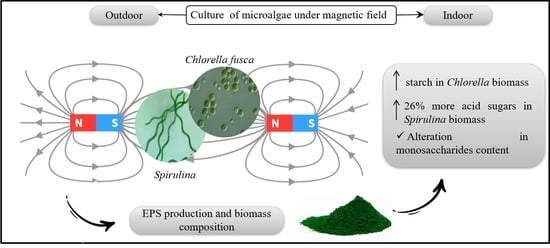
Based on the above, this study aimed to evaluate different conditions of 25–30 mT SMF in Spirulina and Chlorella in indoor and outdoor conditions, in order to confirm the SMF influence on polysaccharide production, evaluate which polysaccharidic fraction is modulated by SMF and highlight a possible modification of EPS composition.

2. Materials and Methods

2.1. Microalgae and Culture Conditions

Arthrospira platensis SAG 21.99 (Göttingen University, Germany), Spirulina sp. LEB 18 [38] and Chlorella fusca LEB 111 (Aidar and Kutner Microorganism Bank-BMA&K) were chosen to be evaluated. A. platensis SAG 21.99 and Spirulina sp. LEB 18 were cultivated in Zarrouk medium [39] and C. fusca in BG 11 medium [40]. These microalgae were selected, since there are previous studies that show the influence of SMF on growth, but the polysaccharides were not evaluated before [16,36,41], besides Spirulina and Chlorella has been widely studied due to their high polysaccharides content [1,7,42].
The A. platensis SAG 21.99 assays were carried out in 1 L glass bottles, illuminated with fluorescent tubular lamps (100 μmolphotons m−2 s−1), aerated by air and maintained at 22 °C under a 16/8 h light/dark photoperiod. Cultures were inoculated at 0.1 g L−1 and run in batch mode for 10 days. C. fusca was cultured in the same devices for 15 days, but maintained at 30 °C, a 12/12 h light/dark cycle and 82 μmolphotons m−2 s−1 for the irradiance [36,41]. Additionally, outdoor conditions were tested in raceway tanks (4.5 L working volume) for Spirulina sp. LEB 18 and C. fusca LEB 111 in a greenhouse, in open PVC raceways with daytime sunlight (mean value 300 μmolphotons m−2 s−1) and the mean temperature in those days were 34 °C. Paddle wheels, set at 24 revs min−1, were used for the mixing. The Figure 1 shows the experiment schemes developed in this study.
Initial biomass concentration was 0.2 g L−1 for Spirulina sp. and 0.3 g L−1 for C. fusca. Daily sampling allowed to follow the biomass concentration (X, g L−1), by measurement of the 670 nm optical density (UV-vis spectrophotometer, QUIMIS Q998U, Brazil) and use of DW/OD correlations established for each microalga. Xmax corresponds to the biomass concentrations obtained at the end of cultures.

2.2. Static Magnetic Fields Application

In A. platensis SAG 21.99 photobioreactor assays, SMF was applied with ferrite magnets (150 × 50 × 10 mm) placed outside the glass bottles. SMF were applied for 1 h d−1 in the light period or for 24 h. For Spirulina sp. LEB 18 and C. fusca LEB 111 raceway cultivations, 6 magnets were placed for each and during the same periods (24 h d−1 and 1 h d−1). The outdoor assays were performed from March to April, at the end of summer and beginning of fall (Southern hemisphere). The growth results are demonstrated and detailed in previous studies [16,36].
For all assays, the magnetic intensity was measured using a Gaussmeter (TLMPHALL-05 k-T0, Cotia, Brazil). To obtain the mean intensity value applied to microalgae, different measurements within the photobioreactor were made, as represented in Figure 2. Magnetic intensity of the magnets was around 30 mT for A. platensis SAG 21.99 cultures and 25 mT for the other strains studied. The magnets used have the following characteristics: barium ferrite, anisotropic and axial bipolar.
For all conditions, control cultures were conducted with the same operating conditions (temperature, irradiance and initial biomass concentration) than the assays with SMF application except that magnetic field were not applied.

2.3. Polysaccharides Extraction and Characterization

2.3.1. Starch from Chlorella fusca Biomass

The centrifuged and lyophilized biomass obtained on the 15th day was used to measure total starch. For this, the total starch assay kit (cat. no. K-TSTA) obtained from Megazyme International Ireland (Bray, Ireland) were used.
The total carbohydrate content was determined by the method described by Dubois, et al. [43] using a glucose standard curve, on biomass extracts obtained from 5 mg of lyophilized biomass resuspended in 10 mL of distilled water and sonicated for 10 min (COLE PARMER, CPX 130, Illinois, CA, USA).

2.3.2. EPS from Spirulina

EPS from Spirulina biomass were extracted according to Filali-Mouhim, et al. [44]. The biomass was resuspended in pH 9.5, 0.05 M TAPS ([tris (hydroxymethyl) methylamino] propanesulfonic acid) buffer containing 0.025 M EDTA and 0.025 M NaCl. The use of this buffer aims to limit cellular lysis during subsequent steps. After heating at 100 °C for 1 h, the suspension was centrifugated (17,000× g, 30 min, 20 °C) and the pellet was re-suspended in the same buffer for a second extraction. The supernatants were combined, and acidic polysaccharides were precipitated with 10 volumes of 3% cetyltrimethylammonium bromide (CTAB) for 24 h. After centrifugation, the pellet was purified by 4 successive cycles of solubilization in KCl 1.5, 0.75 and 0.3 M, and a last resolubilization in milli-Q water. Between each solubilization in KCl, polysaccharides were precipitated with 3 volumes of 96° ethanol (left overnight at −20 °C) and the mixture was centrifugated to recover the polysaccharides pellet. These successive steps aimed to increase the purity of samples by removing most of the proteins and other impurities [45]. The samples were lyophilized for the next analyzes.
Total carbohydrate content of EPS was analyzed by the resorcinol assay as described by Monsigny, et al. [46] using glucose as standard. Another standard curve was performed, using glucuronic acid, in order to access to the neutral sugar content using the corrective formula described by Montreuil, et al. [47]. Uronic acids were evaluated with meta-hydroxyldiphenyl, as described by Blumenkrantz and Asboe-Hansen [48] using glucuronic acid as standard.
Monosaccharide analysis was conducted as previously described [49,50,51,52]. Acid hydrolysis were conducted with 2 M HCl for 90 min at 120 °C. Samples were neutralized with NH4OH 35%, centrifugated (15,000× g, 15 min) and analyzed for their monosaccharide composition by High Pressure Anion Exchange Chromatography (HPAEC) equipped with pulsed amperometric detection (PAD). An ICS 3000 and AS 50 autosampler (Dionex, Sunnyvale, CA, USA) were used to inject 25 µL of samples on a guard CarboPac PA1-column (4 × 50 mm) and analytical CarboPac PA1-column (4 × 250 mm). Elution was performed using 18 mM NaOH for 25 min (to recover neutral sugars), followed by a linear gradient of 1 M sodium acetate in 200 mM NaOH to elute acidic monosaccharides. Data were collected and analyzed with Dionex Chromeleon 6.80 software (Sunnyvale, CA, USA). Quantification of monosaccharides was performed by plotting the response area as function of concentration of injected standards (l-Fuc, l-Ara, l-Rha, d-GlcN, d-Gal, d-Glc, d-Xyl, d-Man, d-GlcA, d-GalA) in the range 0.001 and 0.05 g L−1.

2.4. Statistical Analysis

Analysis of variance (ANOVA) was conducted, followed by the Tukey test at 95% confidence level in order to highlight significance of results.

3. Results

3.1. Polysaccharides in Chlorella Microalgae Biomass

The biomass concentration reached 2.30 g L−1 (Table 1) when C. fusca was cultured outdoor under SMF 24 h, 24% higher than control (1.75 g L−1). When SMF was applied for 1 h d−1, no positive effect on cell growth was noticed, since microalgal growth was similar to the control assay and biomass concentrations showed no significant difference (p ≥ 0.05) at all culture times. However, positive effect was noticed for indoor assays, with a 70% increase when SMF was applied for 1 h d−1 and 85% when SMF was applied for 24 h, compared to control (0.80 g L−1). Concerning total carbohydrates, and contrary to what was previously observed for Spirulina [16,35], only a slight increase was noticed for the cultures conducted with SMF and the statistical analysis revealed no significant difference. The more influent factor on the total carbohydrate content was found to be the indoor vs. outdoor condition, as a significant increase was observed for outdoor cultivation. According to Hu [53], an increase in carbohydrate content is observed with high light intensities, due to more efficient carbon fixation through photosynthesis. In the present study, the outdoor assays were carried out with daytime sunlight (300 μmolphotonsm−2 s−1), while indoor assays illuminated with daylight-type lamps (100 μmolphotonsm−2 s−1). Thus, our results are in accordance with this assumption.
In Chlorophyceae, like Chlorella fusca, the major polysaccharides that can be encountered are the storage polysaccharide (starch, [7]), and those constituting the cell wall (cellulose-like, [54]). A few studies have also described EPS released in the culture medium for some Chlorella species, but at really low yield [37,55]. In our study, we quantified the starch content in each cultivation condition, in order to highlight a potential modulation of the polysaccharide repartition. For the outdoor assays, in both control and SMF application conditions, starch content was found greater than in indoor conditions (p ≤ 0.05). This result seems consistent with the increase of the photosynthesis rate with higher light intensity, as the storage polysaccharide may act as a carbon sink. Starch accumulation acts as a fast stress response [56] and mechanisms of starch accumulation by microalgae as well as the degree of starch polymerization and cellular location vary with species and algae division stage [12].
C. fusca was found to accumulate more starch with SMF action, with 23% increase for continuous application indoor and 13% increase for 1 h d−1 in outdoor condition (Table 1). The main strategy to accumulate starch in microalga cultures is generally nitrogen starvation [7], but other operating conditions can also lead to this overproduction. Ho, et al. [57] have shown that C. vulgaris and C. variabilis biomass contained higher starch amount when the culture was enriched with CO2. However, this appears to be strain dependent as no modification of starch accumulation was noticed for C. sorokiniana and C. minutissima. Luo, et al. [37] studied the influence of SMF on EPS content of Chlorella vulgaris. These authors revealed that the higher the SMF strength, the lower soluble EPS content. In the present study C. fusca was cultivated under SMF that is supposed to act as a stress for the cells, modifying metabolism of microorganisms, affecting cell growth and molecules synthesis, explaining the greater starch content observed when applying SMF. Combining SMF with other stress (for example nitrogen starvation) could then further enhance starch accumulation, that could be of interest as feedstock for bioethanol production. This approach at a larger scale could improve the biomass production and, consequently, increase the biofuel production being feasible outside the laboratory.
Additionally, as the total carbohydrate content was not found significantly increased, it can be supposed that the content in polysaccharides other than starch as thus decreased. As polysaccharides encountered in Chlorella biomass are only starch and cell wall polysaccharides, we can suppose that the content of the latter has decreased. This could also be of great interest for further biomass processing, as it can decrease the cell-wall rigidity and facilitate the downstream processes of cellular lysis and starch extraction.

3.2. Characterization of EPS of Cyanobacteria Strains

The EPS content in the Spirulina and Arthrospira biomass obtained for cultures with SMF or controls is showed in Table 2. While the two cyanobacteria are really closed genetically, Spirulina sp. LEB 18 present higher EPS content in the biomass (49.3 g 100 gbiomass−1) than Arthospira platensis SAG 21.99 (27.9 g 100 gbiomass−1). However, the first one was cultured outdoor, while the latter was cultured indoor. Moreover, the meta-bibliographic data analysis and experiments conducted by Phelippe, et al. [42] has shown that total carbohydrate content is increased at high temperature with a mean of 41% DW (dry weight) for cultures conducted at more than 36 °C, and only 27% DW for lower temperatures. Same trend has been observed for high light intensities, with additionally a modulation between glycogen and EPS repartition and a modification in EPS composition.
Previous studies with Spirulina under SMF demonstrated an increase in total carbohydrates content [16,36]. In the present study, application of SMF on Arthospira platensis SAG 21.99 did not significantly increase the EPS content, and this amount even decreased for Spirulina sp. LEB 18 cultures. In addition to the EPS, these cyanobacteria also produce glycogen as storage polysaccharide. Our results thus suggest that the increase in carbohydrate content previously described [16,36] could be due to a greater accumulation of glycogen within the cells, thus modifying the polysaccharides repartition within cells. This result could also be considered as consistent with what was observed for Chlorella fusca, as in both cases the intracellular polysaccharide seems to be enhanced.
Analysis of the exopolysaccharides from A. platensis 21.99 and Spirulina sp. LEB 18 have shown that they are mainly constituted of neutral sugars (80–86%) and uronic acids (13–19%) (Table 2). The two different cyanobacteria strains studied had no significant different osidic composition (p ≤ 0.05). However, composition was differently affected, as neutral and acidic sugars were not modified for Spirulina sp. LEB 18, while the uronic acids content increased for A. platensis SAG 21.99. The microalgae photochemical reactions are highly affected by irradiance because there is a biologically unbalanced reaction between the light absorbed by photochemistry versus the energy used by metabolism [58]. Phelippe, et al. [42] have shown that an increase in light intensity could enhance this content.
The EPS of cyanobacteria usually includes up to six or more monosaccharides in their structure [11]. Some studies have shown that polysaccharides composition produced by Spirulina is altered by cultivation conditions and extraction methods. These polysaccharides are composed of several monosaccharides, such as rhamnose, ribose, mannose, fucose, galactose, xylose, glucose, glucuronic acid and galacturonic acid [59,60]. In addition, cyanobacteria have the particularity of producing polymers containing sulphate groups and uronic acids. Additionally, pentoses, such as xylose, arabinose and ribose are monosaccharides frequently found in EPS secreted by cyanobacteria [14]. In our study, the monosaccharide composition was investigated after acidic hydrolysis of the EPS, using HPAEC-PAD. The overall composition of the extracted EPS in cyanobacteria strains is shown in Table 3.
The strains studied exhibited eight different monosaccharides in their compositions (fucose, arabinose, rhamnose, galactose, glucose, xylose, glucuronic acid and galacturonic acid). These compositions are thus in accordance with those previously published. The main monosaccharide found in A. platensis 21.99 EPS was glucose, that represents around 70% of the overall composition for the control, while in Spirulina sp. LEB 18 biomass, galactose and glucose are the highest constituents. Phélippé, et al. [42] showed that the cyanobacteria cultured with irradiance up to 800 μmolphotons m−2 s−1 presented strong increase in glucose content, reaching 77.7%, showing that a modulation of EPS composition can be encountered with cultivation conditions. The difference in EPS composition for the controls of the two cyanobacteria could thus be due to the different culture conditions (indoor and outdoor). However, regarding A. platensis SAG 21.99, the SMF applied for 24 h and 1 h showed some monosaccharides that are not found in the control assay, or their amount is higher than control, such as fucose, rhamnose, galactose and glucuronic acid. At the opposite, the SMF application decreased the glucose, arabinose and xylose concentrations. For Spirulina sp. LEB 18 cultured with SMF, differences appeared less significant, but slight increases were observed for fucose, galactose, glucose and glucuronic acid (Table 3).
These cyanobacterial EPS compositions are then in accordance with literature data, especially regarding glucose and galactose content. Glucose is the main monosaccharide in compositions and galactose, the secondary in cyanobacteria and other microalgae strains [61,62]. Xylose is often encountered in EPS compositions, since xylans could be a protective mechanism for cells to resist mechanical stress. Biomass cultured under SMF applied for 1 h produced more xylose, as it may be a protection against SMF stress.
As EPS from microalgae and cyanobacteria are known to exhibit biological activities, the modulation of some monosaccharide that was observed in this study could have significant importance for the potential applications associated to these EPS. Furthermore, in the end of microalgal cultivation when the nutrient content is low and there is high algal density, EPS could be used as carbon and energy source [63,64]. Indeed, even if it is still difficult to rely on a polysaccharide composition and a biological activity, presence of uronic acids, as well as sulphate groups, fucose or rhamnose contents have been suggested to play a role in some activities [42]. On the other hand, the SMF application on microalgae is often studied in laboratory scale and there are some challenges to apply this technique at a pilot scale to achieve high EPS production. Therefore, the present study showed that microalgae cultured in outdoor conditions and open photobioreactors has a good quality biomass and high growth rate, improving the feasibility to test these conditions at large scale, that nowadays is a challenge in SMF approaches.

4. Conclusions

SMF increased the biomass productivities besides the starch content in Chlorella biomass; probably the glycogen content in Spirulina biomass and modifying EPS composition. Outdoor cultures of C. fusca showed 30% more starch than indoor assays, besides, when SMF was applied for 1 h d−1, starch content was higher than control. Regarding uronic acids in the EPS of cyanobacteria, the SMF action significantly increased the content (p ≤ 0.05) in both conditions. For cyanobacteria, SMF application did not significantly modify the EPS amounts, while the increase in total carbohydrate content was already observed suggesting that the SMF action is on the intracellular polysaccharide fraction. However, a modification in the EPS composition was highlighted, that may improve biological activities of EPS. Indeed, the SMF applied for 24 d−1 and 1 h d−1 induced an increase in some monosaccharides content such as fucose, rhamnose, and glucuronic acid that are suggested to be relied to these activities. This study showed thar SMF may alter the composition and concentration of carbohydrate and microalgae metabolism.

Author Contributions

Conceptualization, K.M.D. and C.L.; funding acquisition, J.A.V.C. and C.L.; investigation, K.M.D. and C.G.; methodology, K.M.D.; supervision, J.A.V.C., K.M. and C.L.; validation, C.L.; writing—original draft, K.M.D.; writing—review and editing, K.M.D., M.G.d.M., L.O.S. and C.L. All authors have read and agreed to the published version of the manuscript.

Funding

This research was funded by the Coordination for the Improvement of Higher Education Personnel—Brazil (CAPES), Program CAPES-COFECUB (Sv 945/19) and Ministry of Science, Technology, Innovations and Communication (MCTIC). This study was financed in part by the Coordenação de Aperfeiçoamento de Pessoal de Nível Superior—Brasil (CAPES)—Finance Code 001.

Institutional Review Board Statement

Not applicable.

Informed Consent Statement

Not applicable.

Data Availability Statement

Not applicable.

Conflicts of Interest

The authors declare no conflict of interest.

References

  1. Delattre, C.; Pierre, G.; Laroche, C.; Michaud, P. Production, extraction and characterization of microalgal and cyanobacterial exopolysaccharides. Biotechnol. Adv. 2016, 34, 1159–1179. [Google Scholar] [CrossRef] [PubMed]
  2. Dimitriu, S. (Ed.) Polysaccharides, Structural Diversity and Functional Versatility; Dekker M: New-York, NY, USA, 2004. [Google Scholar]
  3. Dogra, B.; Shaheen, A.; Yong, P.; Jae, P. Biochemical properties of water soluble polysaccharides from photosynthetic marine microalgae Tetraselmis species. Macromol. Res. 2017, 25, 172–179. [Google Scholar] [CrossRef]
  4. Amna Kashif, S.; Hwang, Y.; Park, J. Potent biomedical applications of isolated polysaccharides from marine microalgae Tetraselmis species. Bioproc. Biosyst. Engineer. 2018, 41, 611–1620. [Google Scholar] [CrossRef]
  5. Roussel, M.; Villay, A.; Delbac, F.; Michaud, P.; Laroche, C.; Roriz, D.; el Alaoui, H.; Diogon, M. Antimicrosporidian activity of sulphated polysaccharides from algae and their potential to control honeybee nosemosis. Carbohydr. Polym. 2015, 133, 213–220. [Google Scholar] [CrossRef]
  6. Chen, X.; Song, L.; Wang, H.; Liu, S.; Yu, H.; Wang, X.; Li, R.; Liu, T.; Li, P. Partial characterization, the immune modulation and anticancer activities of sulfated polysaccharides from filamentous microalgae Tribonema sp. Molecules 2019, 24, 322. [Google Scholar] [CrossRef] [Green Version]
  7. Ferreira, A.; Ferreira, S.S.; Correia, A.; Vilanova, M.; Silva, T.H.; Coimbra, M.A.; Nunes, C. Reserve, structural and extracellular polysaccharides of Chlorella vulgaris: A holistic approach. Algal Res. 2020, 45, 101757. [Google Scholar] [CrossRef]
  8. Sekharam, K.M.; Venkataraman, L.V.; Salimath, P.V. Structural studies of a glucan isolated from blue-green alga Spirulina platensis. Food Chem. 1989, 31, 85–91. [Google Scholar] [CrossRef]
  9. Nakamura, Y.J.-I.; Sakurai, A.; Inaba, Y.; Suzuki, E.; Nihei, S.; Fujiwara, S.; Tsuzuki, M.; Miyashita, H.; Ikemoto, H.; Kawachi, M.; et al. Some cyanobacteria synthesize semiamylopectin type α-Polyglucans instead of glycogen. Plant Cell Physiol. 2005, 46, 539–545. [Google Scholar] [CrossRef] [PubMed]
  10. Usov, A.I. Polysaccharides of the red algae. Adv. Carbohydr. Chem. Biochem. 2011, 65, 115–217. [Google Scholar] [PubMed]
  11. Pereira, S.; Zille, A.; Micheletti, E.; Moradas-Ferreira, P.; de Philippis, R.; Tamagnini, P. Complexity of cyanobacterial exopolysaccharides: Composition, structures, inducing factors and putative genes involved in their biosynthesis and assembly. FEMS Microbiol. Rev. 2009, 33, 917–941. [Google Scholar] [CrossRef]
  12. Gaignard, C.; Gargouch, N.; Dubessay, P.; Delattre, C.; Pierre, G.; Laroche, C.; Fendri, I.; Abdelkafi, S.; Michaud, P. New horizons in culture and valorization of red microalgae. Biotechnol. Adv. 2019, 37, 193–222. [Google Scholar] [CrossRef] [PubMed]
  13. Maia, J.L.; Cardoso, J.S.; Mastrantonio, D.J.S.; Bierhals, C.K.; Moreira, J.B.; Costa, J.A.V.; Morais, M.G. Microalgae starch: A promising raw material for the bioethanol production. Int. J. Biol. Macromol. 2020, 165, 2739–2749. [Google Scholar] [CrossRef] [PubMed]
  14. Rossi, F.; de Philippis, R. Role of cyanobacterial exopolysaccharides in phototrophic biofilms and in complex microbial mats. Life 2015, 5, 1218–1238. [Google Scholar] [CrossRef] [Green Version]
  15. Santos, L.O.; Deamici, K.M.; Menestrino, B.C.; Garda, B.J.; Costa, J.A.V. Magnetic treatment of microalgae for enhanced product formation. World J. Microbiol. Biotechnol. 2017, 33, 169–173. [Google Scholar] [CrossRef]
  16. Deamici, K.M.; Costa, J.A.V.; Santos, L.O. Magnetic field action on outdoor and indoor cultures of Spirulina: Evaluation of growth, medium consumption and protein profile. Bioresour. Technol. 2018, 249, 168–174. [Google Scholar] [CrossRef]
  17. Costa, S.S.; Peres, B.P.; Machado, B.R.; Costa, J.A.V.; Santos, L.O. Increased lipid synthesis in the culture of Chlorella homosphaera with magnetic fields application. Bioresour. Technol. 2020, 315, 123880. [Google Scholar] [CrossRef]
  18. Veiga, M.C.; Fontoura, M.M.; Oliveira, M.G.; Costa, J.A.V.; Santos, L.O. Magnetic fields: Biomass potential of Spirulina sp. for food supplement. Bioproc. Biosyst. Engineer. 2020, 43, 1231–1240. [Google Scholar] [CrossRef]
  19. Menestrino, B.C.; Pintos, T.H.C.; Sala, L.; Costa, J.A.V.; Santos, L.O. Application of static magnetic fields on the mixotrophic culture of Chlorella minutissima for carbohydrate production. Appl. Biochem. Biotechnol. 2020, 192, 822–830. [Google Scholar] [CrossRef] [PubMed]
  20. Tu, R.; Jin, W.; Xi, T.; Yang, Q.; Han, S.-F.; Abomohra, A.E.-F. Effect of static magnetic field on the oxygen production of Scenedesmus obliquus cultivates in municipal wastewater. Water Res. 2015, 86, 132–138. [Google Scholar] [CrossRef] [PubMed]
  21. Schenck, J. The role of magnetic susceptibility in magnetic reso- nance imaging: MRI magnetic compatibility of the first and second kinds. Med. Phys. 1996, 23, 815–850. [Google Scholar] [CrossRef]
  22. Rusanowska, P.; Zieliński, M.; Dębowski, M. Impact of static magnetic field on efficiency of fine-bubble aeration of liquid. J. Ecol. Eng. 2017, 2, 130–135. (In Polish) [Google Scholar]
  23. Zieliński, M.; Cydzik-Kwiatkowska, A.; Zielińska, M.; Dębowski, M.; Rusanowska, P.; Kopańska, J. Nitrification in activated sludge exposed to static magnetic field. Water Air Soil Pollut. 2017, 22, 126. [Google Scholar] [CrossRef] [Green Version]
  24. Dini, L.; Abbro, L. Bioeffects of moderate intensity static magnetic fields on cell cultures. Micron 2005, 36, 195–217. [Google Scholar] [CrossRef]
  25. Yadollahpour, A.; Rashidi, S.; Ghotbeddin, Z.; Jalilifar, M.; Rezaee, Z. Electromagnetic fields for treatments of wastewater: A review of applications and future opportunities. J. Pure Appl. Microbiol. 2014, 8, 3711–3719. [Google Scholar]
  26. Repacholi, M.H.; Greenebaum, B. Interaction of static and extremely low frequency electric and magnetic fields with living systems: Health effects and research needs. Bioelectromagnetics 1999, 20, 133–160. [Google Scholar] [CrossRef]
  27. Sahebjamei, H.; Abdolmaleki, P.; Ghanati, F. Effects of magnetic field on the antioxidant enzyme activities of suspension-cultured tobacco cells. Bioelectromagnetics 2007, 28, 42–47. [Google Scholar] [CrossRef]
  28. Wang, H.Y.; Zeng, X.B.; Guo, S.Y. Growth of Chlorella vulgaris under different magnetic treatments. Prog. Mod. Biomed. 2006, 6, 106–108. [Google Scholar]
  29. Zhi-Yong, L.; Si-Yuan, G.; Lin, L.; Miao-Yan, C. Effects of electromagnetic field on the batch cultivation and nutritional composition of Spirulina platensis in an air-lift photobioreactor. Bioresour. Technol. 2007, 98, 700–705. [Google Scholar]
  30. Miyakoshi, J. Effects of static magnetic fields at the cellular level. Prog. Biophys. Mol. Biol. 2005, 87, 213–223. [Google Scholar] [CrossRef]
  31. Niu, C.; Geng, J.; Ren, H.; Ding, L.; Xu, K.; Liang, W. The strengthening effect of a static magnetic field on activated sludge activity at low temperature. Bioresour. Technol. 2013, 150, 156–162. [Google Scholar] [CrossRef]
  32. Filipič, J.; Kraigher, B.; Tepuš, B.; Kokol, V.; Mandic-Mulec, I. Effects of low-density static magnetic fields on the growth and activities of wastewater bacteria Escherichia coli and Pseudomonas putida. Bioresour. Technol. 2012, 120, 225–232. [Google Scholar] [CrossRef]
  33. Bauer, L.M.; Costa, J.A.V.; Rosa, A.P.C.; Santos, L.O. Growth stimulation and synthesis of lipids, pigments and antioxidants with magnetic fields in Chlorella kessleri cultivations. Bioresour. Technol. 2017, 244, 1425–1432. [Google Scholar] [CrossRef]
  34. Shao, W.; Ebaid, R.; Abomohra, A.E.F.; Shahen, M. Enhancement of Spirulina biomass production and cadmium biosorption using combined static magnetic field. Bioresour. Technol. 2018, 265, 163–169. [Google Scholar] [CrossRef]
  35. Deamici, K.M.; Costa, J.A.V.; Santos, L.O. Magnetic fields as triggers of microalga growth: Evaluation of its effect on Spirulina sp. Bioresour. Technol. 2016, 220, 62–67. [Google Scholar] [CrossRef] [PubMed]
  36. Deamici, K.M.; Santos, L.O.; Costa, J.A.V. Use of static magnetic fields to increase CO2 biofixation by the microalga Chlorella fusca. Bioresour. Technol. 2019, 276, 103–109. [Google Scholar] [CrossRef] [PubMed]
  37. Luo, X.; Zhang, H.; Zhang, J. Effects of static magnetic field on Chlorella vulgaris: Growth and extracellular polysaccharide (EPS) production. J. Appl. Phycol. 2020. [Google Scholar] [CrossRef]
  38. Morais, M.G.; Reichert, C.C.; Dalcanton, F.; Durante, A.J.; Marins, L.F.; Costa, J.A.V. Isolation and characterization of a new Arthrospira strain. Z. Naturforsch. C 2008, 63, 144–150. [Google Scholar] [CrossRef]
  39. Costa, J.A.V.; Colla, L.M.; Duarte, F.P. Improving Spirulina platensis biomass yield using a fed-batch process. Bioresour. Technol. 2004, 92, 237–241. [Google Scholar] [CrossRef]
  40. Rippka, R.; Deruelles, J.; Waterbury, J.B.; Herdman, M.; Stanier, R.Y. Generic assignments, strain histories and properties of pure cultures of Cyanobacteria. J. Gen. Microbiol. 1979, 111, 1–61. [Google Scholar] [CrossRef] [Green Version]
  41. Deamici, K.M.; Santos, L.O.; Costa, J.A.V. Magnetic field as promoter of growth in outdoor and indoor assays of Chlorella fusca. Bioprocess Biosyst. Eng. 2021. [Google Scholar] [CrossRef]
  42. Phélippé, M.; Gonçalves, M.; Thouand, G.; Cogne, G.; Laroche, C. Characterization of the polysaccharides chemical diversity of the cyanobacteria Arthrospira platensis. Algal Res. 2019, 33, 101426. [Google Scholar] [CrossRef]
  43. Dubois, M.; Gilles, K.A.; Hamilton, J.K.; Rebers, P.A.; Smith, F. Colorimetric method for determination of sugars and related substances. Anal. Chem. 1956, 28, 350–356. [Google Scholar] [CrossRef]
  44. Filali-Mouhim, R.; Cornet, J.-F.; Fontane, T.; Fournet, B.; Dubertret, G. Production, isolation and preliminary characterization of the exopolysaccharide of the cyanobacterium Spirulina platensis. Biotechnol. Lett. 1993, 15, 567–572. [Google Scholar] [CrossRef]
  45. Patel, A.K.; Laroche, C.; Marcati, A.; Ursu, A.V.; Jubeau, S.; Marchal, L.; Petit, E.; Djelveh, G.; Michaud, P. Separation and fractionation of exopolysaccharides from Porphyridiumcruentum. Bioresour. Technol. 2013, 145, 345–350. [Google Scholar] [CrossRef]
  46. Monsigny, M.; Petit, C.; Roche, A.C. Colorimetric determination of neutral sugars by a resorcinol sulphuric acid micromethod. Anal. Biochem. 1988, 175, 525–530. [Google Scholar] [CrossRef]
  47. Montreuil, J.; Spick, G.; Chosson, A.; Segard, E.; Scheppler, N. Methods of study of the structure of glycoproteins. J. Pharm. Belg. 1963, 18, 529–546. [Google Scholar]
  48. Blumenkrantz, N.; Asboe-Hansen, G. New method for quantitative determination of uronic acids. Anal. Biochem. 1973, 54, 484–489. [Google Scholar] [CrossRef]
  49. Villay, A.; Laroche, C.; Roriz, D.; el Alaoui, H.; Delbac, F.; Michaud, P. Optimisation of culture parameters for exopolysaccharides production by the microalga Rhodella violacea. Biores. Technol. 2013, 146, 732–735. [Google Scholar] [CrossRef]
  50. Soanen, N.; da Silva, E.; Gardarin, C.; Michaud, P.; Laroche, C. Improvement of exopolysaccharide production by Porphyridium marinum. Bioresour. Technol. 2016, 213, 231–238. [Google Scholar] [CrossRef] [PubMed]
  51. Gaignard, C.; Macao, V.; Gardarin, C.; Rihouey, C.; Picton, L.; Michaud, P.; Laroche, C. The red microalga Flintiella sanguinaria as a new exopolysaccharide producer. J. Appl. Phycol. 2018, 30, 2803–2814. [Google Scholar] [CrossRef]
  52. Gaignard, C.; Laroche, C.; Pierre, G.; Dubessay, P.; Delattre, C.; Gardarin, C.; Gourvil, P.; Probert, I.; Dubuffet, A.; Michaud, P. Screening of marine microalgae: Investigation of new exopolysaccharide producers. Algal Res. 2019, 44, 101711. [Google Scholar] [CrossRef]
  53. Hu, Q. Environmental effects on cell composition. In Handbook of Microalgal Culture: Biotechnology and Applied Phycology; Richmond, A., Ed.; Blackwell Publishing Ltd.: Oxford, UK, 2004. [Google Scholar]
  54. Baudelet, P.-H.; Ricochon, G.; Linder, M.; Muniglia, L. New insight into cell walls of Chlorophyta. Algal Res. 2017, 25, 333–371. [Google Scholar] [CrossRef]
  55. Barberousse, H.; Ruiz, G.; Gloaguen, V.; Lombardo, R.J.; Djediat, C.; Mascarell, G.; Castaing, J.C. Capsular polysaccharides secreted by building façade colonisers: Characterisation and adsorption to surfaces. Biofouling 2006, 22, 361–370. [Google Scholar] [CrossRef] [PubMed]
  56. Zhu, S.; Huang, W.; Xu, J.; Wang, Z.; Xu, J.; Yuan, Z. Metabolic changes of starch and lipid triggered by nitrogen starvation in the microalga Chlorella zofingiensis. Biores. Technol. 2014, 152, 292–298. [Google Scholar] [CrossRef]
  57. Ho, S.-H.; Chen, Y.-D.; Chang, C.-Y.; Lai, Y.-Y.; Chen, C.-Y.; Kondo, A.; Ren, N.-Q.; Chang, J.-S. Feasibility of CO2 mitigation and carbohydrate production by microalga Scenedesmus obliquus CNW-N used for bioethanol fermentation under outdoor conditions: Effects of seasonal changes. Biotechnol. Biofuels. 2017, 10, 27–40. [Google Scholar] [CrossRef] [PubMed] [Green Version]
  58. Raven, J.A.; Geider, R.J. Temperature and algal growth. New Phytol. 2006, 110, 441–461. [Google Scholar] [CrossRef]
  59. Hayashi, T.; Hayashi, K.; Maeda, M.; Kojima, I. Calcium spirulan, an inhibitor of enveloped virus replication, from a blue-green alga Spirulina platensis. J. Nat. Prod. 1996, 59, 83–87. [Google Scholar] [CrossRef]
  60. Lee, J.B.; Hayashi, T.; Hayashi, K.; Sankawa, U.; Maeda, M.; Nemoto, T.; Nakanishi, H. Further purification and structural analysis of calcium spirulan from Spirulina platensis. J. Nat. Prod. 1998, 61, 1101–1104. [Google Scholar] [CrossRef]
  61. Richert, L.; Golubic, S.; Guédès, R.L.; Ratiskol, J.; Payri, C.; Guezennec, J. Characterization of exopolysaccharides produced by cyanobacteria isolated from Polynesian microbial mats. Curr. Microbiol. 2005, 51, 379–384. [Google Scholar] [CrossRef]
  62. Rossi, F.; Micheletti, E.; Bruno, L.; Adhikary, S.P.; Albertano, P.; Philippis, R.D. Characteristics and role of the exocellular polysaccharides produced by five cyanobacteria isolated from phototrophic biofilms growing on stone monuments. Biofouling 2012, 28, 215–224. [Google Scholar] [CrossRef] [PubMed] [Green Version]
  63. Sutherland, I.W. Biofilm exopolysaccharides: A strong and sticky framework. Microbiology 2001, 147, 3–9. [Google Scholar] [CrossRef] [PubMed] [Green Version]
  64. Zhang, X.; Bishop, P.L. Biodegradability of biofilm extracellular polymeric substances. Chemosphere 2003, 50, 63–69. [Google Scholar] [CrossRef]
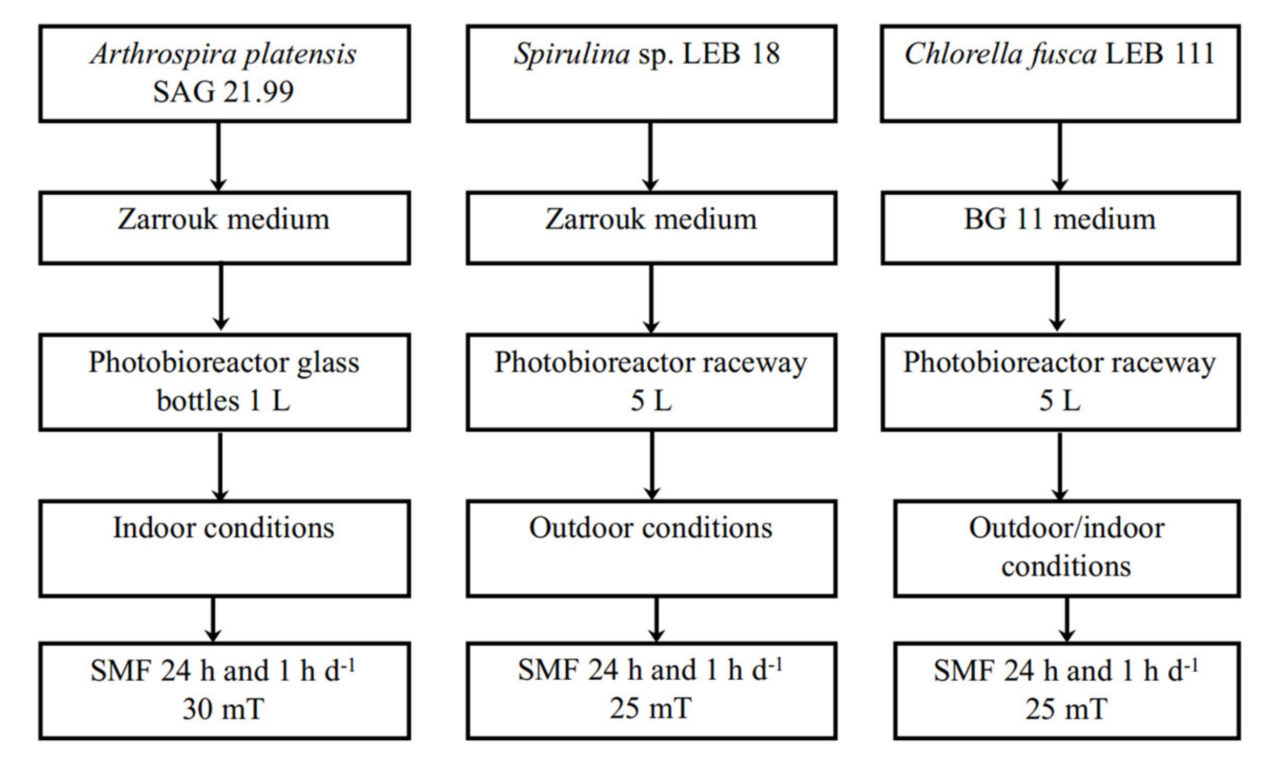
Figure 1. Scheme of research design experiments.
Applsci 11 05299 g001
Figure 2. Scheme of the photobioreactors used for A. platensis SAG 2199 (A), C. fusca and Spirulina sp. (B) assays. 1 represents the ferrite magnets place in the photobioreactors; X represents the SMF measurements points.
Applsci 11 05299 g002
Table 1. Final biomass concentration (Xmax), total carbohydrates and starch contents in Chlorella fusca LEB 111 biomass cultivated under different conditions of static magnetic field (SMF) application.
AssaysStarch (g 100 g−1)Carbohydrates (g 100 g−1)Xmax (g L−1)
IndoorControl6.42 ± 0.26 a24.84 ± 0.11 d0.80 ± 0.01 d
SMF 1 h d−17.28 ± 0.61 a,b24.98 ± 1.00 d1.36 ± 011 c
SMF 24 h d−17.91 ± 0.29 b27.53 ± 0.52 c,d1.48 ± 0.11 c
OutdoorControl 1 h d−19.67 ± 0.82 c31.38 ± 0.28 a,b2.18 ± 0.02 a
SMF 1 h d−110.94 ± 0.55 d34.18 ± 0.68 a2.23 ± 0.07 a
Control 24 h d−110.12 ± 1.27 c,d29.16 ± 1.38 b,c1.75 ± 0.03 b
SMF 24 h d−110.08 ± 0.22 c,d31.45 ± 0.76 a,b2.30 ± 0.20 a
Means ± standard deviations. Different letters in the same column correspond to significant differences (p ≤ 0.05).
Table 2. Exopolysaccharides (EPS) production yield and global osidic composition.
AssaysEPS (g·100 gbiomass−1)% NS in EPS% UA in EPSXmax (g L−1)
Arthrospira platensis SAG 21.99
Control27.9 ± 0.01 a86.83 ± 0.01 b13.17 ± 0.01 a0.59 ± 0.04 a
SMF 24 h d−134.8 ± 0.90 a80.72 ± 2.34 a19.27 ± 2.34 b0.78 ± 0.05 b
SMF 1 h d−119.4 ± 0.06 a81.51 ± 0.01 a18.49 ± 0.01 b0.88 ± 0.04 c
Spirulina sp. LEB 18
Control49.3 ± 0.19 a84.47 ± 2.27 b15.53 ± 0.01 a3.40 ± 0.01 d
SMF 24 h d−133.8 ± 0.49 b85.01 ± 3.10 b15.00 ± 3.10 a3.37 ± 0.01 d
SMF 1 h d−133.1 ± 0.06 b82.98 ± 0.35 b,a17.02 ± 0.35 a3.65 ± 0.06 d
Means ± standard deviations. Different letters in the same column correspond to significant differences for the same microalgae strain (p ≤ 0.05). EPS: exopolysaccharides, NS: Neutral Sugars, UA: Uronic Acids.
Table 3. Monosaccharide composition of EPS from cyanobacteria strains cultivated under different conditions of SMF application.
Monosaccharides (% Molar Ratio)Arthrospira Platensis SAG 21.99Spirulina sp. LEB 18
ControlSMF 24 h d−1SMF 1 h d−1ControlSMF 24 h d−1SMF 1 h d−1
Fucose5.8 ± 0.0110.2 ± 0.017.0 ± 0.0113.34 ± 0.0113.5 ± 0.0215.30 ± 0.01
Arabinose1.7 ± 0.01--1.77 ± 0.012.28 ± 0.011.43 ± 0.01
Rhamnose-4.8 ± 0.024.5 ± 0.029.77 ± 0.016.59 ± 0.014.33 ± 0.01
Galactose7.2 ± 0.0116.9 ± 0.0115.5 ± 0.0418.99 ± 0.0121.05 ± 0.0220.42 ± 0.01
Glucose71.8 ± 0.0152.4 ± 0.0157.2 ± 0.0419.66 ± 0.0123.35 ± 0.0224.51 ± 0.03
Xylose9.9 ± 0.017.8 ± 0.026.2 ± 0.0117.37 ± 0.0117.62 ± 0.0416.78 ± 0.02
Glucuronic acid3.6 ± 0.018.1 ± 0.016.50 ± 0.0213.45 ± 0.0114.30 ± 0.0115.48 ± 0.01
Galacturonic acid---4.18 ± 0.01--
Means ± standard deviations.
Publisher’s Note: MDPI stays neutral with regard to jurisdictional claims in published maps and institutional affiliations.