Review

Extraction of Pigments from Microalgae and Cyanobacteria—A Review on Current Methodologies

by
Fernando Pagels
1,2,3,
Ricardo N. Pereira
3,
António A. Vicente
3 and
A. Catarina Guedes
1,*
1
CIIMAR—Interdisciplinary Centre of Marine and Environmental Research, University of Porto, Novo Edifício do Terminal de Cruzeiros do Porto de Leixões, Av. General Norton de Matos s/n, 4450-208 Matosinhos, Portugal
2
FCUP—Faculty of Sciences, University of Porto, Rua do Campo Alegre s/n, 4169-007 Porto, Portugal
3
CEB—Centre of Biological Engineering, Campus de Gualtar, University of Minho, 4710-057 Braga, Portugal
*
Author to whom correspondence should be addressed.
Appl. Sci. 2021, 11(11), 5187; https://doi.org/10.3390/app11115187
Submission received: 27 April 2021 / Revised: 29 May 2021 / Accepted: 1 June 2021 / Published: 3 June 2021

Abstract

:
Pigments from microalgae and cyanobacteria have attracted great interest for industrial applications due to their bioactive potential and their natural product attributes. These pigments are usually sold as extracts, to overcome purification costs. The extraction of these compounds is based on cell disruption methodologies and chemical solubility of compounds. Different cell disruption methodologies have been used for pigment extraction, such as sonication, homogenization, high-pressure, CO2 supercritical fluid extraction, enzymatic extraction, and some other promising extraction methodologies such as ohmic heating and electric pulse technologies. The biggest constrain on pigment bioprocessing comes from the installation and operation costs; thus, fundamental and applied research are still needed to overcome such constrains and give the microalgae and cyanobacteria industry an opportunity in the world market. In this review, the main extraction methodologies will be discussed, taking into account the advantages and disadvantages for each kind of pigment, type of organism, cost, and final market.

1. Introduction

Microalgae and cyanobacteria are photosynthetic organisms that produce distinct kinds of pigments in order to harvest light. Such pigments are usually grouped in three major classes—chlorophylls, carotenoids, and phycobiliproteins [1]. The presence of different kinds of pigments in microalgae and cyanobacteria varies according to the phylum to which the organism belongs. Chlorophyll is the most fundamental one; it is responsible for oxygenic photosynthetic activity and it is present in all photosynthetic microalgae and cyanobacteria [2]. Carotenoids can be both primary and secondary pigments and the profile changes within the species and growth conditions; the most well known and commercialized ones being astaxanthin, lutein, and β-carotene [3]. Moreover, phycobiliproteins are a special class of pigments present only in cyanobacteria and red algae and can represent the major light absorber in these organisms, where the most well-known phycobiliproteins are phycocyanin and phycoerythrin [4]. Together, the three classes of pigments maximize light harvesting in microalgae and cyanobacteria through the whole visible light range.
Pigments from microalgae and cyanobacteria are known for their highly attractive properties for industrial use in food, feed, pharmaceuticals, nutraceuticals, and cosmetics, mainly due to their color and bioactive properties, but also for being natural and eco-friendly components [3,4]. The demand for natural colors (instead of synthetic ones) is increasing the need for a more sustainable source, however, the use of microalgae and cyanobacteria in the market is still related to high added-value products, in part due to the scale of biomass production being small when compared to synthetic alternatives [5].
Microalgae and cyanobacteria can be sold both as raw biomass (dried) or processed for the obtention of specific compounds. When only a specific compound is the final product, it requires downstream processing of extraction and possibly purification. The cost for the production of purified pigments is still high and it restricts the application to high-value markets, for example as cosmetics [6].
Three cases of successful production of pigments from microalgae and cyanobacteria are known: phycocyanin from Arthrospira platensis [4]; β-carotene from Dunaliella salina [7]; and astaxanthin from Haematococcus pluvialis [8]. Other compounds and sources appear as potential products, such as lutein from Scenedesmus almeriensis [9] and phycoerythrin from Porphyridium spp. [10]. The market for these pigments represents about $1.5 billion (USD), including all sources (natural and synthetic). The phycocyanin market alone has a size of about $100 million (USD), β-carotene ca. $270 million (USD), lutein ca. $350 million (USD), and astaxanthin ca. $800 million (USD) [4]. Pigments are usually found as a part of extracts obtained from the mentioned organisms, because as already mentioned, the purification process leads to unjustifiable costs [8].
Several factors can affect the extraction of pigments, including the target pigment, organism, market trends, available technology, and costs. Extraction usually requires a cell disruption method and a compatible solvent; however, it is also possible to extract some of these compounds without a cell disruption process, using the so-called “cell milking”, where the product is extracted while the culture grows [11].
When it comes to the available technology, many extraction methodologies were developed and optimized in recent years. In general, extraction can be performed using two distinct kinds of methodologies: (i) non-mechanical, such as chemical, thermal, and enzymatic; and (ii) mechanical, such as pressurized systems, ultrasonication, microwave, electric fields, and supercritical extraction. Some systems are based on a synergy between non-mechanical and mechanical characteristics, considering the effects of solvents, temperature and the technology mechanism (e.g., microwave-assisted extraction and ohmic heating). Furthermore, industrial scale extraction methodologies for microalgae and cyanobacteria are not widely described, and scalable downstream processes are still needed.
This review will cover the most commonly used techniques for the extraction of pigments from microalgae and cyanobacteria, paying particular attention to carotenoids and phycobiliproteins. The main advantages and disadvantages of each extraction technique will be highlighted, and some insights into the process optimization for specific cultivation of organisms and production of pigments will be addressed.

2. Classic Extraction

Classic extraction of microalgae and cyanobacteria pigments is performed using a solvent extraction associated, or not, to a thermal treatment (heat or cold), as depicted in Figure 1. The process of solvent extraction entails mixing the two phases (solvent + biomass) so that the solute can come into contact with the solvent until an equilibrium is reached. The transference from one phase to the other is driven by chemical affinities. The solution of solvent plus solute obtained at the end of the process is called “extract”.
The main advantage of this kind of extraction is the reduced cost in terms of infrastructure and operating procedures. Solvent extraction, on the other hand, frequently demands large volumes of organic solvents for carotenoids extraction, or even a lengthy processing time, as in the case of phycobiliproteins extraction, due to cyclic thermal treatment. In both cases, efficiency is usually not enough for industrial application [12]. When selecting a solvent, solubility of the compound, toxicity, and ecological impact of residues are important points to consider.
The extraction performed with organic solvents happens when the solvent is absorbed within the cell wall, causing some rupture and making the cell content available for extraction. The complete extraction can take a long time and usually improved efficiency can be achieved with stirring or mixing [12,13]. Only in the few cases where microalga does not require harsh disruption methods due to cell composition can solvent extraction be as efficient as other methods. For example, Isochrysis galbana is a source of fucoxanthin, which can be obtained via a single solvent extraction with ethanol [14].
Classical extraction of astaxanthin from H. pluvialis is performed by using organic solvents such as chloroform and methanol or more eco-friendly solvents such as acetone, ethyl acetate, and ethanol. The single solvent extraction with acetone can induce cell-wall disintegration and extraction of pigments, although the efficiency is relatively low and the process requires a long time [15,16]. Thus, it is possible to use a mixture of solvents or extraction steps in order to increase efficiency. Zou et al. [17] used both ethanol and ethyl acetate in a 1:1 mixture for the extraction of astaxanthin from H. pluvialis, the optimization was performed varying the percentage of ethanol from 30% to 70%, and consequently changing the polarity of the solvent mixture.
Furthermore, a simple solution for enhancing the extraction rate is to apply a thermal treatment. Carotenoid’s extraction can be more efficient if the process is performed at a temperature between 50 to 65 °C. At these temperatures, the cell wall becomes less resistant, and the process is further enhanced by the increased solubility of pigments in organic solvents at higher temperatures [18]. However, a long exposure to temperatures above 70 °C can cause degradation of carotenoids and chlorophylls that are considered thermally sensitive compounds [19].
When it comes to phycobiliproteins, one of the most common extraction techniques uses the freeze-thaw method, using phosphate buffer or water as a solvent. The freeze-thaw method consists of the crystallization of the intracellular water by freezing the wet biomass, followed by thawing under refrigeration temperatures (4 °C), which causes cell lysis due to the expansion of the ice crystals. The extraction can involve several freeze and thaw cycles, leading to a time-consuming process and lack of reproducibility, which is highly affected by the number of cycles [20,21]. Optimization of the process involves adjustment of solvent, freezing temperature, freezing and thawing time, and biomass-to-solvent ratio, among other factors. The extraction of phycocyanin is not limited by low temperature; Silveira et al. [22] optimized A. platensis phycocyanin extraction at 25 °C. The extraction used water as the extractant, and an orbital mixer for 4 h. Moreover, the freeze-thaw method has also been used as pre-treatment for other extractions. After the freeze-thaw process, the biomass can be submitted to ultrasonication, microwave-assisted extraction, or homogenization. Considering that the cells become more fragile, the extraction is more efficient [12,23].

3. Enzymatic Extraction

The enzymatic extraction is based in the use of hydrolytic enzymes that are able to break the membrane and/or the cell wall of the microalgae or cyanobacteria, exposing their intracellular components to the solvent, as shown in Figure 2.
The main advantage of the use of enzymatic extraction is the specificity of enzymes for cell wall lysis, as in the case of cellulase, once cellulose and hemicellulose are the main components in most cell walls [12,24]. Other advantages can be listed, such as the use of mild reaction conditions in terms of pH and temperature, absence of corrosion, and higher extraction rate, and the fact that this kind of extraction does not require drying steps [25]. On the other hand, some limitations are present and hamper the use of this extraction process, such as the high cost of enzymes and the necessity of ensuring stable conditions during the process [26], once enzymes are very sensitive in terms of temperature and pH changes. In addition, enzymatic reactions may take a long time, becoming less attractive for industrial applications [27].
Most enzymatic extractions in microalgae and cyanobacteria are related to the extraction of lipidic compounds (incl. carotenoids), being usually used as a pre-treatment that requires a solvent extraction afterwards [12,24,28]. Specifically, for pigment extraction, Tavanandi et al. [29] suggested an enzymatic extraction for the obtention of allophycocyanin from A. platensis. In this case, the extraction is performed with lysozyme for 20 h, at 37 °C and pH 7.0. When compared to surfactant-assisted extraction, the use of an enzymatic extraction led to higher purity of allophycocyanin. Furthermore, increasing the extraction efficiency is possible if the biomass is pre-treated prior to enzymatic extraction. Tavanandi et al. [29] have also observed the increase of 30% of the final content of phycocyanin when the biomass is pre-treated with ultrasound prior enzymatic extraction.

4. Pressurized Systems

The use of pressure as a cell disruption technique is one of the most promising methods for the extraction of pigments from microalgae and cyanobacteria. High-pressure homogenization (HPH) and pressurized liquid extraction (PLE) have high extraction capacity and are both scalable and able to be applied in large-scale processes. The effects of pressurized extraction processes on the cell are represented in Figure 3.
HPH forces the suspension (biomass + solvent) through a small orifice, increasing the pressure drastically; the consequent mechanical effects of turbulence, shear stress, and cavitation induce cell disruption [30].
The system must take into account the loading pressure, solvent viscosity, biomass concentration and cell wall composition. However, the increase in the pressure and the shear in the orifice induce a significant increase of the temperature in the system, requiring cooling to prevent pigments degradation [31]. The system is also limited to the high-energy requirement and high cost of implementation and operation [30]. Thus, the pressure required depends on the cell wall composition and varies according to the species. Spiden et al. [32] showed that in the microalgae Nannochloropsis sp., Chlorella sp., and Tetraselmis suecica, the pressure needed to disrupt 50% of the cells per cycle is ca. 2000, 1000, and 170 bar, respectively. A similar result was found by Mulchandani et al. [33], where the optimal condition for lipids extraction from Chlorella saccharophila was 800 bar with 10 cycles through the system. Moreover, Bernaerts et al. [34] showed that HPH using 1000 bar for 4 cycles in Nannochloropsis sp. biomass has enhanced the bioaccessibility of carotenoids for food products, while decreasing the pigment content due to degradation, in particular violaxanthin.
On the other hand, PLE is an alternative to classical thermal solvent extraction. In this method, the suspension (biomass + solvent) is placed in an oven at high temperatures (50 to 200 °C) and increased pressure (100 to 140 bar). The high pressure prevents the solvent from boiling, as the boiling temperature increases; in the liquid state and with high temperature, the solvent has a high diffusion coefficient, low viscosity, and higher solubility strength. At the same time, the high temperature reduces cell wall stability and increases the solvent flux into the cell. After the extraction time, the liquid is pumped out of the extraction chamber by the addition of compressed nitrogen, and the biomass is retained by a filter, reducing the processing steps. The main limitations of this system are, on the one hand, the use of high temperatures, and on the other, the high costs of the facilities [35].
Cha et al. [36] used PLE for the extraction of carotenoids and chlorophylls from Chlorella vulgaris. The optimization was performed through a response surface methodology using temperature and time as factors, and the optimal extraction conditions varied between the target products: 148 °C, 35 min for lutein; 117 °C, 25 min for β-carotene; 173 °C, 15 min for chlorophyll a; and 170 °C, 3 min for chlorophyll b. The pressure in this study was kept constant at 100 bar. Although high temperatures were used, the extraction was more effective when compared to maceration, Soxhlet, and ultrasound extraction. Similar findings were reported to D. salina, Herrero et al. [37] optimized PLE also in terms of temperature and extraction time for the recovery of antioxidant compounds, and in special β-carotene, where the increase in temperature also increased the extraction rate, reaching the optimal extraction at the conditions 160 °C, 30 min, and 100 bar [37]. Moreover, in Phormidium spp., the optimal extraction yield for carotenoids was found at 150 °C; 20 min of time of extraction and pressure were not optimized and were kept constant between treatments at 100 bar [38].
Furthermore, a process of pressurized liquid but using low temperatures (30 to 70 °C) has been suggested by Amaro et al. [39]. The so called continuous pressurized solvent extraction (CPSE) system provides a cheaper and more environmentally friendly approach than the PLE system described above. In this system, the solvent and the extraction chamber, containing the biomass, are heated to the desired temperature; the pressurized solvent is then pumped through the extraction chamber, promoting the contact between solvent and biomass and thus leading to a more efficient extraction. The system has as major advantages the use of mild temperatures (room temperature to 70 °C) and pressures (70 to 260 bar), and also allows recirculation of the solvent, thus reducing its consumption. Amaro et al. [39] used this system with Gloeothece sp. biomass, and the optimal conditions for carotenoid extraction were determined to be 60 °C and 180 bar with 3 cycles of ethanol recirculation.

5. Wave-Energy-Based Cell Disruption

Two wave-based extractions—microwave-assisted extraction (MAE) and ultrasound assisted extraction (UAE)—have been studied and optimized for the extraction of high-value compounds from microalgae and cyanobacteria. Regarding the extraction of pigments, both methods have been suggested, although only a few studies have been carried out regarding their optimization. The effects of wave-based extraction processes in the cell are represented in Figure 4.
MAE occurs due to the heat shock caused when the radiation is applied to the solution, causing cell wall degradation. As a thermal process, the main limitation of its use is the limited thermostability of pigments. The main benefits of MAE are the reduced extraction time and low solvent consumption, making it a green extraction method [30]. Furthermore, because of the short extraction time, this extraction method is not restricted to thermoresistant molecules. Also, low temperatures can be used when performing MAE under vacuum conditions, although this option significantly increases the cost of the extraction process [40].
Juin et al. [40] used MAE to obtain phycobiliproteins from Porphyridium purpureum. Optimal phycoerythrin extraction was achieved at 40 °C in 10 s of microwave application. On the other hand, phycocyanin and allophycocyanin were extracted more effectively at a temperature of 100 °C. The temperature was controlled by oscillating the power applied [40]. In another study, Pasquet et al. [41] also suggest microwave extraction for the obtention of carotenoids and chlorophylls from Cylindrotheca closterium, with an optimal condition at 5 min and 50 W, at 56 °C, with a higher extraction rate when compared with classic thermal extraction and UAE.
Regarding UAE, the process is based in the use of acoustic cavitation for producing cavitation bubbles, that locally increase the pressure when they collapse. The consequent disruption of the cell wall allows the penetration of the solvent, and higher extraction rates are achieved [42]. UAE thus increases extraction yield, reducing the time and energy for processing, while ensuring good reproducibility, low solvent consumption and low temperature ranges (<70 °C), allowing UAE to be associated as a green extraction approach [42,43].
Dey and Rathod [42] optimized the use of UAE for the extraction of β-carotene from A. platensis. Optimal conditions were found by using heptane at a temperature of 30 °C and an electrical acoustic intensity of 167 W.cm−2, over 8 min [42]. The use of a UAE continuous system has been proposed by Natarajan et al. [44] for the extraction of lipids from Chlorella sp., pointing at a possible scaling up of UAE processes. Finally, Zou et al. [17] optimized astaxanthin extraction from H. pluvialis using UAE using a response surface methodology, and the optimal condition was set at an ultrasound power of 200 W and frequency of 40 kHz at 41.1 °C, during 16 min, with a yield 35% higher than classical solvent extraction.

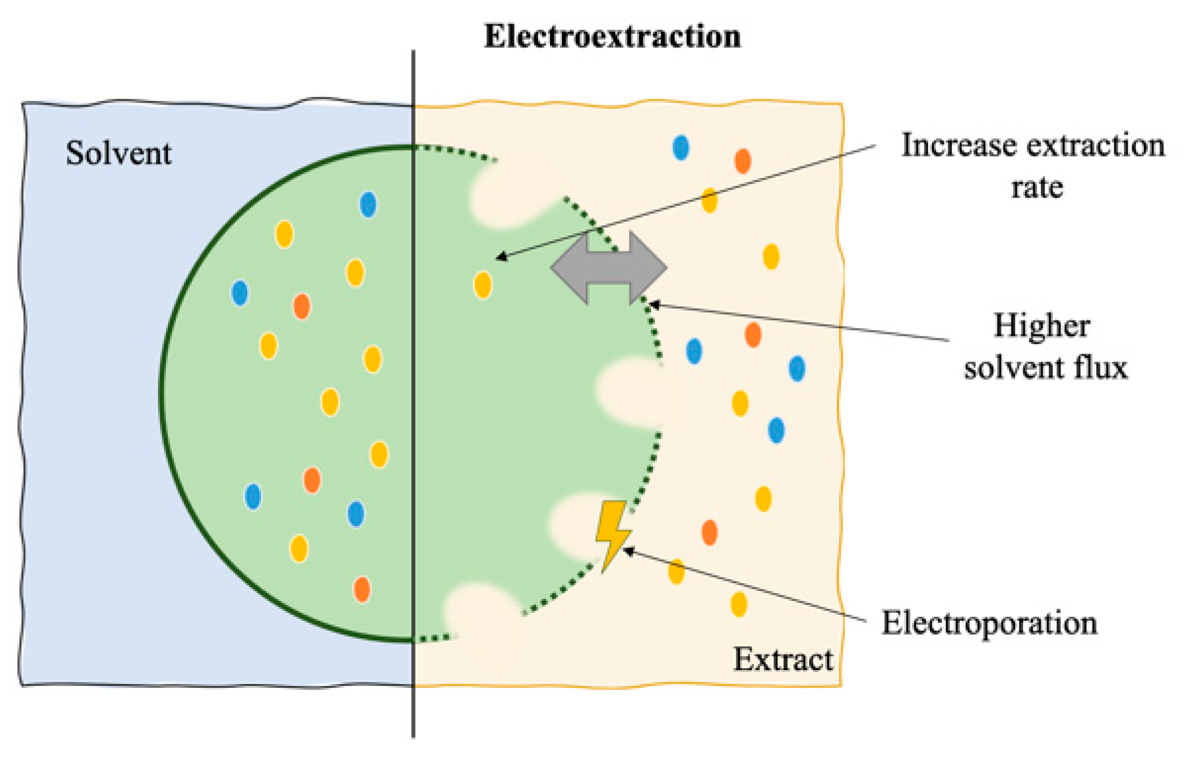
6. Electroextraction

Electric field technologies have been developed and used in food processing since the 1970s, more specifically the pulsed electric fields (PEF), that are based in the application of an electric current through the biomass in contact with a solvent for a certain period of time, in order to induce an electroporation effect [45]. The electric field can be applied continuously or in pulses, applying a charge to the cell membrane and inducing a destabilization of its structure, eventually leading to an electroporation effect by opening pores in the cell (reversibly or irreversibly), allowing solvent penetration and thus facilitating the extraction of intracellular compounds [46]. The effects of electroextraction processes on the cell are depicted in Figure 5.
In microalgae and cyanobacteria, electrotechnologies can be used for bioproduct extraction, but also for harvesting and for production, by inducing stress for the production of secondary metabolites (including carotenoids and total antioxidant compounds) [47,48]. Changes in the intensity, shape of the electrical wave, frequency, temperature, time of extraction, number of pulses, and pulse duration can influence the efficiency of the extraction, which is dependent on the matrix biomass and the desired product or effect [48].
Electro-based methods, in special PEF, have been proposed as suitable extraction methods for several microalgae and cyanobacteria pigments. Luengo et al. [49,50] optimized PEF for the extraction of chlorophylls and carotenoids from C. vulgaris using pulse duration values between microseconds and milliseconds. The optimized treatment (25 pulses of 3 μs at an electric field strength of 20 kV.cm−1) was efficient for both cell permeabilization, cell inactivation, and extraction of pigments. In another study, Luengo et al. [51] suggest the use of a controlled temperature between 25 to 30 °C for the extraction of lutein from C. vulgaris. The temperature range is the same used in the production of the microalgae, thus avoiding extra costs for heating.
In the case of A. platensis, PEF have been suggested by Martínez et al. [52] for the extraction of phycocyanin. The minimum electric field required was 15 kV.cm−1, being the optimal 25 kV.cm−1 associated with a treatment of 50 pulses of 3 μs at temperature of 40 °C. In another study, a higher purity of phycocyanin extracted from Nostoc commune was achieved by using PEF, although at a lower concentration than freeze thawing extraction, using an electric field of 5 kV.cm−1, a frequency of 2 Hz, and 1500 pulses of 1 μs each, at 40 °C [53]. Chittapun et al. [53] also suggest that the PEF methodology seems to be not suitable for some organisms, as in the case of Oscillatoria okeni, where the cell wall is much more resistant. Moreover, for the extraction of phycoerythrin from the red microalgae Porphyridium cruentum, the optimized conditions of PEF were an electric field of 10 kV.cm−1, a frequency of 0.5 Hz, and 50 pulses of 3 μs each with temperature kept between 20 and 30 °C [54].
As a pre-treatment, PEF was used for the extraction of pigments from Nannochloropsis spp. using a two-stage solvent extraction process: the first stage with water as the solvent and the second one with an organic solvent mixture. The PEF treatment (20 kV.cm−1, 400 pulses of 10 μs) is applied only in the first stage and pigments are recovered in the second stage [55]. In addition, PEF pre-treatment was also suggested for the extraction of astaxanthin in H. pluvialis, with a treatment of 1 kV.cm−1, 10 pulses of 5 ms, at 20 °C providing an extraction 1.2-fold more efficient than other disruption methods [56].
As the main advantages, electrotechnologies require much lower energy input, shorter extraction times, and lower amounts of solvent than classical, enzymatic, and wave-based methods [27]. They have advantages in the operational process when compared to traditional mechanical methods; they are scalable, they are considered a “green” method for industrial extraction due to a reduced used of solvents, and they can be operated in continuous mode [46,54]. On the other hand, these technologies can have a higher capital and installation cost. In addition, the use of organic solvents often remains necessary and the extraction parameters such as pulse number and duration and electric field strength must be optimized for each microorganism and desired product [45]. For example, cyanobacteria require much more energy to achieve electroporation than microalgae, due to their smaller size and membrane composition [54].

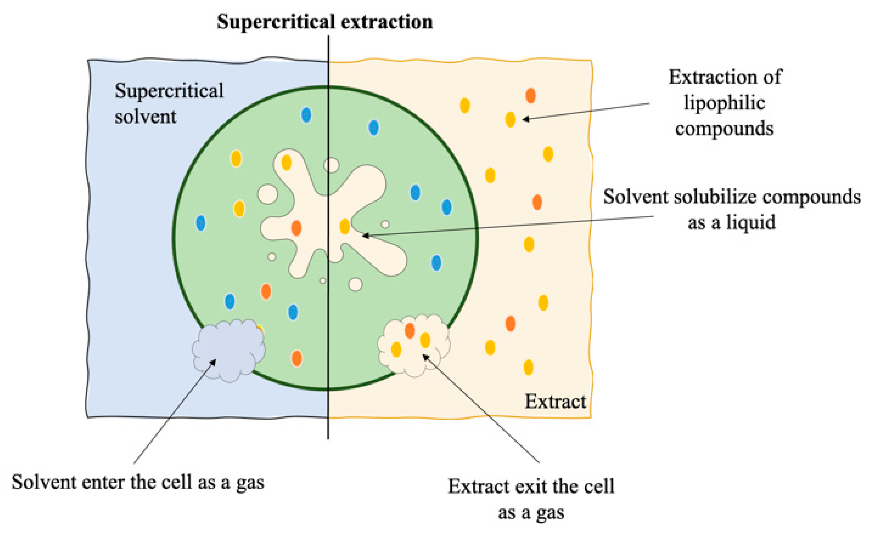
7. Supercritical Fluid Extraction

From scalable techniques, supercritical extraction stands out as an efficient method for obtaining pigments from microalgae and cyanobacteria, especially carotenoids and chlorophylls. Supercritical fluid extraction (SFE) is based on the use of a supercritical solvent, which can permeate the cell by acting like a gas while also solubilizing compounds by acting like a liquid solvent [57]. Because of its low polarity, supercritical CO2 is the most commonly used solvent in carotenoids and chlorophyll extraction, and due to its non-flammable and non-toxic properties, this method is associated with a more environmentally friendly approach [3,58]. In general, SFE is recognized as a green methodology due to lower consumption of organic solvents and shorter extraction time, when compared to classic solvent extraction. However, the main limitation of SFE is still the cost of implementing and operating it [3]. Supercritical extraction may also open the possibility for processes operating under a “biorefinery” approach, once it allows further treatment and extraction of the remaining biomass [59].
The SFE consists of a high–pressure pump for CO2, a heating chamber, an extraction chamber, and a collecting chamber. The CO2 is pumped into the heating chamber where it will reach the supercritical conditions, then it is pumped into the extraction vessel, where it diffuses into the biomass. The extraction occurs as a result of the solvent permeating the biomass, which causes cell expansion and simultaneous solubilization of the target compounds. The extract then goes to the collecting chamber where CO2 is cooled, re-compressed and recycled, leaving an oil-like highly concentrated extract containing carotenoids and chlorophyll [57,60]. Figure 6 shows a schematic representation of SFE and its effects on the cell.
Supercritical CO2 extraction has been applied to the extraction of carotenoids in many microalgae and cyanobacteria. In D. salina, Jaime et al. [61] found a pressure of 443 bar and a temperature of 27.5 °C to be the optimal conditions for the extraction of β-carotene. In another work, Hosseini et al. [62] showed that the optimal operating conditions for extraction of carotenoids from D. salina were 400 bar and 55 °C, although the highest carotenoids/chlorophylls ratio was indeed found at a lower temperature (30 °C). Changes in pressure have been associated to the selective extraction of carotenoids.
Liau et al. [63] optimized supercritical CO2 extraction in Nannochloropsis oculata, and the system used ethanol addition (as co-solvent) to enhance the extraction yield. The optimal condition for lipids and carotenoids extraction was set at 350 bar, 50 °C, and 16.7% of ethanol addition. In the case of Nannochloropsis gaditana, Macías-Sánchez et al. [64] used supercritical CO2 extraction for carotenoids and chlorophyll extraction. The highest yield was obtained at a pressure of 400 bar and a temperature of 60 °C. The best carotenoids/chlorophylls ratio was also obtained by decreasing the pressure (200 bar) and maintaining the temperature at 60 °C. The pressure of 400 bar and a temperature of 60 °C was also the optimal condition for lutein and β-carotene extraction in S. almeriensis. Although the extraction yield of β-carotene is up to 50% of its total content, the percentage of extraction for lutein is very low [65]. For Scenedesmus obliquus, Guedes et al. [66] tested supercritical CO2 extraction of carotenoids and chlorophylls for use in food processing. The highest carotenoid yield and ratio of total carotenoids to chlorophyll was attained at 250 bar and 60 °C, using ethanol (7.7%, v/v) as co-solvent.
Regarding astaxanthin from H. pluvialis, Nobre et al. [67] showed that the highest recovery of carotenoids (92%) was obtained at a pressure of 300 bar and temperature of 60 °C, using ethanol as a co-solvent (10%), while Di Sanzo et al. [68] found the maximum recovery of astaxanthin, at 50 °C and 550 bar, without the addition of a co-solvent.
Supercritical extraction was also applied for the extraction of carotenoids in cyanobacteria. Montero et al. [69] optimized temperature and pressure of Synechococcus sp. extraction, and the optimal settings varied regarding specific carotenoids: 358 bar and 50 °C for β-carotene; 454 bar and 59 °C for cryptoxanthin; and 500 bar and 60 °C for zeaxanthin. Also, Macías-Sánchez et al. [70] studied the SFE of carotenoids and chlorophyll in Synechococcus sp. For the extraction of carotenoids, a pressure of 300 bar and a temperature of 50 °C were suggested, while for chlorophyll the optimal condition was 500 bar and 60 °C. For a higher selectivity of carotenoids, the pressure should be decreased to 200 bar and the temperature kept at 60 °C.

8. Cell Milking

Products coming from microalgae and cyanobacteria traditionally followed the process of biomass cultivation, harvesting, dewatering, extraction, and purification; thus, alternatives to improve the process have been suggested along the years. The term “cell milking” has gained popularity due to the potential of bypassing a portion of the process. Cell milking works on the basis of a two-phase cultivation system that includes an aqueous phase (medium) and an extraction phase with a hydrophobic solvent. The contact of the solvent with the cell creates pores in the membrane, exposing the target compounds that pass to the solvent phase due to their compatibility and solubility in the solvent. The cells are then allowed to recover and produce more of the compound and possibly be milked again in a short time interval [71].
Cell viability is the main factor that distinguishes milking from in situ extraction. In situ extraction uses the same two-phase solvent system, but the cell is not restored after the extraction. The continuous production of a compound through in situ extraction also requires a high productivity in the cultivation phase to overcome cells’ death [11]. Figure 7 shows a schematic representation of cell milking and its effects on the cell.
The main advantage of cell milking is that this process does not require harvesting and the recovery of the products from the hydrophobic phase is relatively simple. Also, the cultivation phase can be maintained continuously, significantly reducing costs [11]. As disadvantages, the solvent needs to be biocompatible, green labelled, and safe, and the solutes can be only extracted in the hydrophobic phase. Moreover, the need of agitation to provoke contact of the two phases requires energy input, increasing costs, although most microalgae and cyanobacteria cultures already have an agitator [11].
The most commonly used solvent in milking is dodecane, approved as food flavoring but with restrictions for other uses. An important factor when considering solvent compatibility is its log P (partition coefficient) value, which will determine its solubility in the cultivation phase. Hejazi et al. [72] used solvents with log P ranging from 3 to 9 in order to extract β-carotene from D. salina and found that solvents with log P > 6 were the most compatible with this microalga. Also, using a higher log P solvent would allow specifying the extraction for β-carotene only, with less chlorophyll contamination. In this study, the optimal extraction was achieved using dodecane. In another study, Kleinegris et al. [73] tested milking with different D. salina strains; interestingly one of the strains (CCAP 19/25) was not milkable, as no carotenoids were extracted. For the other tested strains it was possible to extract different carotenoids profiles by using different mixing methods. Using gentle mixing, it was possible to extract only secondary carotenoids, while when using vigorous mixing primary carotenoids and chlorophyll were mostly extracted instead, although with vigorous mixing the authors reported an increase in cell death.
The use of milking was also described for the obtention of astaxanthin from H. pluvialis by using a two-phase solvent for the extraction phase, after the switch from the green to the red phase. The process uses dodecane for extraction and methanol for recovering the astaxanthin from the dodecane. The addition of NaOH to methanol is suggested in order to increase the stability of astaxanthin [18]. As an alternative to dodecane, Kang and Sim [74] suggested vegetable oils for the extraction of astaxanthin, with an increase in recovery yields.
Milking can be applied for the extraction of other compounds besides pigments, including lipids for biofuel. Jackson et al. [11] suggest the milking of the microalga Botryococcus braunii (Chlorophyta) using hexane, octane or octanol as solvents, due to their biocompatibility, boiling point, extraction effectiveness, cost, and safety.

9. Novel Extraction Methodologies

Novel methodologies have been suggested for cell disruption and extraction of valuable compounds from microalgae and cyanobacteria in special pigments. These methodologies are usually adaptations of the already-mentioned technologies or have the same fundamental principles as a basis. The next few paragraphs will detail the applicability of laser, hydrodynamic cavitation, flotation, high-voltage electrostatic fields, and ohmic heating for the extraction of microalgae and cyanobacteria products.
Laser is a wave-based mechanical method for cell disruption, as with ultrasounds and microwaves. McMillan et al. [75] showed the use of laser as alternative for cell disruption in Nannochloropsis oculata. The laser (1064 nm) used a third of the energy of microwave extraction in order to obtain the same yield of disruption (95%). The biggest issue with using laser for large-scale production is the difficulty of scaling-up. In that study, the laser extraction was performed in only 30 µL of matrix.
Regarding hydrodynamic cavitation, cell disruption occurs after the formation of microbubbles in the medium, which produce shock waves upon collapsing, momentarily inducing higher pressure and temperature. These environmental changes created by those shock waves are responsible for cell breakage. Lee and Han [76] applied hydrodynamic cavitation to N. salina for cell wall disruption and lipid extraction, and this method seems to be as suitable as ultrasonication. On the other hand, hydrodynamic cavitation requires an efficient cooling system, proper facility design, and maintenance of high fluid velocity for the cavitation effect, leading to significant increases in the cost of the process [30].
Also based on bubble formation, the liquid biphasic flotation combines solvent sublation and a liquid biphasic system. The extraction occurs by selective adsorption of target compounds on air bubbles connecting a two-phase system: one salt-rich aqueous phase and the other an organic phase. Liquid biphasic flotation has been suggested for the extraction of astaxanthin from H. pluvialis by using 2-propanol as the organic phase and (NH4)2SO4 as salt for the aqueous phase [77]. The system can be scaled-up, although it requires a large amount of salt and organic solvents.
Moreover, the use of novel methodologies of electric field extraction—high voltage electrical discharge and ohmic heating—seems to present the most scalable and economically viable options. High voltage electrical discharge (HVED) uses continuous electric field of high intensity (E > 10 kV.cm−1) for extraction, which can also preserve the compounds’ stability and the overall quality of the final product. However, only a few studies have used this methodology for extraction [78]. In addition, the requirement for high amounts of energy makes this method less attractive than other electric-based systems. Zhang et al. [79] used HVED for extraction in N. oculata, and suggested a two-phase extraction for chlorophyll, carotenoids, and lipids using HVED as a pre-treatment with aqueous washing, followed by vacuum drying and an organic solvent extraction.
In the case of ohmic heating (OH), an electric field of low to moderate intensity is applied with an alternate current, causing internal heat generation (the Joule effect) that can enhance cell breakage or electroporation [80]. This methodology does not have restrictions in processing times and is currently used in foods thermal processing, mainly due to the uniformity of heating, controllable heating rate, cost, and energy efficiency [47]. Although the use of ohmic heating has been suggested for the extraction of pigments in plants [81,82], only a few studies have suggested the use of this methodology in algae matrices. Yodsuwan et al. [83] suggested the use of OH as a pre-treatment of Chlorella sp. biomass for the production of biodiesel. The optimal pre-treatment was performed at an electric field strength of 15 V.cm−1 and a frequency of 5 Hz, for 2 min at 70 °C.
A summary of techniques discussed in this review for the extraction of pigments from microalgae and cyanobacteria is presented in Table 1.

10. Final Remarks

Microalgae and cyanobacteria pigments show great potential for industrial applications, since there is already a commercial market for β-carotene, astaxanthin, and phycocyanin. However, the process of extraction and the available technologies are still associated with a high cost of implementation and operation. In addition, energetic requirements represent a very significant part of the sustainability of the process [84]. As shown in this review, the standardization of a biomass extraction protocol for obtention of the pigments of interest is still a challenge. The variability of microalgae and cyanobacteria composition needs case-by-case optimization. According to Grima et al. [85], extraction can account for up to 60% of overall expenditures. To lower those expenses, it is still required to optimize existing procedures and develop fresh procedures. The main advantages and limitations of the methods discussed in this review for the extraction of pigments from microalgae and cyanobacteria are summarized in Table 2.
Moreover, the use of an optimized extraction method can reduce the necessity of purification. Depending on the final application (e.g., in the case of food, feed, nutraceuticals, and cosmetics) purification may or may not be needed. The need of purification can increase the final price a hundred fold [86].
Finally, the optimal scenario for microalgae and cyanobacteria valorization is the use of a biorefinery process, which will enhance the economic feasibility by allowing the exploitation of different co-products which can be individually utilized [87]. Nevertheless, only small-scale experiments and theoretical studies can be found in the literature, being mostly inconclusive when larger scale applications are sought. There is, thus, still a gap in technical feasibility studies, being necessary to improve the scale of biorefineries in order to evaluate the viability of the process and its practical implementation.

Author Contributions

Conceptualization, F.P., A.A.V., R.N.P. and A.C.G.; Investigation, F.P.; Writing—original draft preparation, F.P.; Writing—review and editing, A.A.V., R.N.P. and A.C.G.; Supervision, A.A.V. and A.C.G.; Funding acquisition, A.A.V. and A.C.G. All authors have read and agreed to the published version of the manuscript.

Funding

This work was co-supported by a PhD fellowship granted to author F.P. [SFRH/BD/136767/2018] funded by Foundation for Science and Technology (FCT, Portugal) under the auspices of Operational Program Human Capital (POCH), supported by the European Social Fund and Portuguese funds (MECTES); as well as by the national funds through FCT within the scope of UIDB/04423/2020, UIDP/04423/2020 and UIDB/04469/2020.

Institutional Review Board Statement

Not applicable.

Informed Consent Statement

Not applicable.

Data Availability Statement

Not applicable.

Conflicts of Interest

The authors declare no conflict of interest.

References

  1. Richmond, A.; Hu, Q. Handbook of Microalgal Culture: Applied Phycology and Biotechnology, 2nd ed.; Wiley-Blackwell: Hoboken, NJ, USA, 2013; 736p. [Google Scholar] [CrossRef]
  2. Masojdek, J.; Koblek, M.; Torzillo, G. Photosynthesis in Microalgae. In Handbook of Microalgal Culture: Applied Phycology and Biotechnology, 2nd ed.; Richmond, A., Hu, Q., Eds.; Wiley-Blackwell: Hoboken, NJ, USA, 2007; pp. 21–36. [Google Scholar] [CrossRef]
  3. Guedes, A.C.; Amaro, H.M.; Malcata, F.X. Microalgae as sources of carotenoids. Mar. Drugs 2011, 9, 625–644. [Google Scholar] [CrossRef] [PubMed]
  4. Pagels, F.; Salvaterra, D.; Amaro, H.M.; Guedes, A.C. Pigments from microalgae. In Handbook of Microalgae-Based Processes and Products; Jacob-Lopes, E., Queiroz, M.I., Maroneze, M.M., Zepka, L.Q., Eds.; Academic Press: Cambridge, MA, USA, 2020; pp. 465–492. [Google Scholar] [CrossRef]
  5. Acién, F.G.; Fernández, J.M.; Magán, J.J.; Molina, E. Production cost of a real microalgae production plant and strategies to reduce it. Biotechnol. Adv. 2012, 30, 1344–1353. [Google Scholar] [CrossRef] [PubMed]
  6. Mehta, P.; Singh, D.; Saxena, R.; Rani, R.; Gupta, R.P.; Puri, S.K.; Mathur, A.S. High-value coproducts from algae—An innovational way to deal with advance algal industry. In Waste to Wealth; Singhania, R.R., Agarwal, R.A., Kumar, R.P., Sukumaran, R.K., Eds.; Springer: Singapore, 2018; pp. 343–363. [Google Scholar] [CrossRef]
  7. Tafreshi, A.H.; Shariati, M. Dunaliella biotechnology: Methods and applications. J. Appl. Microbiol. 2009, 107, 14–35. [Google Scholar] [CrossRef] [PubMed]
  8. Panis, G.; Carreon, J.R. Commercial astaxanthin production derived by green alga Haematococcus pluvialis: A microalgae process model and a techno-economic assessment all through production line. Algal Res. 2016, 18, 175–190. [Google Scholar] [CrossRef] [Green Version]
  9. Fernández-Sevilla, J.M.; Acién Fernández, F.G.; Molina Grima, E. Biotechnological production of lutein and its applications. Appl. Microbiol. Biotechnol. 2010, 86, 27–40. [Google Scholar] [CrossRef] [PubMed]
  10. Li, S.; Ji, L.; Shi, Q.; Wu, H.; Fan, J. Advances in the production of bioactive substances from marine unicellular microalgae Porphyridium spp. Bioresour. Technol. 2019, 292, 122048. [Google Scholar] [CrossRef] [PubMed]
  11. Jackson, B.A.; Bahri, P.A.; Moheimani, N.R. Repetitive non-destructive milking of hydrocarbons from Botryococcus braunii. Renew. Sustain. Energy Rev. 2017, 79, 1229–1240. [Google Scholar] [CrossRef]
  12. Salinas-Salazar, C.; Saul Garcia-Perez, J.; Chandra, R.; Castillo-Zacarias, C.; Iqbal, H.M.N.; Parra-Saldívar, R. Methods for Extraction of Valuable Products from Microalgae Biomass BT—Microalgae Biotechnology for Development of Biofuel and Wastewater Treatment. In Microalgae Biotechnology for Development of Biofuel and Wastewater Treatment; Alam, M.A., Wang, Z., Eds.; Springer: Singapore, 2019; pp. 245–263. [Google Scholar] [CrossRef]
  13. Grima, E.M.; Fernández, F.G.A.; Medina, A.R. Downstream Processing of Cell Mass and Products. In Handbook of Microalgal Culture: Applied Phycology and Biotechnology, 2nd ed.; Richmond, A., Hu, Q., Eds.; Wiley-Blackwell: Hoboken, NJ, USA, 2013; pp. 266–267. [Google Scholar] [CrossRef]
  14. Kim, S.M.; Kang, S.W.; Kwon, O.N.; Chung, D.; Pan, C.H. Fucoxanthin as a major carotenoid in Isochrysis aff. galbana: Characterization of extraction for commercial application. J. Korean Soc. Appl. Biol. Chem. 2012, 55, 477–483. [Google Scholar] [CrossRef]
  15. Kim, D.-Y.; Vijayan, D.; Praveenkumar, R.; Han, J.-I.; Lee, K.; Park, J.-Y.; Chang, W.-S.; Lee, J.-S.; Oh, Y.-K. Cell-wall disruption and lipid/astaxanthin extraction from microalgae: Chlorella and Haematococcus. Bioresour. Technol. 2016, 199, 300–310. [Google Scholar] [CrossRef] [PubMed]
  16. Mendes-Pinto, M.M.; Raposo, M.F.J.; Bowen, J.; Young, A.J.; Morais, R. Evaluation of different cell disruption processes on encysted cells of Haematococcus pluvialis: Effects on astaxanthin recovery and implications for bio-availability. J. Appl. Phycol. 2001, 13, 19–24. [Google Scholar] [CrossRef]
  17. Zou, T.B.; Jia, Q.; Li, H.W.; Wang, C.X.; Wu, H.F. Response surface methodology for ultrasound-assisted extraction of astaxanthin from Haematococcus pluvialis. Mar. Drugs 2013, 11, 1644–1655. [Google Scholar] [CrossRef] [PubMed] [Green Version]
  18. Kang, C.D.; Sim, S.J.; Chang, D.K.; Sang, J.S. Selective extraction of free Astaxanthin from Haematococcus culture using a tandem organic solvent system. Biotechnol. Prog. 2007, 23, 866–871. [Google Scholar] [CrossRef] [PubMed]
  19. Aguilar-Machado, D.; Morales-Oyervides, L.; Contreras-Esquivel, J.C.; Aguilar, C.; Méndez-Zavala, A.; Raso, J.; Montañez, J. Effect of ohmic heating processing conditions on color stability of fungal pigments. Food Sci. Technol. Int. 2017, 23, 338–348. [Google Scholar] [CrossRef] [PubMed]
  20. Boussiba, S.; Richmond, A.E. Isolation and characterization of phycocyanins from the blue-green alga Spirulina platensis. Arch. Microbiol. 1979, 120, 155–159. [Google Scholar] [CrossRef]
  21. Sumanta, N.; Haque, C.I.; Nishika, J.; Suprakash, R. Spectrophotometric analysis of chlorophylls and carotenoids from commonly grown fern species by using various extracting solvents. Res. J. Chem. Sci. 2014, 4, 63–69. [Google Scholar]
  22. Silveira, S.T.; Burkert, J.F.M.; Costa, J.A.V.; Burkert, C.A.V.; Kalil, S.J. Optimization of phycocyanin extraction from Spirulina platensis using factorial design. Bioresour. Technol. 2007, 98, 1629–1634. [Google Scholar] [CrossRef] [PubMed]
  23. Aarthy, A.; Kumari, S.; Turkar, P.; Subramanian, S. An insight on algal cell disruption for biodiesel production. Asian J. Pharm. Clin. Res. 2018, 11, 21–26. [Google Scholar] [CrossRef] [Green Version]
  24. Kumar, S.; Kumar, G.; Dash, A.; Scholz, P.; Banerjee, R. Sustainable green solvents and techniques for lipid extraction from microalgae: A review. Algal Res. 2017, 21, 138–147. [Google Scholar] [CrossRef]
  25. Sierra, L.S.; Dixon, C.K.; Wilken, L.R. Enzymatic cell disruption of the microalgae Chlamydomonas reinhardtii for lipid and protein extraction. Algal Res. 2017, 25, 149–159. [Google Scholar] [CrossRef]
  26. Vernès, L.; Li, Y.; Chemat, F.; Abert-Vian, M. Biorefinery Concept as a Key for Sustainable Future to Green Chemistry—The Case of Microalgae BT—Plant Based “Green Chemistry 2.0”: Moving from Evolutionary to Revolutionary. In Plant Based “Green Chemistry 2.0.; Li, Y., Chemat, F., Eds.; Springer: Singapore, 2019; pp. 15–50. [Google Scholar] [CrossRef]
  27. Gong, M.; Bassi, A. Carotenoids from microalgae: A review of recent developments. Biotechnol. Adv. 2016, 34, 1396–1412. [Google Scholar] [CrossRef] [PubMed]
  28. Zuorro, A.; Maffei, G.; Lavecchia, R. Optimization of enzyme-assisted lipid extraction from Nannochloropsis microalgae. J. Taiwan Inst. Chem. Eng. 2016, 67, 106–114. [Google Scholar] [CrossRef]
  29. Tavanandi, H.A.; Vanjari, P.; Raghavarao, K.S.M.S. Synergistic method for extraction of high purity Allophycocyanin from dry biomass of Arthrospira platensis and utilization of spent biomass for recovery of carotenoids. Sep. Purif. Technol. 2019, 225, 97–111. [Google Scholar] [CrossRef]
  30. Lee, S.Y.; Cho, J.M.; Chang, Y.K.; Oh, Y.K. Cell disruption and lipid extraction for microalgal biorefineries: A review. Bioresour. Technol. 2017, 244, 1317–1328. [Google Scholar] [CrossRef] [PubMed]
  31. Lee, A.K.; Lewis, D.M.; Ashman, P.J. Disruption of microalgal cells for the extraction of lipids for biofuels: Processes and specific energy requirements. Biomass Bioenergy 2012, 46, 89–101. [Google Scholar] [CrossRef]
  32. Spiden, E.M.; Yap, B.H.J.; Hill, D.R.A.; Kentish, S.E.; Scales, P.J.; Martin, G.J.O. Quantitative evaluation of the ease of rupture of industrially promising microalgae by high pressure homogenization. Bioresour. Technol. 2013, 140, 165–171. [Google Scholar] [CrossRef] [PubMed]
  33. Mulchandani, K.; Kar, J.R.; Singhal, R.S. Extraction of lipids from Chlorella saccharophila using high-pressure homogenization followed by three phase partitioning. Appl. Biochem. Biotechnol. 2015, 176, 1613–1626. [Google Scholar] [CrossRef] [PubMed]
  34. Bernaerts, T.M.M.; Verstreken, H.; Dejonghe, C.; Gheysen, L.; Foubert, I.; Grauwet, T.; Van Loey, A.M. Cell disruption of Nannochloropsis sp. improves in vitro bioaccessibility of carotenoids and ω3-LC-PUFA. J. Funct. Foods 2020, 103770. [Google Scholar] [CrossRef]
  35. Mandal, S.C.; Mandal, V.; Das, A.K. Classification of Extraction Methods. In Essentials of Botanical Extraction; Mandal, S.C., Mandal, V., Das, A.K., Eds.; Academic Press: Boston, MA, USA, 2015; pp. 83–136. [Google Scholar] [CrossRef]
  36. Cha, K.H.; Lee, H.J.; Koo, S.Y.; Song, D.-G.; Lee, D.-U.; Pan, C.-H. Optimization of pressurized liquid extraction of carotenoids and chlorophylls from Chlorella vulgaris. J. Agric. Food Chem. 2010, 58, 793–797. [Google Scholar] [CrossRef] [PubMed]
  37. Herrero, M.; Jaime, L.; Martín-Álvarez, P.J.; Cifuentes, A.; Ibáñez, E. Optimization of the extraction of antioxidants from Dunaliella salina microalga by pressurized liquids. J. Agric. Food Chem. 2006, 54, 5597–5603. [Google Scholar] [CrossRef] [PubMed]
  38. Rodríguez-Meizoso, I.; Jaime, L.; Santoyo, S.; Cifuentes, A.; García-Blairsy Reina, G.; Señoráns, F.J.; Ibáñez, E. Pressurized fluid extraction of bioactive compounds from Phormidium species. J. Agric. Food Chem. 2008, 56, 3517–3523. [Google Scholar] [CrossRef] [PubMed]
  39. Amaro, H.M.; Catarina Guedes, A.; Preto, M.A.C.; Sousa-Pinto, I.; Xavier Malcata, F. Gloeothece sp. as a nutraceutical source-an improved method of extraction of carotenoids and fatty acids. Mar. Drugs 2018, 16, 327. [Google Scholar] [CrossRef] [PubMed] [Green Version]
  40. Juin, C.; Chérouvrier, J.-R.; Thiéry, V.; Gagez, A.-L.; Bérard, J.-B.; Joguet, N.; Kaas, R.; Cadoret, J.-P.; Picot, L. Microwave-assisted extraction of phycobiliproteins from Porphyridium purpureum. Appl. Biochem. Biotechnol. 2015, 175, 1–15. [Google Scholar] [CrossRef] [Green Version]
  41. Pasquet, V.; Chérouvrier, J.R.; Farhat, F.; Thiéry, V.; Piot, J.M.; Bérard, J.B.; Kaas, R.; Serive, B.; Patrice, T.; Cadoret, J.P.; et al. Study on the microalgal pigments extraction process: Performance of microwave assisted extraction. Process Biochem. 2011, 46, 59–67. [Google Scholar] [CrossRef] [Green Version]
  42. Dey, S.; Rathod, V.K. Ultrasound assisted extraction of β-carotene from Spirulina platensis. Ultrason. Sonochem. 2013, 20, 271–276. [Google Scholar] [CrossRef] [PubMed]
  43. Fernandes, A.S.; Nogara, G.P.; Menezes, C.R.; Cichoski, A.J.; Mercadante, A.Z.; Jacob-Lopes, E.; Zepka, L.Q. Identification of chlorophyll molecules with peroxyl radical scavenger capacity in microalgae Phormidium autumnale using ultrasound-assisted extraction. Food Res. Int. 2017, 99, 1036–1041. [Google Scholar] [CrossRef] [PubMed]
  44. Natarajan, R.; Ang, W.M.R.; Chen, X.; Voigtmann, M.; Lau, R. Lipid releasing characteristics of microalgae species through continuous ultrasonication. Bioresour. Technol. 2014, 158, 7–11. [Google Scholar] [CrossRef] [PubMed]
  45. Saini, R.K.; Keum, Y.S. Carotenoid extraction methods: A review of recent developments. Food Chem. 2018, 240, 90–103. [Google Scholar] [CrossRef]
  46. Costa, J.A.V.; Freitas, B.C.B.; Moraes, L.; Zaparoli, M.; Morais, M.G. Progress in the physicochemical treatment of microalgae biomass for value-added product recovery. Bioresour. Technol. 2020, 122727. [Google Scholar] [CrossRef]
  47. Sastry, S.K.; Heskitt, B.F.; Sarang, S.S.; Somavat, R.; Ayotte, K. Why ohmic heating? Advantages, applications, technology, and limitations. In Ohmic Heating in Food Processing; Ramaswamy, H.S., Marcotte, M., Sastry, S., Abdelrahim, K., Eds.; CRC Press: Boca Raton, FL, USA, 2014; pp. 7–14. [Google Scholar] [CrossRef]
  48. Geada, P.; Rodrigues, R.; Loureiro, L.; Pereira, R.; Fernandes, B.; Teixeira, J.A.; Vasconcelos, V.; Vicente, A.A. Electrotechnologies applied to microalgal biotechnology—Applications, techniques and future trends. Renew. Sustain. Energy Rev. 2018, 94, 656–668. [Google Scholar] [CrossRef] [Green Version]
  49. Luengo, E.; Condón-Abanto, S.; Álvarez, I.; Raso, J. Effect of pulsed electric field treatments on permeabilization and extraction of pigments from Chlorella vulgaris. J. Membr. Biol. 2014, 247, 1269–1277. [Google Scholar] [CrossRef]
  50. Luengo, E.; Martínez, J.M.; Bordetas, A.; Álvarez, I.; Raso, J. Influence of the treatment medium temperature on lutein extraction assisted by pulsed electric fields from Chlorella vulgaris. Innov. Food Sci. Emerg. Technol. 2015, 29, 15–22. [Google Scholar] [CrossRef]
  51. Luengo, E.; Martínez, J.M.; Coustets, M.; Álvarez, I.; Teissié, J.; Rols, M.P.; Raso, J. A comparative study on the effects of millisecond- and microsecond-pulsed electric field treatments on the permeabilization and extraction of pigments from Chlorella vulgaris. J. Membr. Biol. 2015, 248, 883–891. [Google Scholar] [CrossRef] [PubMed]
  52. Martínez, J.M.; Luengo, E.; Saldaña, G.; Álvarez, I.; Raso, J. C-phycocyanin extraction assisted by pulsed electric field from Artrosphira platensis. Food Res. Int. 2017, 99, 1042–1047. [Google Scholar] [CrossRef] [PubMed]
  53. Chittapun, S.; Jonjaroen, V.; Khumrangsee, K.; Charoenrat, T. C-phycocyanin extraction from two freshwater cyanobacteria by freeze thaw and pulsed electric field techniques to improve extraction efficiency and purity. Algal Res. 2020, 46, 101789. [Google Scholar] [CrossRef]
  54. Martínez, J.M.; Delso, C.; Álvarez, I.; Raso, J. Pulsed electric field permeabilization and extraction of phycoerythrin from Porphyridium cruentum. Algal Res. 2019, 37, 51–56. [Google Scholar] [CrossRef]
  55. Parniakov, O.; Barba, F.J.; Grimi, N.; Marchal, L.; Jubeau, S.; Lebovka, N.; Vorobiev, E. Pulsed electric field assisted extraction of nutritionally valuable compounds from microalgae Nannochloropsis spp. using the binary mixture of organic solvents and water. Innov. Food Sci. Emerg. Technol. 2015, 27, 79–85. [Google Scholar] [CrossRef]
  56. Martínez, J.M.; Gojkovic, Z.; Ferro, L.; Maza, M.; Álvarez, I.; Raso, J.; Funk, C. Use of pulsed electric field permeabilization to extract astaxanthin from the Nordic microalga Haematococcus pluvialis. Bioresour. Technol. 2019, 289, 121694. [Google Scholar] [CrossRef]
  57. Abrahamsson, V.; Cunico, L.P.; Andersson, N.; Nilsson, B.; Turner, C. Multicomponent inverse modeling of supercritical fluid extraction of carotenoids, chlorophyll a, ergosterol and lipids from microalgae. J. Supercrit. Fluids 2018, 139, 53–61. [Google Scholar] [CrossRef]
  58. Macías-Sánchez, M.D.; Serrano, C.M.; Rodríguez, M.R.; de la Ossa, E.M.; Lubián, L.M.; Montero, O. Extraction of carotenoids and chlorophyll from microalgae with supercritical carbon dioxide and ethanol as cosolvent. J. Sep. Sci. 2008, 31, 1352–1362. [Google Scholar] [CrossRef]
  59. Gallego, R.; Bueno, M.; Herrero, M. Sub- and supercritical fluid extraction of bioactive compounds from plants, food-by-products, seaweeds and microalgae—An update. TrAC—Trends Anal. Chem. 2019, 116, 198–213. [Google Scholar] [CrossRef]
  60. Machmudah, S.; Diono, W.; Kanda, H.; Goto, M. Supercritical fluids extraction of valuable compounds from algae: Future perspectives and challenges. Eng. J. 2018, 22, 13–30. [Google Scholar] [CrossRef]
  61. Jaime, L.; Mendiola, J.A.; Ibáñez, E.; Martin-Álvarez, P.J.; Cifuentes, A.; Reglero, G.; Señoráns, F.J. β-Carotene isomer composition of sub- and supercritical carbon dioxide extracts. Antioxidant activity measurement. J. Agric. Food Chem. 2007, 55, 10585–10590. [Google Scholar] [CrossRef]
  62. Hosseini, S.R.P.; Tavakoli, O.; Sarrafzadeh, M.H. Experimental optimization of SC-CO2 extraction of carotenoids from Dunaliella salina. J. Supercrit. Fluids 2017, 121, 89–95. [Google Scholar] [CrossRef]
  63. Liau, B.C.; Shen, C.T.; Liang, F.P.; Hong, S.E.; Hsu, S.L.; Jong, T.T.; Chang, C.M.J. Supercritical fluids extraction and anti-solvent purification of carotenoids from microalgae and associated bioactivity. J. Supercrit. Fluids 2010, 55, 169–175. [Google Scholar] [CrossRef]
  64. Macías-Sánchez, M.D.; Mantell, C.; Rodríguez, M.; Martínez De La Ossa, E.; Lubián, L.M.; Montero, O. Supercritical fluid extraction of carotenoids and chlorophyll a from Nannochloropsis gaditana. J. Food Eng. 2005, 66, 245–251. [Google Scholar] [CrossRef]
  65. Macías-Sánchez, M.D.; Fernandez-Sevilla, J.M.; Fernández, F.G.A.; García, M.C.C.; Grima, E.M. Supercritical fluid extraction of carotenoids from Scenedesmus almeriensis. Food Chem. 2010, 123, 928–935. [Google Scholar] [CrossRef]
  66. Guedes, A.C.; Gião, M.S.; Matias, A.A.; Nunes, A.V.M.; Pintado, M.E.; Duarte, C.M.M.; Malcata, F.X. Supercritical fluid extraction of carotenoids and chlorophylls a, b and c, from a wild strain of Scenedesmus obliquus for use in food processing. J. Food Eng. 2013, 116, 478–482. [Google Scholar] [CrossRef] [Green Version]
  67. Nobre, B.; Marcelo, F.; Passos, R.; Beirão, L.; Palavra, A.; Gouveia, L.; Mendes, R. Supercritical carbon dioxide extraction of astaxanthin and other carotenoids from the microalga Haematococcus pluvialis. Eur. Food Res. Technol. 2006, 223, 787–790. [Google Scholar] [CrossRef]
  68. Di Sanzo, G.; Mehariya, S.; Martino, M.; Larocca, V.; Casella, P.; Chianese, S.; Musmarra, D.; Balducchi, R.; Molino, A. Supercritical carbon dioxide extraction of astaxanthin, lutein, and fatty acids from Haematococcus pluvialis microalgae. Mar. Drugs 2018, 16, 334. [Google Scholar] [CrossRef] [Green Version]
  69. Montero, O.; Macìas-Sánchez, M.D.; Lama, C.M.; Lubián, L.M.; Mantell, C.; Rodríguez, M.; De La Ossa, E.M. Supercritical CO2 extraction of β-carotene from a marine strain of the cyanobacterium Synechococcus species. J. Agric. Food Chem. 2005, 53, 9701–9707. [Google Scholar] [CrossRef]
  70. Macías-Sánchez, M.D.; Mantell, C.; Rodríguez, M.; Martínez de la Ossa, E.; Lubián, L.M.; Montero, O. Supercritical fluid extraction of carotenoids and chlorophyll a from Synechococcus sp. J. Supercrit. Fluids 2007, 39, 323–329. [Google Scholar] [CrossRef]
  71. Gillet, J.N. Ultrafast molecular dynamics of biofuel extraction for microalgae and bacteria milking: Blocking membrane folding pathways to damaged lipid-bilayer conformations with nanomicelles. J. Biomol. Struct. Dyn. 2015, 33, 690–705. [Google Scholar] [CrossRef]
  72. Hejazi, M.A.; De Lamarliere, C.; Rocha, J.M.S.; Vermuë, M.; Tramper, J.; Wijffels, R.H. Selective extraction of carotenoids from the microalga Dunaliella salina with retention of viability. Biotechnol. Bioeng. 2002, 79, 29–36. [Google Scholar] [CrossRef] [PubMed] [Green Version]
  73. Kleinegris, D.M.M.; Janssen, M.; Brandenburg, W.A.; Wijffels, R.H. The selectivity of milking of Dunaliella salina. Mar. Biotechnol. 2010, 12, 14–23. [Google Scholar] [CrossRef] [PubMed] [Green Version]
  74. Kang, C.D.; Sim, S.J. Direct extraction of astaxanthin from Haematococcus culture using vegetable oils. Biotechnol. Lett. 2008, 20, 441–444. [Google Scholar] [CrossRef] [PubMed]
  75. McMillan, J.R.; Watson, I.A.; Ali, M.; Jaafar, W. Evaluation and comparison of algal cell disruption methods: Microwave, waterbath, blender, ultrasonic and laser treatment. Appl. Energy 2013, 103, 128–134. [Google Scholar] [CrossRef]
  76. Lee, I.; Han, J.-I. Simultaneous treatment (cell disruption and lipid extraction) of wet microalgae using hydrodynamic cavitation for enhancing the lipid yield. Bioresour. Technol. 2015, 186, 246–251. [Google Scholar] [CrossRef]
  77. Khoo, K.S.; Chew, K.W.; Ooi, C.W.; Ong, H.C.; Ling, T.C.; Show, P.L. Extraction of natural astaxanthin from Haematococcus pluvialis using liquid biphasic flotation system. Bioresour. Technol. 2019, 290, 121794. [Google Scholar] [CrossRef] [PubMed]
  78. Barba, F.J.; Grimi, N.; Vorobiev, E. New approaches for the use of non-conventional cell disruption technologies to extract potential food additives and nutraceuticals from microalgae. Food Eng. Rev. 2014, 7, 45–62. [Google Scholar] [CrossRef]
  79. Zhang, R.; Marchal, L.; Lebovka, N.; Vorobiev, E.; Grimi, N. Two-step procedure for selective recovery of bio-molecules from microalga Nannochloropsis oculata assisted by high voltage electrical discharges. Bioresour. Technol. 2020, 302, 122893. [Google Scholar] [CrossRef]
  80. Coelho, M.; Pereira, R.; Rodrigues, A.S.; Teixeira, J.A.; Pintado, M.E. Extraction of tomato by-products’ bioactive compounds using ohmic technology. Food Bioprod. Process. 2019, 117, 329–339. [Google Scholar] [CrossRef] [Green Version]
  81. Mannozzi, C.; Fauster, T.; Haas, K.; Tylewicz, U.; Romani, S.; Dalla Rosa, M.; Jaeger, H. Role of thermal and electric field effects during the pre-treatment of fruit and vegetable mash by pulsed electric fields (PEF) and ohmic heating (OH). Innov. Food Sci. Emerg. Technol. 2018, 48, 131–137. [Google Scholar] [CrossRef]
  82. Yildiz, H.; Icier, F.; Baysal, T. Changes in β-carotene, chlorophyll and color of spinach puree during ohmic heating. J. Food Process Eng. 2010, 33, 763–779. [Google Scholar] [CrossRef]
  83. Yodsuwan, N.; Kamonpatana, P.; Chisti, Y.; Sirisansaneeyakul, S. Ohmic heating pretreatment of algal slurry for production of biodiesel. J. Biotechnol. 2018, 267, 71–78. [Google Scholar] [CrossRef] [PubMed]
  84. Acién, F.G.; Molina, E.; Fernández-Sevilla, J.M.; Barbosa, M.; Gouveia, L.; Sepúlveda, C.; Bazaes, J.; Arbib, Z. Economics of microalgae production. In Microalgae-Based Biofuels and Bioproducts; Woodhead Publishing Series in Energy; Gonzalez-Fernandez, C., Muñoz, R., Eds.; Woodhead Publishing: Cambridge, UK, 2017; pp. 485–503. [Google Scholar] [CrossRef]
  85. Molina Grima, E.; Belarbi, E.-H.; Acién Fernández, F.G.; Robles Medina, A.; Chisti, Y. Recovery of microalgal biomass and metabolites: Process options and economics. Biotechnol. Adv. 2003, 20, 491–515. [Google Scholar] [CrossRef]
  86. De Morais, M.G.; da Fontoura Prates, D.; Moreira, J.B.; Duarte, J.H.; Costa, J.A.V. Phycocyanin from microalgae: Properties, extraction and purification, with some recent applications. Ind. Biotechnol. 2018, 14, 30–37. [Google Scholar] [CrossRef]
  87. Bastiaens, L.; Van Roy, S.; Thomassen, G.; Elst, K. Biorefinery of algae: Technical and economic considerations. In Microalgae-Based Biofuels and Bioproducts; Gonzalez-Fernandez, C., Muñoz, R., Eds.; Woodhead Publishing: Cambridge, UK, 2017; pp. 327–345. [Google Scholar] [CrossRef]
Figure 1. Diagram of classical extraction: solvent extraction, thermal extraction, and freeze−thaw extraction.
Applsci 11 05187 g001
Figure 2. Schematic representation of an enzymatic extraction followed by a solvent extraction.
Applsci 11 05187 g002
Figure 3. Schematic representation of high−pressure homogenization (HPH) and pressurized liquid extraction (PLE) effects in microalgae and cyanobacteria cells.
Applsci 11 05187 g003
Figure 4. Schematic representation of microwave (MAE) and ultrasound assisted extraction (UAE) effects in microalgae and cyanobacteria cells.
Applsci 11 05187 g004
Figure 5. Schematic representation of electroextraction effects in microalgae and cyanobacteria cells.
Applsci 11 05187 g005
Figure 6. Schematic representation of supercritical fluid extraction (SFE) in microalgae and cyanobacteria cells.
Applsci 11 05187 g006
Figure 7. Schematic representation of cell milking in microalgae and cyanobacteria cells.
Applsci 11 05187 g007
Table 1. Extraction parameters and yields of pigments from cyanobacteria and microalgae, using different extraction methods.
Extraction Method aProductSpeciesProcessing Parameters bExtraction Yield (mg.gDW)Reference
Classic solvent extractionFucoxanthinIsochrysis galbanaS: ethanol; T: RT; t: 60 min18.23 ± 0.54[14]
Classic solvent extractionAstaxanthinHaematococcus pluvialisS: acetone; T: RT; t: 16 hca. 5.00[16]
Classic solvent extractionAstaxanthinH. pluvialisS: ethanol:ethyl acetate (1:1, v:v); T: RT; t: 90 min16.48 ± 0.67[17]
Classic solvent extractionPhycobiliproteinsArthrospira platensisS: water or sodium phosphate buffer; T: 25 °C; t: 4 h92.00[22]
EnzymaticAllophycocyaninA. platensisS: lysozyme + surfactants; T: 37 °C; t: 20 h32.27[29]
HPHCarotenoidsNannochloropsis sp.S: water/recovered with hexane:isopropanol (3:2, v:v) p: 1000 bar; T: n.s.; c: 4 cyclesVioloxanthin—2.50 ± 0.24;
Antheraxanthin—1.74 ± 0.34;
Zeaxanthin—1.93 ± 0.24;
β-carotene—10.07 ± 1.70 c
[34]
PLECarotenoids and chlorophyllChlorella vulgarisS: ethanol:water (9:1, v:v); p: 100 bar; Lutein—T: 148 °C; t: 35 min; β-carotene– T: 117 °C; t: 25 min; Chlorophyll aT: 173 °C; t: 15 min; Chlorophyll bT: 170 °C; t: 3 minLutein—3.70
β-carotene—0.67 Chlorophyll a—10.83 Chlorophyll b—6.81
[36]
PLEβ-caroteneDunaliella salinaS: ethanol; p: 100 bar; T: 160 °C; t: 30 min34.6[37]
PLECarotenoidsPhormidium spp.S: ethanol; p: 100 bar; T: 150 °C; t: 20 minn.s.[38]
CPSECarotenoidsGloeothece sp.S: ethanol; p: 180 bar; T: 60 °C; c: 3 cyclesLutein—2.9 ± 0.1;
β-carotene—1.5 ± 0.1 c
[39]
MAEPhycobiliproteinsPorphyridium purpureumS: water; P: n.s.; t: 10 s; Phycoerythrin—T: 40 °C; Phycocyanin/Allophycocyanin—T: 100 °CPhycoerythrin—73.7 ± 2.3;
Phycocyanin—34.8 ± 6.4; Allophycocyanin—32.3 ± 1.2
[40]
MAEFucoxanthinCylindrotheca closteriumS: acetone; P: 50 W; T: 56 °C t: 5 min4.24 ± 0.09[41]
UAEβ-caroteneA. platensisS: heptane; Pd: 167 W.cm−2; T: 30 °C t: 8 minca. 1.00[42]
UAECarotenoidsH. pluvialisS: ethanol:ethyl acetate (1:1, v:v); P: 200 W; F: 40 kHz; T: 41.1 °C t: 16 min27.58 ± 0.40[17]
PEFCarotenoidsC. vulgarisS: citrate- phosphate McIlvaine buffer/recovered with ethanol; E: 20 kV.cm−1; T: n.s.; t: 25 pulses of 3 μsca. 1.00[49]
PEFPhycocyaninA. platensisS: water E: 15 kV.cm−1; T: 40 °C; t: 50 pulses of 3 μs100.00[52]
PEFPhycocyaninNostoc communeS: water E: 5 kV.cm−1; F: 2 Hz; T: 40 °C; t: 1500 pulses of 1 μs29.66 ± 0.52[53]
PEFPhycoerythrinPorphyridium cruentumS: citrate- phosphate McIlvaine buffer; T: 20 to 30 °C; E: 10 kV.cm−1; F: 0.5 Hz; t: 50 pulses of 3 μs32.00[54]
PEF pre-treatmentCarotenoidsNannochloropsis spp.1st stage—S: water; T: 20 °C; E: 20 kV.cm−1, t: 400 pulses of 10 μs) (2nd stage) S: recovered in DMSO or ethanol + water (1:1, v:v); T: 20 °Cn.s.[55]
PEF pre-treatmentAstaxanthinH. pluvialisS: culture medium/recovered with ethanol; T: 20 °C; E: 1 kV·cm−1; t: 10 pulses of 5 ms18.3[56]
SC-CO2β-caroteneD. salinaS: SC-CO2; p: 443 bar; T: 27.5 °C; t: 100 minn.s.[61]
SC-CO2CarotenoidsD. salinaS: SC-CO2; p: 400 bar; T: 30 °C; t: 90 min115.43[62]
SC-CO2CarotenoidsNannochloropsis oculataS: SC-CO2 + Ethanol; p: 350 bar; T: 50 °C; t: 30 min7.61[63]
SC-CO2Carotenoids and chlorophyllNannochloropsis gaditanaS: SC-CO2; p: 400 bar; T: 60 °C; t: 3 hCarotenoids—0.34; Chlorophylls—2.23[64]
SC-CO2Lutein and β-caroteneScenedesmus almeriensisS: SC-CO2; p: 400 bar; T: 60 °C; t: 5 hLutein—0.04; β-carotene—1.50[65]
SC-CO2Carotenoids Scenedesmus obliquusS: SC-CO2; p: 250 bar; T: 60 °C; t: 4 h0.18[66]
SC-CO2AstaxanthinH. pluvialisS: SC-CO2; p: 550 bar; T: 50 °C; t: 100 min19.72[68]
SC-CO2AstaxanthinH. pluvialisS: SC-CO2 with addition of Ethanol; p: 300 bar; T: 60 °C; t: n.s.1.8[67]
SC-CO2β-caroteneSynechococcus sp.S: SC-CO2; p: 358 bar; T: 50 °C; t: 2 h0.49 ± 0.10[69]
SC-CO2CarotenoidsSynechococcus sp.S: SC-CO2; p: 300 bar; T: 50 °C t: 3 h1.51[70]
Cell Milkingβ-caroteneD. salinaS: dodecane0.25 d[72]
Cell Milkingβ-caroteneD. salinaS: dodecane5.30 d[73]
Cell MilkingAstaxanthinH. pluvialisS: dodecane85.00 e[18]
Cell MilkingAstaxanthinH. pluvialisS: vegetable oils76.00 e[74]
Lasern.s.N. oculataS: water; P: 10 W; F: 20 kHz; t: 1 minn.s.[75]
Hydrodynamic cavitationLipidic extractN. salinaS: culture medium/recovered with hexanen.s.[76]
Liquid biphasic flotationAstaxanthinH. pluvialisS: 2-propanol as organic phase and (NH4)2SO4 as salt for the aqueous phase95.11 d[77]
HVEDChlorophyll, carotenoidsN. oculataS: water; E: 40 kV.cm−1; T: 20 to 30 °C; t: 400 pulses of 4msn.s.[79]
OH pre-treatmentLipidic extractChlorella sp.S: water (wet biomass); E: 0.015 kV.cm−1; F: 0.005 kHz; T: 70 °C; t: 2 minn.s.[83]
n.s.—not specified. a HPH—high-pressure homogenization; PLE—pressurized liquid extraction; CPSE—continuous pressurized solvent extraction; MAE—microwave-assisted extraction; UAE—ultrasound-assisted extraction; PEF—pulsed electric fields; SC-CO2—supercritical CO2 extraction; HVED—High voltage electrical discharge; OH—ohmic heating. b S—solvent; T—temperature; t—time; c—cycles of extraction; p—pressure; P—power; Pd—power density; E—electric field; F—frequency; RT—room temperature. c mg.g of extract. d % of produced carotenoids. e mg Lculture−1.
Table 2. Advantages and limitations of the different methods for extraction of pigments in microalgae and cyanobacteria.
MethodologyAdvantagesLimitations
Classic solvent extraction
  • Reduced cost in terms of infrastructure and operating
  • Less efficiency
  • High amount of organic solvents
  • Time-consuming
Enzymatic
  • Specificity
  • High cell disruption
  • High cost of enzymes
  • Requires very controlled process
  • Need of separation of the enzymes
Pressure
  • High cell disruption
  • Reduced need of solvent
  • High energy requirement
  • High cost of infrastructure
Wave energy-based
  • Eco-friendly
  • Reduced time of extraction
  • High extraction rate
  • High cost of infrastructure
  • Hard control of temperature
  • Hard up-scaling process
Electric fields
  • Lower energy input
  • Reduced time of extraction
  • Reduced need of solvent
  • High cost of infrastructure
Supercritical extraction
  • Eco-friendly
  • Optimal temperature below degradation point for pigments
  • High cost of infrastructure
  • Hard operating process
Cell milking
  • Non-destructive method
  • Can operate in continuous
  • Very sensible process to maintain the culture in continuous production
Laser
  • Eco-friendly
  • Reduced time of extraction
  • Lower energy input
  • High extraction rate
  • Only applicable to small volumes
Hydrodynamic cavitation
  • Eco-friendly
  • Reduced time of extraction
  • Lower energy input
  • High extraction rate
  • High cost of infrastructure
  • Hard operating process
Flotation
  • Reduced time of extraction
  • High extraction rate
  • Requires a large amount of salt and organic solvents
High voltage electrical discharge
  • Reduced time of extraction
  • High cell disruption
  • High energy requirement
Ohmic heating
  • Reduced time of extraction
  • Controlled and homogeneous temperature
  • High cell disruption
  • High energy requirement
  • High cost of infrastructure
Publisher’s Note: MDPI stays neutral with regard to jurisdictional claims in published maps and institutional affiliations.

