Article

Antibacterial Activity of TiO2 Nanoparticles Prepared by One-Step Laser Ablation in Liquid

by
Khawla S. Khashan
1,*,
Ghassan M. Sulaiman
2,*,
Farah A. Abdulameer
3,
Salim Albukhaty
4,
Mohammed A. Ibrahem
1,
Tahani Al-Muhimeed
5 and
Abeer A. AlObaid
5
1
Division of Laser Sciences and Technology, Department of Applied Sciences, University of Technology, Baghdad 10066, Iraq
2
Division of Biotechnology, Department of Applied Sciences, University of Technology, Baghdad 10066, Iraq
3
College of Health and Medical Techniques, Gilgamesh Ahliya University, Baghdad 10022, Iraq
4
Department of Chemistry, College of Science, University of Misan, Maysan, Amarah 62001, Iraq
5
Department of Chemistry, College of Science, King Saud University, P.O. Box 22452, Riyadh 11495, Saudi Arabia
*
Authors to whom correspondence should be addressed.
Appl. Sci. 2021, 11(10), 4623; https://doi.org/10.3390/app11104623
Submission received: 20 March 2021 / Revised: 14 May 2021 / Accepted: 17 May 2021 / Published: 19 May 2021
(This article belongs to the Special Issue Synthesis and Biomedical Application of Nanoparticles)

Abstract

:
Laser ablation in liquid was utilized to prepare a TiO2 NP suspension in in deionized distilled water using Q-switch Nd: YAG laser at various laser energies and ablation times. The samples were characterized using UV–visible absorption spectra obtained with a UV–visible spectrophotometer (UV-Vis,) Fourier transform infrared (FTIR), X-ray diffraction (XRD), and transmission electron microscope (TEM). While, UV-Vis spectra showed the characteristic band-to-band absorption peak of TiO2 NPs in the UV range. FTIR analysis showed the existence of O-Ti-O bond. XRD patterns indicated the presence of (101) and (112) plane crystalline phases of TiO2. TEM images showed a spherical-like structure of TiO2 NPs with various size distributions depending on the ablation period. It was also found that there is a relationship between laser ablation time and TiO2 NP size distribution, where longer ablation times led to the smaller size distribution. The antibacterial activity of TiO2 NPs was evaluated with different species of bacteria such as Escherichia coli, Pseudomonas aeruginosa, Proteus vulgaris, and Staphylococcus aureus, using the liquid approach. The optimum activity of TiO2 NPs is found to be against E. coli at 1000 μg mL−1. Furthermore, adding, TiO2 NPs (1000 μg mL−1) in the presence of amoxicillin has a synergic effect on E. coli and S. aureus growth, as measured by the well diffusion method. However, both E. coli (11.6 ± 0.57mm) and S. aureus (13.3 ± 0.57mm) were inhibited by this process.

1. Introduction

The risk of biological and bacterial attacks has grown enormously in the last decade or so, especially in the human consumption sectors such as food, food packaging and water. This increasing risk stimulates scientists to develop new inorganic antibacterial nanoparticle substances that have no side effects and are easy to implement. Reducing the dimensions of certain materials, such as metal oxide semiconductors, to the nanoscale size could result in a wholly new material that has optical and/or electronic properties different from its bulk material. This enables scientists to explore the benefits of nanomaterials in a wide range of scientific fields such as biomedicine, optoelectronics, and the environment [1,2,3,4,5,6]. TiO2 nanoparticles (TiO2 NPs), among other metal oxide semiconductors such as ZnO, MgO, CuO, and Fe2O3, are considered to be the material of choice in biological and environmental remediation applications due to their abundance, low cost, high surface-area-to-volume ratio, non-toxicity, and unique physiochemical properties. It also has a unique photocatalytic activity and good thermal stability and biocompatibility to chemicals. It is well known that the antibacterial activity of TiO2 NPs is highly influenced by their size, shape, and surface chemistry such as the abundance of surface defects which significantly impact the photocatalytic activity of the particles [7,8,9,10,11]. TiO2 has been incorporated with several nanomaterials to work as an antimicrobial agent in several research works. For instance, Fe3O4-TiO2 core/shell NPs were synthesized to restrain the bacterial growth of Staphylococcus aureus which has a survival ratio that decreased from 82.4 to 7.13% after treatment. TiO2 nanotubes were used to restrain Escherichia coli bacteria and their survival ratio reduced by 97.53% [12]. The survival ratio of Pseudomonas aeruginosa was also studied with TiO2 NPs and their survival was found to be significantly reduced [13,14]. Bahjat et al. investigated the remarkable effect of using an external magnetic field during TiO2 NPs preparation on the inhibition zone of S. aureus and E. coli [15].
In addition, a variety of techniques have been utilized to prepare TiO2 nanostructures with different sizes and shapes, among which laser-induced etching, laser ablation, nanolithography, sol-gel methods, and more. Laser ablation in liquids has been considered to be a relatively fast, easy to manipulate, and cost-effective technique as compared to the aforementioned methods [16,17]. For instance, Barreca et al. used the second harmonic generation of Nd: YAG laser working at 532 nm wavelength with a varying fluence of 1 to 10 J cm−2 and ablation time ranging from 10 to 30 min [7]. Their results show particles formation with diameters of 5–7 nm and agglomerations of 100–200 nm, which increases with laser fluence. Laser ablation in liquid has the potential of stimulating chemical reactions during laser irradiation, which can be used to control particles size and functionalizing the surface of nanomaterials to prevent aggregation and precipitation [18,19,20,21,22].
The bactericidal property of TiO2 NPs depends on their size, stability, and concentration added to the growth medium, which provides greater retention time for bacterium NP interaction, allowing them to interact closely with microbial membranes [23,24]. The present study investigates the effect of changing the laser ablation time and energy on the synthesized TiO2 NPs in deionized distilled water. The antibacterial activities of TiO2 NPs were firstly synthesized by 1064 nm Nd: YAG laser ablation and then studied with different kinds of pathogens such as E. coli, P. aeruginosa, Proteus vulgaris, and S. aureus. The antibacterial activity of TiO2 NPs with and without amoxicillin on cultures of gram-negative and gram-positive bacteria was also examined.

2. Materials and Methods

2.1. Preparation of TiO2 NPs

TiO2 NP suspension was synthesized using laser ablation of a titanium mineral pellet (Ti; purity of 99.9%) which fixed in quartz container and immersed in 1 mL of deionized distilled water as shown in Figure 1. Pulsed Nd: YAG laser with Q-switched operation mode was used as the main irradiation source with constant operation parameters fixed: wavelength of 1064 nm, pulse duration of 9 n sec, and 1 Hz repetition rate. The laser beam was focused on the Ti target using a positive lens with a focal length of 8 cm and spot size of 0.3 mm. TiO2 NP suspensions were prepared utilizing different laser energies ranging from 40 to 200 mJ with various ablation times—from 5 to 20 min.

2.2. Characterization of TiO2 NPs

The absorption spectrum of the prepared TiO2 NPs suspensions was measured using a double-beam SP-3000 UV-Vis spectrophotometer and the deionized distilled water was used as blank. A Shimadzu 8000 FTIR Fourier transform infrared spectrometer was used to study a molecular vibration of TiO2 NPs suspension. X-ray diffraction (XRD; Philips PW) was used with Cu-Kα (λ = 1.54060 Å) radiation source to characterize the structural properties of the prepared suspension and determined their grain size utilizing Scherrer equation [25].
D = 0.94 λ β cos θ
where β is the full-width at half maximum and θ is a diffraction angle. Transmission electron microscopy (TEM; EM 208S Philips) was used to evaluate the particle size and shape of TiO2 NPs.

2.3. Antibacterial Activity of TiO2 NPs

The antibacterial activity of nanoparticles (NPs) was tested using the liquid medium method with different species of pathogens such as Escherichia coli, Pseudomonas aeruginosa, Proteus vulgaris, and Staphylococcus aureus. The tested bacteria cells were prepared from (0.5) McFarland turbidity standard (5 × 10⁷ cell mL−1). The inhabitation rate of the bacteria was studied by inoculating the broth with 0.2 and 0.5 mL of bacterial strains following the addition of TiO2 NPs at various concentrations of 400, 600, and 1000 μg mL−1 with and without amoxicillin (30 μg mL−1). The testing tubes were incubated for 24 h at 37 °C. The growth of the bacteria was estimated by its optical density (O.D.) at a wavelength of 600 nm, while its inhibition ratios or efficiency (%) was determined using the following equation [26].
Inhibition   of   Efficiency   ( % ) = ( Control   O . D .   Test   O . D . ) Control   O . D × 100  
The effect of TiO2 NPs with amoxicillin on the E. coli and S. aureus bacteria bacteria was assessed using the agar diffusion method. This process involves spreading a lawn of bacterial culture on the nutrient agar plates and wait for a few minutes. Thereafter, wells of 8 mm in size were punched into the agar using the head of sterile micropipette tips and poured a mixing solution of TiO2 NPs and amoxicillin into the wells. The inhibitory potency was monitored after the incubation process to assess the mixture efficiency by measured inhibition zone diameters from different directions more than once using a ruler.

2.4. Statistical Analysis

Statistical analysis was evaluated using ANOVA and a one-tailed unpaired Student’s t-test.

3. Results and Discussion

The absorption spectra of TiO2 NP colloidal solution, were prepared by laser ablation at 1064 nm in deionized distilled water for 10 min at various laser pulse energies, are shown in Figure 2a. The recorded spectra showed an increase in the absorption intensity in the region from 200 nm to 300 nm with increasing laser energy, most likely resulting from increasing the concentration of TiO2 NPs. Figure 2a showed an absorption peak in the UV region, at around 360 nm, resulting from band-to-band electrons transitions of TiO2. It also depicted a smooth decay at longer wavelengths [5,27]. Moreover, there are many parameters that affected the absorption behavior of TiO2 NPs such as stoichiometry, morphologies, and size distribution [28]. Figure 2b shows the absorption spectra of TiO2 NPs suspension solution synthesized with laser energy of 200 mJ at various irradiation times. The results show an increase in the absorption curves below 370 nm. This blue shift in the absorption values with irradiation times resulting from the quantum confinement effect [29].
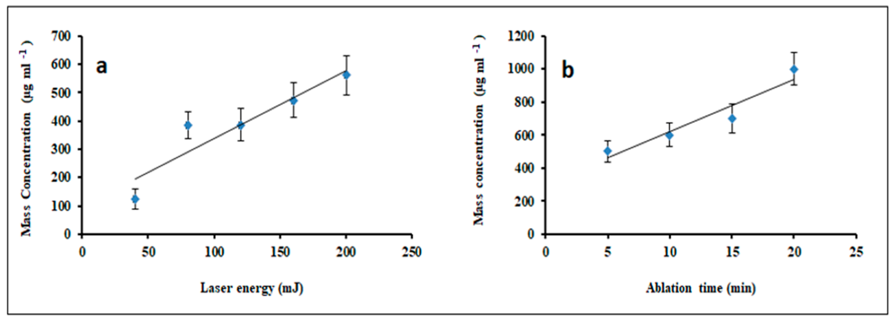
Figure 3a illustrates the impact of irradiation energy on a concentration of prepared TiO2 NPs. The results showed a gradual increase in the NP mass concentration with laser energy, most likely due to target evaporation at higher temperatures caused by high energy. Manufacturing the rate relies on several parameters concerning the optical and thermal properties of the metal target, such as the reflectivity, absorbance, heat capacity, enthalpy of vaporization, boiling point, and thermal conductivity. Figure 3b shows the variation of mass concentration with laser ablation times for the solution prepared at 200 mJ pulse−1 laser irradiation energy. This figure showed a linear increase in mass concentration with increasing ablation time due to high particle concentration in the solution. Additionally, the ablation efficiency decreased with the ablation time due to two reasons: first, the presence of NP in liquid, which could interact with the laser beam and reduce the laser power on the surface; second, the metal surface modification by the irradiation of a pulsed laser, which could reduce the ablation efficiency. This is consistent with the results of Mahfouz et al. [30].
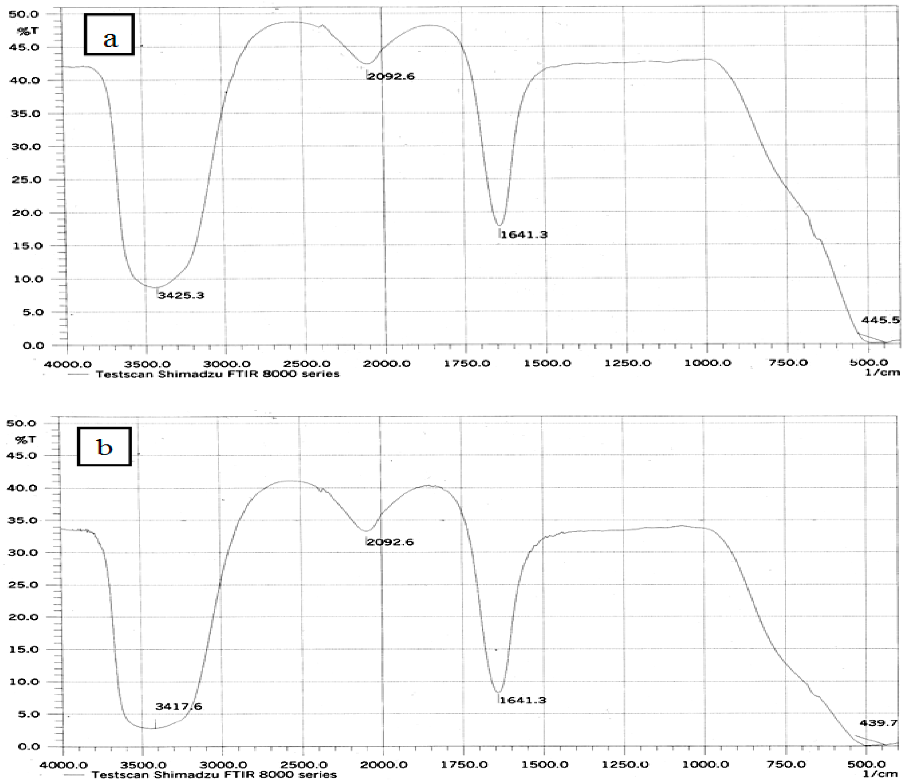
Figure 4a shows the FTIR spectrum of the prepared TiO2 NPs in the transmission mode. Results show that the peaks at 445.5 and 439.7cm−1 are attributed to the O-Ti-O vibration. The board peaks at 3425.3 and 3417.6 cm−1 are assigned to O–H vibrations, while a peak ~1641.3 cm−1 is referred to as the Ti-O-H bending mode [31].
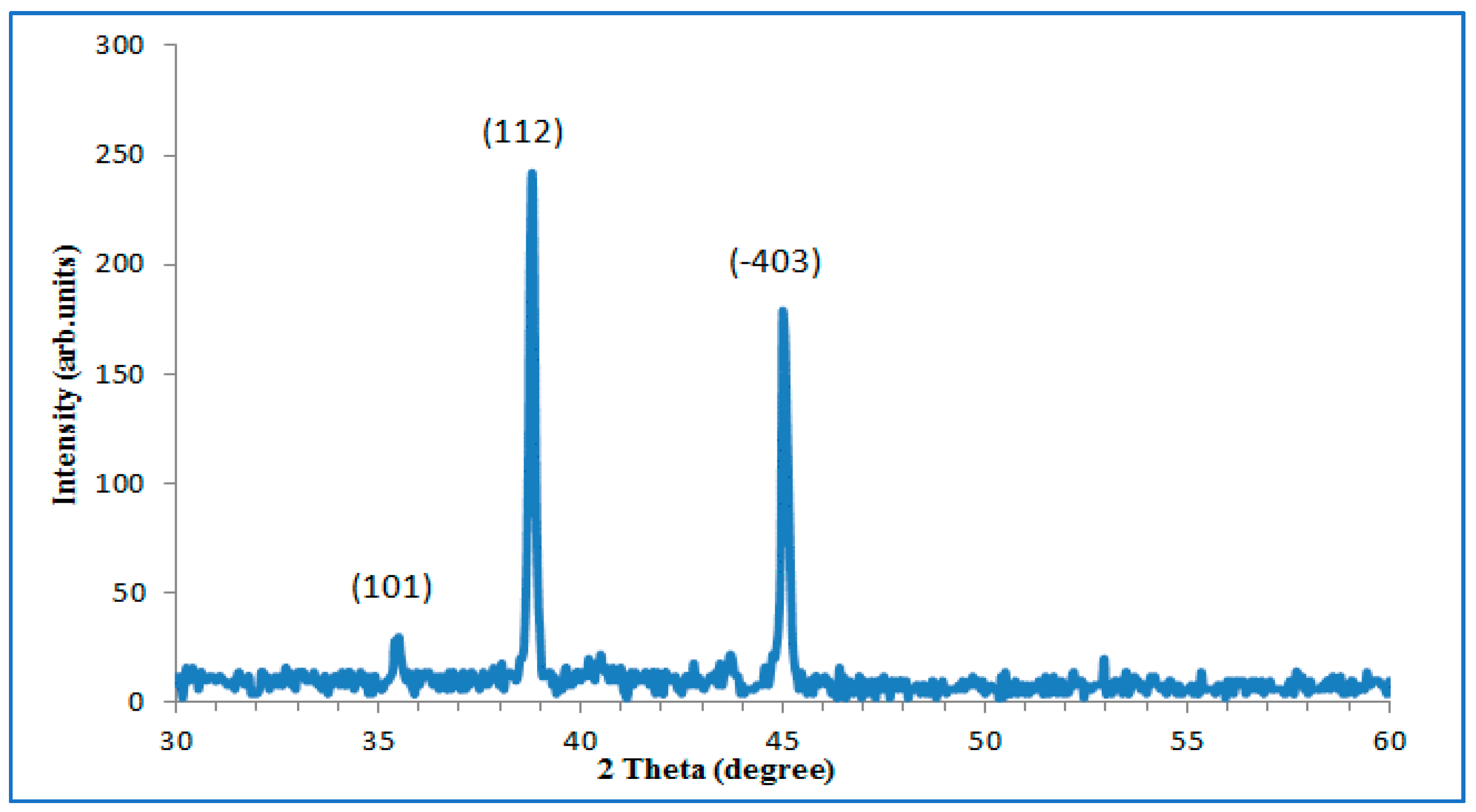
Figure 5 exhibits an XRD patterns of TiO2 NPs synthesized in in deionized distilled water by laser irradiation (200 mJ pulse−1: 10 min). A pattern shows intensity at 2θ = 35.4° (101) and 38.8° (112) corresponding to the crystal formation of rutile and anatase TiO2 (JCPDS: 21-1276 and JCPDS: 21-1272) phase, respectively [18]. Moreover, an additional peak centered at 2θ = 45° corresponding to (−403) plane phase assigned to the β-TiO2 phase (JCPDS No. 46-1238). A grain size value (D) was determined using Scherrer’s formula (aforementioned in the characterization section) are summarized in Table 1.
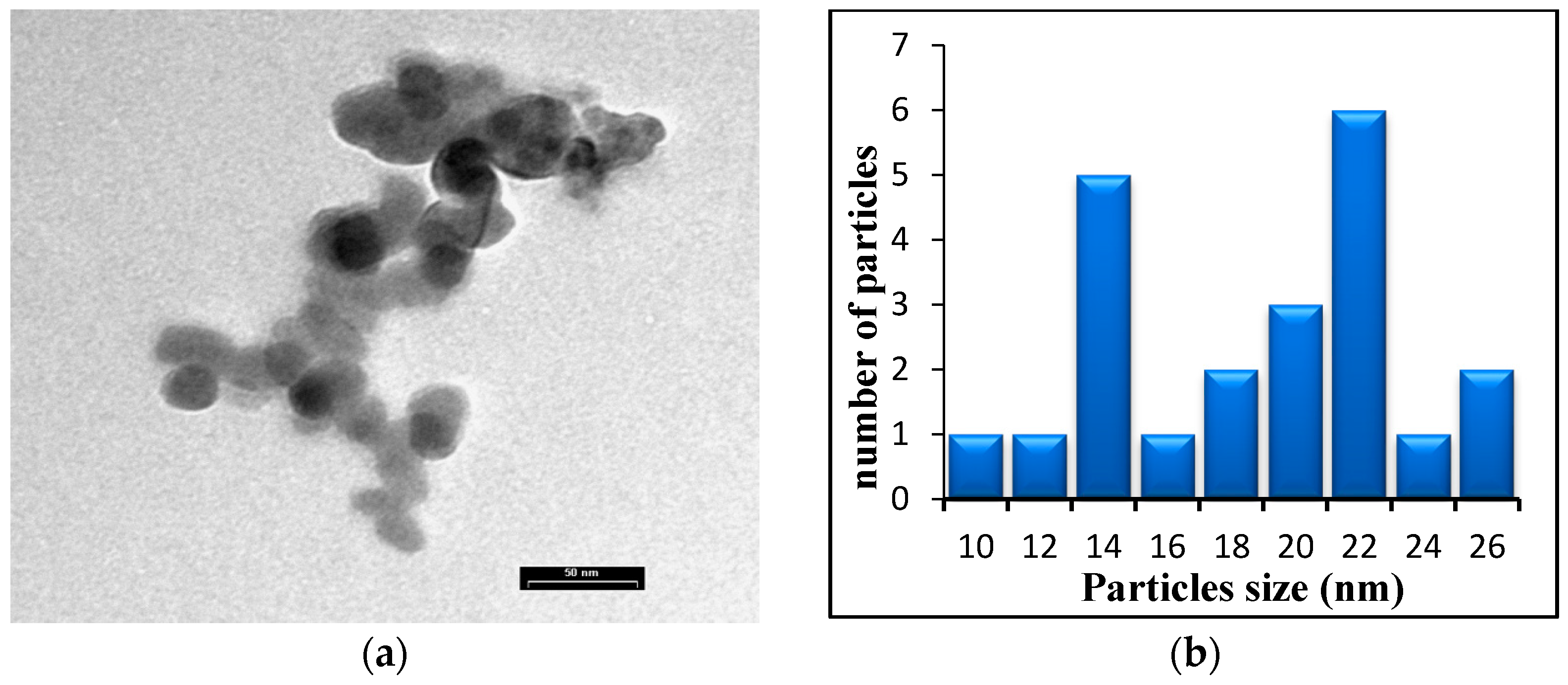
Figure 6 shows TEM images of TiO2 NPs. From the images, it is seen that particle size estimation of 11 to 26 nm and spherical in shape. Also, the same figure shows particle agglomeration due to Ostwald ripening phenomena and the electrostatic attractive force between NPs owing to the electric double layer on NPs surfaces [14,32].
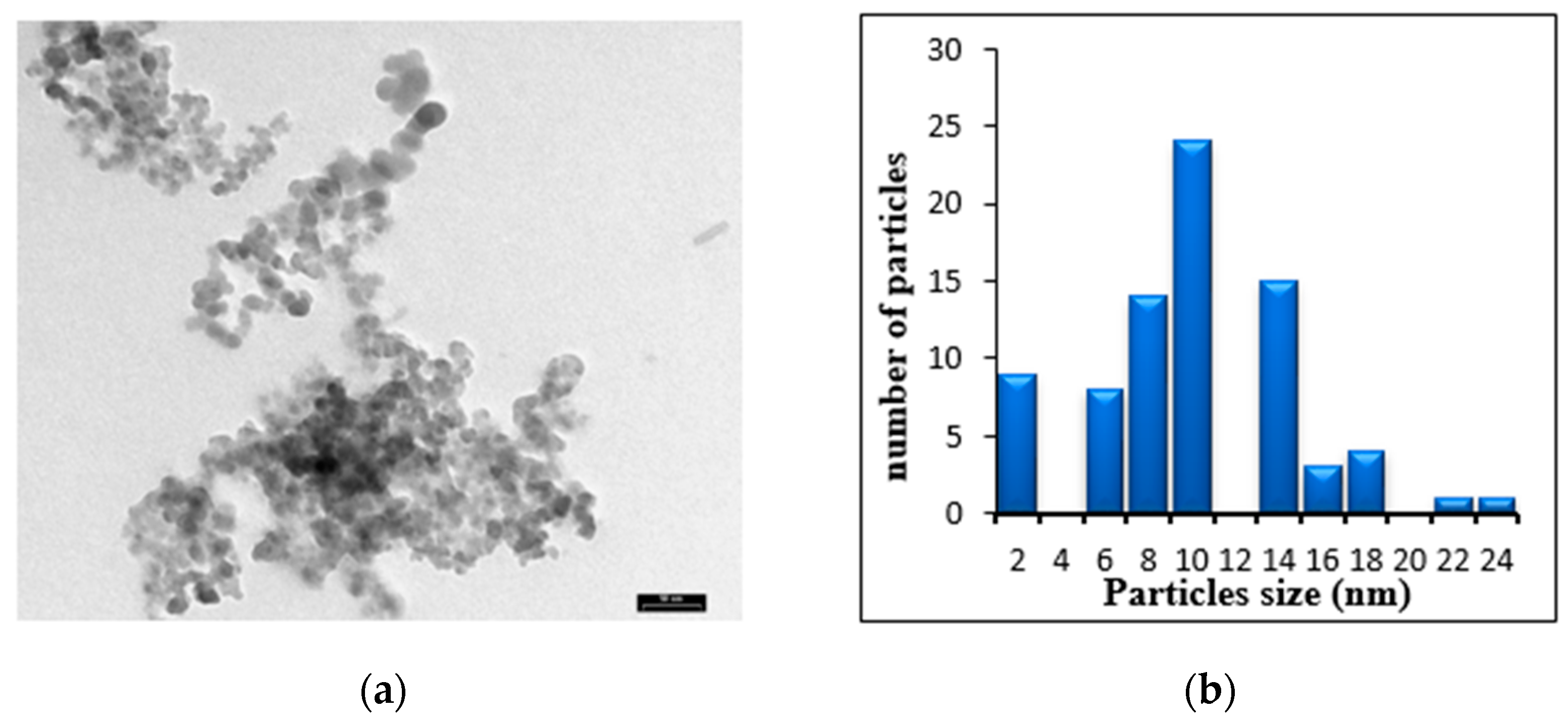
Figure 7 shows TEM image of TiO2 NPs prepared at 80mJ laser energy with a 20 min ablation time. The images revealed that particle size ranged from 2 nm to 23 nm with spherical shaped particles. TEM images in Figure 6 and Figure 7 indicated a reduction in the particle size with increasing laser ablation times. Moreover, the increase in the ablation time results in more ablated mass and high absorption peak, and thereby increases the NPs concentration in the result colloidal solution. The results showed a reduction in the ablation rate with time, which results from the fact that the suspension solution diffracts and/or absorbs a significant portion of incident laser energy and therefore, less energy is absorbed by the target. This process leads to a laser-particles interaction within the solution which causes a smaller particle size formation.
Figure 8a shows the optical density of bacterial culture growths in the presence of TiO2 NPs. The optical density was dropped slightly as the concentrations of TiO2 NPs increased in all types of the tested pathogens. Figure 8b shows that the best-used concentration of TiO2 NPs for inhibiting the growth of both strains was found to be 1000 µg mL−1. The inhibition rates of TiO2 NPs at 400 µg mL−1 were 21%, 3.5%, 5.3%, and 4.9% for E. coli, S. aureus, P. aeruginosa, and P. vulgaris, respectively. The effect of TiO2 NPs against G-positive bacteria was less than for G-negative bacteria, due to the difference in membrane structures of bacteria. The inhibition rates of TiO2 NPs at 600 µg mL−1 are 34%, 13.7%, 8.6%, and 14.04% for E. coli, S. aureus, P. aeruginosa, and P. vulgaris, respectively. On the other hand, the inhibition rates of TiO2 NPs at 1000 µg mL−1 were 42%, 25.5%, 15.05%, and 30.5% for E. coli, S. aureus, P. aeruginosa, and P. vulgaris, respectively. TiO2 was found to be very effective against E. coli among all bacterium types. This effect may be explained by the fact that metal oxide NPs carry a positive charge while the microorganisms carry negative charges; this causes an electromagnetic attraction between microorganisms and metal oxide NPs, leading to the microorganism’s oxidization and finally death [33,34,35].
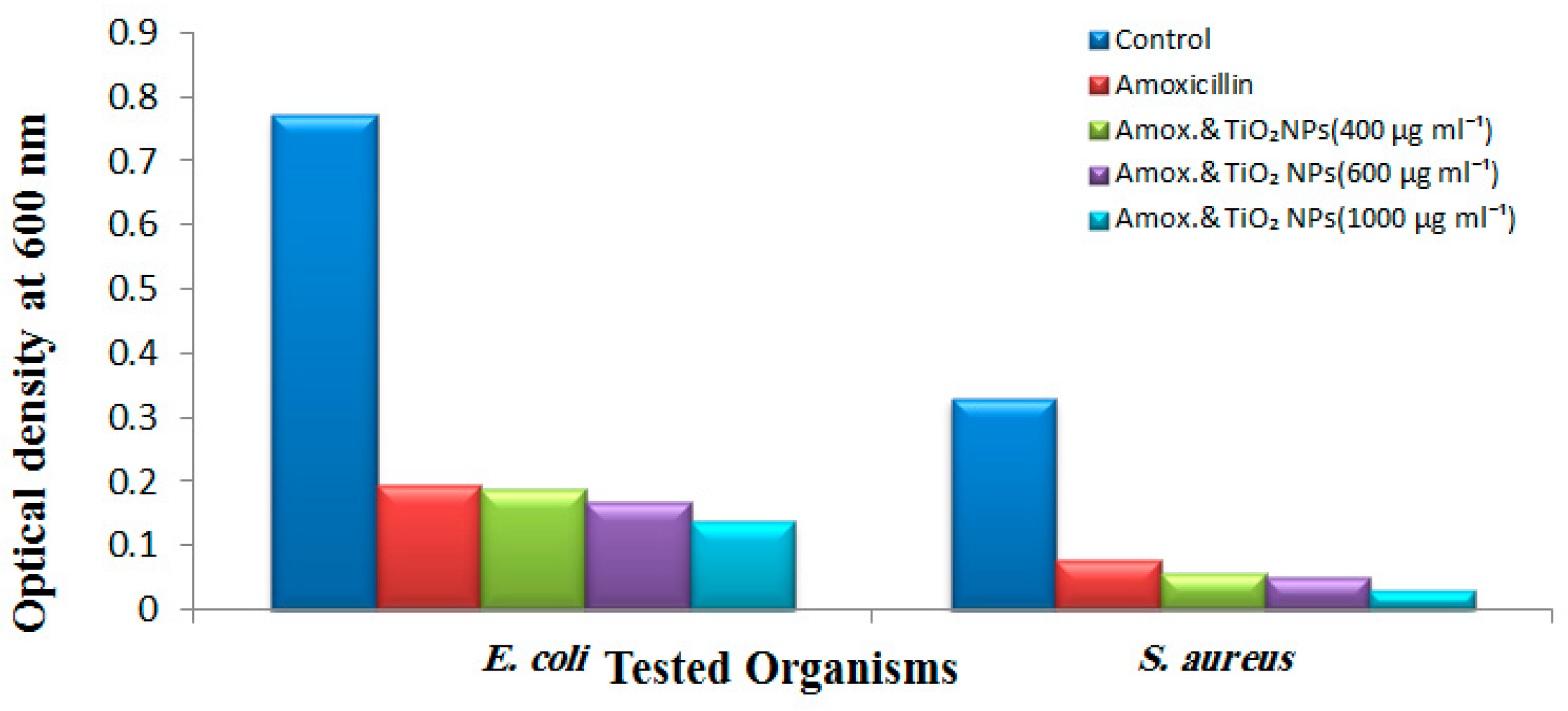
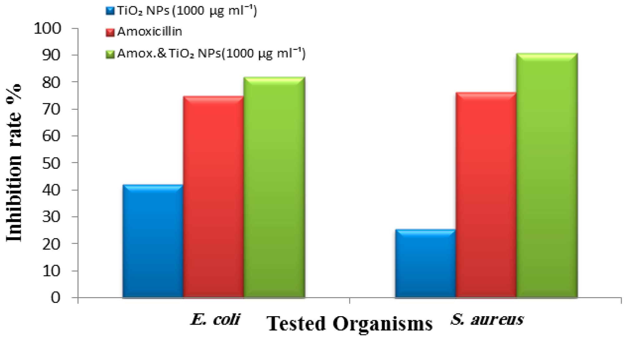
Figure 9 shows the suppressive effect of amoxicillin against E. coli and S. aureus with and without TiO2 NPs. The optical density of cultures was found to break down as the combination of nanoparticles with amoxicillin increased, which confirms the antibacterial effect of amoxicillin enhancement in the presence of nanoparticles. The combined impact of nanoparticles and amoxicillin is due to the cell wall lysis action of the amoxicillin and the DNA binding activity of the nanoparticles [36,37]. Figure 10 shows the inhibition rate of amoxicillin of about 74.8% against E. coli and 76.28% against S. aureus. Meanwhile, in the presence of TiO2 NPs, the activity was enhanced to 81.9% against E. coli and 90.7% against S. aureus.
The crystalline structure and the shape of TiO2 NPs are both considered as the most important conditions that affect its physicochemical factors and therefore its antimicrobial activity [38]. The anatase crystalline structure of TiO2 presents its highest photocatalytic and antimicrobial activities among other crystalline structures. The results showed that structure of anatase can produce OH˙ radicals in the photocatalytic reaction, which will help eliminate the bacteria walls [39].
The antibacterial activity of the TiO2 NPs mixed with amoxicillin was tested against E. coli and S. aureus utilizing the agar well diffusion method. Figure 11 shows the inhibition zones against E. coli and S. aureus. It was noticed that TiO2 NPs improved the activity of amoxicillin, and a higher concentration of nanoparticles had the best eliminating effect along with amoxicillin. The synergetic impact of nanoparticles mixed with amoxicillin was observed via the increase in inhibition zones, as illustrated in Table 2. This is due to the reaction between antibiotics molecules and nanoparticles; these nanoparticles contain hydroxyl groups, which easily react with the antibiotic. Thus, nanoparticles work as an antibiotic carrier and their antibacterial effect can be used.
These results correspond to those of Arora et al., who reported that TiO2 nanoparticles in combination with the antibiotic ceftazidime (CEZ) had an excellent synergistic impact against Pseudomonas spp. [40]. Furthermore, TiO2 and ZnO nanoparticles have shown to promote the antibacterial activity of 8 mm against bacteria (S. aureus—ATCC 25923) and (E. coli—ATCC 25922) [41].
Several studies suggested the possible mechanisms involving the interaction of nanomaterials with biological molecules. It is believed that microorganisms carry a negative charge while metal oxides carry a positive charge. This creates an “electromagnetic” attraction between the microbe and treated surface. Once the contact is made, the microbe is oxidized and dies instantly [42,43]. This synergism may cause cell inactivation at the regulatory network and signaling levels, decrease the activity of the respiratory chain, and inhibit the ability to assimilate and transport iron and phosphorous. These processes with the extensive cell wall and membrane alterations were the main factors that explain the biocidal activity of TiO2 NPs.
IZ: Inhibition Zone.

4. Conclusions

Laser ablation in liquid is a powerful and efficient technique to synthesize TiO2 NPs with a spherical shape and various sizes suspended in deionized distilled water. UV–Vis characterizations of the prepared solutions confirmed the purity of TiO2 NPs and show absorption spectra below 360 nm. FTIR and XRD results confirm the formation of metal oxide NPs. The particles have a spherical shape and mild aggregation was noticed. With the activity of TiO2 NPs against the bacterial strains, the effect of NPs was found to increase with NP concentrations due to the smaller particle sizes of NPs and the best effect was found against E. coli. TiO2 NPs and amoxicillin have a synergic effect on the bacterial cells and the nanoparticles can efficiently improve the permeation and uptake of amoxicillin into the bacterial cells. TiO2 NPs showed an inhibition zone of 11.6 ± 0.57mm against E. coli, while 13.3 ± 0.57mm was observed against S. aureus. Further study is needed to find out the exact reason for the enhancement of the activity of amoxicillin in the presence of TiO2 nanoparticles.

Author Contributions

Conceptualization and methodology, K.S.K., G.M.S. and F.A.A.; formal analysis, S.A., M.A.I. and T.A.-M.; investigation and data curation, K.S.K., G.M.S., F.A.A. and A.A.A.; validation, M.A.I., T.A.-M. and A.A.A.; visualization, all authors; writing—original draft preparation, K.S.K., G.M.S., F.A.A. and S.A.; writing—review and editing, K.S.K., G.M.S., S.A. and M.A.I.; supervision, K.S.K. and G.M.S.; project administration, K.S.K., G.M.S. and T.A.-M. All authors have read and agreed to the published version of the manuscript.

Funding

This research received no external funding.

Institutional Review Board Statement

Not applicable.

Informed Consent Statement

Not applicable.

Data Availability Statement

This study did not report any data.

Acknowledgments

The authors extend their appreciation to the “Research Center of the Female Scientific and Medical Colleges”, Deanship of Scientific Research, King Saud University, for supporting this project. Additionally, the authors extend their appreciation to the University of Technology and the University of Misan for their technical support.

Conflicts of Interest

The authors declare no conflict of interest.

References

  1. Golightly, J.S.; Castleman, A. Analysis of titanium nanoparticles created by laser irradiation under liquid environments. J. Phys. Chem. B 2006, 110, 19979–19984. [Google Scholar] [CrossRef] [PubMed]
  2. Khashan, K.S.; Mohsin, M.H. Characterization of carbon nitride nanoparticles prepared by laser ablation in liquid for optoelectronic application. Surf. Rev. Lett. 2015, 22, 1550055. [Google Scholar] [CrossRef]
  3. Khashan, K.S.; Hadi, A.; Mahdi, M.; Hamid, M.K. Nanosecond pulse laser preparation of InZnO (IZO) nanoparticles NPs for high-performance photodetector. Appl. Phys. A 2019, 125, 51. [Google Scholar] [CrossRef]
  4. Hamad, A.; Khashan, K.S.; Hadi, A. Silver nanoparticles and silver ions as potential antibacterial agents. J. Inorg. Organomet. Polym. Mater. 2020, 20, 8856. [Google Scholar] [CrossRef] [Green Version]
  5. Saimon, J.A.; Madhat, S.N.; Khashan, K.S.; Hassan, A.I. Characterization of CdZnO/Si heterojunction photodiode prepared by pulsed laser deposition. Int. J. Mod. Phys. B 2018, 32, 1850341. [Google Scholar] [CrossRef]
  6. Hadi, A.A.; Badr, B.A.; Mahdi, R.O.; Khashan, K.S. Rapid laser fabrication of Nickel oxide nanoparticles for UV detector. Optik 2020, 219, 165019. [Google Scholar] [CrossRef]
  7. Barreca, F.; Acacia, N.; Barletta, E.; Spadaro, D.; Curro, G.; Neri, F. Small size TiO2 nanoparticles prepared by laser ablation in water. Appl. Surf. Sci. 2010, 256, 6408–6412. [Google Scholar] [CrossRef]
  8. Jadhav, S.; Gaikwad, S.; Nimse, M.; Rajbhoj, A. Copper oxide nanoparticles: Synthesis, characterization and their antibacterial activity. J. Clust. Sci. 2011, 22, 121–129. [Google Scholar] [CrossRef]
  9. Albukhaty, S.; Al-Karagoly, H.; Dragh, M.A. Synthesis of zinc oxide nanoparticles and evaluated it’s activity against bacterial isolates. J. Biotech Res. 2020, 11, 47–53. [Google Scholar]
  10. Pan, Z.; Lee, W.; Slutsky, L.; Clark, R.A.; Pernodet, N.; Rafailovich, M.H. Adverse effects of titanium dioxide nanoparticles on human dermal fibroblasts and how to protect cells. Small 2009, 5, 511–520. [Google Scholar] [CrossRef]
  11. Azam, A.; Ahmed, A.S.; Oves, M.; Khan, M.S.; Memic, A. Size-dependent Antimicrobial Properties of CuO Nanoparticles against Gram-positive and -negative Bacterial Strains. Int. J. Nanomed. 2012, 7, 3527–3535. [Google Scholar] [CrossRef] [Green Version]
  12. Podporska-Carroll, J.; Panaitescu, E.; Quilty, B.; Wang, L.; Menon, L.; Pillai, S.C. Antimicrobial properties of highly efficient photocatalytic TiO2 nanotubes. Appl. Catal. B Environ. 2015, 176, 70–75. [Google Scholar] [CrossRef]
  13. Tsuang, Y.H.; Sun, J.S.; Huang, Y.C.; Lu, C.H.; Chang, W.H.S.; Wang, C.C. Studies of photokilling of bacteria using titanium dioxide nanoparticles. Artif. Organs 2008, 32, 167–174. [Google Scholar] [CrossRef] [PubMed]
  14. Albukhaty, S.; Al-Bayati, L.; Al-Karagoly, H.; Al-Musawi, S. Preparation and characterization of titanium dioxide nanoparticles and in vitro investigation of their cytotoxicity and antibacterial activity against Staphylococcus aureus and Escherichia coli. Anim. Biotechnol. 2020, 1–7. [Google Scholar] [CrossRef] [PubMed]
  15. Bahjat, H.H.; Ismail, R.A.; Sulaiman, G.M.; Jabir, M.S. Magnetic field-assisted laser ablation of titanium dioxide nanoparticles in water for anti-bacterial applications. J. Inorg. Organomet. Polym. Mater. 2021, 1–8. [Google Scholar] [CrossRef]
  16. Yang, G. Laser Ablation in Liquids: Principles and Applications in the Preparation of Nanomaterials; CRC Press: Boca Raton, FL, USA, 2012. [Google Scholar]
  17. Hamad, A.H.; Khashan, K.S.; Hadi, A.A.; Yang, D. Laser Ablation in Different Environments and Generation of Nanoparticles. In Applications of Laser Ablation–Thin Film Deposition, Nanomaterial Synthesis and Surface Modification; IntechOpen: London, UK, 2016; pp. 177–194. [Google Scholar]
  18. Acacia, N.; Barreca, F.; Barletta, E.; Spadaro, D.; Currò, G.; Neri, F. Laser ablation synthesis of indium oxide nanoparticles in water. Appl. Surf. Sci. 2010, 256, 6918–6922. [Google Scholar] [CrossRef]
  19. Khashan, K.S.; Sulaiman, G.M.; Mahdi, R. The effect of laser energy on the properties of carbon nanotube—Iron oxide nanoparticles composite prepared via pulsed laser ablation in liquid. Mater. Res. Express 2018, 5, 105004. [Google Scholar] [CrossRef]
  20. Hameed, R.; Khashan, K.S.; Sulaiman, G.M. Preparation and characterization of graphene sheet prepared by laser ablation in liquid. Mater. Today Proc. 2020, 20, 535–539. [Google Scholar] [CrossRef]
  21. Khashan, K.S.; Abdulameer, F.A.; Jabir, M.S.; Hadi, A.A.; Sulaiman, G.M. Anticancer activity and toxicity of carbon nanoparticles produced by pulsed laser ablation of graphite in water. Adv. Nat. Sci. Nanosci. Nanotechnol. 2020, 11, 35010. [Google Scholar] [CrossRef]
  22. Liu, P.-S.; Cai, W.-P.; Wan, L.-X.; Shi, M.-D.; Luo, X.-D.; Jing, W.-P. Fabrication and characteristics of rutile TiO2 nanoparticles induced by laser ablation. Trans. Nonferrous Met. Soc. China 2009, 19. [Google Scholar] [CrossRef]
  23. Sánchez-López, E.; Gomes, D.; Esteruelas, G.; Bonilla, L.; Lopez-Machado, A.L.; Galindo, R.; Cano, A.; Espina, M.; Ettcheto, M.; Camins, A.; et al. Metal-based nanoparticles as antimicrobial agents: An overview. Nanomaterials 2020, 10, 292. [Google Scholar] [CrossRef] [PubMed] [Green Version]
  24. Han, C.; Romero, N.; Fischer, S.; Dookran, J.; Berger, A.; Doiron, A.L. Recent developments in the use of nanoparticles for treatment of biofilms. Nanotechnol. Rev. 2017, 6, 383–404. [Google Scholar] [CrossRef]
  25. Swarnkar, R.; Singh, S.; Gopal, R. Effect of aging on copper nanoparticles synthesized by pulsed laser ablation in water: Structural and optical characterizations. Bull. Mater. Sci. 2011, 34, 1363–1369. [Google Scholar] [CrossRef]
  26. Mahdy, S.A.; Raheed, Q.J.; Kalaichelvan, P. Antimicrobial activity of zero-valent iron nanoparticles. Int. J. Mod. Eng. Res. 2012, 2, 578–581. [Google Scholar]
  27. Nikolov, A.; Atanasov, P.; Milev, D.; Stoyanchov, T.; Deleva, A.; Peshev, Z. Synthesis and characterization of TiOx nanoparticles prepared by pulsed-laser ablation of Ti target in water. Appl. Surf. Sci. 2009, 255, 5351–5354. [Google Scholar] [CrossRef]
  28. Ola, O.; Maroto-Valer, M.M. Review of material design and reactor engineering on TiO2 photocatalysis for CO2 reduction. J. Photochem. Photobiol. C Photochem. Rev. 2015, 24, 16–42. [Google Scholar] [CrossRef] [Green Version]
  29. Hong, S.M.; Lee, S.; Jung, H.J.; Yu, Y.; Shin, J.H.; Kwon, K.-Y.; Choi, M.Y. Simple preparation of anatase TiO2 nanoparticles via pulsed laser ablation in liquid. Bull. Korean Chem. Soc. 2013, 34, 279–282. [Google Scholar] [CrossRef] [Green Version]
  30. Mahfouz, R.; Aires, F.C.S.; Brenier, A.; Jacquier, B.; Bertolini, J. Synthesis and physico-chemical characteristics of nanosized particles produced by laser ablation of a nickel target in water. Appl. Surf. Sci. 2008, 254, 5181–5190. [Google Scholar] [CrossRef]
  31. León, A.; Reuquen, P.; Garín, C.; Segura, R.; Vargas, P.; Zapata, P.; Orihuela, P.A. FTIR and Raman characteriztion of TiO2 nanoparticles coated with polyethyleneglycol as carrier for 2-methoxyestradiol. Appl. Sci. 2017, 7, 49. [Google Scholar] [CrossRef]
  32. Baladi, A.; Mamoory, R.S. Effect of Laser Wavelength and Ablation Time on Pulsed Laser Ablation Synthesis of Al Nanoparticles in Ethanol; World Scientific: Singapore, 2012; pp. 58–65. [Google Scholar]
  33. Rosická, D.; Šembera, J. Changes in the nanoparticle aggregation rate due to the additional effect of electrostatic and magnetic forces on mass transport coefficients. Nanoscale Res. Lett. 2013, 8, 1–9. [Google Scholar] [CrossRef] [Green Version]
  34. Al-Nori, T.M. Antibacterial activity of silver and gold nanoparticles against Streptococcus, Staphylococcus aureus and E. coli. Al-Mustansiriyah J. Sci. 2012, 23, 45–54. [Google Scholar]
  35. Haghi, M.; Hekmatafshar, M.; Janipour, M.B.; Gholizadeh, S.S.; Faraz, M.; Sayyadifar, F.; Ghaedi, M. Antibacterial effect of TiO2 nanoparticles on pathogenic strain of E. coli. Int. J. Adv. Biotechnol. Res. 2012, 3, 621–624. [Google Scholar]
  36. Murthy, P.S.; Venugopalan, V.; Arunya, D.D.; Dhara, S.; Pandiyan, R.; Tyagi, A. Antibiofilm Activity of Nano Sized CuO. In Proceedings of the International Conference on Nanoscience, Engineering and Technology, Chennai, India, 28–30 November 2011; IEEE: Chennai, India, 2011; pp. 580–583. [Google Scholar]
  37. Rajawat, S.; Qureshi, M.S. Comparative study on bactericidal effect of silver nanoparticles, synthesized using green technology, in combination with antibiotics on Salmonella typhi. J. Biomater. Nanobiotechnol. 2012, 3, 480. [Google Scholar] [CrossRef] [Green Version]
  38. de Dicastillo, C.L.; Correa, M.G.; Martínez, F.B.; Streitt, C.; Galotto, M.J. Antimicrobial Effect of Titanium Dioxide Nanoparticles. In Antimicrobial Resistance-A One Health Perspective; IntechOpen: London, UK, 2020. [Google Scholar]
  39. Ziental, D.; Czarczynska-Goslinska, B.; Mlynarczyk, D.T.; Glowacka-Sobotta, A.; Stanisz, B.; Goslinski, T.; Sobotta, L. Titanium dioxide nanoparticles: Prospects and applications in medicine. Nanomaterials 2020, 10, 387. [Google Scholar] [CrossRef] [PubMed] [Green Version]
  40. Arora, B.; Murar, M.; Dhumale, V. Antimicrobial potential of TiO2 nanoparticles against MDR Pseudomonas aeruginosa. J. Exp. Nanosci. 2015, 10, 819–827. [Google Scholar] [CrossRef] [Green Version]
  41. Nguyen, V.T.; Vu, V.T.; Nguyen, T.A.; Tran, V.K.; Nguyen-Tri, P. Antibacterial activity of TiO2-and ZnO-decorated with silver nanoparticles. J. Compos. Sci. 2019, 3, 61. [Google Scholar] [CrossRef] [Green Version]
  42. Savi, G.D.; Trombin, A.C.; da Silva Generoso, J.; Barichello, T.; Possato, J.C.; Ronconi, J.V.V.; da Silva Paula, M.M. Antibacterial acivity of gold and silver nanoparticles impregnated with antimicrobial agents. Saúde Pesqui. 2013, 6, 227–235. [Google Scholar]
  43. Wang, L.; Hu, C.; Shao, L. The antimicrobial activity of nanoparticles: Present situation and prospects for the future. Int. J. Nanomed. 2017, 12, 1227. [Google Scholar] [CrossRef] [Green Version]
Figure 1. Schematic illustration of the experimental set-up of TiO2 NPs suspension preparation at ambient temperature.
Applsci 11 04623 g001
Figure 2. UV–Vis absorption of TiO2 NP solution synthesized at different (A) laser pulse energies and (B) ablation times.
Applsci 11 04623 g002
Figure 3. Mass concentration of TiO2 NPs at different (a) laser energies and (b) ablation time.
Applsci 11 04623 g003
Figure 4. FTIR spectra of TiO2 NPs synthesized: (a) 80 mJ; (b) 200 mJ.
Applsci 11 04623 g004
Figure 5. XRD patterns of TiO2 NPs.
Applsci 11 04623 g005
Figure 6. (a) TEM image of TiO2 NPs prepared at the following laser conditions (80 mJ; 10 min) and (b) size distribution histogram.
Applsci 11 04623 g006
Figure 7. (a) TEM image of TiO2 NPs prepared under the following laser conditions (80 mJ; 20 min) and (b) size distribution histogram.
Applsci 11 04623 g007
Figure 8. Growth curves of tested bacteria in the presence TiO2 NPs: (a) optical density, (b) cell viability.
Applsci 11 04623 g008aApplsci 11 04623 g008b
Figure 9. Growth of the tested bacteria in the presence of amoxicillin and TiO2 NPs measured at 600 nm.
Applsci 11 04623 g009
Figure 10. Inhibition rate (%) of the tested bacteria in the presence of TiO2 NPs, amoxicillin alone and a combination of amoxicillin and TiO2 NPs.
Applsci 11 04623 g010
Figure 11. The antibacterial activity of TiO2 NPs with (A) amoxicillin against E. coli, and (B) S. aureus. A (yellow color): amoxicillin used as control, while 1,2, and 3 were 400, 600, and 1000 µg mL1, respectively.
Applsci 11 04623 g011
Table 1. XRD parameters of TiO2 NPs.
Ablation Condition2θ (Degree)Orientation (hkl)G.S (nm)FWHM (Degree)Phase
200 mJ 10 min35.410141.670.2anatase TiO2
38.811246.770.18anatase TiO2
45−40344.460.19β-TiO2
Table 2. Inhibition zone in mm (mean ± standard deviation) of TiO2 NPs with amoxicillin.
OrganismsTiO2 NPs Along with AmoxicillinIZ (mm) Mean ± S.D.
E. coliControl (amoxicillin)8.0 ± 0.57
400 µg mL−110.3 ± 0.57
600 µg mL−111.3 ± 0.57
1000 µg mL−111.6 ± 0.57
S. aureusControl (amoxicillin)9.3 ± 1.15
400 µg mL−112.3 ± 0.57
600 µg mL−111.6 ± 1.15
1000 µg mL−113.3 ± 0.57
IZ: Inhibition Zone.
Publisher’s Note: MDPI stays neutral with regard to jurisdictional claims in published maps and institutional affiliations.