Article

Microcystis aeruginosa and M. wesenbergii Were the Primary Planktonic Microcystin Producers in Several Bulgarian Waterbodies (August 2019)

1
Faculty of Biology, Department of Botany, Sofia University, 8 Blvd. Dragan Zankov, 1164 Sofia, Bulgaria
2
AgroBioInstitute, Bulgarian Agricultural Academy, 8 Blvd. Dragan Zankov, 1164 Sofia, Bulgaria
3
Unité d’Océanographie Chimique, Université de Liège, Sart Tilman, 4000 Liège, Belgium
4
National Center of Public Health and Analyses, “I. Geshov” str. 15, 1431 Sofia, Bulgaria
5
Institut für Botanik, Universität Innsbruck, Sternwartestrasse 15, 6020 Innsbruck, Austria
*
Author to whom correspondence should be addressed.
Appl. Sci. 2021, 11(1), 357; https://doi.org/10.3390/app11010357
Submission received: 30 November 2020 / Revised: 19 December 2020 / Accepted: 28 December 2020 / Published: 31 December 2020
(This article belongs to the Special Issue Harmful Cyanobacteria and Their Metabolites)

Abstract

:

Featured Application

The present work provides new data on the spread of toxic strains of Microcystis aeruginosa and M. wesenbergii in Bulgaria, important for further revision of the notable microcystin producing genus Microcystis.

Abstract

The rising interest in harmful cyanoprokaryote blooms promotes an increase of phycological and ecological research on potentially toxic species and their hazardous substances. The present study aimed to identify the main microcystin (MC) producers and their contribution to the phytoplankton of shallow waterbodies in Bulgaria, applying different methods. The sampling was performed in August 2019 in nine lakes and reservoirs, two of which (reservoirs Kriva Reka and Izvornik 2) were studied for the first time. The high contribution of cyanoprokaryotes to the total species composition and phytoplankton abundance was proved by light microscopic (LM) observations and HPLC analysis of marker pigments. The LM identification of potential MC-producers was supported by PCR amplification of mcyE and mcyB genes. The MCs amounts, detected by HPLC-DAD, varied by sites with a range from undetectable concentrations to 0.46 µg L−1 with only one recorded variant, namely MC-LR. It was found only in the reservoirs Mandra and Durankulak, while toxigenic MC-strains were obtained by PCR from five more waterbodies. Both LM and PCR demonstrated that the MC-producers were Microcystis aeruginosa and M. wesenbergii, despite their occurrence in low amounts (<0.5–5% of the total biomass) when filamentous cyanoprokaryotes dominated.

1. Introduction

During the last decades many countries throughout the world experienced incidences related with mass developments of both marine and freshwater toxic algae [1,2]. These harmful algal blooms, formed by eukaryotic algae (commonly abbreviated as HABs) or by cyanoprokaryotes/cyanobacteria (known as CyanoHABs, or CHABs) are anticipated to expand their distribution, promoted mainly by global warming, population growth and increasing nutrient loading [1,3,4,5].
Simultaneously with the increased awareness of CyanoHABs at a global scale [2] there is a rising interest in phycological and ecological studies of potentially toxic species and their hazardous substances [4,6]. However, many questions related to the taxonomy, ecology, distribution and toxicity of some important bloom-causative cyanoprokaryotes are still awaiting their answers.
Microcystis, despite being one of the first and best-known toxin producers and successful phytoplankton competitors due to numerous adaptive features (e.g., [7]), is among these genera with unresolved taxonomy and disputable species in regard to their toxicity [8,9]. It is the most often reported toxic genus worldwide [2] and besides the ability to produce renowned potent hepatotoxic cyclic peptides microcystins (MCs), its species have biosynthesis genes for other peptides (aeruginosins, anabaenopeptins, microginins, microviridins, and cyanopeptolins), some of which can deleteriously affect aquatic ecosystems [6]. Additional concern comes from studies indicating that future elevated temperatures can positively influence MC gene expression and favor toxic Microcystis genotypes over their nontoxic counterparts [10,11]. There is also evidence that higher concentrations of nutrients may promote toxic genotypes and MC production [5,12].
The discussions of all accumulated data on Microcystis beyond different levels of its investigation at a global scale, are complicated because: (1) the main morphological features are variable, can overlap and transitional colonies between different morphospecies have been observed (for details see [9]); (2) this prokaryotic genus contains both toxigenic and non-toxigenic strains; (3) its typical toxins, MCs, are known to be produced by other algae which have MC synthase (mcy) genes. The most frequently reported planktonic genera with mcy genes are Anabaena s.l. (recently including Dolichospermum, Sphaerospermopsis, etc.), Planktothrix (former Oscillatoria p.p.) and, occasionally, Nostoc [2,3,6,13,14,15]. Additionally, there is evidence that some species of Aphanizomenon s.l. (including A. flos-aquae Ralfs ex Bornet & Flahault) and of Raphidiopsis (including Raphidiopsis raciborskii (Woloszynska) Aguilera, Berrendero Gómez, Kastovsky, Echenique & Salerno) also have mcy genes [16,17]. Therefore, the application of different methods in studies of toxigenic cyanoprokaryotes carried on the background of total phytoplankton composition and abundance, combined with environmental data, have been encouraged (e.g., [9,14]).
This approach was applied in the present study aimed to identify the main MC producers and their contribution to the phytoplankton of different waterbodies in Bulgaria. The background lies in the: (1) proven CyanoHABs threat for the wetlands of the country, which cover less than 0.1% of Bulgarian territory and are mainly shallow [18,19], and (2) the recognized role of cyanotoxins as a potential risk factor for national security [20] and for malignant diseases like cancer in the country [21]. Because Microcystis is one of the most important bloom-forming genera in Bulgaria [19,22,23], the second purpose was to continue our polyphasic studies of this genus and its toxigenic strains [9]. Therefore, in August 2019 we performed phytoplankton sampling in 28 different water bodies of the country. Here we report phytoplankton data on nine shallow lakes and reservoirs (two of them sampled for the first time), in six of which Microcystis has already been detected and examined by the polyphasic approach [9].
With this paper we demonstrate the role of two Microcystis species identified by light microscopic (LM) and PCR, namely M. aeruginosa and M. wesenbergii, as primary MC-producers in the studied shallow water bodies with high total cyanoprokaryote contribution to phytoplankton species composition and biomass dominated mainly by filamentous heterocytous taxa. The results indicated the high genetic diversity of toxigenic Microcystis strains, based on mcyE and mcyB sequences, despite their occurrence in low amounts (<0.5–5% of the total biomass). However, our study once more demonstrated that currently available genomic methods and supplied to NCBI sequences are still insufficient to fully resolve the species identification among cyanoprokaryotes and specifically in the genus Microcystis.

2. Materials and Methods

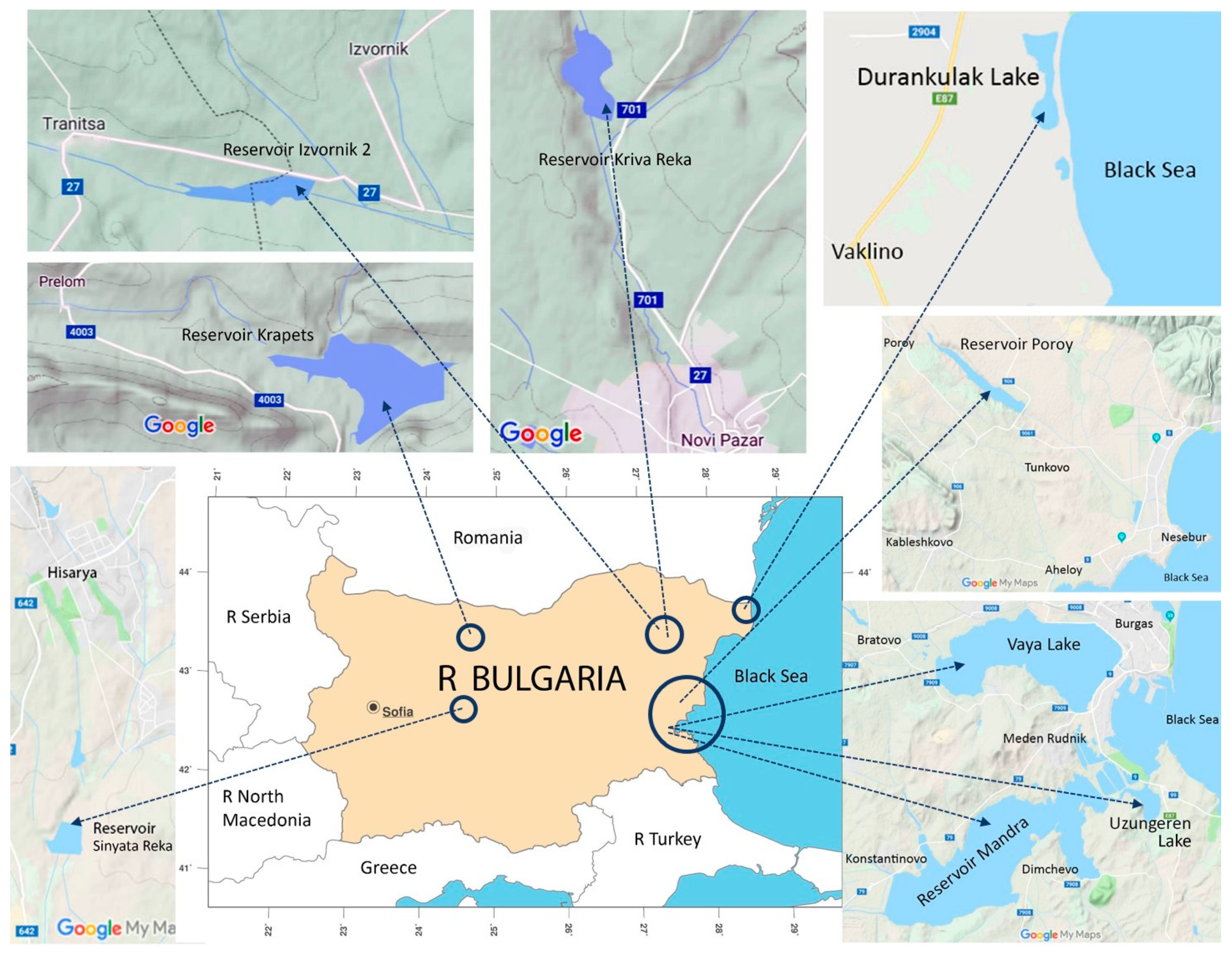
2.1. Sites and Sampling

The sampling was carried out between the 14th and 21st August 2019 in nine shallow waterbodies situated in Central and Eastern Bulgaria (Figure 1, Table 1). In Table 1 we provide the unique number (IBWXXXX) from the Inventory of Bulgarian wetlands [18] where detailed descriptions of the waterbodies are available. However, it is worth mentioning that: (1) the reservoirs Krapets, Kriva Reka, Izvornik 2 and Sinyata Reka are inland, while the lakes Durankulak, Vaya and Uzungeren, and the reservoirs Poroy and Mandra are coastal; (2) three waterbodies are included in the Red List of Bulgarian Wetlands [18] as critically endangered (lakes Vaya and Durankulak) and endangered (reservoir Mandra); (3) reservoirs Kriva Reka and Izvornik 2 were sampled for first time.
The sampling was preceded by the use of a drone DJI Mavic 2 Enterprise Dual Pro (DJI Technology Co., Ltd. Shenzhen, China), supplied by a photo camera and with the possibility to measure the surface water temperature. In this way we observed in real time the whole waterbody and spots with visible differences in the color were chosen for sampling of cyanoblooms from inflatable boats in accordance with our former study [23]. In cases of visible water homogeneity, the sites from our previous studies were repeated (for details see [23]). Therefore, the Mandra reservoir was sampled in two sites—in its eastern part, and its western part, presented hereafter as Mandra East and Mandra West, respectively. The coordinates, altitude above sea level, water temperature, pH, total dissolved solids, oxygen concentration, and conductivity were measured in situ by Aquameter AM-200 and Aquaprobe AP-2000 from Aquaread water monitoring instruments (Aquaread Ltd., Broadstairs, UK). The water transparency was estimated using a Secchi disk, while total nitrogen (TN) and total phosphorus (TP) were measured ex situ with the Aqualytic AL410 Photometer from AQUALYTIC® (Dortmund, Germany) [23]. Data from all these measurements are shown in Table 1.
Phytoplankton samples for taxonomic identification and counts, for pigment analysis and for molecular-genetic studies (each in a volume of 0.5 L) and for toxin identification (in a volume of 1 L) were collected from the water surface (0–20 cm) due to the holo-polymictic character of the studied wetlands. The samples for phytoplankton identification and counts were fixed immediately with 2% formalin and transported to the lab, where they were further concentrated by sedimentation [23]. Additionally, from each site a living sample in a volume of 0.05 L was inoculated in plastic tubes containing Bristol media and Allen–Arnon solution (50:50) [24] for stimulation of cyanoprokaryote growth and further cultivation in the Algal Collection of Sofia University ACUS [25]. The samples for toxin analyses were immediately placed in a box with dry ice and were provided frozen to the relevant lab. The water samples for pigment analyses and PCR-studies were filtered under a mild vacuum, using Macherey–Nagel GF5 filters porosity 0.4 µm (MACHEREY-NAGEL GmbH & Co. KG, Düren, Germany), and Whatman® 0.45 µm cellulose filters (Merck KGaA, Darmstadt, Germany), respectively, within a few hours after collection. The filters were placed in 15 mL sterile plastic tubes (Falcon) and preserved in dry ice until further treatment (see below).

2.2. Phytoplankton Species Composition and Abundance Assessment by Conventional Light Microscopy (LM)

In the lab the taxonomic work and counts were done on non-permanent slides on a Motic B1 microscope under magnification 100× and immersion. The microphotographs were taken by a Moticam 2.0 mp camera supplied by Motic Images 2 Plus software program. From each site at least four slides were examined.
The identification of algae followed the standard European taxonomic literature with updates from AlgaeBase [26] and relevant modern taxonomic papers. For cyanoprokaryotes, in particular, determination was based on [27,28,29,30,31,32,33] and additional checks were made in CyanoDB [34]. The following morphological features were used for species distinguishing in the genus Microcystis: general shape of the colony and presence/absence of subcolonies, density of cells inside the colony, structure of colonial mucilage, cell size, facultative or obligatory occurrence of gas vesicles aggregated in aerotopes. The potential MC-producers were outlined after [16,35,36].
The quantitative contribution of each species was estimated using the method of the stereometrical approximations [37] on Thoma blood-counting chamber with cell considered as the main counting unit [23,38].

2.3. Phytoplankton According to HPLC Marker Pigment Analysis

Phytoplankton marker pigments were extracted from the filters using 90% acetone, with two 15 min sonication steps in melting ice separated by overnight at 4 °C in the dark. Pigment analysis was performed on a Waters HPLC system equipped with a photodiode array detector, and concentrations were determined from calibration with chlorophyll and carotenoid standards (DHI, Hórsholm, Denmark). The contribution of different phytoplankton groups to chlorophyll a was calculated by application of CHEMTAX [23,39], which uses a steepest descent algorithm to optimize the marker pigments to chlorophyll a ratios, given an input ratio matrix [40]. The initial ratio matrix used in this study was derived from [41] and CHEMTAX processing was run until stability of the pigment ratios in the output ratio matrix was reached. With this procedure we determined the chlorophyll a biomass of green algae (chlorophytes and streptophytes), cryptophytes, pyrrhophytes (dinoflagellates), euglenophytes and cyanoprokaryotes (two pigment types, cyanoprokaryotes T1 and T2), as well as from two classes of ochrophytes—golden algae (chrysophyceans) and diatoms (bacillariophyceans). This technique was applied in a similar study on cyanoprokaryotes [42] which demonstrated an excellent fit of pigment estimates and LM counts.

2.4. Identification of Toxins

Detection of MCs followed the procedures completely compatible with those from our previous studies (for details see references in [19] and [23]), performed by HPLC according to ISO 20179:2005, validated for MC–RR, MC–YR, and MC–LR and applicable for several variants of these MCs [43,44]. The method is updated every five years, and the latest revision with confirmation of its current character dates 2019 [43]. After obtaining cell lysis through triple freezing and defrosting, samples were filtered through nylon membrane filter 0.45 µm (Supelco, Inc. Bellefonte, PA, USA) and threated by solid-phase extraction with ENVI™-18 DSK SPE. Cyanotoxins were eluted with methanol with subsequent drying by a gentle stream of nitrogen, re-dissolved in 500 µL of 50% methanol (v/v), and then filtered through 0.22 µm PTFE syringe filters (Thermo Fisher Scientific, Waltham, MA, USA).
The HPLC-DAD system for quantitative and qualitative analyses contains Agilent 1200 Series coupled with a Diode Array Detector (DAD; Agilent Technologies, Santa Clara, CA, USA). Analyses were performed on a Supelcosil ABZ + Plus column (150 × 4.6 mm, 5 µm, Supelco). The binary gradient of mobile phase consisted of deionized water + 0.1% TFA (A) and acetonitrile + 0.1% TFA (B) (linear increase from 20% B at 0 min to 46% B at 25 min and stop time 30 min); the flow rate was 1 mL min−1 at 25 °C. Cyanotoxins were identified based on characteristic UV absorption spectra from 200 to 300 nm on chromatograms at 238 nm with purified MC–LR, –RR, –YR (Eurofins Abraxis, Inc. Warminster, PA, USA) used as external standards.

2.5. Molecular-Genetic Studies

The sequence analysis of PCR amplified fragments of mcyB and mcyE genes from the mcyA-J gene cluster involved in the biosynthesis of MCs [45,46] was performed. The single copy genes mcyA, mcyB, mcyD and mcyE are the most commonly used target mcy genes for detection of MC-producers (even when the toxin concentrations are too low to be detected) and primers for different regions of these genes can be generalists for any MC producer, or can be specific for Microcystis [46,47]. Considering the lack of universal primers (e.g., [48,49]) we decided to combine primers for two genes from both different operons of the mcyA-J gene cluster, namely mcyA-C and mcyD-J [47]. In our previous study we pointed out the reliable MC-detective biomarker character of the mcyE gene from the mcyD-J operon and its strong indicator properties for identifying of potential risk from MCs in water bodies comprising mixed assemblages of toxic and non-toxic species (for details see [9]) and in samples with low cyanoprokaryote biomass [50]. The recent choice for additional application of primers for mcyB gene from the mcyA-C operon was based on the reported results of its successful application [51].
A few hours after collection, the samples for molecular-genetic analysis were filtered through 0.45 µm cellulose filters Whatman NC45 ST/Sterile EO (Merck KGaA, Darmstadt, Germany). In the lab, the total DNA was isolated from the filters following the protocol of GeneJETTM Plant Genomic DNA Purification Mini Kit (Thermo Scientific, Waltham, MA, USA).
For PCR amplification of the mcyE gene the following steps were used: the DNA amplification was achieved by application of HEPF (5′TTTGGGGTTAACTTTTTTGGGCATAGTC-3′) × HEPR (5′AATTCTTGAGGCTGTAAATCGGGTTT-3′) synthetase-gene-specific pair of primers [52]. Then PCR amplification was performed in a 25 µL volume containing 10 pmol primers; 5 μL of 5× HF PCR buffer (2 mM MgCl2), 2.5 μM deoxynucleoside triphosphates and 0.2 U/µL Phusion™ High-Fidelity DNA Polymerase, supplied by Thermo Scientific, Waltham, MA, USA). The amplification of DNA was done in a thermal cycler QB-96 (Qianta Biotech) under the following PCR conditions: denaturation at 98 °C for 3 min, 35 cycles of denaturation (10 s at 98 °C), annealing at 57 °C for 30 s, extension at 72 °C for 15 s, and a final extension at 72 °C for 3 min. The resulting PCR products were purified using GeneJET™ Gel Extraction Kit (Thermo Fisher Scientific, Waltham, MA, USA). CloneJET ™PCR Cloning Kit (Thermo Fisher Scientific, Waltham, MA, USA) was used for obtaining of clones of the mcyE amplification products, and the Sigma Plasmid Miniprep Kit (Sigma, Taufkirchen, Germany) was used for isolation of recombinant plasmids. For each site or waterbody between nine and fourteen clones were selected and sequenced by Macrogen Inc. (Seoul, South Korea).
The fragments of mcyB gene were selectively amplified from the purified metagenomics DNA by PCR using the following pair of primers: MIf GCAGCGAACTCTTGAAGGGTTTATG and Mir GCGGATTCTGTGCAGCTTGTTCTTC [53] in the above-described reaction mixtures. The amplification program included denaturation at 98 °C for 3 min, 35 cycles of denaturation (10 s at 98 °C), annealing at 60 °C for 30 s, extension 72 °C for 40 s, and a final extension at 72 °C for 3 min. The resulting PCR products were purified using the GeneJET ™ Gel Extraction Kit (Thermo Scientific), following the manufacturer’s instructions and directly sequenced by Macrogen Inc. (Seoul, South Korea).
The obtained sequences from both mcyE and mcyB genes were processed with Vector NTI 11.5 software and used for BLAST search [54] in the NCBI database [55]. The phylogenetic tree, based on neighbor-joining method, was constructed by Mega 6.06 software [56]. There, the accession numbers of 34 mcyE-based strains (MW187787-MN187820) and five mcyB-based strains (MW218898-MW218902) newly submitted by us to NCBI, are shown in brackets, while in the text, following the NCBI requirements, when available, the taxonomic identification number in NCBI is also indicated (NCBItxidXXXXXX).

3. Results

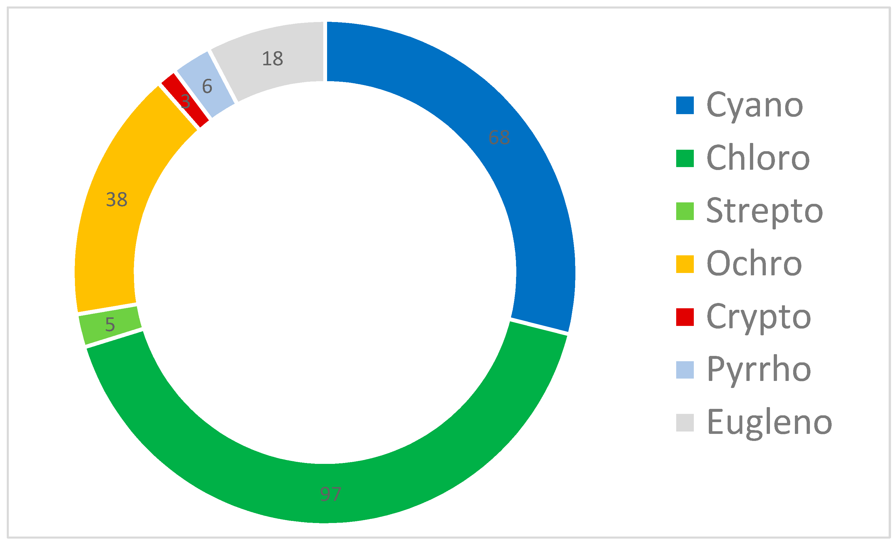
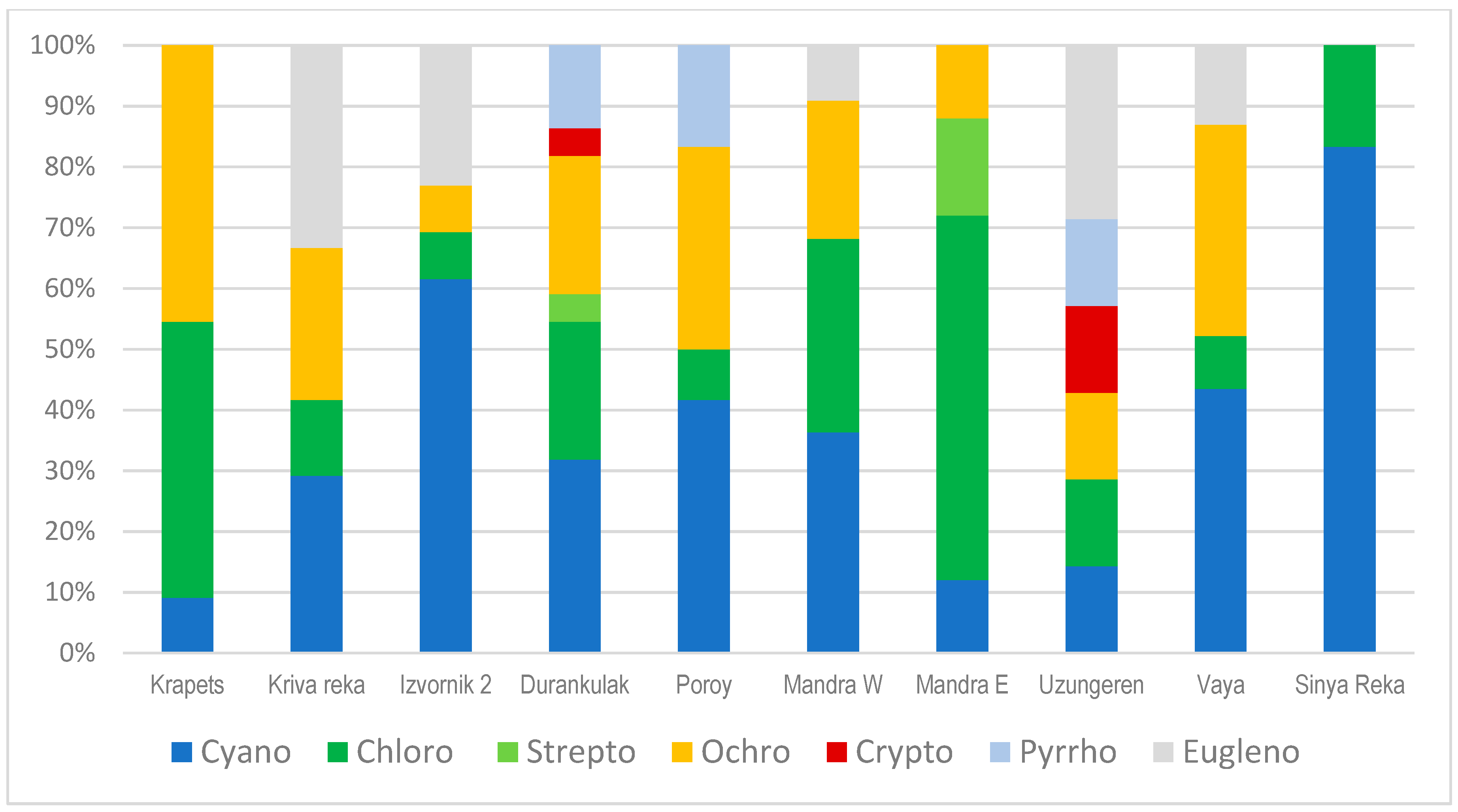
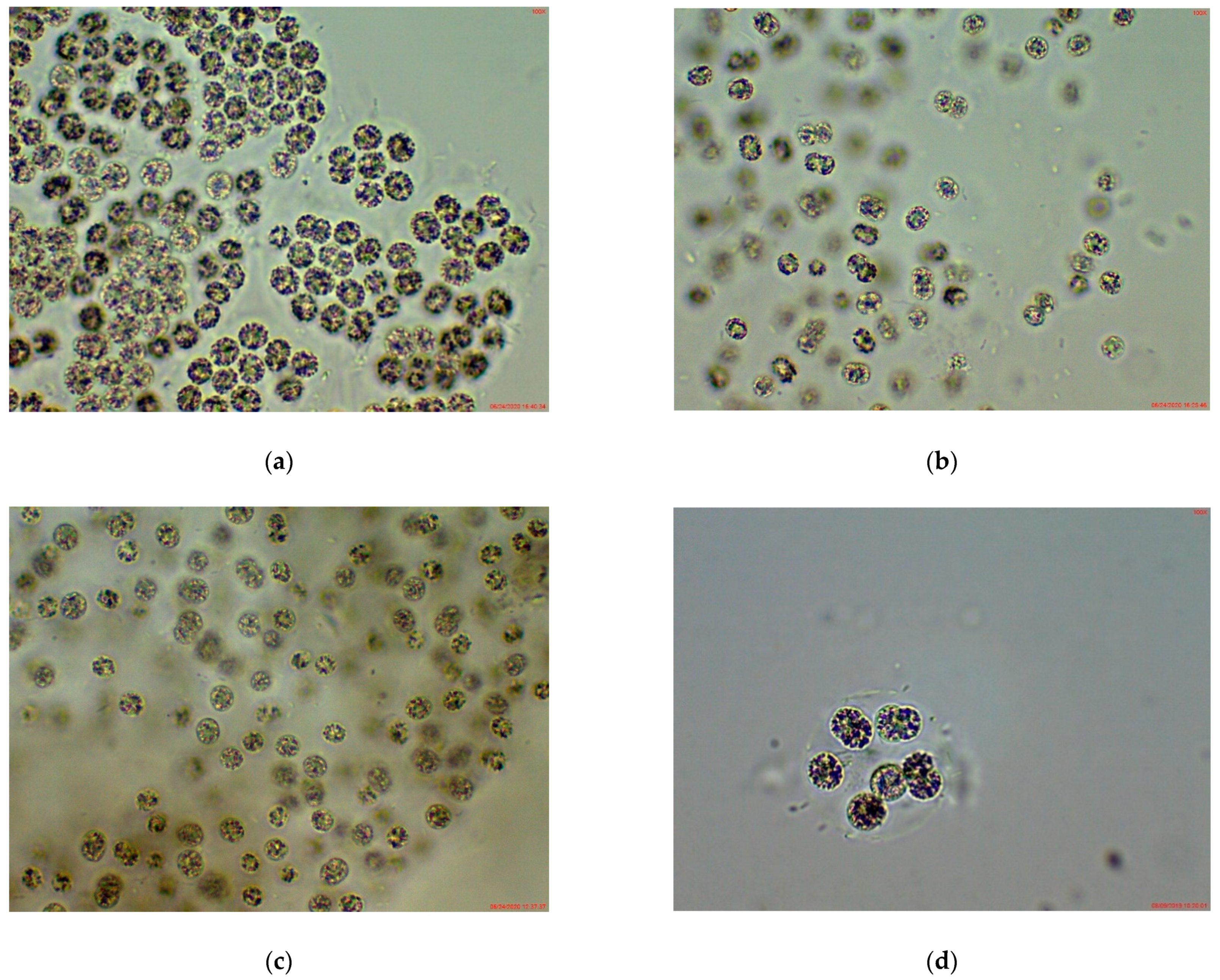
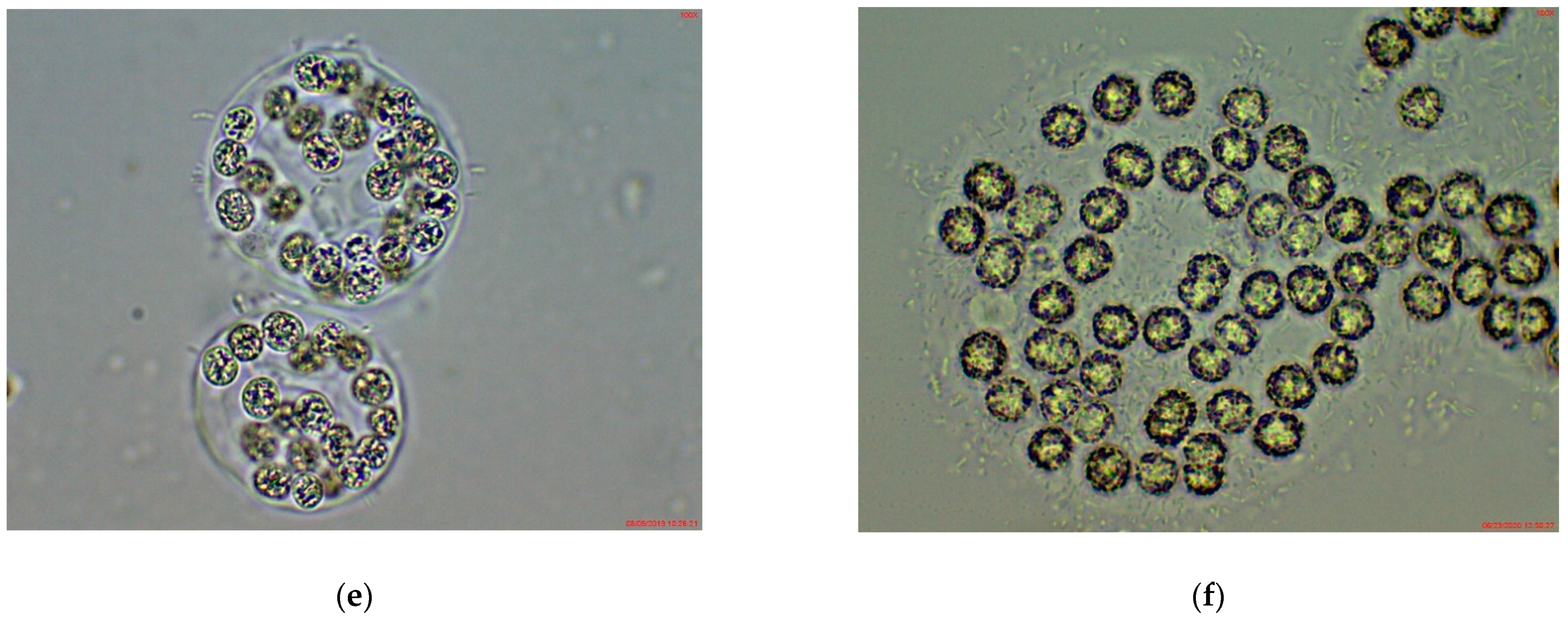
3.1. Phytoplankton Species Composition and Abundance, Obtained by Light Microscopy (LM)

In total, 235 species from seven algal phyla (Cyanoprokaryota, Chlorophyta, Streptophyta, Ochrophyta, Cryptophyta, Pyrrhophyta and Euglenophyta) were identified using LM in the phytoplankton of the nine studied wetlands (Figure 2). Among them 68, or 29% of all taxa were cyanoprokaryotes from 30 genera of four orders: Chroococcales, Synechococcales, Oscillatoriales and Nostocales. In the studied samples we found different potential MC-producers from the genera Anathece, Aphanothece and Microcystis (Chroococcales), Synechococcus, Jaaginema and Pseudanabaena (Synechococcales), Geitlerinema, Oscillatoria, Phormidium and Planktothrix (Oscillatoriales), Anabaena s.str. and Dolichospermum (Nostocales), which occurred separately or in different combinations. Some species from the nostocalean genera Aphanizomenon s.l. (which recently belong to different genera Aphanizomenon s.str., Chrysosporum, Cuspidothrix) and Raphidiopsis (incl. former Cylindrospermopsis) which may contain mcy genes, were also identified in the studied wetlands. All observed species had different contribution to the phytoplankton biomass, with Aphanizomenon s.l., Dolichospermum, and more rarely Planktothrix, recorded as dominants.
The highest number of cyanoprokaryote species was recorded in Vaya (10), Izvornik 2 and Mandra West (8 in each), followed by Durankulak and Kriva Reka (7 in each), Poroy and Sinya Reka (5 in each), Mandra East (3), Krapets and Uzungeren (1 in each). The relative contribution of these species to the phytoplankton composition in the studied sites ranged between 10 and 83% (Figure 3).
Cyanoprokaryotes comprised from 11 to 99% of the total phytoplankton biomass, with highest relative contribution in the reservoir Sinyata Reka (99%) and lake Vaya (95%). Their lowest relative contribution was in the lake Durankulak (11%) and in the reservoir Krapets (20%) (Figure 4), where dominants were diatoms (Cyclotella) and green algae (Elakatothrix), respectively.
Considering the results from the PCR analysis, provided below, we will present in detail the LM data obtained for the genus Microcystis. Based on the classical taxonomic criteria [27,28,29,30,31,32,33], we identified only two species: M. aeruginosa (Kützing) Kützing and M. wesenbergii (Komárek) Komárek in Kondratieva 1964 (Figure 5; Table 2). In some samples we also found cells from disintegrated colonies and a few compact colonies with small cells (ca. 2 µm in diameter) without gas vesicles, which could not be reliably identified using traditional morphological diagnostic features. The relative contribution (Table 2) of Microcystis was very low (<1% of the total phytoplankton biomass, except for the reservoir Mandra where it comprised <5% of the biomass). In the eastern part of this reservoir some small juvenile transitional colonies between both species with thin mucilage margin were also observed. In the reservoir Poroy and in the lake Durankulak M. wesenbergii was rarely detected and seen only in juvenile colonies, which were better developed and contained higher number of cells in Durankulak (Figure 5d,e).

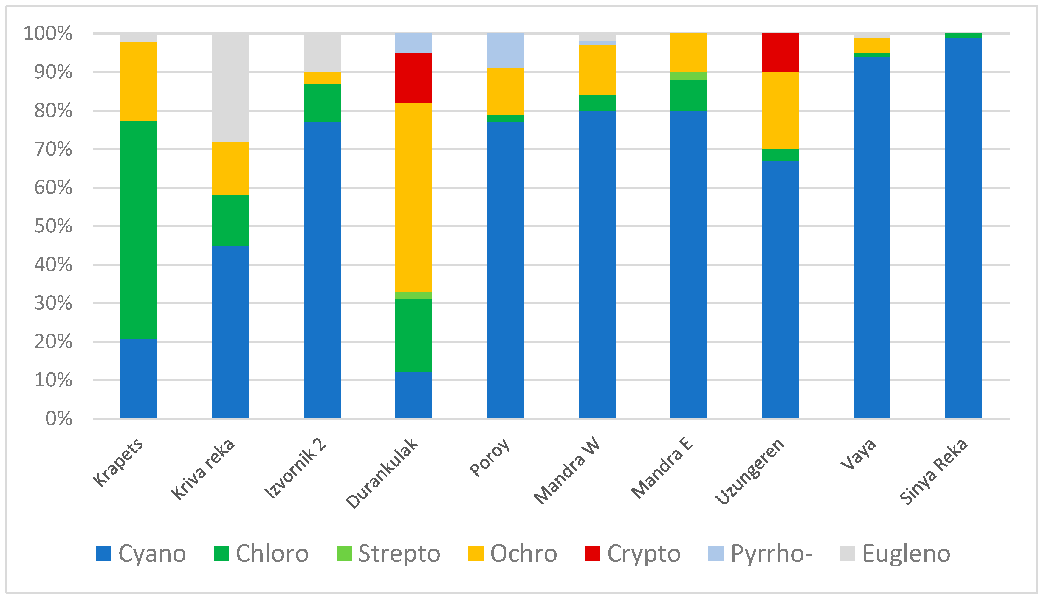
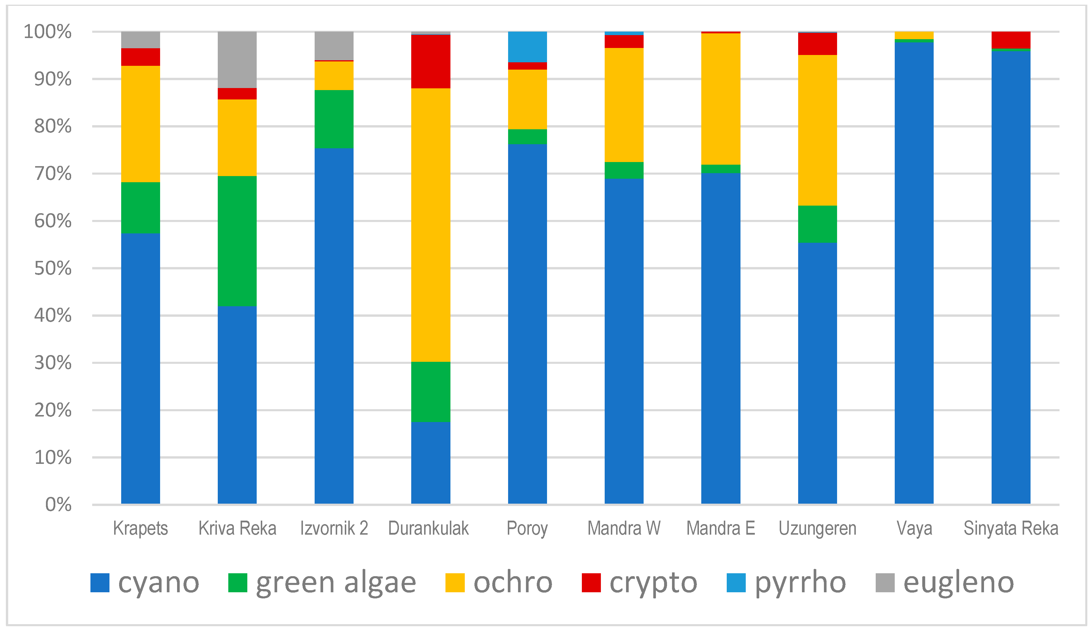
3.2. Results from Marker Pigment Analysis

According to HPLC data on pigment markers, the phytoplankton of the studied waterbodies was composed mainly of cyanoprokaryotes, ochrophytes (mainly diatoms and less chrysophyceans/golden algae), followed by green algae (chlorophytes and streptophytes) with lower contribution of cryptophytes, pyrrhophytes and euglenophytes (Figure 6). The highest dominance of Cyanoprokaryota was found in the coastal lake Vaya (up to 98%) and in the inland reservoir Sinyata Reka (96%), followed by the coastal reservoir Poroy (76%) and inland reservoir Izvornik 2 (72%). The only exception was the relatively low amount (18%) of cyanoprokaryotes in the coastal lake Durankulak, where, according to the LM observations, they were represented by high cell numbers, but large centric diatoms of the genus Cyclotella dominated by biomass.
The values of chlorophyll a, measured by HPLC, ranged from 0.9 to 856 µg L−1. All waterbodies, except the Krapets reservoir, had chlorophyll a concentration >10 µg L−1, indicating their eutrophic to strongly hypertrophic status, in agreement with the Secchi disk depth and TP data (Table 1).

3.3. Results from Toxin Analysis

Toxin analyses proved the presence of MCs only in two of the studied wetlands, namely the coastal lakes Mandra and Durankulak, with a difference in their concentrations by sites (Table 3).

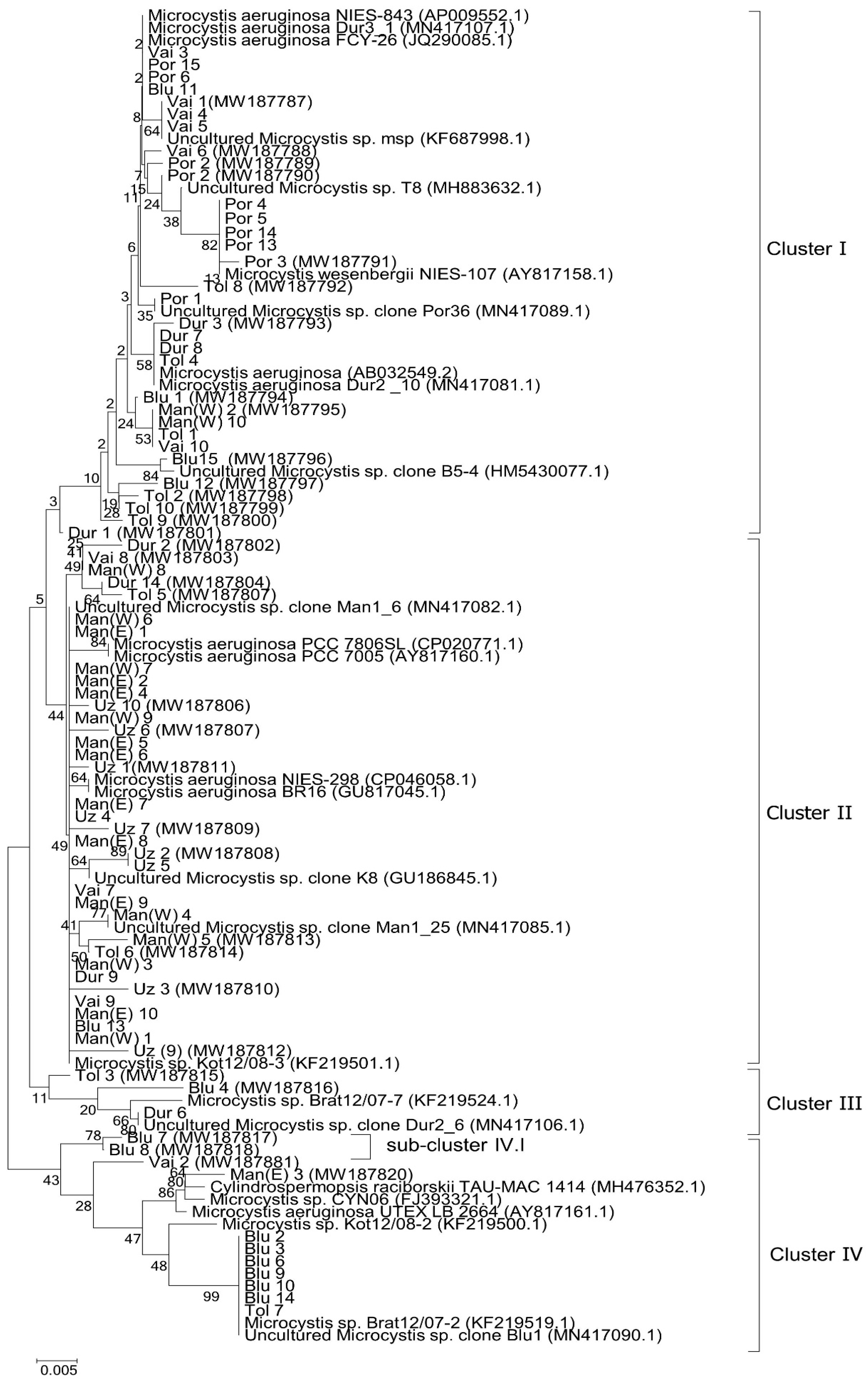
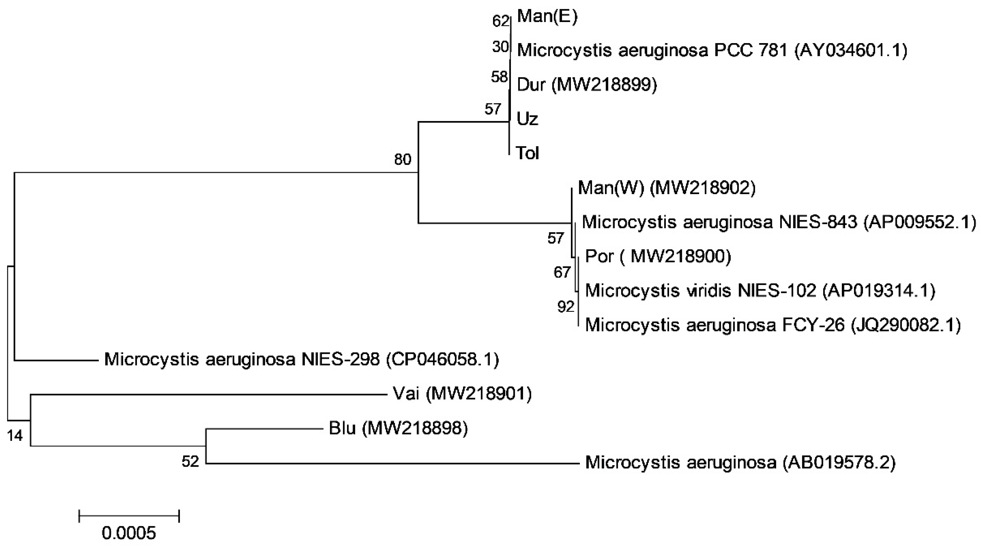
3.4. Results from PCR Analysis for Microcystin-Producing Strains

After the application of HEPF × HEPR synthetase-gene-specific pair of primers, isolation of DNA fragments with expected length of 470 bp and their cloning into plasmid vector, we constructed eight mcyDNA-clone libraries. They reflect data on all 81 mcyE sequences obtained from eight of the ten studied sites: Izvornik 2, Durankulak, Poroy, Mandra (with sites in its both parts—Mandra East and Mandra West), Uzungeren, Vaya and Sinyata Reka (Figure 7). PCR products were not obtained from the reservoirs Krapets and Kriva Reka.
According to the BLAST search [54], 43 (53%) of all 81 obtained sequences had 100% identity with the NCBI [55] mcyE sequences of known strains and could be affiliated to them. Another 31 sequences (38%) showed high level homology (>99% identity) and only seven strains, namely Blu4, Blu7, Blu8, Tol3, Uz9, Uz3, and Dur2 (Figure 7), were more distant, showing lower identity (>98%) to the known mcyE sequences. All the analyzed 81 mcyE sequences and their corresponding high homology NCBI sequences were used in the phylogenetic assay. In the constructed phylogenetic tree, four main clusters were formed (Figure 7).
According to the combined results from the NCBI search and phylogenetic analysis, it was not possible to affiliate correctly to specific genera the seven strains mentioned above with lower identity, but 74 from all 81 obtained sequences clearly belonged to the genus Microcystis. Most of these 74 sequences (41, or 55%) were affiliated to uncultured or unidentified to species level strains of the genus and only 11 of them (15%) could be referred to two distinct species, with named sequences in NCBI—M. aeruginosa and M. wesenbergii (Figure 7). On the basis of Microcystis strains, submitted by us to NCBI, sequenced in 2018 and described morphologically in [9], we can suppose that in the processed samples we found seven more sequences coinciding with M. wesenbergii (with 100% homology). They are grouped together in cluster IV with NCBI clone Microcystis sp. Brat 12/07-2 (KF219519.1) and are allied to the species M. wesenbergii on the basis of the submitted by us strain Uncultured Microcystis clone Blu1. MN417090.1 (Figure 7). In this way, during this study, we identified M. wesenbergii as present in the reservoirs Sinyata Reka and Izvornik 2 (cluster IV), and in the reservoir Poroy, the last according to the sequences in cluster I of Figure 7. It is notable that the affiliation to the species in cluster I is based on the same strain Microcystis wesenbergii NIES-107, NCBI:txid315483, which was involved in the construction of the phylogenetic tree from June 2018 and is similar to the strain Microcystis wesenbergii submitted by us (Vai1-2_11, NCBI:txid2703880 with number MN417095.1) [9].
Comparison of data from this study and our previous results [9] allowed us to suppose that cluster II on Figure 7 contains 19 strains identical (100%) to Microcystis sp. Kot12/08-3 (NCBI:txid1402958), which we tentatively related with Microcystis novacekii (Komárek) Compère identified by LM and submitted to NCBI as Microcystis sp. Man1_6 (MN417082.1). These strains are spread in the reservoirs Mandra (West—5 strains, and East -9) and Sinyata Reka (1), and in the lakes Durankulak (1), Vaya (2), and Uzungeren (1). However, cluster II is quite complicated and contains more sequences, grouped in clear subclusters, the affiliation of which so far was not possible and requires further studies.
Up-to-now it was also not possible to affiliate with certainty to any species the six sequences of 100% homology with NCBI, namely Vai1, Vai4, Vai5, Por1, ManW4 and Dur 6 (Figure 7). However, according to the presence of NCBI strain Microcystis sp. Brat12/07-7 (NCBI:txid1402954), similarity with which was found in 2018 [9], we provisionally suppose that one of the six strains mentioned above, namely Dur 6, is similar with the strain of Microcystis natans (observed by LM in the same waterbody—Durankulak in 2018 [9] and submitted by us to NCBI as Uncultured Microcystis sp. clone Dur2_6 (MN417106.1).
The mcyE phylogenetic tree (Figure 7) demonstrated the rich biodiversity of Microcystis strains in the studied sites. The highest diversity was detected in the small inland reservoir Izvornik 2; there, all ten sequences were distant from each other and were distributed in four different clusters (I–IV). Rich diversity was found also in the coastal lake Durankulak, the eight sequences of which were distributed in three different clusters (I-III). From the coastal lake Vaya and reservoir Poroy a total of 15 sequences were obtained and most of them (11) belonged to cluster I. Much less heterogeneity was found in the small inland reservoir Sinyata Reka, from which eight sequences (out of 14) were concentrated in the single cluster IV. The lowest biodiversity was found in the coastal lake Mandra, most sequences of which (16 out of 19) were distributed in cluster II.
By applying direct sequencing of PCR products with expected length of 850 bp and mcyB marker, eight sequences were obtained and were included in the constructed phylogenetic tree (Figure 8). They belonged to two main clusters and were affiliated mainly to M. aeruginosa strains registered in NCBI [55].
These results showed the diversity of the strains of M. aeruginosa and demonstrated that by application of mcyB a better grouping (80% high bootstrap value at branch point) was achieved. It allowed distinguishing the sequences isolated from the western and eastern parts of the reservoir Mandra (Figure 8), but also to separate them from the NCBI strain M. aeruginosa NIES-298 (CP046058.1) with NCBI:txid449468, which is close to and grouped together with almost all sequences isolated from Mandra in the mcyE-based phylogenetic tree (Figure 8).

4. Discussion

The HPLC data on marker phytoplankton pigments revealed high chlorophyll a concentrations (with very high values in the small inland reservoirs Sinyata Reka and Izvornik 2) and demonstrated high proportion of cyanoprokaryotes in the quantitative structure of the summer phytoplankton of the studied waterbodies (Figure 6). The contribution of cyanoprokaryotes to the total biomass exceeded 95% in the Lake Vaya and Sinyata Reka reservoir, and was over 70% in the reservoirs Izvornik 2 and Poroy (Figure 6). All these data are in accordance with the maximum abundance of cyanoprokaryotes in the late summer phytoplankton of the shallow and highly eutrophic waterbodies of the world [57,58], and of Bulgaria in particular [19,22,23].
The results on taxonomic phytoplankton structure obtained during this study by conventional LM (Figure 2, Figure 3 and Figure 4) demonstrated a high contribution of cyanoprokaryotes to the phytoplankton species composition and abundance. These data coincide with data assessed from the phytoplankton pigment concentrations (Figure 6) and chemically detected cyanotoxins—MCs (Table 3).
According to the toxin analyses, MC concentrations varied by sites with a range from undetectable concentrations to 0.46 µg L−1, with only one recorded type, namely MC–LR (Table 3). This MC is renowned because it has been considered as the most dangerous in the WHO Guidelines for Drinking-water Quality with a provisional value of 1 μg L−1 [59]. This is the second finding of MCs, and of MC–LR in particular, in the reservoir Mandra and the fourth record of MCs (incl. MC–LR) from the lake Durankulak (for details see [19]). The absence of detectable amounts of MCs in the reservoir Sinyata Reka, in which MC-RR (0.09 μg L−1) and MC-LR (0.3 μg L−1) were detected in June 2018 [23] is explainable by the different dominants in both studied periods. In June 2018, in Sinyata Reka Microcystis wesenbergii dominated, reaching over 90% of the total biomass [9,23], while in August 2019 heterocytous filamentous Dolichospermum dominated and M. wesenbergii occurred rarely in the samples, in combination with even more sparse M. aeruginosa. The difference in dominants and frequent changes of phytoplankton assemblages are typical for shallow waterbodies (e.g., [57]). But in this particular case we have to also mention the strong, atypical for Bulgaria, rains from May until the first decade of August 2019, which preceded the sampling and caused serious dilution of the reservoir waters with lower amounts of TP and TN, the values of which were twice less in comparison with previous year [23].
The results from conducted molecular-genetic analysis (Figure 7 and Figure 8) demonstrated that toxigenic mcy sequences were found in all three sites, where MC-LR was detected (Mandra West, Mandra East and Durankulak—Table 3). The lack of measurable amounts of MCs (<LOD) in all other wetlands, besides the strong rains mentioned above, can be explained by: (1) the fact that the methods applied by us have limited scope while types of MCs are highly diverse with increasing number of found congeners [13]. Therefore, it is possible to suppose that in shallow Bulgarian waterbodies there are more MC types which have not been detected during the study; (2) quite low Microcystis amounts recorded by LM (Table 2), which do not allow the chemical toxin detection; (3) the lack of gene expression during the sampling period due to the specific temporal patterns of mcy gene expression (e.g., [60]); (4) the influence of other possible factors on toxin production, like the growth phase of the populations [61]. The absence of mcyE and mcyB sequences and of detected MCs in the reservoirs Krapets and Kriva Reka is supported by the fact that no known MC producers were identified in the phytoplankton samples.
Considering Microcystis in particular, both LM and molecular-genetic approaches demonstrated its presence with uneven distribution of species and toxigenic strains in eight of the ten studied sites (except in the mentioned above reservoirs Krapets and Kriva Reka). The results indicated low taxonomic (2 species; Table 2, Figure 5) but high genetic diversity (Figure 7 and Figure 8). Most strains (81) were identified according to their homology with mcyE-based sequences and grouped in four clusters (Figure 7), and five strains homologous with mcyB were spread in two clusters (Figure 8). Despite the high homology (100%) of more than 50% of the obtained sequences, only eleven (15%) of them were affiliated to the NCBI strains of both distinct species M. aeruginosa and M. wesenbergii. For seven more strains we assume belonging to M. wesenbergii based on our former polyphasic study and relevant sequences provided to NCBI [9] (Figure 7). In the same study the reasons for the low number of certain affiliations were discussed in detail, stressing that many NCBI strains are not supplied with relevant morphological descriptions, and that the value of some phylogenetic constructions is reduced because of using sequences from strains which have not passed taxonomic revision and therefore have incorrect or arbitrary names [9,62]. Here we shall note also that many NCBI strains have only generic names, which obviously complicates the affiliation to distinct species.
The phylogenetic trees, constructed from the analyzed mcyE and mcyB sequences and their homologous representatives from NCBI (Figure 7 and Figure 8), demonstrated the complexity of the isolated sequences pool. This complexity reflects the relevant rich biodiversity of Microcystis strains in the studied waterbodies and is on accordance with our previous mcyE-based data, obtained from the same wetlands [9].
With this study we demonstrate for second time the presence of mcy genes in the strains of M. wesenbergii from Bulgaria. The potential toxicity of this species has long been debated (for details see [9,19,62]). The mcyE toxigenic sequences allied to this species were found for first time in the reservoirs Poroy and Izvornik 2, and were confirmed for the reservoir Sinyata Reka. Thus, considering our former data [9], MC-producing strains of M. wesenbergii were found altogether in four Bulgarian waterbodies, where the species was also detected by LM observations at: Sinyata Reka, Vaya, Izvornik 2 and Poroy. The rare initial colonies of the species seen by LM in the samples from Poroy in August 2019 (Figure 5d) coincide with the undetectable amounts of MCs in this reservoir (Table 3). Our results on mcyE sequences, LM observations and toxin analyses are consistent with the findings of mcyE genes in a single strain of M. wesenbergii isolated from Taskisi Lake (Turkey), which demonstrated MC production [63]. They coincide also with the records of mcy genes A and B in M. wesenbergii [64], with detected MC production of its different strains [65] and its findings in field samples containing MCs (for details see [19,62,66]).
Considering that the mcyE gene has been successfully used to reveal the presence of potential MC-producing genera other than Microcystis [2,3,6,13,14,15,16,17,63] and that species of such genera (e.g., Anabaena, Dolichospermum, Sphaerospermopsis, Oscillatoria, Planktothrix, Raphidiopsis and Aphanizomenon s.l.) were identified by conventional LM in the phytoplankton of the studied waterbodies (in some of which they dominated or were abundant), we can argue that in the processed samples MC-producing strains of all these genera were not found by molecular-genetic methods. The appearance of Raphidiopsis raciborskii strain NCBI:txid2217671 (provided as Cylindrospermopsis raciborskii Tau-Mac 1414 in NCBI and in Cluster IV on Figure 7) also does not show the occurrence of MC-producing strains of this species in our waterbodies due to its low similarity with the newly obtained sequence ManE3 (MW187820). Moreover, R. raciborskii was not found by LM in the phytoplankton of the Mandra reservoir, where Aphanizomenon cf. flos-aquae dominated. However, our data support the findings of mcyE genes in this species by [17].
It is broadly accepted that the high diversity and occurrence of different MC congeners is due to variability in the coding of the mcy genes among cyanobacterial strains [13]. Taking this into account, we cannot argue that during the present study we detected all MCs and their causative agents, but after juxtaposing of all obtained data we demonstrated that M. aeruginosa and M. wesenbergii were the main reliably identified MC producers in the studied samples from August 2019. This result is in agreement with our earlier data on LM identification of these species in the samples and waterbodies where MCs were detected [19] and with the first finding of their toxigenic strains [9].

5. Conclusions

This study demonstrates the notable role of cyanoprokaryotes in the late summer phytoplankton of 2019 and high diversity of toxic Microcystis strains in seven (from the nine studied) shallow Bulgarian waterbodies of high trophic state. All reliably identified strains belonged to two species, namely M. aeruginosa and M. wesenbergii. If M. aeruginosa is worldwide accepted as an MC-producer, the toxic potential of the easily morphologically recognized M. wesenbergii has long been debated. This work provides more evidence on the existence of toxic strains in M. wesenbergii. The lack of toxigenic mcyE and mcyB sequences from other genera known as potential MC-producers, together with conventional LM observations, allows us to conclude that M. aeruginosa and M. wesenbergii were the primary MC-producers in the investigated wetlands in August 2019. Moreover, the fact that only MC-LR was detected in water samples from the sites where both species were found (separately or together), suggests that they were associated with the production of this dangerous cyanotoxin. This finding is important as both Microcystis species occurred in low amounts in waterbodies dominated by the filamentous heterocytous Dolichospermum or Aphanizomenon s.l. or, more rarely, of the filamentous non-heterocytous Planktothrix. On the background of notable quantitative role of cyanoprokaryotes assessed by pigment marker analysis and conventional LM observations, the proved high genetic diversity of marker mcy genes-bearing strains demonstrates the need for diversification of methods applied for cyanotoxin detection in the studies of vulnerable to CyanoHABS shallow Bulgarian waterbodies. On the other side, the comparison of results from the applied LM and PCR methods once more proved the possibility genetically to detect toxigenic algae, even when they occur in low amounts, but also showed again that currently available genomic sequences are still insufficient to fully resolve the species identification among cyanoprokaryotes and specifically in the genus Microcystis.

Author Contributions

Conceptualization, M.S.-G. and B.U.; sample collection, B.U., M.S.-G., J.-P.D., G.G. and M.R.; molecular genetic analyses, K.S. and M.R.; investigations by LM, M.S.-G., G.G. and B.U.; HPLC pigment marker analyses, J.-P.D.; HPLC toxin analyses, V.P. and M.M.; resources, B.U. and M.S.-G.; writing, original draft preparation, M.S.-G.; writing—review and editing, M.S.-G., J.-P.D., G.G., B.U., M.R. and K.S.; funding acquisition—B.U., M.S.-G., M.R. and G.G.; project administration, B.U. All authors have read and agreed to the published version of the manuscript.

Funding

This research was funded by the Scientific Research Fund of the Ministry of Education and Science of Bulgaria (SRF-MESB), grant numbers KP-06-OPR06/2/18.12.2018 and KP-06-OPR03/18/19.12.2018.

Institutional Review Board Statement

Not applicable.

Informed Consent Statement

Not applicable.

Data Availability Statement

Data available in a publicly accessible repository. The data presented in this study are openly available in [NCBI] at [https://www.ncbi.nlm.nih.gov/], reference number [MW187787-MN187820 and MW218898-MW218902].

Acknowledgments

The authors would like once more to acknowledge SRF-MESB for projects granting and the European Cooperation in Science and Technology, COST Action ES 1105 “CYANOCOST—Cyanobacterial blooms and toxins in water resources: occurrence, impacts and management” for adding value to this paper through networking and knowledge sharing with European experts and researchers in the field.

Conflicts of Interest

The authors declare no conflict of interest. The funders had no role in the design of the study, in the collection, analyses, interpretation of data, in the writing of the manuscript, and in the decision to publish the results.

References

  1. Anderson, D. HABs in a changing world: A perspective on harmful algal blooms, their impacts, and research and management in a dynamic era of climactic and environmental change. Harmful Algae 2014, 2012, 3–17. [Google Scholar]
  2. Svirčev, Z.; Lalić, D.; Savić, G.B.; Tokodi, N.; Backović, D.D.; Chen, L.; Meriluoto, J.; Codd, G.A. Global geographical and historical overview of cyanotoxin distribution and cyanobacterial poisonings. Arch. Toxicol. 2019, 93, 2429–2481. [Google Scholar] [CrossRef] [PubMed]
  3. Mitrovic, S.M.; Kobayashi, T.; Roelke, D.L. Cyanobacteria in inland waters: New monitoring, reporting, modelling and ecological research. Mar. Freshw. Res. 2020, 71, i–iv. [Google Scholar] [CrossRef]
  4. Meriluoto, J.; Blaha, L.; Bojadzija, G.; Bormans, M.; Brient, L.; Codd, G.A.; Drobac, D.; Faassen, E.J.; Fastner, J.; Anastasia, H.; et al. Toxic cyanobacteria and cyanotoxins in European waters—Recent progress achieved through the CYANOCOST Action and challenges for further research. Adv. Oceanogr. Limnol. 2017, 8, 161–178. [Google Scholar] [CrossRef] [Green Version]
  5. Jankowiak, J.T.; Hattenrath-Lehmann, B.J.; Kramer, M.L.; Gobler, C.J. Deciphering the effects of nitrogen, phosphorus, and temperature on cyanobacterial bloom intensification, diversity, and toxicity in western Lake Erie. Limnol. Oceanogr. 2019, 64, 1347–1370. [Google Scholar] [CrossRef] [Green Version]
  6. Janssen, E.M.L. Cyanobacterial peptides beyond microcystins—A review on co-occurrence, toxicity and challenges for risk assessment. Water Res. 2019, 151, 488–499. [Google Scholar] [CrossRef]
  7. Ghaffar, S.; Stevenson, R.J.; Khan, Z. Effect of phosphorus stress on Microcystis aeruginosa growth and phosphorus uptake. PLoS ONE 2017, 12, e0174349. [Google Scholar] [CrossRef]
  8. Xiao, M.; Li, M.; Reynolds, C.S. Colony formation in the cyanobacterium Microcystis. Biol. Rev. 2018, 93, 1399–1420. [Google Scholar] [CrossRef] [Green Version]
  9. Radkova, M.; Stefanova, K.; Uzunov, B.; Gärtner, G.; Stoyneva-Gärtner, M. Morphological and molecular identification of microcystin-producing cyanobacteria in nine shallow Bulgarian water bodies. Toxins 2020, 12, 39. [Google Scholar] [CrossRef] [Green Version]
  10. Scherer, P.I.; Raeder, U.; Geist, J.; Zwirglmaier, K. Influence of temperature, mixing, and addition of microcystin-LR on microcystin gene expression in Microcystis aeruginosa. MicrobiologyOpen 2017, 6, e00393. [Google Scholar] [CrossRef]
  11. Davis, T.W.; Berry, D.L.; Boyer, G.L.; Gobler, C.J. The effects of temperature and nutrients on the growth and dynamics of toxic and non-toxic strains of Microcystis during cyanobacteria blooms. Harmful Algae 2009, 8, 715–725. [Google Scholar] [CrossRef]
  12. Vézie, C.; Rapala, J.; Vaitomaa, J.; Seitsonen, J.; Sivonen, K. Effect of nitrogen and phosphorus on growth of toxic and nontoxic Microcystis strains and on intracellular microcystin concentrations. Microb. Ecol. 2002, 43, 443–454. [Google Scholar] [CrossRef] [PubMed]
  13. Puddick, J.; Prinsep, M.R.; Wood, S.A.; Kaufononga, S.A.F.; Cary, S.C.; Hamilton, D.P. High levels of structural diversity observed in microcystins from Microcystis CAWBG11 and characterization of six new microcystin congeners. Mar. Drugs 2014, 12, 5372–5395. [Google Scholar] [CrossRef] [PubMed]
  14. Rantala, A.; Rajaniemi-Wacklin, P.; Lyra, C.; Lepisto, L.; Rintala, J.; Mankiewicz-Boczek, J.; Sivonen, K. Detection of microcystin-producing cyanobacteria in Finnish lakes with genus-specific microcystin synthetase gene E (mcyE) PCR and associations with environmental factors. Appl. Environ. Microbiol. 2006, 72, 6101–6110. [Google Scholar] [CrossRef] [PubMed] [Green Version]
  15. Hisbergues, M.; Christiansen, G.; Rouhiainen, L.; Sivonen, K.; Börner, T. PCR-based identification of microcystin-producing genotypes of different cyanobacterial genera. Arch. Microbiol. 2003, 180, 402–410. [Google Scholar] [CrossRef] [PubMed]
  16. Lyon-Colbert, A.; Su, S.; Cude, C. A systematic literature review for evidence of Aphanizomenon flos-aquae toxigenicity in recreational waters and toxicity of dietary supplements: 2000–2017. Toxins 2018, 10, 254. [Google Scholar] [CrossRef] [Green Version]
  17. Panou, M.; Zervou, S.-K.; Kaloudis, T.; Hiskia, A.; Gkelis, S. A Greek Cylindrospermopsis raciborskii strain: Missing link in tropic invader’s phylogeography tale. Harmful Algae 2018, 80, 96–106. [Google Scholar] [CrossRef]
  18. Michev, T.; Stoyneva, M. (Eds.) Inventory of Bulgarian Wetlands and Their Biodiversity; Elsi-M: Sofia, Bulgaria, 2007. [Google Scholar]
  19. Stoyneva-Gärtner, M.P.; Descy, J.-P.; Latli, A.; Uzunov, B.; Pavlova, V.; Bratanova, Z.; Babica, P.; Maršálek, B.; Meriluoto, J.; Spoof, L. Assessment of cyanoprokaryote blooms and of cyanotoxins in Bulgaria in a 15-years period (2000–2015). Adv. Oceanogr. Limnol. 2017, 8, 131–152. [Google Scholar] [CrossRef] [Green Version]
  20. Stoyneva-Gärtner, M.; Uzunov, B.; Dimitrova, P.; Pavlova, V. Algal toxins—New risk factors for national security in Bulgaria. In Proceedings of the Actual Problems of the Security, Veliko Turnovo, Bulgaria, 26–27 October 2017; Publishing House Complex of NVU “Vasil Levski”: Veliko Turnovo, Bulgaria, 2017; pp. 271–281, Electronic Publication. [Google Scholar]
  21. Stoyneva-Gärtner, M.P.; Uzunov, B.A.; Dimitrova, P. Pilot assessment of cyanotoxins as potential risk factors for cancer in Bulgaria. BioDiscovery 2017, 20, e20501. [Google Scholar] [CrossRef] [Green Version]
  22. Descy, J.-P.; Stoyneva-Gärtner, M.P.; Uzunov, B.A.; Dimitrova, P.H.; Pavlova, V.T.; Gärtner, G. Studies on cyanoprokaryotes of the water bodies along the Bulgarian Black Sea Coast (1890–2017): A review, with special reference to new, rare and harmful taxa. Acta Zool. Bulgar. Suppl. 2018, 11, 43–52. [Google Scholar]
  23. Stoyneva-Gärtner, M.P.; Uzunov, B.A.; Descy, J.-P.; Gärtner, G.; Draganova, P.H.; Borisova, C.I.; Pavlova, V.; Mitreva, M. Pilot application of drone-observations and pigment marker detection by HPLC in the studies of CyanoHABs in Bulgarian inland waters. Mar. Freshw. Res. 2019, 71, 606–616. [Google Scholar] [CrossRef]
  24. Andersen, R.A. Algal Culturing Techniques; Elsevier Academic Press: Burlington, NJ, USA, 2005. [Google Scholar]
  25. Uzunov, B.; Stoyneva, M.; Mancheva, A.; Gärtner, G. ACUS—The new collection of living aeroterrestrial algae of the University of Sofia ‘St Kliment Ohridski’. In Proceedings of the VII National Botanical Conference, Sofia, Bulgaria, 29–30 September 2011; Petrova, A., Ed.; Bulgarian Botanical Society: Sofia, Bulgaria, 2012; pp. 271–274. [Google Scholar]
  26. AlgaeBase. Available online: http://www.algaebase.org/ (accessed on 28 October 2020).
  27. Geitler, L. Cyanophyceae. In Rabenhorst's Kryptogamenflora von Deutschland, Österreich und der Schweiz, 2nd ed.; Rabenhorst, L., Ed.; Akademische Verlagsgesellschaft: Leipzig, Germany, 1932; Volume 14, pp. 1–1196. [Google Scholar]
  28. Geitler, L. Schizophyta: Klasse Schizophyceae. In Die natürlichen Pflanzenfamilien, Zweite Auflage; Engler, A., Prantl, K., Eds.; Wilhelm Engelmann: Leipzig, Germany, 1942; Volume 1b, pp. 1–232. [Google Scholar]
  29. Gollerbakh, M.M.; Kossinskaya, E.K.; Polyanskiy, V.I. Manual of Freshwater Algae of the USSR. Volume 2. Blue-Green Algae; Sovetskaya Nauka: Moscow, Russia, 1953. [Google Scholar]
  30. Starmach, K. Cyanophyta-Sinice. Glaucophyta-Glaukofity; Państwowe Wydawnictwo Naukowe: Warszawa, Poland, 1966. [Google Scholar]
  31. Komárek, J.; Anagnostidis, K. Cyanoprokaryota. Teil 1/Part 1: Chroococcales. In Süßwasserflora von Mitteleuropa; Ettl, H., Gerloff, J., Heynig, H., Mollenhauer, D., Eds.; Spektrum Akademischer Verlag: Heidelberg, Germany, 2008; Volume 19/1, pp. 1–556. [Google Scholar]
  32. Komárek, J.; Komárková, J. Review of the European Microcystis morphospecies (Cyanoprokaryotes) from nature. Czech Phycol. 2002, 2, 1–24. [Google Scholar]
  33. Šejnohová, L.; Maršálek, B. Microcystis. In Ecology of Cyanobacteria II. Their Diversity in Space and Time; Whitton, B.A., Ed.; Springer: Dordrecht, The Netherlands, 2012; pp. 195–228. [Google Scholar]
  34. CyanoDB 2.0. Available online: http://www.cyanodb.cz (accessed on 29 October 2020).
  35. Catherine, Q.; Wood, S.; Echenigue-Subiabre, I.; Heath, M.; Villeneuve, A.; Humbert, J.-F. A review of current knowledge on toxic benthic freshwater cyanobacteria—Ecology, toxin production and risk management. Water Res. 2013, 47, 5464–5479. [Google Scholar] [CrossRef]
  36. Bernard, C.; Ballot, A.; Thomazeau, S.; Maloufi, S.; Furey, A.; Mankiewicz-Boczek, I.; Pawlik-Skowrońska, B.; Capelli, C.; Salmazo, N. Appendix 2. Cyanobacteria associated with the production of cyanotoxins. In Handbook of Cyanobacterial Monitoring and Cyanotoxin Analysis; Meriluoto, J., Spoof, L., Codd, J., Eds.; John Wiley & Sons, Ltd.: Chichester, UK, 2017; pp. 501–525. [Google Scholar]
  37. Rott, E. Some results from phytoplankton counting intercalibration. Schweiz. Z. Hydrol. 1981, 43, 34–62. [Google Scholar] [CrossRef]
  38. Stoyneva, M.P.; Descy, J.-P.; Vyverman, W. Green algae in Lake Tanganyika: Is morphological variation a response to seasonal changes? Hydrobiologia 2007, 578, 7–16. [Google Scholar] [CrossRef]
  39. Descy, J.P. SOP5: Estimation of cyanobacteria biomass by marker pigment analysis. In Handbook of Cyanobacterial Monitoring and Cyanotoxin Analysis; Meriluoto, J., Spoof, L., Codd, J., Eds.; John Wiley & Sons, Ltd.: Chichester, UK, 2017; pp. 343–349. [Google Scholar]
  40. Mackey, M.D.; Mackey, D.J.; Higgins, H.W.; Wright, S.W. CHEMTAX—A program for estimating class abundances from chemical markers: Application to HPLC measurements of phytoplankton. Mar. Ecol. Prog. Ser. 1996, 144, 265–283. [Google Scholar] [CrossRef] [Green Version]
  41. Sarmento, H.; Descy, J.-P. Use of marker pigments and functional groups for assessing the status of phytoplankton assemblages in lakes. J. Appl. Phycol. 2008, 20, 1001–1011. [Google Scholar] [CrossRef]
  42. Van Wichelen, J.; Van Gremberghe, I.; Vanormelingen, P.; Debeer, A.-E.; Leporcq, B.; Menzel, D.; Codd, G.A.; Descy, J.-P.; Vyverman, W. Strong effects of amoebae grazing on the biomass and genetic structure of a Microcystis bloom (Cyanobacteria). Environ. Microbiol. 2010, 12, 2797–2813. [Google Scholar] [CrossRef]
  43. International Standard Organization. ISO 20179:2005. Water Quality—Determination of Microcystins—Method Using Solid Phase Extraction (SPE) and High Performance Liquid Chromatography (HPLC) with Ultraviolet (UV) Detection. Available online: https://www.iso.org/standard/34098.html (accessed on 18 December 2020).
  44. Hiskia, A.; Spoof, L.; Kaloudis, T.; Meriluoto, J. Determination of cyanotoxins by High-Performance Liquid Chromatography with Photodiode Array. In Handbook of Cyanobacterial Monitoring and Cyanotoxin Analysis; Meriluoto, J., Spoof, L., Codd, J., Eds.; John Wiley & Sons, Ltd.: Chichester, UK, 2017; pp. 205–211. [Google Scholar]
  45. Heck, K.; Alvarenga, D.O.; Shishido, T.K.; Varani, A.M.; Dorr, F.A.; Pinto, E.; Rouhiainen, L.; Jokela, J.; Sivonen, K.; Fiore, M.F. Biosynthesis of microcystin hepatotoxins in the cyanobacterial genus Fischerella. Toxicon 2017, 141, 43–50. [Google Scholar] [CrossRef] [Green Version]
  46. Humbert, J.-F. Molecular tools for the detection of toxigenic Cyanobacteria in natural ecosystems. In Handbook of Cyanobacterial Monitoring and Cyanotoxin Analysis; Meriluoto, J., Spoof, L., Codd, J., Eds.; John Wiley & Sons, Ltd.: Chichester, UK, 2017; pp. 280–283. [Google Scholar]
  47. Pacheco, A.B.F.; Guedes, I.A.; Azevedo, S.M.F.O. Is qPCR a reliable indicator of cyanotoxin risk in freshwater? Toxins 2016, 8, 172. [Google Scholar] [CrossRef] [Green Version]
  48. Vaitomaa, J.; Rantala, A.; Halinen, K.; Rouhiainen, L.; Tallberg, P.; Mokelke, L.; Sivonen, K. Quantitative real-time PCR for determination of microcystin synthetase E copy numbers for Microcystis and Anabaena in lakes. Appl. Environ. Microbiol. 2003, 69, 7289–7297. [Google Scholar] [CrossRef] [PubMed] [Green Version]
  49. Moffitt, M.C.; Neilan, B.A. Characterization of the nodularin synthetase gene cluster and proposed theory of the evolution of cyanobacterial hepatotoxins. Appl. Environ. Microb. 2004, 70, 6353–6362. [Google Scholar] [CrossRef] [PubMed] [Green Version]
  50. Mankiewicz-Boczek, J.; Izydorczyk, K.; Romanowska-Duda, Z.; Jurczak, T.; Stefaniak, K.; Kokocinski, M. Detection and monitoring toxigenicity of cyanobacteria by application of molecular methods. Environ. Toxicol. 2006, 21, 380–387. [Google Scholar] [CrossRef] [PubMed]
  51. Nonneman, D.; Zimba, P.V. A PCR-based test to assess the potential for microcystin occurrence in Channel catfish production ponds. J. Phycol. 2002, 38, 230–233. [Google Scholar] [CrossRef]
  52. Jungblut, A.-D.; Neilan, B.A. Molecular identification and evolution of the cyclic peptide hepatotoxins, microcystin and nodularin synthetase genes in three orders of cyanobacteria. Arch. Microbiol. 2006, 185, 107–114. [Google Scholar] [CrossRef]
  53. Foulds, I.V.; Granacki, A.; Xiao, C.; Krull, U.J.; Castle, A.; Horgen, P.A. Quantification of microcystin-producing cyanobacteria and E. coli in water by 5′-nuclease PCR. J. Appl. Microbiol. 2002, 93, 825–834. [Google Scholar] [CrossRef] [Green Version]
  54. Basic Local Alignment Search Tool (BLAST). Available online: https://blast.ncbi.nlm.nih.gov/Blast.cgi (accessed on 24 October 2019).
  55. National Centre for Biotechnology Information (NCBI). Available online: https://www.ncbi.nlm.nih.gov/ (accessed on 24 October 2019).
  56. Tamura, K.; Stecher, G.; Peterson, D.; Filipski, A.; Kumar, S. MEGA6: Molecular Evolutionary Genetics Analysis Version 6.0. Mol. Biol. Evol. 2013, 30, 2725–2729. [Google Scholar] [CrossRef] [Green Version]
  57. Reynolds, C.S. The Ecology of Phytoplankton; Cambridge University Press: Cambridge, UK, 2006. [Google Scholar]
  58. Whitton, B.A. (Ed.) Ecology of Cyanobacteria II: Their Diversity in Space and Time, 2nd ed.; Springer: Dordrecht, the Netherlands, 2012. [Google Scholar]
  59. Chorus, I.; Bartram, J. (Eds.) Toxic Cyanobacteria in Water: A Guide to Their Public Health Consequences, Monitoring and Management; E & FN Spon: London, UK, 1999. [Google Scholar]
  60. Tang, X.; Krausfeldt, L.E.; Shao, K.; LeCleir, G.R.; Stough, J.M.A.; Gao, G.; Boyer, G.L.; Zhang, Y.; Paerl, H.W.; Qin, B.; et al. Seasonal Gene Expression and the ecophysiological implications of toxic Microcystis aeruginosa blooms in Lake Taihu. Environ. Sci. Technol. 2018, 52, 11049–11059. [Google Scholar] [CrossRef]
  61. Vasconcelos, V. Global changes and the new challenges in the research on cyanotoxin risk evaluation. Limnetica 2015, 34, 149–158. [Google Scholar]
  62. Komárek, J. A polyphasic approach for the taxonomy of cyanobacteria: Principles and applications. Eur. J. Phycol. 2016, 51, 346–353. [Google Scholar] [CrossRef]
  63. Köker, L.; Akçaalan, R.; Albay, M.; Neilan, B.A. Molecular detection of hepatotoxic cyanobacteria in inland water bodies of the Marmara Region, Turkey. Adv. Oceanogr. Limnol. 2017, 8, 52–60. [Google Scholar] [CrossRef] [Green Version]
  64. Gkelis, S.; Zaoutsos, N. Cyanotoxin occurrence and potentially toxin producing cyanobacteria in freshwaters of Greece: A multi-disciplinary approach. Toxicon 2014, 78, 1–9. [Google Scholar] [CrossRef] [PubMed]
  65. Yasuno, M.; Sugaya, Y.; Kaya, K.; Watanabe, M.M. Variations in the toxicity of Microcystis species to Moina macrocopa. Phycol. Res. 2006, 46, 31–36. [Google Scholar] [CrossRef]
  66. Christophoridis, C.; Zervou, S.-K.; Manolidi, K.; Katsiapi, M.; Moustaka-Gouni, M.; Kaloudis, T.; Triantis, T.M.; Hiskia, A. Occurrence and diversity of cyanotoxins in Greek lakes. Sci. Rep. 2018, 8, 17877. [Google Scholar] [CrossRef] [Green Version]
Figure 1. Map of R. Bulgaria with indication of the studied waterbodies (modified after (http://www.ginkgomaps.com and Google Maps). Explanations are provided in the text.
Applsci 11 00357 g001
Figure 2. General phytoplankton species composition in nine shallow Bulgarian wetlands sampled in August 2019. The number of identified taxa in each phylum is shown. Legend: Cyano—Cyanoprokaryota, Chloro—Chlorophyta, Strepto—Streptophyta, Ochro—Ochrophyta (incl. classes Bacillariophyceae (diatoms)—28, Chrysophyceae (golden algae)—4, Xanthophyceae (yellow-green algae)—5, Eustigmatophyceae −1, Crypto—Cryptophyta, Pyrrho—Pyrrhophyta and Eugleno—Euglenophyta.
Applsci 11 00357 g002
Figure 3. Relative contribution of different taxonomic groups to the phytoplankton biodiversity according to the number of species, obtained by light microscopy, in the studied Bulgarian water bodies (August 2019). Legend: Mandra W—western part of the reservoir Mandra, Mandra E—eastern part of the reservoir Mandra; Cyano—Cyanoprokaryota, Chloro—Chlorophyta, Strepto—Streptophyta, Ochro—Ochrophyta, Crypto—Cryptophyta, Pyrrho—Pyrrhophyta and Eugleno—Euglenophyta.
Applsci 11 00357 g003
Figure 4. Relative contribution of different taxonomic groups to phytoplankton biomass, obtained by light microscopy, in the phytoplankton of the studied Bulgarian water bodies (August 2019). Legend: Mandra W—western part of the reservoir Mandra, Mandra E—eastern part of the reservoir Mandra; Cyano—Cyanoprokaryota, Chloro—Chlorophyta, Strepto—Streptophyta, Ochro—Ochrophyta (mainly diatoms), Crypto—Cryptophyta, Pyrrho—Pyrrhophyta and Eugleno—Euglenophyta.
Applsci 11 00357 g004
Figure 5. Examples of Microcystis from different Bulgarian waterbodies (under immersion and objective 100×): (a)—M. wesenbergii from the reservoir Sinyata Reka; (b) —M. aeruginosa from the reservoir Sinyata Reka; (c) —M. aeruginosa from the reservoir Mandra (Eastern part); (d)—M. wesenbergii from the reservoir Poroy; (e)—M. wesenbergii from the lake Durankulak; (f)—M. wesenbergii from the reservoir Izvornik 2.
Applsci 11 00357 g005aApplsci 11 00357 g005b
Figure 6. General phytoplankton composition according to the pigment markers analyzed by HPLC (expressed as percentage contribution to chlorophyll a, calculated using CHEMTAX) in the studied Bulgarian water bodies (August 2019). Legend: Mandra W—western part of the reservoir Mandra, Mandra E—eastern part of the reservoir Mandra; cyano—cyanoprokaryotes, green algae—chlorophytes and streptophytes, ochro—ochrophyta (incl. diatoms and golden algae), crypto—cryptophytes, pyrrho—pyrrhophytes and eugleno—euglenophytes.
Applsci 11 00357 g006
Figure 7. Neighbor-joining phylogenetic tree constructed using nucleotides sequences from eight library samples and closest sequences retrieved after BLAST search in NCBI database with indication of their accession number in NCBI. Bootstrap values are shown at branch points (percentage of 1050 resamplings). Legend: Tol—Reservoir Izvornik 2 (=Tulumdere), Dur—Lake Durankulak, Por—Reservoir Poroy, Man (E)—Reservoir Mandra (site East), Man (W)—Reservoir Mandra (site West), Vai—Lake Vaya, Uz—Lake Uzungeren, Blu—Reservoir Sinyata Reka (=Blue River). For the identical sequences (IS), obtained during this study, only one accession number received from NCBI is provided in each cluster in Bold. The IS from Mandra (W), Izvornik 2 and Vaya in Cluster I have accession number MW187795. The IS from Mandra (W) and Vaya in Cluster II have accession number MW187803. The IS from Poroy (Por 4, 5, 13, 14) in Cluster I have accession number MN417095.1 because these sequences are identical with a strain submitted earlier by us, Vai1-2_11, NCBI:txid2703880 with accession number MN417095.1 [9].
Applsci 11 00357 g007
Figure 8. Neighbor-joining phylogenetic tree constructed from nucleotides sequences from direct sequencing of PCR products and mcyB marker retrieved after BLAST search in NCBI database with the accession number presented. Bootstrap values are shown at branch points (percentage of 1050 resampling). Legend: Tol—Reservoir Izvornik 2 (=Tulumdere), Dur—Lake Durankulak, Por—Reservoir Poroy, Man (E)—Reservoir Mandra (site East), Man (W)—Reservoir Mandra (site West), Vai—Lake Vaya, Uz—Lake Uzungeren, Blu—Reservoir Sinyata Reka (=Blue River). For the identical sequences (IS), obtained during this study, only one accession number received from NCBI is provided in each cluster or subcluster. The IS from Durankulak, Mandra East, Izvornik 2 and Uzungeren have accession number MW218899.
Applsci 11 00357 g008
Table 1. Sampling sites and their main environmental parameters in August 2019. Legend: IBWXXXX–number of the waterbody in the Inventory of Bulgarian wetlands [18]; SD–sampling date; Alt–altitude (m a.s.l.); WT—water temperature (°C), SD—Secchi depth (m); CN—conductivity (µS); TD—total dissolved solids (µg L−1); DO—oxygen concentration (mg L−1); TP—total phosphorus (mg L−1), TN—total nitrogen (mg L−1). For details see the text of the paper.
WaterbodyAltLatitudeLongitudeWTpHSDCNTDDOTPTN
Res. Krapets (IBW2000)41043°04.0316′24°52.3835′28.78.265.08705647.740.11
Res. Kriva
Reka (IBW3071)
13343°22.6573′27°10.9807′23.78.380.36624286.241.09
Res. Izvornik 2 (IBW3082)25543°27.3838′ 27°21.111′24.59.440.1538925313.268.964.8
Lake Durankulak (IBW0216)443°40.5355′28°33.0806′26.78.910.610486806.040.330.63
Res. Poroy (IBW3038)4342°43.3403′27°37.5255′27.58.050.46444167.60.100.31
Res. Mandra (IBW1720)
- West
742°24.0295′27°19.1194′25.887.90.456764367.930.660.46
- East842°25.9303′27°26.7652′27.28.460.455783757.871.51.8
Lake Uzungeren (IBW0710)−342°26.1551′27°27.2235′27.68.450.45174811329.70.400.28
Lake Vaya (IBW0191)−242°30.5940′27°22.075′27.99.220.15490177.690.500.26
Res. Sinyata Reka (IBW1793)31742°28.1518′24°42.0159′28.210.390.449031714.761.00.23
Table 2. Distribution of Microcystis taxa in the studied Bulgarian waterbodies (August 2019) with their contribution to the total phytoplankton biomass (n.d.—not detected).
WaterbodyMicrocystis aeruginosaMicrocystis wesenbergii
Reservoir Izvornik 2<0.5%<0.5%
Lake Durankulakn.d.<1%
Reservoir Poroyn.d.<0.5%
Reservoir Mandra West<1%<0.5%
Reservoir Mandra East<5%<0.5%
Reservoir Sinyata Reka<0.5%<1%
Table 3. Microcystins (MC–RR, -YR and -LR) in water samples from nine shallow Bulgarian waterbodies in August 2019. Limit of detection (LOD) of the applied method is 0.08–0.15 µg L−1.
WaterbodySampling DateMC–RR, –YR, –LR
Reservoir Krapets14 August 2019<LOD
Reservoir Kriva Reka16 August 2020<LOD
Reservoir Izvornik 216 August 2019<LOD
Lake Durankulak16 August 2019LR 0.15 µg L−1
Reservoir Poroy17 August 2019<LOD
Reservoir Mandra (site West)18 August 2019LR 0.24 µg L−1
Reservoir Mandra (site East)19 August 2019LR 0.46 µg L−1
Lake Uzungeren19 August 2019<LOD
Lake Vaya19 August 2019<LOD
Reservoir Sinyata Reka20 August 2019<LOD
Publisher’s Note: MDPI stays neutral with regard to jurisdictional claims in published maps and institutional affiliations.

