Atomic, Molecular and Cluster Science with the Reaction Microscope Endstation at FLASH2

 , , , , , , , and
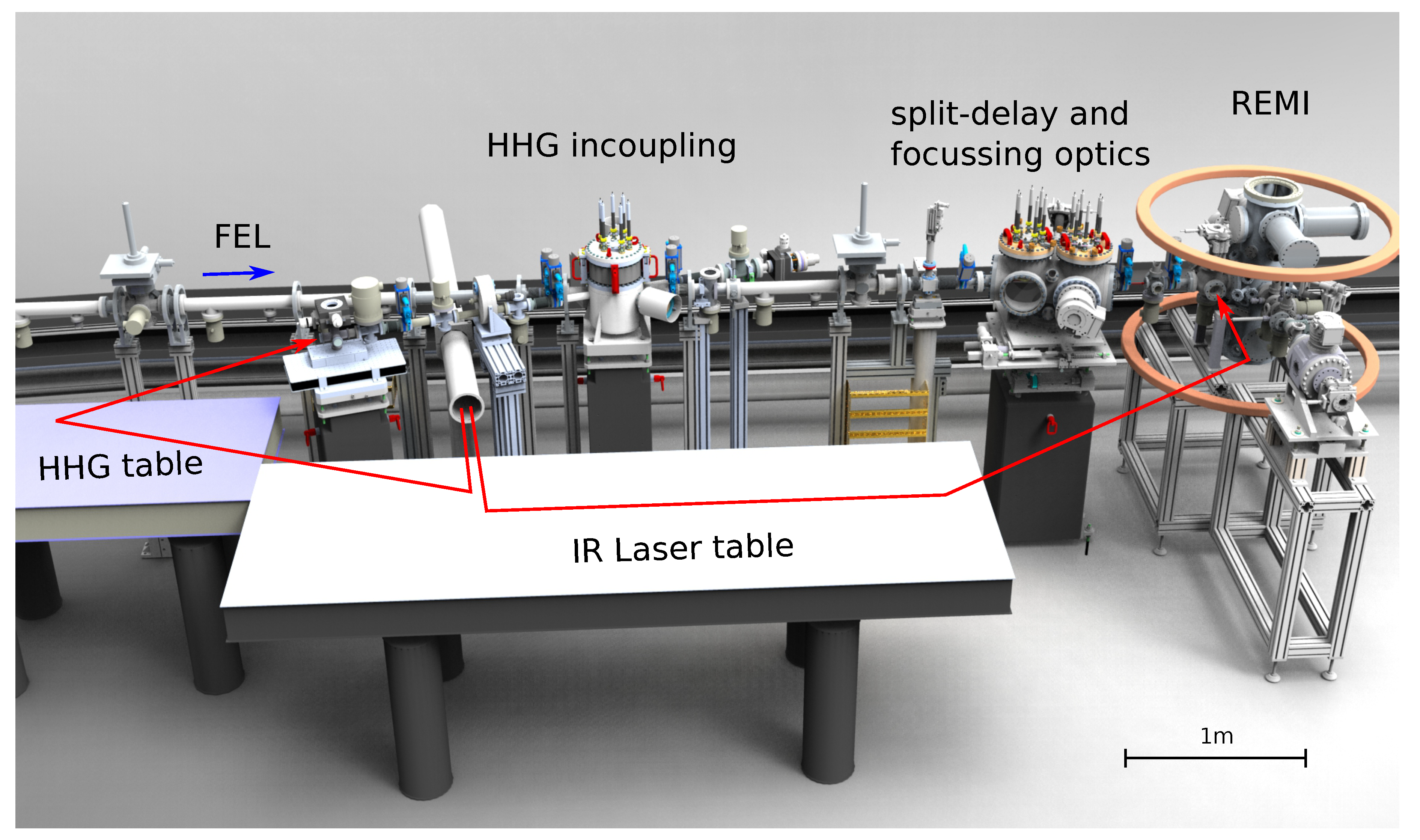
Abstract
1. Introduction
2. Experimental Setup
2.1. The Free-Electron Laser FLASH2
2.2. IR Laser
2.3. HHG Radiation
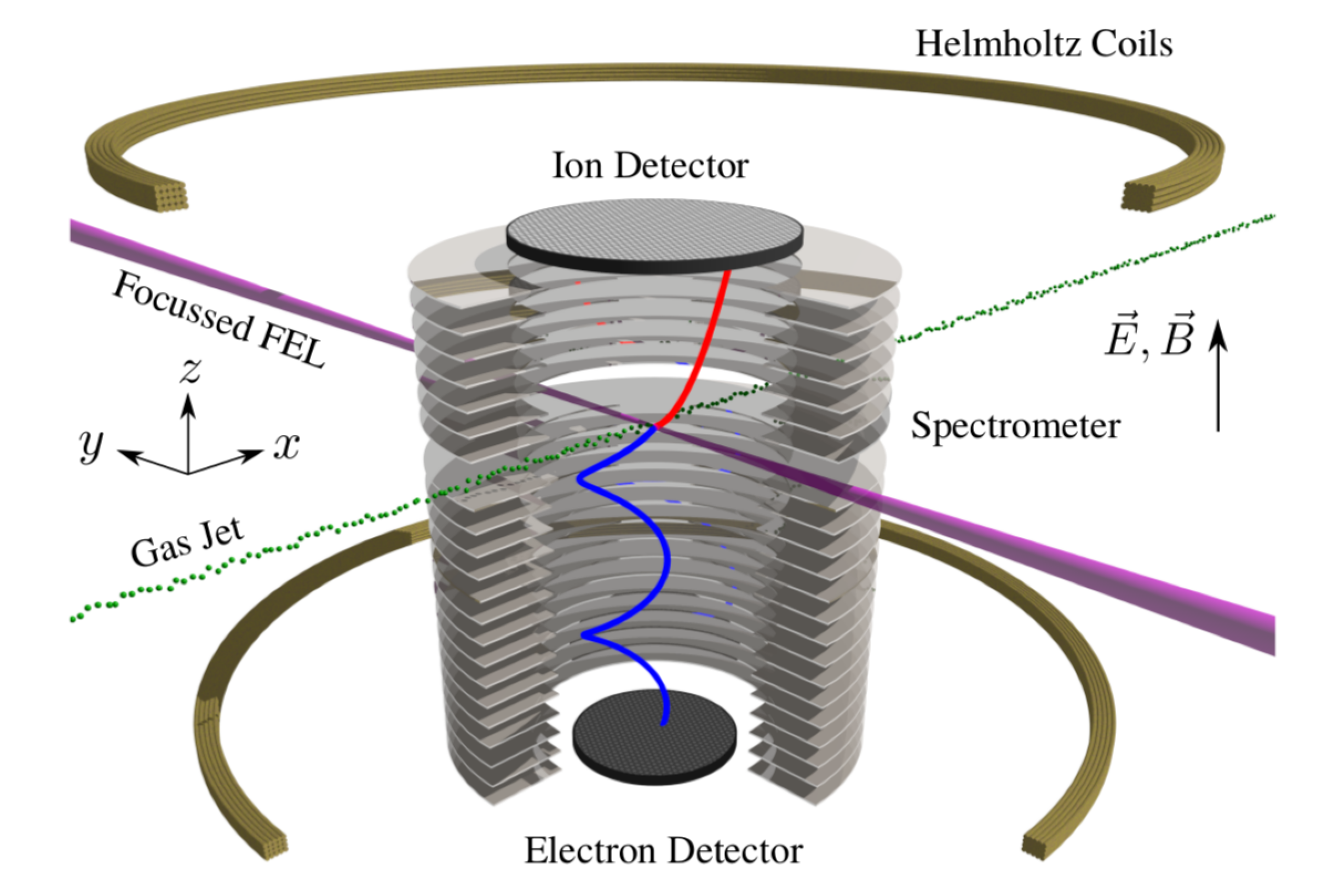
2.4. Reaction Microscope
3. Selected Results
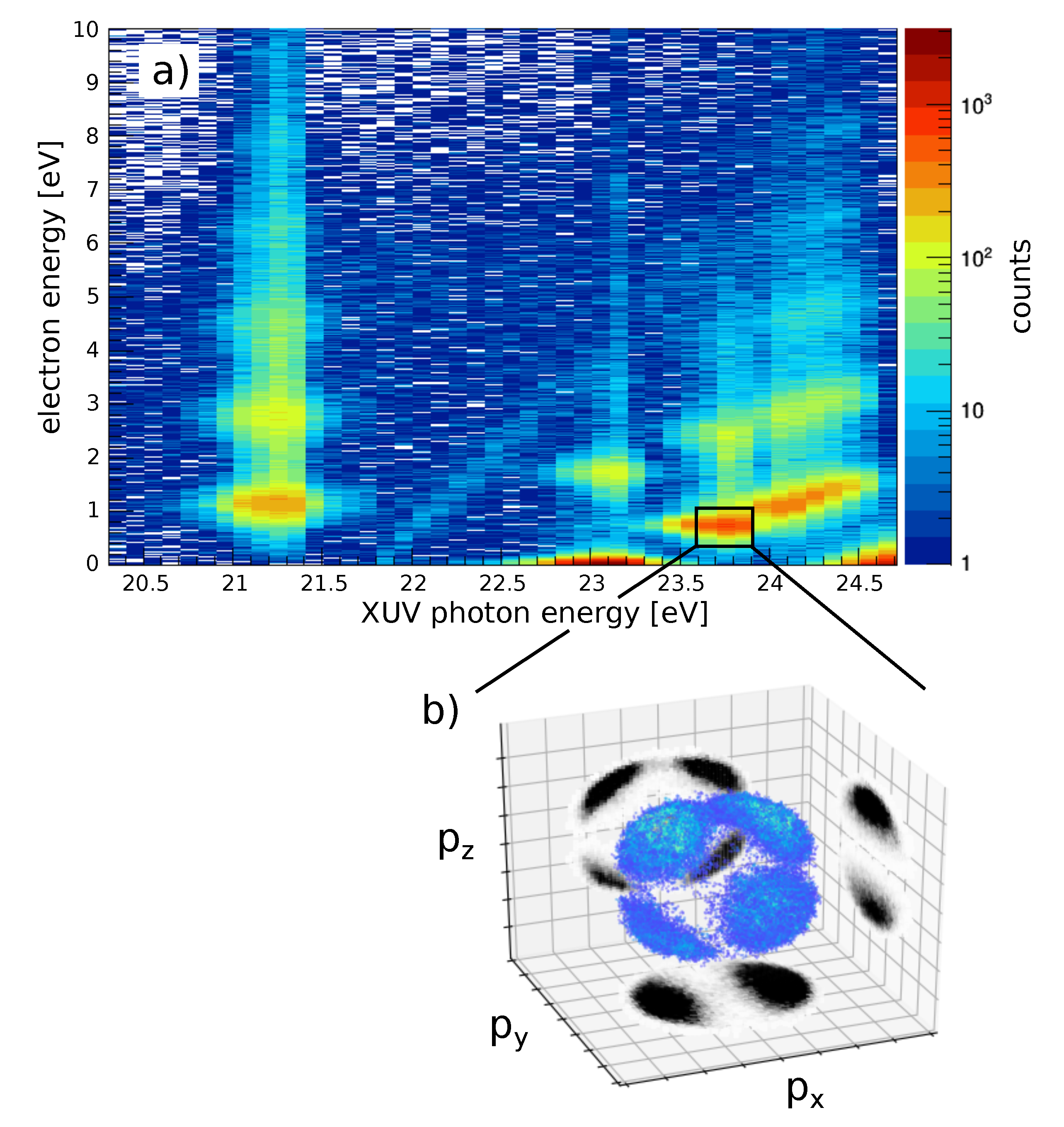
3.1. Angle-Resolved Wavelength Scan: XUV+IR in Helium
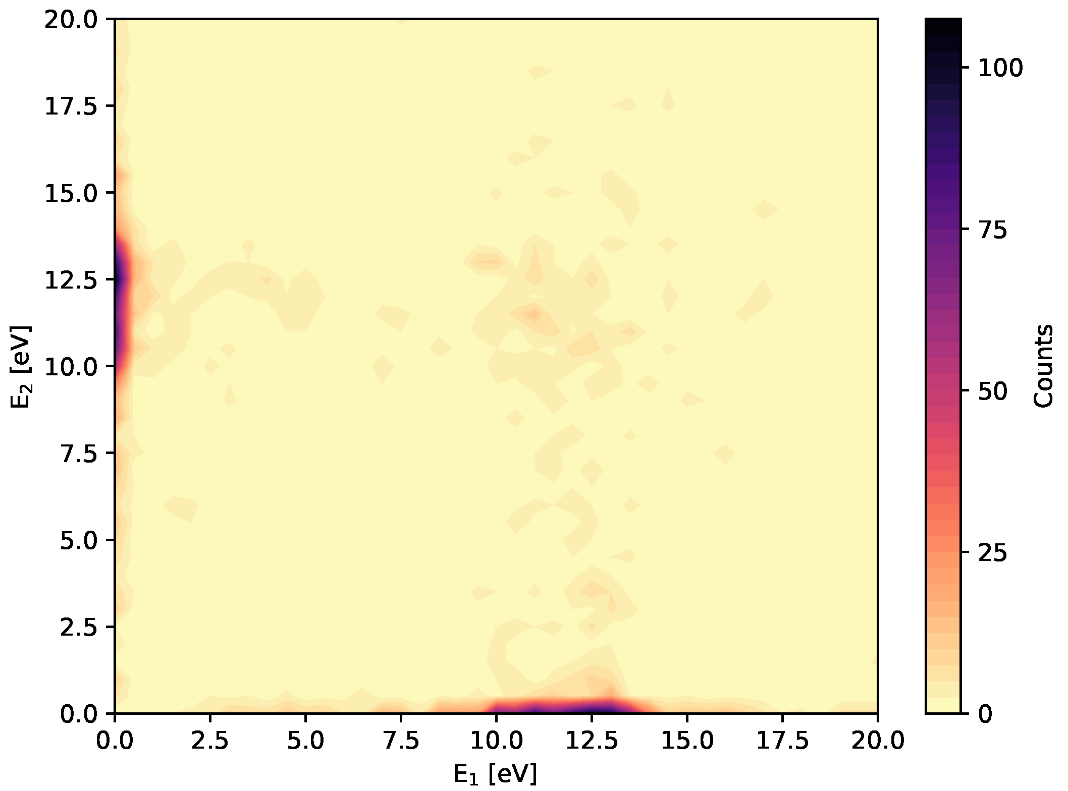
3.2. Two-Photon Double Ionization in Argon
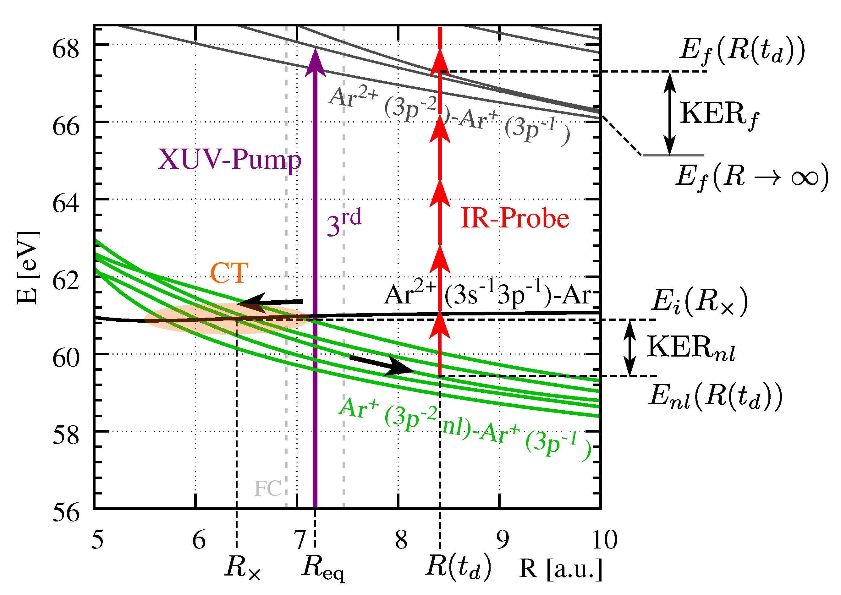
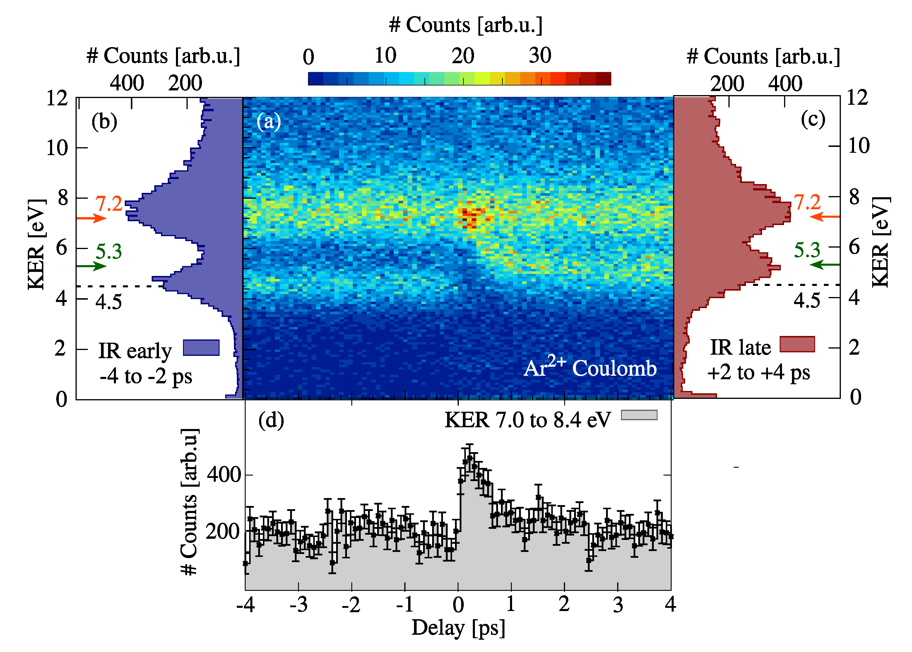
3.3. Molecular Dynamics in Ar Dimers
4. Conclusions
Author Contributions
Funding
Conflicts of Interest
References
- Strickland, D.; Mourou, G. Compression of amplified chirped optical pulses. Opt. Commun. 1985, 55, 447–449. [Google Scholar] [CrossRef]
- Zewail, A.H. Laser Femtochemistry. Science 1988, 242, 1645–1653. [Google Scholar] [CrossRef] [PubMed]
- Ullrich, J.; Rudenko, A.; Moshammer, R. Free-Electron Lasers: New Avenues in Molecular Physics and Photochemistry. Annu. Rev. Phys. Chem. 2012, 63, 635–660. [Google Scholar] [CrossRef] [PubMed]
- Jiang, Y.H.; Senftleben, A.; Kurka, M.; Rudenko, A.; Foucar, L.; Herrwerth, O.; Kling, M.F.; Lezius, M.; Tilborg, J.V.; Belkacem, A.; et al. Ultrafast dynamics in acetylene clocked in a femtosecond XUV stopwatch. J. Phys. B At. Mol. Opt. Phys. 2013, 46, 164027. [Google Scholar] [CrossRef]
- Schmid, G.; Schnorr, K.; Augustin, S.; Meister, S.; Lindenblatt, H.; Trost, F.; Liu, Y.; Miteva, T.; Gisselbrecht, M.; Düsterer, S.; et al. Tracing charge transfer in argon dimers by XUV-pump IR-probe experiments at FLASH. J. Chem. Phys. 2019, 151, 084314. [Google Scholar] [CrossRef]
- Schnorr, K.; Senftleben, A.; Kurka, M.; Rudenko, A.; Schmid, G.; Pfeifer, T.; Meyer, K.; Kübel, M.; Kling, M.F.; Jiang, Y.H.; et al. Electron Rearrangement Dynamics in Dissociating Molecules Accessed by Extreme Ultraviolet Pump-Probe Experiments. Phys. Rev. Lett. 2014, 113, 073001. [Google Scholar] [CrossRef]
- Jiang, Y.H.; Rudenko, A.; Herrwerth, O.; Foucar, L.; Kurka, M.; Kühnel, K.U.; Lezius, M.; Kling, M.F.; van Tilborg, J.; Belkacem, A.; et al. Ultrafast Extreme Ultraviolet Induced Isomerization of Acetylene Cations. Phys. Rev. Lett. 2010, 105, 263002. [Google Scholar] [CrossRef]
- Orzechowski, T.J.; Anderson, B.; Fawley, W.M.; Prosnitz, D.; Scharlemann, E.T.; Yarema, S.; Hopkins, D.; Paul, A.C.; Sessler, A.M.; Wurtele, J. Microwave radiation from a high-gain free-electron laser amplifier. Phys. Rev. Lett. 1985, 54, 889–892. [Google Scholar] [CrossRef]
- Emma, P.; Akre, R.; Arthur, J.; Bionta, R.; Bostedt, C.; Bozek, J.; Brachmann, A.; Bucksbaum, P.; Coffee, R.; Decker, F.J.; et al. First lasing and operation of an ångstrom-wavelength free-electron laser. Nat. Photonics 2010, 4, 641–647. [Google Scholar] [CrossRef]
- Ackermann, W.A.; Asova, G.; Ayvazyan, V. Operation of a free-electron laser from the extreme ultraviolet to the water window. Nat. Photonics 2007, 1, 336–342. [Google Scholar] [CrossRef]
- Brabec, T.; Krausz, F. Intense few-cycle laser fields: Frontiers of nonlinear optics. Rev. Mod. Phys. 2000, 72, 545–591. [Google Scholar] [CrossRef]
- Cerullo, G.; Baltuška, A.; Mücke, O.; Vozzi, C. Few-optical-cycle light pulses with passive carrier-envelope phase stabilization. Laser Photonics Rev. 2011, 5, 323–351. [Google Scholar] [CrossRef]
- Corkum, P.B. Plasma perspective on strong field multiphoton ionization. Phys. Rev. Lett. 1993, 71, 1994–1997. [Google Scholar] [CrossRef] [PubMed]
- Lewenstein, M.; Balcou, P.; Ivanov, M.Y.; L’Huillier, A.; Corkum, P.B. Theory of high-harmonic generation by low-frequency laser fields. Phys. Rev. A 1994, 49, 2117–2132. [Google Scholar] [CrossRef]
- McPherson, A.; Gibson, G.; Jara, H.; Johann, U.; Luk, T.S.; McIntyre, I.A.; Boyer, K.; Rhodes, C.K. Studies of multiphoton production of vacuum-ultraviolet radiation in the rare gases. J. Opt. Soc. Am. B 1987, 4, 595–601. [Google Scholar] [CrossRef]
- Calegari, F.; Sansone, G.; Stagira, S.; Vozzi, C.; Nisoli, M. Advances in attosecond science. J. Phys. B At. Mol. Opt. Phys. 2016, 49, 062001. [Google Scholar] [CrossRef]
- Vozzi, C.; Calegari, F.; Frassetto, F.; Poletto, L.; Sansone, G.; Villoresi, P.; Nisoli, M.; De Silvestri, S.; Stagira, S. Coherent continuum generation above 100 eV driven by an IR parametric source in a two-color scheme. Phys. Rev. A 2009, 79, 033842. [Google Scholar] [CrossRef]
- Boutet, S.; Yabashi, M. X-Ray Free Electron Lasers and Their Applications. In X-Ray Free Electron Lasers: A Revolution in Structural Biology; Springer International Publishing: Cham, Switzerland, 2018; pp. 1–21. [Google Scholar] [CrossRef]
- Schmid, G.; Schnorr, K.; Augustin, S.; Meister, S.; Lindenblatt, H.; Trost, F.; Liu, Y.; Braune, M.; Treusch, R.; Schröter, C.D.; et al. Reaction microscope endstation at FLASH2. J. Synchrotron Radiat. 2019, 26, 854–867. [Google Scholar] [CrossRef]
- Ayvazyan, V.; Baboi, N.; Bähr, J.; Balandin, V.; Beutner, B.; Brandt, A.; Bohnet, I.; Bolzmann, A.; Brinkmann, R.; Brovko, O.I.; et al. First operation of a free-electron laser generating GW power radiation at 32 nm wavelength. Eur. Phys. J. D At. Mol. Opt. Plasma Phys. 2006, 37, 297–303. [Google Scholar] [CrossRef]
- Faatz, B.; Plönjes, E.; Ackermann, S.; Agababyan, A.; Asgekar, V.; Ayvazyan, V.; Baark, S.; Baboi, N.; Balandin, V.; von Bargen, N.; et al. Simultaneous operation of two soft X-ray free-electron lasers driven by one linear accelerator. New J. Phys. 2016, 18, 062002. [Google Scholar] [CrossRef]
- Faatz, B.; Braune, M.; Hensler, O.; Honkavaara, K.; Kammering, R.; Kuhlmann, M.; Ploenjes, E.; Roensch-Schulenburg, J.; Schneidmiller, E.; Schreiber, S.; et al. The FLASH Facility: Advanced Options for FLASH2 and Future Perspectives. Appl. Sci. 2017, 7, 1114. [Google Scholar] [CrossRef]
- Deutsches Elektronen-Synchrotron DESY. FLASH Parameters. Available online: https://photon-science.desy.de/facilities/flash/flash_parameters/index_eng.html (accessed on 9 March 2020).
- Lang, T.; Alisauskas, S.; Große-Wortmann, U.; Hülsenbusch, T.; Manschwetus, B.; Mohr, C.; Müller, J.; Peters, F.; Schirmel, N.; Schulz, S.; et al. Versatile OPCPA Pump-Probe Laser System for the FLASH2 XUV FEL Beamline at DESY. In Proceedings of the 2019 Conference on Lasers and Electro-Optics Europe European Quantum Electronics Conference (CLEO/Europe-EQEC), Munich, Germany, 23–27 June 2019; p. 1. [Google Scholar] [CrossRef]
- Appi, E.; Papadopoulou, C.; Mapa, J.; Wesavkar, N.; Jusko, C.; Mosel, P.; Alisauskas, S.; Lang, T.; Heyl, C.; Manschwetus, B.; et al. A synchronized VUV light source based on high-order harmonic generation at FLASH. Sci. Rep. 2020, 10, 6867. [Google Scholar] [CrossRef]
- Heyl, C.M.; Arnold, C.L.; Couairon, A.; L’Huillier, A. Introduction to macroscopic power scaling principles for high-order harmonic generation. J. Phys. B At. Mol. Opt. Phys. 2016, 50, 013001. [Google Scholar] [CrossRef]
- Ullrich, J.; Moshammer, R.; Dorn, A.; Dörner, R.; Schmidt, L.P.H.; Schmidt-Böcking, H. Recoil-ion and electron momentum spectroscopy: Reaction-microscopes. Rep. Prog. Phys. 2003, 66, 1463–1545. [Google Scholar] [CrossRef]
- Jagutzki, O.; Dangendorf, V.; Lauck, R.; Czasch, A.; Milnes, J. A position- and time-sensitive photon-counting detector with delay- line read-out. Opt. Sens. Technol. Appl. 2007. [Google Scholar] [CrossRef]
- Kurka, M.; Rudenko, A.; Foucar, L.; Kühnel, K.U.; Jiang, Y.H.; Ergler, T.; Havermeier, T.; Smolarski, M.; Schössler, S.; Cole, K.; et al. Two-photon double ionization of Ne by free-electron laser radiation: A kinematically complete experiment. J. Phys. B At. Mol. Opt. Phys. 2009, 42, 141002. [Google Scholar] [CrossRef]
- Augustin, S.; Schulz, M.; Schmid, G.; Schnorr, K.; Gryzlova, E.V.; Lindenblatt, H.; Meister, S.; Liu, Y.F.; Trost, F.; Fechner, L.; et al. Signatures of autoionization in the angular electron distribution in two-photon double ionization of Ar. Phys. Rev. A 2018, 98, 033408. [Google Scholar] [CrossRef]
- National Institute of Standards and Technology (NIST). NIST Atomic Spectra Database Levels Data. Available online: https://physics.nist.gov/cgi-bin/ASD/energy1.pl?encodedlist=XXT2&de=0&spectrum=He+I&submit=Retrieve+Data&units=1&format=0&output=0&page_size=15&multiplet_ordered=0&conf_out=on&term_out=on&level_out=on&unc_out=1&j_out=on&lande_out=on&perc_out=on&biblio=on&temp= (accessed on 16 April 2020).
- Agostini, P.; Fabre, F.; Mainfray, G.; Petite, G.; Rahman, N.K. Free-Free Transitions Following Six-Photon Ionization of Xenon Atoms. Phys. Rev. Lett. 1979, 42, 1127. [Google Scholar] [CrossRef]
- Corkum, P.B.; Burnett, N.H.; Brunel, F. Above-threshold ionization in the long-wavelength limit. Phys. Rev. Lett. 1989, 62, 1259–1262. [Google Scholar] [CrossRef]
- Kerbstadt, S.; Pengel, D.; Johannmeyer, D.; Englert, L.; Bayer, T.; Wollenhaupt, M. Control of photoelectron momentum distributions by bichromatic polarization-shaped laser fields. New J. Phys. 2017, 19, 103017. [Google Scholar] [CrossRef]
- Fechner, L.; Camus, N.; Ullrich, J.; Pfeifer, T.; Moshammer, R. Strong-Field Tunneling from a Coherent Superposition of Electronic States. Phys. Rev. Lett. 2014, 112, 213001. [Google Scholar] [CrossRef]
- Fechner, L.; Camus, N.; Krupp, A.; Ullrich, J.; Pfeifer, T.; Moshammer, R. Creation and survival of autoionizing states in strong laser fields. Phys. Rev. A 2015, 92, 051403. [Google Scholar] [CrossRef]
- Busto, D.; Vinbladh, J.; Zhong, S.; Isinger, M.; Nandi, S.; Maclot, S.; Johnsson, P.; Gisselbrecht, M.; L’Huillier, A.; Lindroth, E.; et al. Fano’s Propensity Rule in Angle-Resolved Attosecond Pump-Probe Photoionization. Phys. Rev. Lett. 2019, 123, 133201. [Google Scholar] [CrossRef] [PubMed]
- Braune, M.; Hartmann, G.; Ilchen, M.; Knie, A.; Lischke, T.; Reinköster, A.; Meissner, A.; Deinert, S.; Glaser, L.; Al-Dossary, O.; et al. Electron angular distributions of noble gases in sequential two-photon double ionization. J. Mod. Opt. 2016, 63, 324–333. [Google Scholar] [CrossRef]
- Grum-Grzhimailo, A.N.; Gryzlova, E.V.; Strakhova, S.I.; Kabachnik, N.M.; Fritzsche, S. Angular distributions and correlations in sequential two-photon atomic double ionization. J. Phys. Conf. Ser. 2009, 194, 012004. [Google Scholar] [CrossRef]
- Redlin, H.; Al-Shemmary, A.; Azima, A.; Stojanovic, N.; Tavella, F.; Will, I.; Düsterer, S. The FLASH pump–probe laser system: Setup, characterization and optical beamlines. Nucl. Instrum. Methods Phys. Res. Sect. A Accel. Spectrom. Detect. Assoc. Equip. 2011, 635, S88–S93. [Google Scholar] [CrossRef]


© 2020 by the authors. Licensee MDPI, Basel, Switzerland. This article is an open access article distributed under the terms and conditions of the Creative Commons Attribution (CC BY) license (http://creativecommons.org/licenses/by/4.0/).
Share and Cite
Meister, S.; Lindenblatt, H.; Trost, F.; Schnorr, K.; Augustin, S.; Braune, M.; Treusch, R.; Pfeifer, T.; Moshammer, R. Atomic, Molecular and Cluster Science with the Reaction Microscope Endstation at FLASH2. Appl. Sci. 2020, 10, 2953. https://doi.org/10.3390/app10082953
Meister S, Lindenblatt H, Trost F, Schnorr K, Augustin S, Braune M, Treusch R, Pfeifer T, Moshammer R. Atomic, Molecular and Cluster Science with the Reaction Microscope Endstation at FLASH2. Applied Sciences. 2020; 10(8):2953. https://doi.org/10.3390/app10082953
Chicago/Turabian StyleMeister, Severin, Hannes Lindenblatt, Florian Trost, Kirsten Schnorr, Sven Augustin, Markus Braune, Rolf Treusch, Thomas Pfeifer, and Robert Moshammer. 2020. "Atomic, Molecular and Cluster Science with the Reaction Microscope Endstation at FLASH2" Applied Sciences 10, no. 8: 2953. https://doi.org/10.3390/app10082953
APA StyleMeister, S., Lindenblatt, H., Trost, F., Schnorr, K., Augustin, S., Braune, M., Treusch, R., Pfeifer, T., & Moshammer, R. (2020). Atomic, Molecular and Cluster Science with the Reaction Microscope Endstation at FLASH2. Applied Sciences, 10(8), 2953. https://doi.org/10.3390/app10082953