Review

Challenges for the Implantation of Symbiotic Nanostructured Medical Devices

Univ. Grenoble Alpes, CNRS, Grenoble INP, TIMC-IMAG/SyNaBi (UMR 5525), 38000 Grenoble, France
*
Author to whom correspondence should be addressed.
Appl. Sci. 2020, 10(8), 2923; https://doi.org/10.3390/app10082923
Submission received: 26 March 2020 / Revised: 15 April 2020 / Accepted: 21 April 2020 / Published: 23 April 2020
(This article belongs to the Special Issue Advances in Translational Nanotechnology)

Abstract

:
We discuss the perspectives of designing implantable medical devices that have the criterion of being symbiotic. Our starting point was whether the implanted device is intended to have any two-way (“duplex”) communication of energy or materials with the body. Such duplex communication extends the existing concepts of a biomaterial and biocompatibility to include the notion that it is important to consider the intended functional use of the implanted medical device. This demands a biomimetic approach to design functional symbiotic implantable medical devices that can be more efficient in mimicking what is happening at the molecular and cellular levels to create stable interfaces that allow for the unfettered exchanges of molecules between an implanted device and a body. Such a duplex level of communication is considered to be a necessary characteristic of symbiotic implanted medical devices that are designed to function for long periods of time inside the body to restore and assist the function of the body. We illustrate these perspectives with experience gained from implanting functional enzymatic biofuel cells.

1. Introduction

The foundation to harness nanotechnology for the engineering of biomedical systems was pioneered by Richard Zsigmondy in 1903, when he first used the term “nanometer” in the characterization of the gold nanoparticles he made and then measured with his ultramicroscope [1]. Subsequently, the introduction of the concept of “biomimetics” by Otto Schmitt in the 1960s has stimulated a great many possible applications for the engineering of biomedical devices [2]. Though nanotechnology was not explicitly mentioned in the text of the famous lecture by Richard Feynman, he described a future where small-scale machines might be constructed, controlled, and manipulated. He highlighted the medical relevance for these types of machines by quoting his friend, Albert R. Hibbs, who suggested the concepts of a miniaturized surgical device and “small machines that might be permanently incorporated in the body to assist some inadequately-functioning organ” [3]. All of those previous concepts introduced by Zsigmondy, Schmitt, Hibbs, and Feynman have provided pillars upon which we can advance our ideas about nanostructured medical devices for implantation.
In this short review, we introduce the concept of “symbiotic nanostructured medical devices,” since our starting point was whether the implanted device is intended to have any two-way communication of energy or materials with the body. If the intended application is only one-way communication from the medical device to the body, such as for the delivery of a drug, the challenge is to avoid the degradation or encapsulation of the medical device. However, if the implanted medical device is intended to properly integrate with the body, then it needs to mimic the two-way (duplex) communication that is required for transplanted living organs or cells [4]. Such an implanted medical device is one that we consider symbiotic. Examples of such implantable duplex communicating systems include biofuel cells or open-loop feedback devices, where a molecule from the body is utilized by the duplex communicating system to produce a different material (e.g., molecule or energy) for use by the body (Figure 1). There is clearly a major technical component in engineering such duplex communicating systems to provide the appropriate performance and lifetime when implanted inside the body. Nonetheless, those functions of such duplex systems depend on maintaining the unfettered two-way transport communication of materials when the system is implanted in the body or, indeed, if the device is externally positioned to maintain tight contact with the body. Maintaining such an unfettered two-way transport in order to design long-term functional performance requires overcoming the challenges of biocompatibility. We discuss the relative significance of those two challenges from the perspective of an implanted enzymatic biofuel cell. We have several years of expertise in enhancing its performance inside the body of mammals for this duplex symbiotic system.

2. Is it Sufficient to Rely Only on Technological Advances?

Here, we consider the example of an enzymatic biofuel cell as a symbiotic implanted medical device. The present challenges in the field of implanted enzymatic biofuel cells are to improve the performance and life-time of the biofuel cells operating inside the body. The energy density can be increased with the utilization of enzyme cascades to ensure the complete oxidation of fuels [5]. In addition to the challenge of increasing the energy density in enzymatic biofuel cells, Xiao et al. [6] summarized the other main technological challenges to be overcome in order to improve the performance of the enzymatic fuel cells as the needs to increase power density, operational stability, and voltage output. To increase power density, Xiao et al. [6] proposed to increase enzyme activities, to facilitate electron transfers, to use nanomaterials, to design more efficient enzyme–electrode interfaces, and to combine enzymatic fuel cells with (super) capacitors. To improve stability, they proposed the use of different enzyme immobilization approaches, the tuning of enzyme properties, the designing of protective matrixes, and the use of microbial surface displaying enzymes. To improve voltage output, they proposed optimizing mediators, serial connection, and external boost converter electronic circuits [6]. However, the implantation of devices brings the dimension of biocompatibility as perhaps the key challenge that needs to be overcome to increase the life-time of implanted enzymatic biofuel cells. Our experience from implanting enzymatic biofuel cells in mammals has shown that producing a biocompatible system is the major issue to solve for enhancing the performance and life-time of implanted enzymatic biofuel cells [7].

3. Biocompatibility is the Major Challenge for Symbiotic Implanted Medical Devices

Indeed, the issues of biocompatibility have remained largely unsolved since scientists such as B. Ratner, R. Langer, J.M. Anderson, and D. Williams pioneered the fields of modern biomaterials and biocompatibility issues [8,9,10,11,12,13,14]. Current thinking about biocompatibility pathways for a biomaterial have included the ways in which the components of the biomaterial actually interact with cells of the body [15]. Williams (2014) identified those components acting on the cell as having an origin from the biomaterial (e.g., metal ions, polymer additives, nanoparticles, and contaminants), mechanical and biophysical mediators of interactions (e.g., electromagnetic fields and structural/fluid stresses), internalization mechanisms (e.g., phagocytosis, endocytosis, and pinocytosis), or material mediators of interactions (chemical structure, elasticity, shape, volume, and topography). However, those mediating components were considered as originating from, or derived from, the biomaterial. Nonetheless, such a concept of biocompatibility subsumes a collection of individual phenomena that make it impossible to consider that biocompatibility can be graduated into some form of a scale. Furthermore, even by referring to a “biocompatible system” rather than a “biocompatible material,” Williams concluded that the term “biocompatible system” remained imprecise and potentially misleading [16].
We suggest that such imprecision can be avoided if biocompatibility is defined by the intended functional properties of the implanted device. Such a definition implies that the implanted device would need the characteristics of duplex communication with the body in order for it to be a symbiotic device and, hence, biocompatible. The value of re-defining the framework of a “biocompatible system” is in considering the next generation of such symbiotic devices where long-term function is the focus of the implanted system. An example of such a symbiotic device is a biofuel cell that can also produce potentially irritant chemicals from its catalytic reactions. The enzymatic biofuel cell needs to be designed to include a stable and permeable interface with the body. Nonetheless, an implanted enzymatic biofuel cell will inevitably be enclosed by a fibrous capsule that follows the usual foreign-body reaction (FBR), as is the case for all non-biodegradable implanted devices [17]. The formation of a fibrous capsule could be initiated by adsorption of a cluster of inflammatory proteins [18]. However, that hypothesis does not completely explain the differences in biocompatibility amongst all types of biomaterials. Indeed, the same material can initiate an in vivo reaction that may not only include the encapsulation that follows the FBR [17]. Ratner’s group found that an implanted non-porous slab of poly(hydroxyethylmethacrylate) hydrogel heals via the prototypical FBR. In comparison, an implanted 34-µm porous poly(hydroxyethylmethacrylate) hydrogel has the additional feature that the pores are infiltrated with a mostly cellular and slightly fibrous tissue substance, but the implant is surrounded by a fibrous capsule, albeit one that is thinner and less dense than the capsule surrounding the non-porous implant. For larger pores of 160-µm, there was a greater fraction of fibrous tissue inside the pores [19]. Those observations provide a snapshot of the FBR, since the different hydrogel samples were implanted for only three weeks. Nonetheless, those results raised several interesting directions for finding a solution to minimize and/or eliminate the fibrous encapsulation of the enzymatic biofuel cell in order to maintain a stable and permeable interface for the exchange of molecules of fuel.
The consideration of such directions is also important for the more general issue of implanting a symbiotic device where there is the need to maintain a stable and porous interface between the device and the body. Ratner did not use the term symbiotic, but he previewed the need to develop biomaterials that heal and integrate by minimizing the FBR capsule. Minimizing such FBR encapsulation would lead to better implant electrodes, longer-lasting implanted biosensors, consistent release for implanted drug delivery systems, improved vascular prostheses, capsular contraction-free breast implants, hydrocephalous shunts and glaucoma drainage shunts that do not fail fibrotically, and many other advances for the millions of devices implanted in humans each year [17].
We can also gain further insights into the search for solutions to solve such biocompatibility issues for symbiotic devices by referring to discussions about the challenges for implantation strategies to treat diabetes [20]. The challenges in that field are to implant cells, tissues, or whole organs for the long-term control of glucose–insulin metabolism, with the major biocompatibility challenge being to protect the implants from the immune system. For that, potent immunosuppressing drugs are used for either solid organ pancreas transplantation or pancreatic islet transplantation. Though immunosuppression protects the grafts from the body’s immune system, the major side-effect problem is the vulnerability to infections and malignancies [20]. Sneddon et al. described the approach of using a semipermeable membrane/capsule to protect transplanted beta cells from the patient’s immune system while allowing for sufficient mass transfer to maintain cell viability and ensuring favorable insulin secretion kinetics. They also raised the major challenges for the successful design of next-generation cell encapsulation devices as being the provisions of effective immunoprotection and of sufficient mass transfer between the body environment and the encased islets. An interesting way to meet those challenges has been to use a polymer-coated silicon membrane that includes nanopores formed by microelectromechanical systems (MEMS) technology [21].
For several types of implanted devices, the largely unsolved requirement to maintain the transfer of materials with the body leads to the more general challenge that the practical frontier for the biocompatibility of a symbiotic implanted device continues to be the design of the membrane that is at the interface of the implanted device and the body. Such a membrane needs to maintain an unfettered exchange of materials to enhance the lifetime and performance of the implanted symbiotic device [4].

4. Choices of Membranes for Symbiotic Implanted Medical Devices

We illustrate this challenge by returning to the problem of an enzymatic biofuel cell. The membrane needs to have appropriate mechanical properties, have selective permeability to biological “fuel” molecules, and be easily constructed. Those requirements imply the usage of some form of a gel-like polymer. Recent examples include a composite material that comprised PDMS (polydimethylsiloxane) functionalized with a stable hydrogel layer of MPC (2-methacryolyl phosphorylcholine). This composite gel can potentially improve the long-term biocompatibility of a medical device, since the composite material is nontoxic to mammalian cells and strongly resists macrophage attachment and bacterial colonization [22,23]. Additionally, a porous chitosan membrane coating, in combination with either PTFE (polytetrafluoroethylene) or Nafion (perfluorosulfonic acid) supporting polymers, was subcutaneously implanted in rats, and, after 100 days, it did not enhance the deposition of collagen but did promote angiogenesis [24]. In that context of utilizing an appropriate polymer membrane, we successfully implanted enzymatic biofuel cells and bioelectrodes that were enclosed using a chitosan membrane [25,26,27]. An important consideration for the design of the chitosan membrane is the environment of the bioelectrode. For example, an anode constructed using glucose oxidase releases gluconic acid, which induces a decrease in pH that can hydrolyze the chitosan. The fabrication of the chitosan membrane using cross-linking agents can help in stabilizing the membrane. Such a chitosan membrane allowed for the optimization of the bioelectrode coating. The thinness (10–15 μm) and the hydrophilicity of the chitosan membrane allowed for the rapid wetting of the bioelectrode. This rapid wetting effectively resolved the hydration problems of bioelectrodes that were previously constructed using a cellulose acetate membrane [28,29]. Though successful for implantations up to six months, those chitosan membranes may not provide the perfect solution for the challenge of biocompatibility.
Nonetheless, if not perfectly suitable as a protective membrane for an implanted enzymatic biofuel cell, our recent results showed that chitosan provides an open structure that allows for the ingrowth and integration of cells. Cells were able to migrate into the chitosan structure within three days in culture, and they were able to adhere to the gel structure of the chitosan. These are properties of chitosan that can be exploited for future enzymatic biofuel cell systems that require integration and biocompatibility with tissues of the body. Indeed, a previous report indicated that treating the chitosan with cold atmospheric plasma could enhance the infiltration and ingrowth of mesenchymal stem cells for application in bone regeneration [30]. Such an exploitation of the capability of chitosan to form an open architecture to provide conditions for the infiltration and ingrowth of cells is a novel application compared to the usage of dense forms of chitosan as bioabsorbable implants for orthopedic applications [31].
We can further consider several directions in which the membrane can be improved in order to increase the lifetime, again using the example of improving the performance of the implanted enzymatic biofuel cell. The key direction is to fabricate a membrane that should remain stable when implanted for long periods. It is important to consider that even cross-linked chitosan is not the basis for a perfectly stable membrane when implanted, because 100% crosslinking cannot be guaranteed over the entirety of the membrane surface. For example, we observed that a biocathode can be implanted for up to six months in a rat. However, even if the chitosan membrane looks intact macroscopically and since it is difficult to ensure that there is 100% crosslinking in all regions, it is not possible to avoid the degradability 100% of the time for such long period of implantation [32].
Though not directly for enzymatic biofuel cells and only for short-term in vivo usage, combining chitosan with alginate to produce an outer protective membrane was shown to improve the response of a glucose biosensor used for short-term in vivo sensing in a mouse [33]. An important consideration is the time-course for implantation experiments to test the biocompatibility of membranes. In many reports, the duration of implantation of membrane materials has been too short to evaluate the long-term application for designing an implanted biofuel cell. For example, a macromolecule cross-linked biocomposite scaffold composed of hydroxyapatite, alginate, chitosan, and fucoidan was only assessed for a maximum of eight weeks when implanted in a rat [34]. Another potentially interesting membrane that was formed from alkyl cross-linking of chitosan was only tested for seven weeks implanted in rats [35].
An alternative is to use synthetic materials in designing a biocompatible system. Letourneur’s team built vascular grafts made in polyvinyl alcohol (PVA) [36]. They showed that a cross-linked PVA combined with gelatin slowed down platelet adhesion phenomena and improved the endothelialization of a vascular prosthesis [37]. Inspired by Letourneur’s results, we designed hydrogels of PVA formed by a freeze/thaw method [38], with the PVA membranes tuned to the needs of the enzymatic biofuel cell. Indeed, the semi-crystalline structure of PVA allowed for the modification of the porosity and mechanical properties by varying the parameters of the production process. We tuned the PVA membranes to the needs of the enzymatic biofuel cell, which relied upon duplex communication for its function. We analyzed the performance of PVA to provide an optimized coating that provided (i) a solid and easily handled membrane, (ii) a porous membrane optimized for the diffusion of glucose and oxygen to the bioelectrodes, and (iii) a protective membrane against proteins of larger dimensions than the glucose (that was required to permeate the PVA) [38].
The PVA membrane has advantages in creating a biocompatible system and in retaining the permeability to glucose. Our freeze/thaw protocol produced a PVA membrane that had a stable permeability to glucose and that was similar to chemically cross-linked PVA [39]. As previously described above, there is also an advantage in incorporating chitosan in the bioelectrodes for enzymatic biofuel cells, since the open architecture assists in immobilizing the enzymes [25,27], and its three-dimensional structure encourages the ingrowth of cells. Indeed, the use of multiple cross-linking agents with combined collagen/chitosan gels [40] could be useful in combination with protective PVA membranes for fabricating stable implanted enzymatic biofuel cells.

5. Perspectives

For the appropriate long-term stable function of implanted symbiotic medical devices, it is essential to develop a system that includes a membrane interface that maintains unfettered duplex communication of materials with the body; this remains a major challenge for the field of implantable enzymatic biofuel cells. Clearly, it is necessary to consider the technical electrochemical aspects to improve the energy density, power density, and voltage output of the enzymatic biofuel cell. However, the technical aspects of operational stability must be considered in parallel with developing the enzymatic biofuel cell as a biocompatible system for implantation. Such a biocompatible system for stable long-term function requires the combination of a 3D structured bioelectrode with a stable porous protective membrane. The most promising directions are for the integration of 3D bioelectrodes that utilize some form of cross-linked chitosan to provide the optimal environment for the stabilization of enzymes combined with a protective membrane, such as those formed with PVA as the basis. This type of approach will most likely optimize the design of gel-like polymer membranes to minimize the fibrous encapsulation of implanted enzymatic biofuel cells [41]. Such an approach could include the possibilities of either excluding biofouling with the PVA-based membrane or promoting cell adhesion and hence integration with the body, e.g., by using modified chitosan or polyelectrolyte coatings [42,43]. Indeed, even materials like porous silicon could contribute to biocompatible systems after the porous silicon is stabilized to avoid degradation in the body [44].
This practical experience of designing enzymatic biofuel cells for long-term and stable performance when implanted provides directions to the general challenge of biocompatibility for symbiotic implanted devices. Such symbiotic systems extend the classical biocompatibility concept to include this functional requirement for the continuous two-way (duplex) communication of materials with the body. It is probable that utilizing such a biomimetic approach could enable scientists to be more efficient in mimicking what is happening at the molecular and cellular levels to create a porous membrane that allows for the long-term exchanges of molecules between an implanted device and the body (see Figure 1). Having this concept in mind will guide the research in a new field between medical implant and regenerative medicine to create functional symbiotic devices for long periods of implantation.

Author Contributions

All authors contributed information that guided the perspectives discussed in this article. The initial draft was written by D.K.M. All authors contributed to the editing of revised drafts. All authors have read and agreed to the published version of the manuscript.

Funding

This work was supported financially by grants from the French Agence Nationale de la Recherche (ANR-15-CE05-0003-01, and ANR-16-CE19-0007-03) and the French Investissements d’Avenir (ANR-10-NANO-03-01).

Conflicts of Interest

The authors declare no conflict of interest.

References

  1. Zsigmondy, R.A. Properties of Colloids. In Nobel Lectures, Chemistry 1922–1941; Elsevier: Amsterdam, The Netherlands, 1966. [Google Scholar]
  2. Schmitt, O.H. Some interesting and useful biomimetic transforms. In Proceedings of the 3rd Inter Biophysics Congress, Boston, MA, USA, 29 August–3 September 1969. [Google Scholar]
  3. Feynman, R.P. There’s plenty of room at the bottom. Calif. Inst. Technol. Eng. Sci. 1960, 23, 22–36. [Google Scholar]
  4. Alcaraz, J.P.; Cinquin, P.; Martin, D.K. Tackling the concept of symbiotic implantable medical devices with nanobiotechnologies. Biotechnol. J. 2018, 13, 1800102. [Google Scholar] [CrossRef]
  5. Sokic-Lazic, D.; De Andrade, A.R.; Minteer, S.D. Utilization of enzyme cascades for complete oxidation of lactate in an enzymatic biofuel cell. Electrochim. Acta 2011, 56, 10772–10775. [Google Scholar] [CrossRef]
  6. Xiao, X.; Xia, H.Q.; Wu, R.; Bai, L.; Yan, L.; Magner, E.; Cosnier, S.; Lojou, E.; Zhu, Z.; Liu, A. Tackling the challenges of enzymatic (bio)fuel cells. Chem. Rev. 2019, 119, 9509–9558. [Google Scholar] [CrossRef]
  7. Zebda, A.; Alcaraz, J.P.; Vadgama, P.; Shleev, S.; Minteer, S.D.; Boucher, F.; Cinquin, P.; Martin, D.K. Challenges for successful implantation of biofuel cells. Bioelectrochemistry 2018, 124, 57–72. [Google Scholar] [CrossRef]
  8. Langer, R.; Folkman, J. Polymers for the sustained release of proteins and other macromolecules. Nature 1976, 263, 797–800. [Google Scholar] [CrossRef]
  9. Langer, R.; Brem, H.; Tapper, D. Biocompatibility of polymeric delivery systems for macromolecules. J. Biomed. Mater. Res. 1981, 15, 267–277. [Google Scholar] [CrossRef] [PubMed]
  10. Ratner, B.D.; Horbett, T.; Hoffman, A.S.; Hauschka, S.D. Cell adhesion to polymeric materials: Implications with respect to biocompatibility. J. Biomed. Mater. Res. 1975, 9, 407–422. [Google Scholar] [CrossRef] [PubMed]
  11. Ratner, B.D.; Weathersby, P.K.; Hoffman, A.S.; Kelly, M.A.; Scharpen, L.H. Radiation-grafted hydrogels for biomaterial applications as studied by the ESCA technique. J. Appl. Polym. Sci. 1978, 22, 643–664. [Google Scholar] [CrossRef]
  12. Williams, D.F. Biomaterials and biocompatibility. Med. Prog. Technol. 1976, 4, 31–42. [Google Scholar]
  13. Anderson, J.M.; Hiltner, A.; Schodt, K.; Woods, R. Biopolymers as biomaterials: Mechanical properties of gamma-benzyl-L-glutamate-L-leucine copolymers. J. Biomed. Mater. Res. 1972, 6, 25–35. [Google Scholar] [CrossRef] [PubMed]
  14. Anderson, J.M.; Gibbons, D.F.; Martin, R.L.; Hiltner, A.; Woods, R. The potential for poly-α-amino acids as biomaterials. J. Biomed. Mater. Res. 1974, 8, 197–207. [Google Scholar] [CrossRef] [PubMed]
  15. Williams, D.F. Biocompatibility pathways: Biomaterials-induced sterile inflammation, mechanotransduction, and principles of biocompatibility control. ACS Biomater. Sci. Eng. 2017, 3, 2–35. [Google Scholar] [CrossRef]
  16. Williams, D.F. Phase changes in biomaterials. Biomaterials 2014, 35, 10007–10008. [Google Scholar] [CrossRef]
  17. Ratner, B.D. Biomaterials: Been there, done that, and evolving into the future. Annu. Rev. Biomed. Eng. 2019, 21, 171–191. [Google Scholar] [CrossRef]
  18. Romero-Gavilan, F.; Sánchez-Pérez, A.M.; Araújo-Gomes, N.; Azkargorta, M.; Iloro, I.; Elortza, F.; Gurruchaga, M.; Goni, I.; Suay, J. Proteomic analysis of silica hybrid sol-gel coatings: A potential tool for predicting the biocompatibility of implants in vivo. Biofouling 2017, 33, 676–689. [Google Scholar] [CrossRef]
  19. Sussman, E.M.; Halpin, M.C.; Muster, J.; Moon, R.T.; Ratner, B.D. Porous implants modulate healing and induce shifts in local macrophage polarization in the Foreign Body Reaction. Ann. Biomed. Eng. 2014, 42, 1508–1516. [Google Scholar] [CrossRef]
  20. Sneddon, J.B.; Tang, Q.; Stock, P.; Bluestone, J.A.; Roy, S.; Desai, T.; Hebrok, M. Stem cell therapies for treating diabetes: Progress and remaining challenges. Cell Stem Cell 2018, 22, 810–823. [Google Scholar] [CrossRef] [Green Version]
  21. Li, L.; Marchant, R.E.; Dubnisheva An Roy, S.; Fissell, W.H. Anti-biofouling sulfobetaine polymer thin films on silicon and silicon nanopore membranes. J. Biomater. Sci. 2011, 22, 91–106. [Google Scholar] [CrossRef]
  22. Nowak, T.; Nishida, K.; Shimoda, S.; Konno, Y.; Ichinose, K.; Sakakida, M.; Shichiri, M.; Nakabayashi, N.; Ishihara, K. Biocompatibility of MPC: In vivo evaluation for clinical application. J. Artif. Org. 2000, 3, 39–46. [Google Scholar] [CrossRef]
  23. Qin, X.H.; Senturk, B.; Valentin, J.; Malheiro, V.; Fortunato, G.; Ren, Q.; Rottmar, M.; Maniuraweber, K. Cell-membrane-inspired silicone interfaces that mitigate proinflammatory macrophage activation and bacterial adhesion. Langmuir 2019, 35, 1882–1894. [Google Scholar] [CrossRef] [PubMed]
  24. Liu, B.J.; Ma, L.N.; Su, J.; Jing, W.W.; Wei, M.J.; Sha, X.Z. Biocompatibility assessment of porous chitosan-Nafion and chitosan-PTFE composites in vivo. J. Biomed. Mater. Res. A 2014, 102, 2055–2060. [Google Scholar] [CrossRef] [PubMed]
  25. El Ichi, S.; Zebda, A.; Alcaraz, J.P.; Laaroussi, A.; Boucher, F.; Boutonnat, J.; Reverdy-Bruas, N.; Chaussy, D.; Belgacem, M.N.; Cinquin, P.; et al. Bioelectrodes modified with chitosan for long-term energy supply from the body. Energy Environ. Sci. 2015, 8, 1017–1026. [Google Scholar] [CrossRef]
  26. El Ichi-Ribault, S.; Alcaraz, J.P.; Boucher, F.; Boutaud, B.; Dalmolin, R.; Boutonnat, J.; Cinquin, P.; Zebda, A.; Martin, D.K. Remote wireless control of an enzymatic biofuel cell implanted in a rabbit for 2 months. Electrochim. Acta 2018, 269, 360–366. [Google Scholar] [CrossRef]
  27. El Ichi, S.; Zebda, A.; Laaroussi, A.; Reverdy-Bruas, N.; Chaussy, D.; Belgacem, M.N.; Cinquin, P.; Martin, D.K. Chitosan improves stability of carbon nanotube biocathodes for glucose biofuel cells. Chem. Commun. 2014, 50, 14535–14538. [Google Scholar] [CrossRef]
  28. Cinquin, P.; Gondran, C.; Giroud, F.; Mazabrard, S.; Pellissier, A.; Boucher, F.; Alcaraz, J.; Gorgy, K.; Lenouvel, F.; Mathe, S.; et al. A glucose biofuel cell implanted in rats. PLoS ONE 2010, 5, e10476. [Google Scholar] [CrossRef]
  29. Zebda, A.; Cosnier, S.; Alcaraz, J.P.; Holzinger, M.; Le Goff, A.; Gondran, C.; Boucher, F.; Giroud, F.; Gorgy, K.; Lamraoui, H.; et al. Single glucose biofuel cells implanted in rats power electronic devices. Sci. Rep. 2013, 3, 1516. [Google Scholar] [CrossRef] [Green Version]
  30. Wang, M.; Cheng, X.; Zhu, W.; Holmes, B.; Keidar, M.; Zhang, I.G. Design of biomimetic and bioactive cold plasma-modified nanostructured scaffolds for enhanced osteogenic differentiation of bone marrow-derived mesenchymal stem cells. Tissue Eng. A 2014, 20, 1060–1071. [Google Scholar] [CrossRef]
  31. Guitian Oliveira, N.; Sirgado, T.; Reis, L.; Pinto, L.F.V.; da Silva, C.L.; Ferreira, F.C.; Rodrigues, A. In vitro assessment of three dimensional dense chitosan-based structures to be used as bioabsorbable implants. J. Mech. Behav. Biomed. Mater. 2014, 40, 413–425. [Google Scholar] [CrossRef]
  32. Alcaraz, J.P. Conception Guidée par la Physiologie de Biopiles Bioinspirées Implantables. Ph.D. Thesis, Université Grenoble Alpes, Grenoble, France, 2016. [Google Scholar]
  33. Zhou, J.; Ma, Z.; Hong, X.; Wu, H.M.; Ma, S.Y.; Li, Y.; Chen, D.J.; Yu, H.Y.; Huang, X.J. Top-down strategy of implantable biosensor using adaptable, porous hollow fibrous membrane. ACS Sens. 2019, 4, 931–937. [Google Scholar] [CrossRef] [PubMed]
  34. Sumayya, A.S.; Muraleedhara Kurup, G. Biocompatibility of subcutaneously implanted marine macromolecules cross-linked bio-composite scaffold for cartilage tissue engineering applications. J. Biomater. Sci. Polym. Ed. 2018, 29, 257–276. [Google Scholar] [CrossRef] [PubMed]
  35. Li, L.; Cao, X.; Shen, X.; Yu, S.; Su, F.; Liu, S.; Liu, F.; Xie, C. Alkyl chitosan film-high strength, functional biomaterials. J. Biomed. Mater. Res. A 2017, 105, 3034–3041. [Google Scholar]
  36. Chaouat, M.; Le Visage, C.; Baille, W.E.; Escoubet, B.; Chaubet, F.; Mateescu, M.A.; Letourneur, D. A novel cross-linked poly(vinyl alcohol) (PVA) for vascular grafts. Adv. Funct. Mater. 2008, 18, 2855–2861. [Google Scholar] [CrossRef]
  37. Ino, J.M.; Sju, E.; Ollivier, V.; Yim, E.K.F.; Letourneur, D.; Le Visage, C. Evaluation of hemocompatibility and endothelialization of hybrid poly(vinyl alcohol) (PVA)/gelatin polymer films. J. Biomed. Mater. Res. B 2013, 101, 1549–1559. [Google Scholar] [CrossRef]
  38. Penven, G. Encapsulation de Dispositifs Symbiotiques Implantables: Évaluation de la Biocompatibilité et des Performances. Ph.D. Thesis, Université Grenoble Alpes, Grenoble, France, 2016. [Google Scholar]
  39. Burczak, K.; Gamian, E.; Kochman, A. Long-term in vivo performance and biocompatibility of poly(vinyl alcohol) hydrogel macrocapsules for hybrid-type artificial pancreas. Biomaterials 1996, 17, 2351–2356. [Google Scholar] [CrossRef]
  40. Shah, R.; Stodulka, P.; Skopalova, K.; Saha, P. Dual crosslinked collagen/chitosan film for potential biomedical applications. Polymers 2019, 11, 2094. [Google Scholar] [CrossRef] [PubMed] [Green Version]
  41. Anderson, D.E.; Truong, K.P.; Hagen, M.W.; Yim, E.K.; Hinds, M.T. Biomimetic modification of poly (vinyl alcohol): Encouraging endothelialization and preventing thrombosis with antiplatelet monotherapy. Acta Biomater. 2019, 86, 291–299. [Google Scholar] [CrossRef]
  42. Dolega, M.E.; Wagh, J.; Gerbaud, S.; Kermarrec, F.; Alcaraz, J.P.; Martin, D.K.; Gidrol, X.; Picollet-d’hahan, N. Facile bench-top fabrication of enclosed circular microchannels provides 3D confined structure for growth of prostate epithelial cells. PLoS ONE 2014, 9, e99416. [Google Scholar] [CrossRef]
  43. Picollet-d’hahan, N.; Gerbaud, S.; Kermarrec, F.; Alcaraz, J.P.; Obeid, P.; Bhajun, R.; Guyon, L.; Sulpice, E.; Cinquin, P.; Dolega, M.E.; et al. The modulation of attachment, growth and morphology of cancerous prostate cells by polyelectrolyte nanofilms. Biomaterials 2013, 34, 10099–10108. [Google Scholar] [CrossRef]
  44. Baraket, A.; Alcaraz, J.P.; Gondran, C.; Costa, G.; Nonglaton, G.; Gaillard, F.; Cinquin, P.; Cosnier, M.; Martin, D.K. Long duration stabilization of porous silicon membranes in physiological media: Application for implantable reactors. Mater. Sci. Eng. C 2020, 108, 110359. [Google Scholar] [CrossRef]
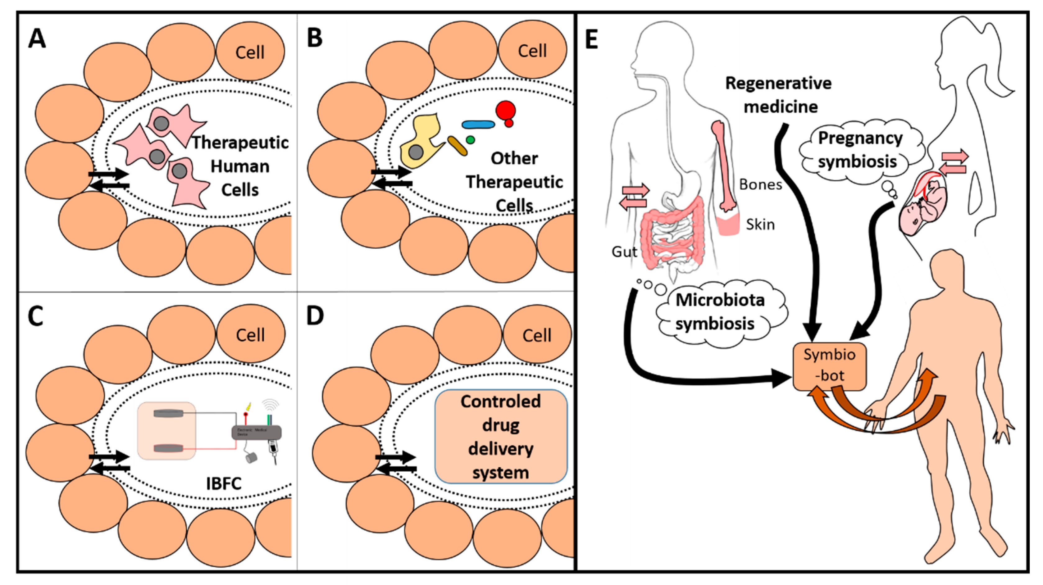
Figure 1. Examples of symbiotic devices (symbio-bots) (A)–(D) that can be developed using bioinspiration from existing natural symbiotic systems (E). Each device is separated with a smart porous packaging that allows for duplex communication. Therapeutic cells (A) and (B) need a porous encapsulation that avoids an immune reaction and allows for protection from both sides. They may be human mesenchymal stem cells (MSC), specialized cells (such as β-cell from Langerhans islets) (A), other eukaryotic or prokaryotic cells (B), an implantable biofuel cell (IBFC) linked to an electronic medical device (C), or a generic device delivering a therapeutic molecule (D). Existing symbiosis (e.g., microbiota or pregnancy) is a source of bio-inspiration to establish duplex communication between the body and its implants (E). Regenerative medicine should embrace this concept of bioinspiration for the better design and integration of implants, especially for future symbio-bots. (Figure reproduced with permission from [4]).
Applsci 10 02923 g001